GUIDELINES
Recommendations on nuclear and multimodality imaging in IE
and CIED infections
Paola Anna Erba1,2 &Patrizio Lancellotti3,4&Isidre Vilacosta5&Oliver Gaemperli6&Francois Rouzet7,8&
Marcus Hacker9&Alberto Signore10&Riemer H. J. A. Slart2,11&Gilbert Habib12,13
Received: 10 April 2018 / Accepted: 13 April 2018 / Published online: 24 May 2018 # Springer-Verlag GmbH Germany, part of Springer Nature 2018
Abstract
In the latest update of the European Society of Cardiology (ESC) guidelines for the management of infective endocarditis (IE), imaging is positioned at the centre of the diagnostic work-up so that an early and accurate diagnosis can be reached. Besides echocardiography, contrast-enhanced CT (ce-CT), radiolabelled leucocyte (white blood cell, WBC) SPECT/CT and [18F]FDG PET/CT are included as diagnostic tools in the diagnostic flow chart for IE. Following the clinical guidelines that provided a straightforward message on the role of multimodality imaging, we believe that it is highly relevant to produce specific recommendations on nuclear multimodality imaging in IE and cardiac implantable electronic device infections. In these procedural recommendations we therefore describe in detail the technical and practical aspects of WBC SPECT/CT and [18F]FDG PET/CT, including ce-CT acquisition
Preamble The European Association of Nuclear Medicine (EANM) is a professional nonprofit medical association that facilitates communication worldwide among individuals pursuing clinical and research excellence in nuclear medicine. The EANM was founded in 1985.
These recommendations are intended to assist practitioners in providing appropriate nuclear medicine care for patients. They are not inflexible rules or requirements of practice and are not intended, nor should they be used, to establish a legal standard of care.
The ultimate judgment regarding the propriety of any specific procedure or course of action must be made by medical professionals taking into account the unique circumstances of each case. Thus, there is no impli-cation that an approach differing from the recommendations, standing alone, is below the standard of care. To the contrary, a conscientious practitioner may responsibly adopt a course of action different from that set out in the guidelines when, in the reasonable judgment of the practi-tioner, such course of action is indicated by the condition of the patient, the limitations of available resources or advances in knowledge or tech-nology subsequent to publication of the recommendations.
The practice of medicine involves not only the science but also the art of dealing with the prevention, diagnosis, alleviation and treatment of dis-ease. The variety and complexity of human conditions make it impossible to always reach the most appropriate diagnosis or to predict with certainty a particular response to treatment. Therefore, it should be recognized that adherence to these recommendations will not ensure an accurate diagno-sis or a successful outcome. All that should be expected is that the prac-titioner will follow a reasonable course of action based on current knowl-edge, available resources and the needs of the patient to deliver effective and safe medical care. The sole purpose of these recommendations is to assist practitioners in achieving this objective.
* Paola Anna Erba [email protected]
protocols. We also discuss the advantages and limitations of each procedure, specific pitfalls when interpreting images, and the most important results from the literature, and also provide recommendations on the appropriate use of multimodality imaging.
Keywords Infective endocarditis . Cardiac implantable electronic device . Infection . Radiolabelled WBC .18F PET/CT . Echocardiography . Cardiac CT
Introduction
Over the last decades, there have been a series of tech-nology improvements that have significantly changed the role of clinical imaging in healthcare. Evolving tech-nologies such as multimodality imaging have gained a central key role in the evaluation of several disease entities. Infectious endocarditis (IE), on both native valve (NVE) and prosthetic valve (PVE), and cardiovas-cular implantable electronic device (CIED) infections are examples of such diseases in which multimodality imaging is used effectively in decision-making.
Extensive clinical use of radiolabelled leucocyte (white blood cell, WBC) SPECT/CT and [18F]FDG PET/CT in the imaging of IE has provided robust evidence of a major impact on patient management in terms of early diagnosis and the selection of optimal treatment strategies [1]. Therefore, in the latest update of the European Society of Cardiology (ESC) guidelines for the management of IE [2] both tech-niques are included as diagnostic tools in the diagnostic flow chart for PVE in particular. In these guidelines, imaging is a fundamental backbone of the diagnostic strategy, due to the concept that IE, rather than a single disease, may present with very different clinical patterns depending on the first organ involved, the underlying cardiac disease (if any), the microor-ganism involved, the presence or absence of complications, and the patient’s characteristics. Therefore, imaging specialists with a high level of expertise need to participate in a multidis-ciplinary team together with practitioners from several other specialties, including cardiologists, microbiologists, clinicians and surgeons.
Following the clinical guidelines that provide a straightforward message on the role of multimodality imaging, we believe that it is highly relevant to produce specific recommendations on nuclear and multimodality imaging in IE and CIED infections. Therefore, in these procedural recommendations, we describe in detail the technical and practical aspects of WBC SPECT/CT and [18F]FDG PET/CT, including contrast-enhanced CT (ce-CT) acquisition protocols. We also discuss the advan-tages and limitations of each procedure, specific pitfalls when interpreting images, the most important results from the literature and recommendations on the appro-priate use of multimodality imaging.
Definition and clinical challenges in IE
and CIED infections
IE is a life-threatening disease associated with a high mortality rate, difficult diagnosis and controversial man-agement [3, 4]. The clinical history of IE is highly variable and depends on the causative microorganism, the presence or absence of preexisting cardiac disease, the presence or absence of prosthetic valves or cardiac devices, and the mode of presentation. IE may present as an acute, rapidly progressive infection, but also as a subacute or chronic disease, and should be suspected in a variety of very different clinical scenarios [2].
Echocardiography and blood cultures are the cornerstone of IE diagnosis. Three echocardiographic findings are consid-ered as major criteria in the diagnosis of IE: vegetation, ab-scess or pseudoaneurysm, and new dehiscence of a prosthetic valve. However, the diagnostic accuracy of echocardiography may be challenging in PVE and intracardiac device infection, even with the use of transoesophageal echocardiography (TOE). Similarly, false diagnosis of IE may occur, and in some instances it may be difficult to differentiate vegetations from thrombi or other noninfective valvular lesions. Therefore, the results of the echocardiographic study must be interpreted with caution, taking into account the patient’s clinical presen-tation and the likelihood of IE.
CIED infection is a dreadful complication of cardiac device implantation with a high mortality rate [5], and is increasingly observed in elderly patients [6]. The reported incidence of permanent pacemaker infection varies widely among studies [7]. A distinction should be made between local device infec-tion and cardiac device-related IE (CDRIE). Local device in-fection is defined as an inin-fection limited to the pocket of the cardiac device and/or extravascular lead infection, while CDRIE is defined as an infection extending to the intravascu-lar and/or intracardiac lead, cardiac valve leaflets, or endocar-dial surface. Both diagnosis and therapeutic strategy are par-ticularly challenging in these patients [2].
As in NVE, echocardiography plays a key role in CDRIE and is helpful in the diagnosis of lead vegetations and tricuspid involvement, quantification of tricuspid regurgitation, sizing of vegetations, and follow-up after lead extraction [2]. However, false-negative and false-positive echo studies are not rare, and the Duke criteria are difficult to apply in these
patients because of lower sensitivity [5], even when the mod-ified Duke criteria are used.
Because of the frequently difficult diagnosis of the disease, and because of some limitations of echocardiography, partic-ularly in patients with PVE and CDRIE, other imaging niques have recently been applied, including nuclear tech-niques and ce-CT [8].
Summary of the 2015 ESC guidelines
for the management of infective endocarditis
The recently published 2015 ESC guidelines on the manage-ment of IE [2] propose important new features, including the need for a collaborative approach (the‘Endocarditis Team’), the emergence of nuclear imaging techniques in the early di-agnosis of IE, and the refinement of surgical indications.The Endocarditis Team
An interdisciplinary approach is required for the successful treatment of patients with IE and CIED infection. This team approach involves specialists in imaging, cardiologists, cardi-ac surgeons, specialists in infectious disease, and others. This multidisciplinary approach has already shown significant ad-vantages in the management of valvular heart disease (the ‘Heart Valve Clinic’), particularly in the selection of patients for transcatheter aortic valve implantation procedures (‘Heart Team’ approach) [9]. Such a team approach has been recom-mended recently as class IB in the 2014 American Heart
Association/American College of Cardiology (AHA/ACC) guidelines for the management of patients with valvular heart disease [10] as well as a class IIa, level B recommendation in the 2015 ESC guidelines [2]. In patients with IE, a team ap-proach has been shown to significantly reduce 1-year mortal-ity [11]. In the setting of the Endocarditis Team, a new entity, the‘imaging specialist’, plays a fundamental role: specialists in echocardiography, but also experts in nuclear medicine im-aging, cardiac CT, and magnetic resonance imaging (MRI) are increasingly included in the team. Decisions about how to diagnose, manage, treat and follow-up patients should be tak-en in refertak-ence ctak-entres based on constak-ensus decisions by the Endocarditis Team (Table1).
Cardiac imaging in the early diagnosis of IE
Early diagnosis of IE is a major challenge. In 2000, the mod-ified Duke criteria were recommended for diagnostic classifi-cation, and are mainly based upon echocardiography and blood culture results [12]. This classification has a sensitivity of approximately 80% for the diagnosis of IE [13]. However, the modified Duke criteria have a lower diagnostic accuracy in clinical practice, especially in patients with PVE and CDRIE, in up to 30% of whom echocardiography is normal or incon-clusive, and in patients with blood culture-negative IE. Recent advances in imaging techniques, including cardiac/whole-body CT, cerebral MRI, [18F]FDG PET/CT and radiolabelled WBC SPECT/CT, have resulted in improved identification of endocardial involvement and extracardiac complications of IE [8]. Recent studies have demonstrated that the addition of a
Table 1 Characteristics of the Endocarditis Team
When to refer a patient with IE to an Endocarditis Team in a reference centre
Patients with complicated IE, i.e. endocarditis with HF, abscess, or embolic or neurological complication or CHD, should be referred early and managed in a reference centre with immediately available surgical facilities.
Patients with uncomplicated IE can be initially managed in a nonreference centre, but with regular communication with the reference centre, consultations with the multidisciplinary Endocarditis Team and, when needed, with external visits to the reference centre.
Characteristics of the reference centre
Immediate access to diagnostic procedures should be possible, including TTE, TOE, multislice CT, MRI, and nuclear imaging.
Immediate access to cardiac surgery should be possible during the early stage of the disease, particularly in patients with complicated IE (HF, abscess, large vegetation, or neurological and embolic complications).
Several specialists should be present on site (the Endocarditis Team), including at least cardiac surgeons, cardiologists, anaesthesiologists, infectious disease specialists and microbiologists, and when available specialists in valve diseases, CHD, pacemaker extraction, echocardiography and other cardiac imaging, and neurologists (together with facilities for neurosurgery and interventional neuroradiology).
Role of the Endocarditis Team
The Endocarditis Team should have meetings on a regular basis to discuss cases, take surgical decisions, and define the type of follow-up. The Endocarditis Team chooses the type, duration, and mode of follow-up of antibiotic therapy, according to a standardized protocol, following current
guidelines.
The Endocarditis Team should participate in national and international registries, publicly report the mortality and morbidity of their centre, and be involved in a quality improvement programme (certifications?), as well as in a patient education programme.
The follow-up should be organized on an outpatient visit basis at a frequency depending on the patient’s clinical status (ideally at 1, 3, 6 and 12 months after hospital discharge, since the majority of events occur during this period).
Reproduced from Rundstrom et al. [5], with permission
positive [18F]FDG PET/CT scan to diagnostic criteria in-creases sensitivity without significantly decreasing specificity [14]. The value of cardiac CT was underlined in the AHA/ ACC guidelines [10] and the ESC guidelines [2]. The ESC considers that the published data are sufficiently strong and convincing to propose new criteria (the 2015 ESC Modified Diagnostic Criteria) including these new imaging techniques as new criteria for the diagnosis of IE. Three items are added in the ESC diagnostic criteria (Table2):
1. The identification of paravalvular lesions by cardiac CT should be considered as a major criterion.
2. In the setting of the suspicion of PVE, abnormal uptake of [18F]FDG on PET/CT or WBC on SPECT/CT should be considered as a major criterion.
3. The identification by imaging of recent embolic events or infectious aneurysms (silent events) should be considered as a minor criterion.
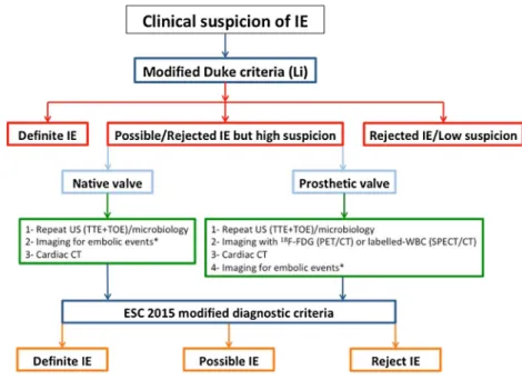
Figure1presents the proposed ESC diagnostic algorithm including the 2015 ESC modified diagnostic criteria. The di-agnosis of IE is still based upon the Duke criteria (blood cul-tures and echocardiography). However, when the diagnosis remains doubtful, other imaging techniques should be used, either for diagnosis of cardiac involvement or for imaging embolic events.
Cardiac imaging in treatment and follow-up of IE
Indications for surgery in IE may be subdivided into three categories: haemodynamic, infectious and embolic [2]. The decision to operate is frequently challenging and must be discussed on an individual basis using a multidisciplinary ap-proach, i.e. by the Endocarditis Team. Imaging including echocardiography, cardiac CT and nuclear imaging, plays a central role in this decision, along with the clinical presentation.
Table 2 The 2015 ESC modified criteria for diagnosis of IE Major criteria
Blood cultures positive for IE Typical microorganisms consistent with IE from two separate blood cultures
Viridans streptococci
Streptococcus gallolyticus (formerly S. bovis) HACEK group
Staphylococcus aureus or
Community-acquired enterococci, in the absence of a primary focus
or
Microorganisms consistent with IE from persistently positive blood cultures
Two or more positive blood cultures of blood samples drawn >12 h apart
or
All of three or a majority of four or more separate cultures of blood (with first and last samples drawn≥1 h apart)
or
Single positive blood culture for Coxiella burnetii or phase I IgG antibody titre >1:800
Imaging positive for IE
Echocardiogram positive for IE Vegetation
Abscess, pseudoaneurysm, intracardiac fistula Valvular perforation or aneurysm
New partial dehiscence of prosthetic valve
Abnormal activity around the site of prosthetic valve implantation detected by [18F]FDG PET/CT (only if the prosthesis was implanted >3 months previously) or radiolabelled WBC SPECT/CT Abnormal activity around the site of prosthetic valve implantation detected by [18F]FDG PET/CT (only if the prosthesis was implanted for >3 months) or radiolabelled WBC SPECT/CT
Definite paravalvular lesions on cardiac CT Minor criteria
Predisposition such as predisposing heart condition, or injection drug use Fever defined as temperature >38 °C
Vascular phenomena (including those detected only by imaging): major arterial emboli, septic pulmonary infarcts, infectious (mycotic) aneurysm, intracranial haemorrhage, conjunctival haemorrhages, and Janeway’s lesions
Immunological phenomena: glomerulonephritis, Osler’s nodes, Roth’s spots, and rheumatoid factor
Microbiological evidence: positive blood culture but does not meet a major criterion as noted above or serological evidence of active infection with organism consistent with IE
In summary, the 2015 ESC guidelines on the management of IE provide a novel approach in the diagnosis and manage-ment of this life-threatening disease. However, they are main-ly based on expert opinion because of the low incidence of the disease, the few available randomized trials and the limited number of meta-analyses. The sensitivity of the Duke criteria can be improved by new imaging modalities (MRI, CT, PET/ CT, SPECT/CT) that allow the diagnosis of embolic events and cardiac involvement when transthoracic echocardiogra-phy (TTE) or TOE are negative or doubtful. These criteria are useful, but they do not replace the clinical judgment reached and agreed by the Endocarditis Team.
Echocardiography for diagnosis of IE
and CIED infections
Echocardiography is still the main diagnostic tool in the as-sessment of patients with IE [2]. It is essential for diagnosis, initial evaluation of the risk of complications and the need for surgery, assessment and monitoring of in-hospital complica-tions, and morphological and functional assessment of the patient’s heart condition before hospital discharge.
Diagnosis
TTE should be performed as soon as IE is suspected. TOE must be performed when TTE is negative and the clinical suspicion of left-sided IE is high [15]. In patients with an initially negative echocardiographic examination, TTE and TOE should be repeated 5–7 days later if the clinical suspicion of IE remains high [2]. Specific procedural recommendations for TTE and TOE have been addressed previously [16].
Vegetations are the hallmark lesions of IE, but other echo-cardiographic findings are also considered major criteria for the diagnosis of IE; for example, perivalvular abscesses, perivalvular pseudoaneurysms, intracardiac fistulas, valvular perforations, valvular aneurysms, and new dehiscence of a prosthetic valve [2]. The detection of these lesions in patients with prosthetic valves is more difficult than in patients with native valves. Currently, the sensitivity of TTE and TOE for the diagnosis of vegetations is 70% and 96%, respectively, in native valves and 50% and 92%, respectively, in prosthetic valves. Regarding abscesses, the sensitivity of TTE is about 50%, compared with 90% for TOE [2,15]. The specificity for the detection of abscesses is higher than 90% with both echo-cardiographic modalities. Therefore, when IE is suspected in patients with prosthetic valves, both TTE and TOE must be done systematically.
It is worth emphasizing that the detection of a new large peri-prosthetic dehiscence should be considered a major crite-rion of IE even in the absence of other clinical signs or echo-cardiographic findings of IE [2].
TOE must always be performed when there is clinical sus-picion of IE in prosthetic valve or CIED carriers [2]. In these patients, TOE is also clearly superior to TTE in the detection and sizing of vegetations [16]. TOE allows visualization of lead vegetations in the right atrium/superior vena cava area and in other regions less well visualized by TTE. Both echo-cardiographic modalities are complementary in the assess-ment of tricuspid valve involveassess-ment, and quantification of tricuspid valve regurgitation and pulmonary hypertension. Another echocardiographic technique, intracardiac echocardi-ography, may be considered in patients with suspected CIED infection, positive blood cultures, and negative TTE and TOE studies [2].
Fig. 1 Algorithm for the diagnosis of IE according to the 2015 ESC guidelines (reproduced from Habib et al. [2] with permission). IE infective endocarditis, TTE transthoracic echocardiography, TOE transoesophageal echocardiography, US ultrasonography; asterisks may include cerebral MRI and/or whole-body CT and/or FDG PET/ CT or labelled WBC SPECT/CT
In patients with Staphylococcus aureus bacteraemia, the fre-quency of IE is high. Therefore, TTE or TOE should be per-formed according to the patient’s clinical profile and risk fac-tors for IE [17]. TOE is not mandatory in isolated right-sided NVE with good quality TTE and clear-cut echocardiographic findings [18]. Real-time three-dimensional (3D) TOE is useful for the assessment of vegetation morphology and size, and this may lead to a better prediction of the embolic risk in IE [19]. This echocardiographic technique is particularly useful in the assessment of perivalvular extension of the infection, prosthetic valve dehiscence, and leaflet perforation [20].
It is important to remember that a negative echocardiogra-phy study (TTE and TOE) does not rule out the diagnosis of IE. The negative predictive value (NPV) of a second TOE study in patients with the suspicion of NVE is extremely high. On the contrary, the NPV of TOE in patients with prosthetic valves is modest, and in many cases a second diagnostic im-aging technique will be needed [21].
Initial evaluation of the risk of complications
and need for surgery
The risk in patients with left-sided IE can be formally assessed according to clinical, microbiological and echocardiographic var-iables. Early TOE (during the first 48 h after admission) is ad-visable in most patients with left-sided IE in order to better assess vegetation size, degree of valvular regurgitation, and local perivalvular complications [22]. Patients with periannular com-plications, severe left-sided valve regurgitation, large vegetations, severe prosthetic valve dysfunction, low left ventricular ejection fraction, pulmonary hypertension or premature mitral valve clo-sure are at highest risk of death, stroke, and the need for surgery in the active phase of the disease. All these parameters can be easily and rapidly obtained by echocardiography [23].
Assessment and monitoring of in-hospital
complications
Local infection follow-up should be performed even when the clinical course of the patient with IE is good. Thus, in order to monitor vegetation size and to detect new silent complica-tions, repeated TTE and TOE during in-hospital follow-up (7–10 days) of uncomplicated IE is recommended [2]. TTE and TOE must be repeated as soon as a new clinical compli-cation appears during the patient’s in-hospital clinical course (new murmur, heart failure, embolism, persisting fever, atrio-ventricular block).
Echocardiographic assessment before hospital
discharge
TTE is recommended at completion of antibiotic therapy to assess left ventricular function, pulmonary pressure, and
valvular morphology and function. For better comparison, TOE is needed during follow-up in some patients (PV carriers, patients with complex surgery) before hospital discharge. In patients with CIED infection, TTE before hospital discharge is also recommended to detect the presence of retained segments of the pacemaker lead, and to assess tricuspid valve function, right ventricular function, and pulmonary hypertension. In addition, TOE after percutaneous lead extraction should be considered to detect residual infected material and potential tricuspid valve complications [15].
Radiolabelled white blood cell imaging
for diagnosis of IE and CIED infections
Radiopharmaceutical preparation and acquisition
protocol
W B C c a n b e r a d i o l a b e l l e d e i t h e r w i t h 9 9 mTc -hexamethylpropyleneamine oxime (99mTc-HMPAO, 370– 555 MBq) or with111In-oxine (10–18.5 MBq), as detailed in the specific European Association of Nuclear Medicine (EANM) guidelines [24,25]. Briefly, the procedure consists of isolation, radiolabelling and reinjection of autologous WBC obtained from the patient’s blood (about 50 mL). Therefore, strict aseptic conditions are required for the label-ling procedure. During labellabel-ling, care should be taken to avoid damage to leucocytes, as this would result in leakage of the radioactivity from the cell, adhesion of labelled leucocytes to the vascular endothelium and loss of motility. To avoid deg-radation of the radiopharmaceutical and radiation damage to labelled cells, radiolabelled WBC should be reinjected as soon as possible, and not later than 1 h after labelling. 99m Tc-HMPAO is generally preferred, because of the better image quality (higher count statistics and spatial resolution), and lower radiation exposure (0.011 mSv/MBq of 99m Tc-HMPAO versus 0.36 mSv/MBq111In-oxine). Patient prepara-tion is equivalent to that for any other clinical indicaprepara-tion. At least 2 × 10⁸leucocytes are required to achieve good labelling efficiency.
The image acquisition protocol includes planar acquisitions at 30 min (early images), 4–6 h (delayed images), and 20–24 h (late images) after reinjection of99mTc-HMPAO/111In-oxine WBC. A SPECT/CT acquisition is mandatory as part of the standard imaging protocol (as discussed in more detail in the sectionTechnical issues) and it is usually acquired 4–6 h and/
or 20–24 h after injection. Planar acquisitions will always include whole-body images (at least at 30 min) and anterior and posterior views of the thorax and any other region of interest (i.e. CNS, abdomen) when searching for septic embo-li. In patients with CIED infection, care should be taken to ensure that the generator site is included in the field of view, considering all the possible generator positions (i.e.
abdomen). Late acquisitions are particularly relevant in car-diovascular infections since background activity related to blood pool spill-over strongly hampers the detectability of lesions. These should be acquired with a“time corrected for isotope decay” modality as described in the EANM guide-lines. SPECT/CT performed at 4–6 h provides better image quality and might be repeated at 20–24 h if planar images (and SPECT images) at 4–6 h are negative.
A low-dose CT transmission scan is acquired while the patient continues tidal or shallow breathing, and is used for attenuation correction (CT-AC) and for colocalization (see below; on average 0.5–1.0 mSv radiation burden). Transmission data are reconstructed using filtered back-projection to produce cross-sectional images. With the avail-ability of cutting-edge SPECT/CT systems, administration of contrast medium to perform ce-CT is also feasible, despite the fact that experience in this specific setting is very limited. Recently, a study including a small sample of patients com-pared the diagnostic performance of WBC SPECT acquired on a conventional (NaI) camera to a cardiac-dedicated high-sensitivity cadmium-zinctelluride (CZT) camera[26]. Using 111
In-oxine-labelled WBC, it has been shown that target-to-background contrast increases with the CZT camera. This ap-proach has the advantages of overcoming the limitation of low count statistics with late acquisitions and reducing image noise due to better energy resolution. The field of acquisition of such cameras is limited to the cardiac area, but all-purpose CZT cameras are becoming commercially available and will be an attractive solution for late imaging in WBC SPECT.
Patient preparation
Preparation of patients with IE and CIED infection for WBC scintigraphy follow the general recommendations for any oth-er nuclear medicine procedure and the genoth-eral rules for WBC preparation. The major goals are to minimize tracer uptake in normal tissues, while maintaining uptake in target tissues. Because the effect of antibiotics on radiolabelled WBC uptake is unknown, it is important to be aware of ongoing antibiotic treatment, but no general recommendation on withdrawal can currently be made.
Image postprocessing and interpretation criteria
Both CT-AC and noncorrected SPECT images have to be evaluated in the coronal, transaxial and sagittal planes, as well as in 3D maximum intensity projection (MIP) cine mode. Misalignment between emission and transmission data may generate erroneous correction and thus data misinterpretation. Careful attention should be paid to quality control to avoid reconstruction artefacts. Noncorrected SPECT images be-come significantly important in the presence of prosthetic
valves, generators and electrocatheters due to possible overcorrection artefacts on SPECT/CT images.
The interpretation of WBC scintigraphy should always be-gin with a visual quality control performed on whole-body images and chest planar acquisitions to check for: (1) the ab-sence of high blood pool activity (suggesting the labelling of a substantial amount of erythrocytes) hampering interpretation even on delayed and late acquisitions, (2) liver uptake higher than spleen uptake, and (3) persistent pulmonary uptake (both 2 and 3 suggesting WBC damage prior to reinjection). The signal kinetics between the acquisitions at 4–6 h and 20–24 h are important features for interpretation: any stable or increased site of uptake (either intensity or size) over time, confirmed on SPECT/CT, is highly suggestive of infection. Overall pooled sensitivity and specificity have been reported to be 80–86% and 97–100%, respectively, with an AUC of 0.957 [27,28]. Such a high specificity is also maintained in patients with very early IE [29–32], a clinical setting in which WBC should represent the imaging modality of choice. Semiquantitative evaluation of WBC is also feasible, despite the fact that it has been validated in musculoskeletal infections [33] and no data are currently available for IE/CIED infections. Figures2and3show exam-ples of WBC SPECT/CT imaging in NVE and PVE.
In patients with suspected CIED infection and in patients implanted with a left-ventricular-assist device, WBC imaging revealed similar figures with a consistent high specificity [32–34]. In these studies, WBC SPECT/CT was found to be able to identify and define the precise anatomical location and extent of a suspected infection, improving patient management (Fig.4). Additionally, WBC SPECT/CT allows the detection of additional unsuspected extracardiac sites of infection in up to 23% of patients with device-related sepsis [33,35].
Abnormalities detected on WBC imaging should be local-ized as precisely as possible on SPECT/CT images since: (1) their colocalization with a structural abnormality considered as doubtful on echocardiography will support the hypothesis of infection, and (2) the localization and extent of the disease, on prosthetic material particularly, may help guide the surgical procedure. Localization of the sites of concomitant extracardiac infection from septic embolism is also possible on SPECT/CT images, influencing the Duke score and con-sequently the diagnostic certainty.
Pitfalls and differential diagnosis of IE/CIED infections
with WBC imaging
False-positive WBC imaging findings in IE and CIED infec-tions have been rarely described. On the other hand, false-negative scans have been observed in the presence of IE caused by some strains such as Candida spp. and Enterococcus spp. possibly due to the ability of these micro-organisms (as well as others such as Staphylococcus epidermidis) to form a“biofilm” that results in resistance to
antimicrobial treatment and escape from the host defence mechanisms. Additionally, altered neutrophil recruitment at
the primary site of IE by Enterococcus faecalis extracellular proteases constitutes a further mechanism of innate immune response impairment. Such mechanisms might reduce the sen-sitivity of scintigraphy with radiolabelled leucocytes in pa-tients with IE. The same limitation has always to be consid-ered in patients with CIED infection, in particular in the pres-ence of very small vegetations along the electrocatheter.
Embolisms on WBC imaging might appear either as areas of increased uptake over time, for example in brain, lung and soft tissue embolism, or as cold spots, for example in spleen embolism and spondylodiscitis. This latter appearance has to be considered nonspecific for infectious embolism since it might be present in other benign or malignant conditions. Therefore, despite the fact that these findings in patients with IE are high-ly suggestive of septic embolism, they should be con-firmed by additional diagnostic imaging tests. In addi-tion, reduced sensitivity has been reported in patients with small embolisms [29].
[
18F]FDG PET/CT for diagnosis of IE and CIED
infections
Patient preparation and acquisition protocol
When [18F]FDG PET/CT is used to diagnose cardiac and pericardial infection, patient preparation is very important due to the possible presence of physiological uptake of [18F]FDG in normal myocardium. Notably, these protocols differ fundamentally from cardiac viability imaging protocols in which myocardial glucose uptake is intentionally enhanced and homogenized by a combination of glucose loading and insulin. The current Society of Nuclear Medicine and Molecular Imaging (SNMMI)/American Society of Nuclear Cardiology (ASNC)/Society of Cardiovascular CT (SCCT) guidelines recommend preparation with a fat-enriched diet lacking carbohydrates for 12–24 h prior to the scan, a fast of 12–18 h, and/or the administration of intravenous heparin ap-proximately 15 min prior to [18F]FDG injection [36]. Fig. 3 99mTc-HMPAO WBC SPECT/CT imaging in a patient with aortic
NVE showing an increase in uptake of the radiopharmaceutical at the valve site (from left to right coronal, sagittal and transaxial superimposed (fused) SPECT/CT images)
Fig. 2 99mTc-HMPAO WBC SPECT/CT imaging in a patient with PVE. The emission image (middle) shows an area of increased radiopharmaceutical uptake which on the superimposed SPECT/CT image (bottom) corresponds to the prosthetic aortic valve. Top low-dose CT image
A recent review provides a unique overview of the avail-able literature regarding preparation for cardiac [18F]FDG PET imaging [37]. The data support the use of a high-fat, low-carbohydrate (HFLC) diet for at least two meals with a fast of at least 4 h for optimal suppression of physiological myocardial glucose utilization. Because there is no single su-perior patient preparation technique, in each institution image quality data should be continually evaluated to ensure that adequate suppression of [18F]FDG is achieved in more than 80% of the scans (Table3). Finally, following [18F]FDG in-jection and before the images are obtained, the patient should continue to fast and should not be physically active, as either of these will enhance myocardial glucose uptake.
Although antimicrobial treatment is considered to decrease the intensity of [18F]FDG accumulation [38], there is no evi-dence at this stage to routinely recommend treatment discontin-uation before performing PET/CT. On the contrary, steroid treat-ment should be discontinued or at least reduced to the lowest possible dose in the 24 h preceding the examination [39].
Blood glucose levels should always be checked and record-ed, keeping in mind that, in contrast to tumour imaging, nei-ther diabetes nor hyperglycaemia at the time of the study has been demonstrated to increase the false-negative rate in pa-tients with infection or inflammation [40]. Therefore, although efforts should be made to decrease blood glucose to the lowest possible level, hyperglycaemia should not represent an abso-lute contraindication to performing the study [41]. [18F]FDG imaging can be performed in patients with kidney failure, although the image quality may be suboptimal and prone to interpretation pitfalls [42].
The administered activity does not crucially affect the results of the examination within a certain range and also depends on the type of PET scanner. The EANM guidelines on [18F]FDG
PET imaging in inflammation/infection suggest a dose of 2.5– 5.0 MBq/kg, that is 175–350 MBq or 4.7–9.5 mCi in a 70-kg standard adult. In the US, the [18F]FDG administered activity should be 370–740 MBq (10–20 mCi) in adults and 3.7– 5.2 MBq/kg (0.10–0.14 mCi/kg) in children [43].
Technical issues
The acquisition is usually performed according to routine pro-tocols, 45–60 min after intravenous injection of [18
F]FDG, with an emission time/bed position depending on the sensitiv-ity of the scanner. One report suggests that delayed imaging acquired 3 h after injection (while maintaining the count Table 3 Recommendations for patient preparation for cardiac [18F]FDG PET/CT imaging for IE/CIED infections
Recommendation
Intervention
High evidence High-fat no-carbohydrate diet for at least two meals Fast for at least 4 h prior to examination
Avoid carbohydrate consumption Optimize fat intake
Avoid vigorous exercise during the 24 h before the examination
Intermediate evidence
Heparin given intravenously 15 min before [18F]FDG with dietary preparation/fasting Low evidence Any food or drink during the 4 h before the
examination Unrestricted diet Isolated fasting (<12 h) Calcium channel blockers Adapted from Osborne et al. [37]
Fig. 4 99mTc-HMPAO WBC SPECT/CT images in a patient with suspected CIED infection showing radiopharmaceutical uptake at the pocket site (top transaxial images, from left to right low-dose CT, emission and superimposed SPECT/CT images, respectively) and at the
intracardiac portion of the leads (bottom coronal images, from left to right low-dose CT, emission and superimposed SPECT/CT images, respectively)
statistic by doubling the time per bed position) is associated with greater contrast and improves the accuracy in diagnosing pacing lead infections in comparison with the standard proto-col [44]. However, recently and in a small series of patients images acquired at late time-points (150 min) after injection in patients with PVE have been found to be more prone to false-positive interpretation in both visual and semi-quantitative analyses [45].
The field of acquisition is usually derived from oncol-ogy studies from the skull base to the mid-thighs. Cerebral complications are frequent in left-sided IE and MRI studies have shown that early brain imaging can affect the diagnosis and management of patients [46]. Due to the low sensitivity of [18F]FDG PET in the detec-tion of brain lesions, inclusion of the brain in the field of acquisition is not recommended. A series of case reports suggest that the extension of the field of acquisition to the lower limbs allows the detection of complications of IE such as mycotic aneurysms that may require specific treat-ment by embolization to prevent rupture [47]. All these data are preliminary, based on small population samples, and require further validation.
The majority of PET/CT studies involve the use of a protocol comprising a scanogram/scout scan/topogram and CT-AC. Overall, the CT scan parameters should be such that patient exposure is the minimum necessary to provide diagnostic information. The simultaneous ac-quisition of a standard diagnostic CT scan with intrave-nous contrast agent is possible and should be preferred when appropriate in order to maximize the diagnostic information provided by the examination. Different im-aging protocols might be suggested for a PET/ce-CT scan (Tables 4 and 5 and Addendum 1).
In a series of patients with suspected PVE or CIED infection, Pizzi et al. showed that the addition of ce-CT to the standard [18F]FDG PET/CT protocol results in a high rate of patients reclassified from “possible” IE to “definite” IE, thus improving the overall diagnostic ac-curacy as compared with PET/CT without contrast en-hancement combined or not with the Duke score [49]. The main additional information provided by ce-CT was: better discrimination of the origin of FDG uptake between prosthetic valves or incomplete myocardial sup-pression; better coregistration between PET and ECG-gated CT angiography; identification of a greater num-ber of anatomical lesions in the valve area and of periannular complications; and the preoperative evalua-tion of coronary artery disease. Though not recommend-ed for routine use, ce-CT combinrecommend-ed with PET may prove useful in selected patients, particularly when echocardiography is of poor quality or did not allow precise evaluation of the periannular area. Limiting the wider use of ce-CT is the deleterious impact of contrast
agents on kidney function. Patients with IE or CIED infection are likely to receive high doses of antibiotics, some of them nephrotoxic, over long periods of time. It is therefore crucial to avoid any unnecessary exposure to additional nephrotoxic agents.
Table 4 Protocol for WBC SPECT/CT Acquisition time Acquisition
30 min (early) Whole-body and/or planar thorax/upper abdomen
followed by
4–6 h (delayed images) Planar images of the thorax followed by
Planar images of any additional FOV followed by
SPECT/CT acquisitions of the thorax with patient continuing tidal or shallow breathing followed by
SPECT/CT acquisitions of any additional FOV followed by
20–24 h (late images) Planar images of the thorax followed by
Planar images of any additional FOV followed by
SPECT/CT if needed FOV field of view
Table 5 Protocols for [18F]FDG PET/CT and for [18F]FDG PET with ce-CT
Protocol Acquisitions
1. [18F]FDG PET/CT when CT is used for attenuation correction and localization only (not intended as a clinically diagnostic CT scan)
CT topogram followed by
Low-dose CT scan (continuous tidal or shallow breathing)a followed by PET acquisition 2. [18F]FDG PET/CT with diagnostic CT scan [45] CT topogram followed by Whole-body CT-AC followed by Whole-body PET followed by Gated cardiac PET followed by
ECG-gated cardiac CT angiography 3. [18F]FDG PET/CT with
diagnostic CT scan [48]
CT topogram followed by
Thoracic CT in deep inspiration to acquire images of the arterial phase
followed by
Whole-body CT in portal phase followed by
Whole-body PET a
In the case of CT systems with up to six rings, a protocol using breath-hold in normal expiration is preferred
Imaging postprocessing and interpretation criteria
It is recommended that reconstructions be performed with and without attenuation correction to identify potential reconstruc-tion artefacts. Such artefacts have been well investigated in CIEDs, including pacing lead artefacts [50,51]. Metal artefact reduction techniques are useful for minimizing overcorrection artefacts, despite not always being successful in annulling their impact on PET image quality. The CT data acquired during the PET/CT study are usually reconstructed using fil-tered back projection. Recently introduced iterative recon-struction methods for CT data may be applied, if available on the PET/CT system. Depending on the CT protocol and the clinical case, separate CT reconstructions may be per-formed for diagnostic purposes.
PET images have to be visually evaluated for increased [18F]FDG uptake, taking into consideration the pattern (focal,
linear, diffuse), intensity and relationship to areas of physiolog-ical distribution. PET information is compared with morpho-logical information obtained by CT (Figs.5 and 6). Several recent meta-analyses have indicated that the overall pooled sensitivity of [18F]FDG PET/CT in IE is 61% [1], increasing to 73% when only PVE are considered and to 76% [27] or 81% with good overall accuracy (AUC 0.897) [28] when including only studies reporting adequate cardiac preparation. Thus, even if the PET results are negative (including whole-body evalua-tion for embolism detecevalua-tion), thorough interpretaevalua-tion of the echocardiography and CT scan is essential.
The pooled specificity of [18F]FDG PET/CT in patients with adequate cardiac preparation has been reported to be be-tween 85% and 90% [27,28]. Indeed. in the absence of infec-tion, [18F]FDG uptake around the prosthetic valve might be visible particularly early after surgery, and has different causes. Faint and homogeneous [18F]FDG uptake strictly limited to the
Fig. 5 Examples of different patterns of99mTc-HMPAO WBC and [18F]FDG uptake in patients with confirmed PVE: a typical focal pattern at99mTc-HMPAO WBC imaging; b focal pattern of [18F]FDG;
c diffuse [18F]FDG uptake of; d focal uptake over an area of diffuse [18F]FDG uptake. From left to right transaxial emission, low-dose CT and superimposed SPECT/CT or PET/CT images
valve annulus or around the struts of a bioprosthetic valve, very similar to the pattern observed in prosthetic vascular grafts [52], is most likely to have resulted from a persistent host reaction against the biomaterial coating the sewing ring of the prosthetic valve and chronic tension or friction exerted on these anchor points [53,54]. Such [18F]FDG uptake seems to be slightly greater in mechanical than in biological prostheses and in patients with vasculitis [55]. To prevent misinterpreta-tion of a positive scan due to imaging too early after valve implantation, the ESC guidelines recommend that [18F]FDG PET results should not be considered in the 3-month period following prosthetic valve implantation [2]. However, if sur-gery was uncomplicated, imaging before the recommended 3 months might be considered in an individual patient with awareness of this possible limitation [55].
In patients with NVE, interpretation of [18F]FDG uptake when the HFLC diet has been successfully adhered to is more straightforward than in those with PVE since any focal [18F]FDG uptake should be considered as abnormal. However, the diagnostic value of [18F]FDG PET in patients with NVE has not been well determined, due to higher rates of patients with prosthetic valves or mixed patients with native and prosthetic valves included in most studies. Recently, Granados et al. found that [18F]FDG PET was negative in six of six patients diagnosed with definite NVE [56]. In a recent meta-analysis pooled sensi-tivity for NVE was 71% [27]. The lack of sensitivity of
[18F]FDG PET in NVE is probably related to: (1) the size of the lesion (NVE is generally limited to the presence of a vege-tation, whereas in PVE generally spreads along the sewing ring or leads to abscess formation), and (2) the fact that blurring artefacts due to motion are more important at the tip of a valve leaflet than at the annulus. In this regard, ECG-gated acquisi-tions could help. This further emphasizes the need for a multimodality approach in which each imaging modality over-comes the other’s possible limitations in this clinical setting.
Semiquantitative analysis using the standardized uptake value (SUV) is also possible. However, in contrast to its ap-plication in oncology, SUV has not been validated in inflam-mation and infection. The additional value of quantitative pa-rameters (SUVmax normalized or not to the blood pool activ-ity, referred to as the target-to-background ratio) in differenti-ating between infected and non-infected material is a matter of debate. Whereas some literature tentatively provides values likely to identify infection with high specificity, it seems that the overlap of SUV between infected and non-infected pros-thetic valves precludes determination of a threshold. In this regard, all the factors influencing SUV quantification should be carefully considered, including those related to patient preparation (glycaemia, concurrent treatment, etc), time of uptake and the use of positive contrast.
The value of [18F]FDG PET in the diagnosis of CIED in-fection is substantiated by a large body of literature. After some Fig. 6 Same patient as in Fig.5c. In addition to the diffuse uptake of mild
intensity at the valve prosthesis (a from left to right transaxial emission, superimposed PET/CT, low-dose CT and ce-CT images), combined [18F]FDG PET/CT with ce-CT allows the identification of spleen
embolism as shown on both the PET/CT and ce-CT images (b from left to right transaxial emission, superimposed PET/CT, low-dose CT and ce-CT images), and brain embolism as shown only on the ce-ce-CT images (c, ce-CT transaxial image)
case reports and the seminal work of Ploux et al. [57], a larger scale study compared three groups of patients implanted with CIEDs: patients with suspicion of device infection, patients with a recently implanted device, and a control group of pa-tients without infection [58]. Images without attenuation cor-rection were used for final interpretation and to determine a parameter referred to as the semiquantitative ratio (SQR; the maximum count rate of the pocket device divided by the mean count rate of the lung parenchyma). The study showed the presence of mild postoperative residual inflammation up to 2 months after device implantation, whereas infected devices showed a significantly greater SQR. Both the sensitivity and specificity were >85% in a population with a high prevalence of infection. Finally, those patients with suspicion of infection but without [18F]FDG uptake had a favourable outcome under antibiotic therapy, suggesting the absence of bacterial coloni-zation of the CIEDs. It is noteworthy that no abnormal uptake was detected in the control group. A prospective study further supported the value of SQR for diagnosing CIED infection, and suggested that this parameter could help identify patients requiring device extraction [59]. More recently, normalization of SUVmax around the CIED to the mean hepatic blood pool activity has been shown to be an accurate and consistent quan-titative parameter for discriminating infected from asymptom-atic and symptomasymptom-atic non-infected devices. In the same study, however, no significant differences were found between sev-eral different SQRs with the exception of the contralatsev-eral ref-erence region that showed the lowest values. Metabolic uptake was increased at later acquisition time-points (90 and 180 min), suggesting potential usefulness of delayed imaging in terms of visual assessment and increasing reporter confidence [60].
Recent meta-analyses have shown a pooled sensitivity of [18F]FDG PET/CT of 83–87% and a pooled specificity of 89– 94%. These values were higher for generator/pocket infec-tions (93–96% and 97–98%, respectively) than for infecinfec-tions at the side of electrocatheters, where they were 65–76% and 83–88%, respectively [27,61,62], as a consequence of the very small size of the vegetations along the leads, which are often smaller than the spatial resolution of the system [57]. Therefore, [18F]FDG PET/CT has been suggested to be of value in guiding the clinician in choosing the most suitable treatment, i.e. conservative treatment (antimicrobial agents alone, or removal of just the generator) versus device removal. This is especially relevant considering the ongoing debate concerning the observation that novel antimicrobial agents can penetrate the bacteria-produced biofilm [63], thus poten-tially decreasing the need of hardware removal in CIED infec-tion [64]. However, considering that the differential diagnosis between an infection limited to the skin/pocket and more se-vere infection that involves the device over the pocket is the clue to choosing medical or surgical treatment in CIED infec-tion, further investigation is needed before [18F]FDG PET/CT can be introduced into the routine diagnostic work-up to guide
such a clinical decision. Figure 7 shows an example of [18F]FDG PET/CT in a patient with CIED infection.
One of the main features of [18F]FDG PET/CT is the ability to perform whole-body evaluation for abnormal focal uptake of [18F]FDG in a single scan with very high sensitivity [65]. In the setting of IE/CIED infection, the detection of septic emboli affects the Duke score and consequently the diagnostic certain-ty [44,56,65–69]. This comprehensive evaluation of the dis-ease extent will affect therapeutic management and lead to a reduction in the risk of relapse [59,70]. This has been shown to be particularly useful in the identification of embolisms in un-expected locations, such as mycotic aneurysms [47], a potential life-threatening complication requiring specific treatment. Similarly, in right-sided IE or CIED infection the detection of lung embolisms, considered as a major criterion of the Duke score, increases the diagnostic sensitivity [71]. In addition, identification of the infection portal of entry on [18F]FDG PET/CT is critical to prevent IE relapse. This primary infectious site may be orientated by the common biotope of the bacteria strain (digestive, skin, catheter) and should be part of the report. Finally, an additional promising role of [18F]FDG PET/CT is in patients with established IE, in whom it can be used to monitor response to antimicrobial treatment. Indeed, consider-ing the difficulties in the choice of type, dose and duration of antimicrobial treatment, the possibility of using PET/CT imag-ing to distimag-inguish between patients who respond favourably to treatment from those who require intensified administration or alternative treatment options is extremely attractive. However, significant data in this regard are scarce and the use of PET/CT in this clinical scenario can be suggested only as part of a case-based discussion within the Endocarditis Team.
Pitfalls and differential diagnosis of IE/CIED infections
with [
18F]FDG PET/CT
As discussed in detail, the diagnostic performance of [18F]FDG PET in IE and CIED infections is highly dependent on the background activity from physiological [18F]FDG myocardial uptake, and accordingly on the correct adherence to the HFLC diet followed by a fast of >12 h as discussed above in more detail. This is critical for the optimal analysis of valvular regions. Another potential confounding factor for [18F]FDG PET/CT is increased metabolic activity along the posterior part of the heart, where lipomatous hypertrophy of the interatrial septum may appear as a fat-containing mass with increased [18F]FDG uptake [72]. Early PET/CT scanning following valve implantation is not recommended since per-sistent reaction of the host against the synthetic component of the sewing ring might cause false-positive results. The persis-tent host reaction against the biomaterial coating of the sewing ring of the prosthetic valve may persist for years after valve implantation and should always be considered as a source of misinterpretation.
A wide range of pathological conditions can mimic the pattern of focally increased [18F]FDG uptake. The follow-ing conditions might present with focal [18F]FDG uptake: active thrombi [73], soft atherosclerotic plaques [74], vas-culitis [75], primary cardiac tumours [76] and cardiac me-tastasis [77], postsurgical inflammation [78] and foreign body reactions (such as a reaction to BioGlue, a surgical adhesive used to repair the aortic root) [79], and stitches
[80]. Recently intense [18F]FDG uptake has also been found in a patient with Libman-Sacks endocarditis [81]. Therefore, to maintain the high specificity of [18F]FDG for IE, it is essential that patient selection and inclusion criteria, as well as image reading, are accurate. In this regard, the CT component of PET/CT plays a crucial role by accurately localizing vascular wall uptake and intracar-diac lesions.
Fig. 7 99mTc-HMPAO WBC (a) and [18F]FDG PET/CT (b) in patients with CIED infection. Faint WBC uptake is prentent at the pocket site which is an artifact due to attenuation correction as demonstrated by the disappearnce of the uptake at the non-attenuated corrected images(a upper panel from left to right transaxial emission, superimposed SPECT/CT and non-attenuated corrected), whereas the uptake at the intracardiac portion of the leads, is consistent with infection (lower panel, coronal view emision and fused SPECT/CT). In b increased
[18F]FDG uptake at both the pocket site and at the intravascular portion of the leads, persisten at the non-attenuated corrected images (upper, from left to right transaxial emission, superimposed PET/CT and non-attenuated corrected) and at the intracardiac portion of the leads (lower panel from left to right coronal emissiona nd fused PET/CT) confirming the presence of device infection. From left to right transaxial images and coronal images emission, low-dose CT, superimposed PET/CT and non-attenuated emission images, respectively
As already discussed, antimicrobial therapy and/or veg-etation size can account for false-negative [18F]FDG PET/ CT results.
Cardiac CT for diagnosis of IE and CIED
infections
Since the introduction of the first 64-slice CT devices in 2005, cardiac CT has evolved to be one of the most important struc-tural imaging techniques of the heart. Its strengths lie in the high isotropic (i.e. in all three axes of 3D space) spatial reso-lution in the range of 0.2 to 0.3 mm with the most recent devices, and its ease of use and wide availability. The draw-backs of cardiac CT are the need for iodinated contrast agent administration and ionizing radiation. The latter, however, can be limited with appropriate imaging protocols and newer de-vices to a radiation dose in the low single-digit millisievert range (depending on the field of view). Temporal resolution has also been improved considerably (from 250 ms with the first generation four-slice CT scanners down to 66 ms with the most recent dual-source devices), allowing high-quality im-ages to be obtained in patients with faster heart rates or even in selected patients with rate-controlled atrial fibrillation [82].
In the context of IE and CIED infections, cardiac CT may serve two different purposes. First, cardiac CT almost invari-ably complements every radionuclide imaging study (e.g. [18F]FDG PET or WBC scintigraphy) to provide an anatomical map for coregistration with radionuclide signals. This allows identification of the particular anatomical structures with path-ological uptake and improves the diagnostic accuracy of the technique. Most often, this objective is achieved with a native low-dose CT scan, although some have suggested that performing ECG-gated ce-CT angiography may improve the diagnostic accuracy of the combined hybrid study (as men-tioned before) [83]. Second, the anatomical information pro-vided by CT may itself allow the diagnosis of IE or CIED infection, particularly in the presence of vegetations and com-plications such as abscesses, pseudoaneurysms, fistulas or sep-tic embolization. Moreover, cardiac CT, if performed on a high-end CT device, allows the assessment of coronary arteries prior to any surgical procedure. This avoids preoperative inva-sive coronary angiography, which (in the case of aortic valve endocarditis) carries a certain risk of vegetation embolization due to manipulation of the catheter in the aortic root [84].
Patient preparation
Only minor patient preparation is required prior to cardiac CT. In fact, cardiac CT offers the advantage of also being available for urgent indications or in critically ill patients. The level of patient preparation depends solely on the type of scan envis-aged, and the latter in turn depends on the clinical situation
and the question of interest. In the simplest case, where only a native (noncontrast) nongated CT scan is required (e.g. for simple coregistration with radionuclide imaging according to a standard PET/CT protocol), no particular patient preparation is needed. The scan is usually performed immediately before or after the acquisition of PET emission data. Occasionally, however, if indicated for IE and/of CIED infection, a high-resolution contrast-enhanced ECG-gated scan will be pre-ferred to resolve anatomical details of moving heart structures. In this case, it is generally recommended that the patient should fast for 3–4 h prior to the scan [85]. A careful history with regard to potential pregnancy, allergy to contrast agents, and impaired renal function should be taken.
Despite the high temporal resolution of current CT devices, image quality still depends considerably on heart rate. In pa-tients with heart rates >65 bpm, pretreatment with a beta-receptor antagonist (either intravenously or orally depending on the clinical setting) is recommended to lower the heart rate as much as possible, preferably below 60 bpm, providing there is no contraindication to the use of these agents [85]. Although achieving diagnostic quality for the assessment of coronary arteries may not be the primary goal of the scan, it may still be advisable to lower the heart rate and administer nitrates prior to image acquisition. Special care should be taken in patients with severe aortic regurgitation, in whom lowering the heart rate may have detrimental haemodynamic effects.
Imaging protocols and technical issues
As mentioned above, different image acquisition protocols may be used depending on the scanner platform and the type of scan ordered. To obtain high-quality images in re-gions affected by cardiac motion, ECG gating by either retrospective or prospective triggering is indispensable. Retrospective gating offers the advantage of allowing re-construction of CT images over all phases of the cardiac cycle. This allows the demonstration of the oscillation of masses, visualization of native or prosthetic valvular func-tion, and identification of rocking motion in prosthetic dys-function. However, the radiation dose is considerably higher with retrospective ECG triggering (depending on scan parameters and the use or not of dose-modulation algorithms, up to 20 mSv). On the other hand, prospective ECG gating triggers image acquisition during a predefined interval of the RR cycle and therefore is obtained with a fraction of the exposure radiation of retrospective helical scans. Radiation dose also depends on the longitudinal field of view of the scanner and is higher for partial-body CT angiography than for cardiac CT angiography. High-end CT devices with 64 slices or more are generally pre-ferred due to their higher spatial resolution, larger axial coverage and shorter scan time. Good quality images are difficult to obtain in patients with atrial fibrillation or
tachycardia due to the deleterious effect on image quality of motion artefacts. In these patients, it is recommended that the heart rate is lowered as much as possible using oral or intravenous beta-blockers (if clinically feasible) and that devices with the highest temporal resolution are used (e.g. dual-source scanners, if available).
Imaging postprocessing and interpretation criteria
Images are generally reconstructed as transaxial images, multiplanar reformations, maximum intensity projections, and 3D volume renderings. For fusion with radionuclide images, axial and coronal source images are usually the preferred mode
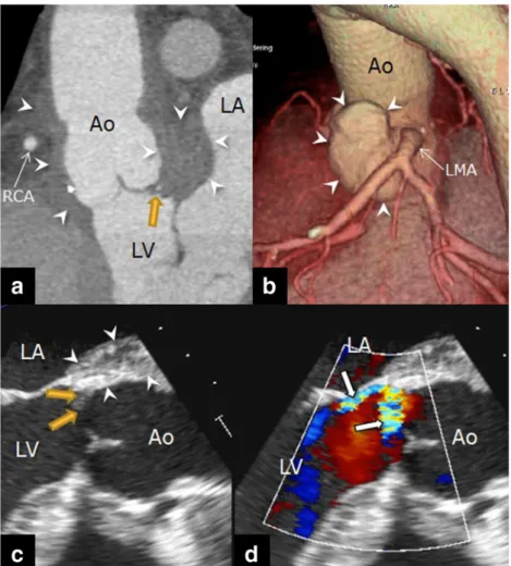
Fig. 9 Same patient as in Fig.8. a Double-oblique multiplanar reformatted cardiac CT image shows a perivalvular abscess (arrowheads) surrounding the entire aortic root (Ao) with the largest collection between the left atrium (LA) and the left coronary cusp. b 3D volume-rendered CT image shows the relationship between the pseudoaneurysm (arrowheads) and the other anatomical structures: The pseudoaneurysm is located just anterior of the left main coronary artery (LMA) but does not compromise the vessel. c Transoesophageal
echocardiography (TOE) image shows a hypoechogenic abscess between the aortic root and the left atrium. d The colour Doppler image confirms the presence of at least two small perforations of the left coronary cusps (white arrows, yellow arrows in c) which are not clearly visible on the CT image (yellow arrow in a)
Fig. 8 A 52-year-old man with prosthetic aortic valve endocarditis (coagulase-negative staphylococci) 4 years after aortic root replacement with a 25-mm Freestyle biological valve, replacement of the ascending aorta with a 28-mm Dacron graft and reconstruction of the posterior sinus with xenopericardium. a ECG-gated contrast-enhanced cardiac CT image (GE VCT 64-slice CT scanner) of the aortic root (Ao) shows a
pseudoaneurysm of the left coronary sinus (arrowheads) with a small 3–4 mm vegetation (yellow arrow) attached to the anterior aspect of the neck of the pseudoaneurysm. b Transoesophageal echocardiography (TOE) image confirms the CT findings. Note that the CT and TOE images have opposite anteroposterior orientation (LA left atrium, RCA right coronary artery)
of display. Multiplanar reformations of ce-CT images allow reconstruction of the structure of interest in every possible plane, and thereby allow assessment of its structure, size and extent. Vegetations appear as irregularly shaped, hypodense, oscillating structures adherent to the endocardium (Fig.8). Abscesses are defined as irregularly shaped, inhomogeneous masses within the paravalvular myocardium or pericardium, while a pseudoaneurysm is defined as space filled with contrast medium with connection to any of the cardiac chambers (Fig.
9). CT has shown very high sensitivity in comparison with TOE or surgery in detecting the presence and extent of paravalvular complications such as abscesses and pseudoaneurysms [86,
87]. TOE, on the other hand, is probably superior to CT for the diagnosis of small vegetations and for detecting leaflet per-forations, particularly if they are smaller than 2 mm (Fig.9).
However, in the setting of PVE, the diagnostic yield of TOE is lower than in NVE as previously mentioned. In PVE, the onset of infection may be more insidious, extension of infec-tion into the perivalvular tissue more common, and acoustic shadowing from metallic prostheses interferes with TOE im-aging (particularly in the detection of paravalvular complica-tions located in or close to the right coronary cusp). In this setting, CT may even be superior to TOE in detecting vegeta-tions, abscesses, and pseudoaneurysms [88]. TOE remains more sensitive than CT for valvular dehiscence, although using retrospective ECG triggering and image reconstruction at dif-ferent time intervals during the RR cycle after a full helical CT scan, even rocking motion from a severely detached prosthesis can be visualized. However, large comparisons between CT and TOE are lacking, therefore CT and TOE are considered complementary techniques, but it is recommended that TOE remain the first-line test in suspected PVE. Moreover, CT al-lows the assessment of systemic complications from IE includ-ing septic embolization (e.g. lung, spleen, brain), mycotic an-eurysms, and intracranial bleeding, which may all have impor-tant implications for patient prognosis and management and may add to the diagnostic criteria for IE.
In recognition of the increasing role of cardiac CT, the 2015 ESC guidelines for the management of IE have been modified by adding to the traditional Duke criteria“definite para valvu-lar lesions by cardiac CT” as a major criterion, and “vascuvalvu-lar phenomena detected by CT including arterial emboli, septic pulmonary infarcts, infectious (mycotic) aneurysm, intracra-nial haemorrhage, conjunctival haemorrhages, and Janeway’s lesions” as a minor criterion for endocarditis [2].
In CIED infections, cardiac CT probably has a more limited role than radionuclide imaging, except for providing anatomical maps to coregister signal from radionuclide imaging with ana-tomical structures (e.g. device pocket or leads). In the majority of cases, nongated native CT scans are considered sufficient for this purpose. Small vegetations on pacemaker leads may be difficult to detect on ce-CT angiography and the generator often causes significant blooming and beam hardening artefacts in
the pacemaker pocket. However, as mentioned before in the field of IE, CT angiography may add important remote infor-mation on vascular complications such as mycotic aneurysms, arterial emboli, and septic pulmonary infarcts, which adds to the diagnostic criteria and affect the overall treatment strategy.
Pitfalls, differential diagnosis of IE/CIED infections
with CT
Even though CT has been demonstrated to be more accurate in the presence of metallic prostheses, it is not devoid of artefacts. Some tilting-disc metallic prostheses, such as the Björk-Shiley valve, have been associated with severe beam-hardening arte-facts, which affect the correct evaluation of the perivalvular region [89]. Increased wall thickness of the aorta has been pro-posed as a sign of early aortic root infection after surgery. However, there is no consensus about the upper limits of the normal aortic root thickness after aortic valve surgery. In addi-tion, in the early postoperative period, the aortic wall may be thickened from haematoma or oedema, which may resolve with-in 3 to 6 months, requirwith-ing close imagwith-ing follow-up and with- inte-gration of clinical findings. The differential diagnosis of valvular vegetations includes mobile strands or free sutures arising from sewing rings or prosthetic valves, and cardiac (mostly benign) masses such as thrombus, fibroelastoma and myxoma.
The
“imaging specialist” in the Endocarditis
Team: role, challenges and education
The new ESC guidelines for the management of IE introduce the Endocarditis Team as the basis for maximizing the likelihood of success in managing patients with IE and CIED infections. They delineate the wide scope and complexity of the Endocarditis Team and provide a general framework for its functioning, cluding recommending the professionals who should be in-volved in the decision making process. Given the complexity of the topic, a team approach provides the most logical solution to delivering interdisciplinary competence. The benefits of team-work are obvious. The team approach leads professionals to evaluate IE as a whole complex, multisite disease that should be addressed with an integrated diagnostic-therapeutic strategy rather than a single isolated medical action. The diverse range of professionals working together, bringing together diverse knowledge and skills, results in higher levels of innovation in patient care and faster decision making. From the patients’ (and also the families’) perspective, teamwork affects compliance since it is easier to communicate with a cohesive team than with practitioners who work in isolation.
Efficient team functioning is challenging. Building well-functioning teams requires time, education and support. With time, team members get to know each other and learn about each other’s professional work and attitudes to change and


![Table 5 Protocols for [ 18 F]FDG PET/CT and for [ 18 F]FDG PET with ce-CT](https://thumb-eu.123doks.com/thumbv2/123doknet/6526647.175383/10.892.456.818.623.1034/table-protocols-fdg-pet-ct-fdg-pet-ct.webp)
![Fig. 5 Examples of different patterns of 99m Tc-HMPAO WBC and [ 18 F]FDG uptake in patients with confirmed PVE: a typical focal pattern at 99m Tc-HMPAO WBC imaging; b focal pattern of [ 18 F]FDG;](https://thumb-eu.123doks.com/thumbv2/123doknet/6526647.175383/11.892.122.766.434.1020/examples-different-patterns-patients-confirmed-typical-pattern-imaging.webp)
![Fig. 7 99m Tc-HMPAO WBC (a) and [ 18 F]FDG PET/CT (b) in patients with CIED infection](https://thumb-eu.123doks.com/thumbv2/123doknet/6526647.175383/14.892.86.809.74.755/fig-hmpao-wbc-fdg-pet-patients-cied-infection.webp)