1 Contribution à la connaissance de l’épidémiologie de la fièvre aphteuse au Niger
Contribution to the knowledge of the epidemiology of Foot and Mouth Disease in Niger
Bachir SOULEY KOUATO
THESE PRESENTEE EN VUE DE L’OBTENTION DU GRADE DE
DOCTEUR EN SCIENCES VETERINAIRES
ORIENTATION MEDECINE VETERINAIRE ANNEE ACADEMIQUE 2016-2017
ACADEMIE UNIVERSITAIRE WALLONIE-EUROPE
UNIVERSITE DE LIEGE-FACULTE DE MEDECINE VETERINAIRE
DEPARTEMENT DES MALADIES INFECTIEUSES ET PARASITAIRES
FUNDAMENTAL AND APPLIED RESEARCH ON ANIMAL HEALTH
UNITE DE RECHERCHE EN EPIDEMIOLOGIE ET ANALYSE DE RISQUES APPLIQUEES AUX SCIENCES VETERINAIRES
2
3
Académie Universitaire Wallonie-Europe Université de Liège - Faculté de Médecine Vétérinaire
Département des Maladies Infectieuses et Parasitaires
Fundamental and Applied Research on Animal and Health (FARAH) Unité de Recherche en Epidémiologie et Analyse de Risques
Appliquées aux Sciences Vétérinaires (UREAR-ULg)
Institut de Médecine Tropicale d’Anvers Département de Sciences Biomédicales
Institut National de la Recherche Agronomique du Niger (INRAN), Niamey, Niger
Contribution à la connaissance de l’épidémiologie de la fièvre aphteuse au Niger
Bachir SOULEY KOUATO Promoteur :
Prof. Dr. Claude SAEGERMAN
Unité de Recherche en Epidémiologie et Analyse de Risques Appliquées aux Sciences Vétérinaires (UREAR-ULg)
Fundamental and Applied Research on Animal and Health (FARAH) Département des Maladies Infectieuses et Parasitaires
Faculté de Médecine Vétérinaire-Université de Liège
Co-promoteur : Dr. Thys Eric
Institut de Médecine Tropicale d’Anvers Département de Sciences Biomédicales
4
Wallonia-Europe University Academy University of Liege-Faculty of Veterinary Medicine
Department of Infectious and Parasitic Diseases
Fundamental and Applied Research on Animal and Health (FARAH) Research Unit of Epidemiology and Risk Analysis Applied to Veterinary Sciences
(UREAR-ULg)
Institute of Tropical Medicine of Antwerp Department of Biomedical Sciences
National Institute of Agronomic Research of Niger (INRAN), Niamey, Niger
Contribution to the knowledge of the epidemiology of Foot and Mouth Disease in Niger
Bachir SOULEY KOUATO Promoter:
Prof. Dr. Claude SAEGERMAN
Research Unit of Epidemiology and Risk Analysis Applied to Veterinary Sciences (UREAR-ULg)
Fundamental and Applied Research on Animal and Health (FARAH) Department of Infectious and Parasitic Diseases
Faculty of Veterinary Medicine - University of Liege
Co-promoteur: Dr. Thys Eric
Institut de Médecine Tropicale d’Anvers Département de Sciences Biomédicales
5
Abstract
FMD is a severe, highly contagious viral disease affecting domestic and wild ruminants and pigs. FMD is endemic in Niger with potential impact on the national economy because of its negative effect on animal production. However, there is evidence that FMD is poorly investigated in Niger as the prevalence as well as the associated risk factors of the disease and serotypes circulating are not well known. These informations are of key importance to implement appropriate and efficient prevention and control measures against FMD. Therefore, the research presented in this thesis aimed to contribute to a better understanding of the epidemiology of FMD in Niger.
Firstly, two prerequisites systematic review studies were performed on FMD risk factors modelling and molecular epidemiology of FMD in Africa respectively. The findings of the first systematic review showed that the most reported factors related to FMD were the uncontrolled animal movement and the mixing of animals around water and grazing points. Depending on the model used, the included articles in this review presented some limitations. The lack of reliable data especially from endemic settings to perform these epidemiological modelling studies was also highlighted. On the other hand, the second systematic review showed an increasing interest from African countries to conduct research on molecular epidemiology of FMD. The identification and molecular characterization studies of African FMDV strains showed the complexity of the genetic relationships between circulating strains as reflected by the diversity and transboundary mobility of FMDV in the continent.
Further, an original study to get insight in the economic impact and the spatiotemporal pattern of transmission of FMD outbreaks in Niger was performed based on the retrospective analysis of 9-year (2007-2015) outbreak data. This study revealed that FMD outbreaks occurred in all regions but affecting more the districts bordering neighbouring countries. The animal density was the important predictor variable of outbreaks occurrence. The seasonal trend of FMD outbreak occurrence was confirmed by this study with most outbreaks occurring during the cold and dry season and starting at the end of the rainy season. This study revealed that at outbreak level, the mean stochastic estimates were respectively 52.33 cattle affected by the disease and 4.33 cattle assumed to die from FMD. In this analysis, the cost of FMD consists of the cost due to the morbidity assumed to be the loss of milk production and the cost of mortality of young animals. Thereby, the average total cost of FMD at herd level was estimated at 732.72
6
euros. The cost of mortality of young bulls was the largest portion of the total cost, contributing to 41.55%, while costs related to heifer mortality and reduced milk production were respectively 35.36% and 23.09% of the total cost of FMD at outbreak level. To estimate the cost of vaccination at FMD outbreak level, one scenario was considered consisting in vaccinating each animal with 2 doses of vaccine allowing to estimate a cost of vaccination at FMD outbreak level at 315.27 euros on average at herd level. Consequently, the average ratio total costs of FMD/ cost of vaccination at outbreak level was estimated at 2.31.
The performed field outbreak study in southwestern Niger indicated that 70% (158/227) of the sera were positive for the presence of antibodies against FMDV through NSP ELISA. Multivariate logistic regression analysis revealed that only the herd composition (presence of both cattle and small ruminants) was significantly associated with FMDV seropositivity (P-value = 0.006). Among the NSP ELISA positive sera tested by LPBE, 86% (136/158) were positive for one or more serotypes (A, O, SAT 1 and SAT 2). Additionally, either as single or as multiple serological reactions, there was a clear dominance of serotype O followed by serotypes A and SAT1. Moreover, FMDV serotype O was isolated and characterised within the O/WEST AFRICA topotype. One of the FMDV isolates from Niger (O/NGR/4/2015) showed a close antigenic match with three FMDV serotype O reference vaccine strains. The phylogenetic results showed a strong relation amongst and between collected samples from Niger and the result revealed that these isolates are closely related to strains previously isolated in some West African countries including Benin, Togo and Ghana.
In conclusion, the results of the field outbreak study together with the spatiotemporal distribution of FMD outbreaks confirm the endemic nature of the disease in Niger. Furthermore, the molecular characterization highlights the complex transboundary nature of FMD in Africa through uncontrolled animal movement, cross bordering transhumance and live animal trade. The key messages for decision makers resulting from this thesis are the need for further countrywide comprehensive epidemiological research on the epidemiology of FMD and the launching of a strategic plan for disease control in Niger. Moreover, the major implication of this study is the requirement for integrated and regional FMD control strategies with the aim to more effectively prevent or control FMD in Africa.
7
Résumé
La fièvre aphteuse est une grave maladie virale et hautement contagieuse qui affecte les ruminants domestiques et sauvages et les porcs. La fièvre aphteuse est endémique au Niger avec potentiellement un impact sur l'économie nationale en raison de ses effets néfastes sur les productions animales. Cependant, il est évident que la fièvre aphteuse est très peu étudiée au Niger car la prévalence ainsi que les facteurs de risque associés à la maladie d’une part et d’autre part les sérotypes circulants ne sont pas bien connus. Or, ces informations sont d'une importance capitale pour mettre en œuvre des mesures de prévention et de lutte adaptées et efficaces contre la fièvre aphteuse. Par conséquent, les recherches présentées dans cette thèse visent à contribuer à une meilleure compréhension de l'épidémiologie de la fièvre aphteuse au Niger.
D’emblée, le besoin d’effectuer deux revues systématiques s’est fait sentir et ce, respectivement sur la modélisation des facteurs de risque de la fièvre aphteuse et l'épidémiologie moléculaire de la fièvre aphteuse en Afrique. Les résultats de la première revue systématique ont montré que les facteurs les plus rapportés qui sont liés à la fièvre aphteuse sont le mouvement non contrôlé des animaux et le mélange des troupeaux autour des points d’eau et des pâturages. Il a été rapporté certaines limites selon le modèle utilisé et décrit dans les articles inclus dans cette revue. Il a également été mis en évidence le manque de données fiables pour effectuer ces études de modélisation épidémiologique et particulièrement dans le contexte des pays endémiques. Par ailleurs, la deuxième revue systématique a montré un intérêt croissant des pays africains à mener des recherches sur l'épidémiologie moléculaire de la fièvre aphteuse. L'identification et les études de caractérisation moléculaire des souches africaines du virus de la fièvre aphteuse ont mis en évidence la complexité des relations génétiques entre les souches virales circulantes, se traduisant par la diversité et la mobilité transfrontière du virus de la fièvre aphteuse au sein du continent.
Une étude a ensuite été conduite et est basée sur une analyse rétrospective de neuf années (2007-2015) de données sur des foyers de fièvre aphteuse. L’objectif de cette étude était d’avoir un aperçu de l’impact économique mais aussi de connaitre les caractéristiques spatiotemporelles de transmission de la maladie au Niger. Ainsi, cette étude a révélé l’apparition des foyers de fièvre aphteuse dans toutes les régions, avec les départements frontaliers avec les pays voisins étant les plus affectés. Il s’est également avéré que la densité animale était la principale variable prédictive de l'apparition de ces foyers. En outre, la tendance
8
saisonnière de l'apparition des foyers de fièvre aphteuse a été confirmée par cette étude, la plupart des épidémies se produisant pendant la saison sèche et froide et débutant à la fin de la saison des pluies. Cette étude a révélé qu'à l’échelle du foyer, les estimations stochastiques moyennes étaient respectivement de 52,33 bovins affectés par la maladie et de 4,33 bovins supposés mourir de cette maladie. Pour cette analyse, le coût estimé de la fièvre aphteuse est composé du coût dû à la morbidité qui est ici représenté par la perte de production laitière et le coût de la mortalité des jeunes animaux. Ainsi, le coût total moyen de la fièvre aphteuse au niveau du troupeau était estimé à 732,72 euros. Le coût de la mortalité des jeunes taureaux contribuant à 41,55% du coût total de la maladie représentait la plus grande part de ce coût estimé de la fièvre aphteuse, tandis que les coûts liés à la mortalité des génisses et à la réduction de la production laitière étaient respectivement de 35,36% et 23,09% du coût total de la maladie. Pour estimer le coût de la vaccination au niveau d’un foyer de fièvre aphteuse, un seul scénario consistant à vacciner chaque animal avec 2 doses de vaccin, a été pris en compte. Ainsi, le coût de la vaccination à l’échelle d’un foyer a été estimé en moyenne à 315,27 euros. Par conséquent, le ratio moyen du coût total de la maladie /coût de la vaccination pour un foyer est estimé à 2.31.
Une étude sur des foyers de fièvre aphteuse survenus dans le Sud-Ouest du Niger a révélé que 70% (158/227) des sérums étaient positifs pour la présence d'anticorps viraux par la méthode NSP ELISA. L'analyse de régression logistique multivariée a révélé que seule la composition du troupeau (présence de bovins et de petits ruminants) était significativement associée à la séropositivité (valeur P = 0,006). Parmi les sérums positifs à la NSP ELISA et testés par LPBE, 86% (136/158) étaient positifs pour un ou plusieurs sérotypes (A, O, SAT 1 et SAT 2). En outre, pour les réactions sérologiques spécifiques (à un seul sérotype) ou multiples (plusieurs sérotypes), il y avait une nette prédominance du sérotype O suivi des sérotypes A et SAT 1. Par ailleurs, le sérotype O du virus de la fièvre aphteuse a été le seul isolé et dont les résultats de la caractérisation moléculaire indique qu’il appartient au topotype ouest Africain (WA : West Africa). Un des isolats de ce virus du sérotype O (O/NGR/4/2015) a montré une étroite relation antigénique avec trois souches de vaccin de référence du même sérotype. Les résultats phylogénétiques ont montré une forte relation génétique entre les souches virales isolées au Niger et par ailleurs, ces souces virales sont étroitement liées à des souches isolées précédemment dans certains pays ouest africains à savoir le Bénin, le Togo et le Ghana.
9
En conclusion, les résultats sérologiques obtenus sur le terrain ainsi que la distribution spatiotemporelle des foyers de fièvre aphteuse confirment la nature endémique de la maladie au Niger. En outre, les résultats de la caractérisation moléculaire mettent en évidence le caractère transfrontalier et complexe de la fièvre aphteuse en Afrique à travers le mouvement non contrôlé des animaux et la transhumance transfrontalière ainsi que le commerce du bétail sur pied. Il résulte de cette thèse, un vibrant appel adressé aux décideurs politiques concernant le besoin urgent de mener plus de recherches épidémiologiques de la fièvre aphteuse sur tout le territoire et le besoin d’élaboration et la mise en œuvre d’un plan stratégique de lutte contre la maladie au Niger. En outre, l'implication majeure de cette étude est la nécessité absolue de mettre en place une stratégie intégrée et régionale de lutte contre la fièvre aphteuse visant à prévenir ou à combattre plus efficacement la maladie en Afrique.
10
Acknowledgements
I would like first to express my deep thanks and gratitude to my supervisor Prof. Dr. Claude Saegerman, Faculty of Veterinary Medicine, University of Liege, for his constant cooperation, inspiring guidance, encouragement, patience and human qualities. You have assisted me closely and guided with consistency, I really appreciate.
I also offer my special gratitude and sincere thanks to my co-promoter Dr Eric Thys for your support during all the process of this PhD thesis. I very much appreciate your devotion to repeatedly review my papers until they get right.
My thanks and deepest gratitude also go to my local supervisor Prof. Hamani Marichatou from University of Niamey, for his generous assistance and piece of advises during the application and execution of my PhD project.
I owe deep gratitude to Dr Issa Salissou, Scientific director of INRAN, Niamey whose logistic support during field data collection in Niger has been a crucial input for the success of my research.
My sincere thanks go to the members of my doctoral committee, Prof. Etienne Thiry and Prof. Frédéric Farnir. It is a great honour for me by accepting to be members of this committee, thanks much for your encouragement and moral support.
Specials thanks also to the members of my jury, Prof. Benjamin Dewals (President of the Jury), Prof. Dirk Berkvens (ITM, Antwerp), Dr. Labib Bakkali Kassimi (Anses, Paris), Prof. Fabienne Fecher-Bourgeois (ULg), Prof. Alain Vanderplasschen (ULg), Prof. Jean-Luc Hornick (ULg), Prof. Frédéric Rollin (ULg), for accepting spontaneously to evaluate this work, it is for me a great honour.
I deep appreciate Elliot M. Fana, Joseph Hyera, George Matlho, Mbaakanyi Mazwiduma and all their colleagues from Botswana Vaccine Institute, for their warm welcome and attention during my stay in Botswana. Many thanks for the technical support during my lab work.
11
I would like to extend my appreciation and gratitude to Donald King and Nick Knowles of World reference laboratory for FMD, Pirbright Institute, for their advice and cooperation from their wide experiences in FMD.
My grateful thanks and appreciation to Dr Kris De Clercq for the quality of guidance, advice and encouragement during all steps of this study.
My sincere thanks go Dr. Emmanuel Nji Abatih, for reviewing papers, for all the valuable inputs.
Deep thanks and gratitude to Marie-France Humblet, Véronique Renault, Noémie El Agrebi, Bianchini Juana, Ludovic Martinelle, Fabiana Dal Pozzo, Nemery Quentin and Jorge Ron Román for the good times spent together at the Research Unit of Epidemiology and Risk Analysis Applied to Veterinary Sciences, and your encouragement.
I would like to acknowledge the staff of the secretariat of the Faculty of Veterinary Medicine - University of Liege for their cooperation and support. Special thanks extended to Christina Espert Sanchez for supporting the layout and formatting of the thesis document.
This thesis would not have been possible without the emotional support and patience of my family, my mother, my father, my aunt Aissa and my other aunts and uncles, brothers, sisters and cousins.
Many Thanks to my lovely wife Nana Hadiza Chaibou for your patience and invaluable support.
To my children, Raouda and Abdoul-Rahim, through this work, I would like to express you my full affection.
Big thanks extended to my colleagues and friends from Niger (all of you) for their advices and moral support. It was so helpful to have you close.
12
This work was made possible through to the funding of WAAPP Niger (PPAAO-Niger), my deepest gratitude to all the project staff including Prof. Abdoulaye Gouro, Mourtala Kabirou, Ms Seydou Mariama Altiné, Dr Adakal, Chamsou Maigari, etc.
13
“It always seems impossible until it's done” Nelson Mandela
14
List of Abbreviations
°C: Degree Celsius
BHK: Baby hamster kidney BVI: Botswana Vaccine Institute
CART: Classification and regression tree analysis CBPP: Contagious Bovine Pleuro Pneumonia CFA: Communauté Financière d’Afrique CFT: Complement fixation test
CPE: Cytopathic effect EA: East Africa
ECOWAS: Economic Community of West African States FAO: Food and Agriculture Organization
FAOSTAT: Food and Agriculture Organization Statistics Division FMD: Foot-and-Mouth Disease
FMDV: Foot-and-Mouth Disease virus g: gravitational force
GDP: Gross Domestic Product GLM: Generalized linear models IB-RS-2: Swine kidney epithelial cells
ICTV: International Committee on Taxonomy of Viruses Ig : Immunoglobulin
INS : Institut National de la Statistique Km²: Square kilometre
KOH: Potassium hydroxide
LABOCEL : Laboratoire Central de l’Elevage LFD: Lateral flow devices
LPBE: Liquid-phase blocking ELISA MEL : Ministère de l’Elevage
ME-SA: Middle East-South Asia Na OH: Sodium hydroxide Na2Co3: Sodium Carbonate NGR: Niger
15
nt: Nucleotide
OIE: World Organization for Animal Health, former Organisation Internationale des Epizooties OPF: Oropharyngeal fluid
PCP: Progressive Control Pathway PPR : Peste des Petits Ruminants RNA: Ribonucleic acid
RT PCR: Reverse transcription polymerase chain reaction s/n/y: substitutions per nucleotide site per year
SAT: Southern African Territories
SONERA : Société Nationale d'Exploitation des Ressources Animales du Niger SP: Structural Proteins
SPCE: Solid-phase competition ELISA SSA: Sub Saharan Africa
SVD: Swine vesicular disease
SVDV : Swine vesicular disease virus TCID50: Tissue Culture Infective Doses 50 UNDP: United Nations Development Programme VNT: Virus Neutralisation Tests
VP: Viral Protein (major capsid protein) WA: West Africa
WAAPP: West African Agricultural Productivity Program WRLFMD: World Reference Laboratory for FMD
16
Content
Abstract 5
Résumé 7
General introduction 18
Part 1 – Literature review 27
Chapter 1: Foot-and-Mouth Disease : Etiological agent, clinical signs and pathology, Epidemiology, Diagnosis, Surveillance, prevention and control of FMD with focus on Africa
28
Chapter 2: Objectives of the thesis 65
Part 2 – Experimental section 69
Chapter 3 : Review of epidemiological risk modelling of foot-and-mouth diseases : implications for prevention strategies and perspectives with focus on Africa
69
Chapter 4 : Spatiotemporal patterns of FMD transmission in cattle between 2007 and 2015 and quantitative assessment of the economic impact of the disease in Niger
102
Chapter 5 : Systematic review of molecular epidemiology of foot-and-mouth disease in Africa : implications for more integrated control and regional strategies
138
Chapter 6 : Outbreak investigations and molecular characterization of foot-and-mouth disease viruses circulating in southwest Niger
17 Part 3 – General discussion, conclusions and perspectives 249
Chapter 7 : General discussion, conclusions and perspectives 250
18
19
General introduction
Niger is a Sahelian country in West Africa located between the longitude 0°16’ and 16° East and the latitude 11°1’ and 23°17’ North. The country covers a surface area of 1,267,000 square kilometres (Km²). The three fourths of the country are occupied by deserts. This makes it the world's twenty-second largest country and the largest country in West Africa. Niger borders Nigeria and Benin to the South, Burkina Faso and Mali to the West, Algeria and Libya to the North and Chad to the East (Figure 1). The climate is characterized by a short rainy season from three to four months (from May-June to September), and a dry season from eight to nine months (from September-October to May-June). The country is usually divided into four ecological zones: the “Sahara” zone, the arid central zone (Sahelo-Saharan), the “Sahel” and the “Sudan” zone (Figure 2).
Niger’s economy is mainly based on agriculture and livestock (Figure 3). Livestock in Niger is the main or the secondary activity of around 87% of the population, and it contributes to their financial resources as well as to their food security. Livestock production contributes up to 35% of the agricultural gross domestic product (GDP) and 12 % of the total GDP (INS, 2010). After uranium, livestock production is the second largest export product of the country, which is believed to have one of the largest livestock population in West Africa comprising approximately 10.3 million of cattle, 25.02 million of sheep and 27.88 million of goats (MEL, 2012). The livestock system in Niger could be classified into three systems: the pastoral system, including transhumance and nomadism; the agropastoral system; and the peri-urban system (Lhoste, 1984, Bernus and Boutrais, 1994). The peri-urban farming system mainly consists of dairy farms. Agropastoral farming is mostly practised by sedentary people, but transhumance is also common in this type of breeding. In the pastoral system, the main feature is animal mobility. Pastures generally correspond to areas unsuitable for crop production in the northern part of the country. Over the eras, pastoralists have developed some strategies adapted to the difficult climatic conditions of semi-arid environments including the scarcity of pastoral resources. One of the approaches is relative to the mobility of pastoralists with their herds (Benoit, 1998; Convers et al., 2007; Leclerc and Sy, 2011). It takes three main forms: (1) transhumance, or cyclic seasonal mobility between an initial point or locality (within or outside the country so-called an “attachment point”) (in the rainy season) and "host" terroirs during the dry season, this mobility is performed over long distances that can range from a dozen to several hundred kilometers; (2) nomadism, characterized by mobility without an attachment point for
20
the whole household; (3) migration, which involves the change of attachment land of the whole household, which may include short-term "test" movements (Turner, 1999). This pastoral livestock system has indeed some advantages. For instance, each transhumant herdsman has its own motivations that guide its choices for moving. The major reasons include the search for water, pastures or crop residues (after the rainy season). Another advantage of this mobility is the existence of markets for dairy products and opportunities for livestock trade (especially the small ruminants). There is also the mineral complementation of their animals on salted land. The example of “cure salée” of Ingall in the region of Agadez in Niger is an illustration of this practice at the end of the rainy season and which allow several herdsmen to naturally feed their animal with mineral salts found in ground deposit as well as water or grasses and plant. However, the pastoral system has also some disadvantages including the frequent and violent conflicts between herdsmen and crop-farmers, and the introduction and/or reintroduction of animal diseases in a given area or region through livestock movements (Abiola et al., 2005).
Thereby, Nigerien1 livestock production based on extensive grazing is continuously challenging with climatic vagaries, pastures scarcity, and sanitary constraints that set limits to its performance. The animal health constraints include, inter alia, the persistence and/or resurgence of transboundary diseases, including foot-and-mouth disease (FMD).
1 Nigerien' is used here to mean ‘pertaining to Niger' and should not be confused with ‘Nigerian', i.e., 'pertaining to Nigeria'.
21 Figure 1: Administrative map of Niger (Source: http://www.nationsonline.org/oneworld/map/niger-political-map.htm)
22 Figure 2: Climatic zones in Niger (Adapted from http://unfccc.int/resource/docs/napa/ner01e.pdf) Latitude
23 Figure 3: Spatial distribution of economic activity in Niger (Source:
24
FMD is a severe, highly contagious viral disease of livestock with significant economic impact (James & Rushton, 2002; Thompson et al., 2002). The disease affects domestic and wild ruminants and pigs. FMD is the most feared infectious animal disease owing to nearly 100% morbidity, rapid spread, severe decrease in livestock production, and mortality in young animals (Grubman & Baxt, 2004). Accordingly, FMD is a disease listed in the World Organization for Animal Health (OIE) Terrestrial Animal Health Code and the disease must be strictly reported to that organization. Moreover, FMD is the first disease for which the OIE established an official list of free countries and zones with or without vaccination. FMD remains widespread throughout the world, and is endemic particularly in Asia, the Middle East and Africa (OIE, 2016; WRLFMD, 2016). Indeed, FMD is endemic to most of sub-Saharan Africa (SSA), except in a few countries in southern Africa, where the disease is controlled by the separation of infected wildlife from susceptible livestock as well as by intensive vaccination. Historically, FMD has been reported in many West African countries. FMD virus (FMDV) was identified in Nigeria (1955), Burkina Faso (1964), Ghana (1958), Niger (1971), Côte d'Ivoire and Niger (1971), as well as in Senegal, Mauritania and Liberia (Habou, 1976). Due to the permeability of the borders and uncontrolled animal movements between countries, the existence of FMD in other West African countries cannot be excluded at that time. The identified FMDV serotypes were O, A, C, SAT1 and SAT2. However earlier in 1945, FMDV was already isolated in Niger and the virus which belonged to serotype C was typed by the Laboratoire Central de Recherches Vétérinaires of Maisons-Alfort in France (Pagot J, 1948). The occurrence of FMD outbreaks in Niger had important economic repercussions, notably in lucrative market access of live cattle but also in meat. About forty years ago, Niger was one of the largest meat exporters in West Africa through its company called "National Society of Export of Animal Resources" with French acronym "SONERA". The Niger exported meat to other African countries such as Ghana, Gabon, Benin, Togo, Libya, and even to the Caribbean. But since the multiple occurrence of FMD in Niger, the Libyan and Caribbean markets were closed to Niger, resulting in a significant slowdown of the company's activities (Habou, 1976).
Mainly due to the endemicity of the disease, and the fact that FMD does not normally cause high rate of mortality in adult animals as other animal epizootics do, FMD outbreaks were not perceived as important and consequently were not reported or further investigated to determine the causative serotypes. However, a number of countries within African continent realise at present that FMD is one of the transboundary diseases that should be controlled to
25
ensure economic stability and access to lucrative international export markets for animal and animal products. Furthermore, moving towards the global control of FMD has been considered as a priority for international donors. Therefore, interventions must fall within the framework of programmes developed by intercontinental organisations, such as the Food and Agriculture Organization of the United Nations (FAO) and the OIE, through the FAO/OIE Global Framework for the Progressive Control Pathway (PCP) of FMD and other transboundary animal diseases (Forman et al., 2009). Such interventions should specifically focus on thoroughly work including, epidemiological surveillance, communication, monitoring and evaluation, continuous strengthening of veterinary services and research activities.
Hence, an understanding of the epidemiology of the disease is critical for the implementation of efficient control programs and further eradication of the disease. For FMD, one of the important aspects of combating the disease is virus characterization, where the study of relationships between field isolates using reference and historical viruses is used to investigate the possible origins of the disease and to select suitable vaccine (Knowles & Samuel, 1998; Knowles & Samuel, 2003; Sahle et al., 2007). Unlike southern Africa and parts of central and East Africa, little is known about the FMD situation in West Africa. Although, recently, a few studies on FMD were conducted in that part of the continent (Ehizibolo et al., 2014; Fasina
et al., 2013; Gorna et al., 2014; Sangare et al., 2001; Sangare et al., 2003; Sangare et al., 2004;
Ularamu et al., 2016; WRLFMD, 2016). In Niger, despite the endemicity of FMD, the prevalence of the disease and serotypes circulating are not well known.Moreover, until at the time of this study, there are no scientific evidence on the spatiotemporal patterns of FMD occurrence as well as on the associated risk factors. Consequently, at present there is no possibility of preventing and controlling effectively the disease such as by vaccination. It is in this context that the West African Agricultural Productivity Program (WAAPP2) has selected FMD as one of the priority areas of research in Niger and has accordingly funded this thesis.
This thesis is structured in three main parts and is presented in seven chapters. It aims to improve the current knowledge on the epidemiological status of FMD in Niger. The introduction part includes two chapters. Chapter 1 gives an overview on the disease, its etiological agent, clinical signs and pathology, epidemiology, and diagnosis. In addition,
2 WAAPP is funded by World bank, globally, the aim of the program is to achieve agricultural growth and increased food production and availability in West Africa. Thirteen west African countries are included in this program.
26
prevention and control of FMD with focus on Sub Saharan Africa are presented in this chapter too. Chapter 2 presents the objectives of the experimental part of the thesis whose research contributions are outlined in chapters 3, 4, 5 and 6. Chapter 3 includes a review of risk models for FMD providing a synopsis of the strengths and weaknesses of these models and their relevance to FMD prevention policy, focusing on their use in African countries where the disease remains enzootic. In relation to the use of epidemiological modelling, a retrospective study was performed and reported in chapter 4. Chapter 5 provides a systematic review of molecular epidemiology of FMD in Africa. It gives an overview of the distribution and diversity of FMDV, pointing out the need to develop more comprehensive surveillance and reporting systems for effective prevention and control of FMD in Africa with the respect of the PCP-FMD. In the respect of molecular epidemiology, an outbreak investigation and molecular characterization of FMD was conducted and described in Chapter 6. Finally, the last part includes chapter 7 presenting a general discussion on the overall contribution of the thesis as well as the conclusion and recommendations that arise from this research work and the perspectives to be considered.
27
Part one: literature review
Chapter 1: Foot-and Mouth Disease: Etiological agent, clinical signs and pathology, Epidemiology, Diagnosis, Surveillance, prevention and control of FMD with focus on Africa
28
Part one: Literature review
Chapter 1: Foot-and Mouth Disease: Etiological agent, clinical signs and pathology, Epidemiology, Diagnosis, Surveillance, prevention and control of FMD with focus on Africa
1.1 Etiological agent of FMD
1.1.1 Brief History of foot-and-mouth disease virus
The earliest description of what was probably Foot-and-mouth disease (FMD) was proposed by Hieronymi Fracastorii in 1546. He described the disease, which occurred in Northern Italy in 1514, as being unusual and affecting only cattle. In 1780, in Southern Africa, Le Vaillant described in 1795 a disease in cattle which "attacked the feet of oxen causing them
to swell prodigiously and after producing suppuration, sometimes the hooves dropped off". In
1897, Loeffler and Frosch proved that a filterable agent caused FMD (Brown, 2003). This was the first demonstration that animal disease was caused by a filterable agent and marked the beginning of the era of animal virology. This happened after Ivanovski had shown in 1892 that the agent of tobacco mosaic disease would pass through a bacteria-proof filter candle but before Beijerinck developed the concept of a filterable virus that he called contagium vivum fluidum (Bos, 2000; Mahy, 2004). For many years after its discovery, research on FMD virus (FMDV) was inhibited by the lack of a suitable experimental animal model to study the disease. Subsequently, Waldmann and Pape discovered in 1920 the sensitivity of guinea pigs to FMD. In 1922, a new progress was made when Vallée and Carrée demonstrated that there were different antigenic FMDV types (serotypes) suggesting the possibility of the same animal to be infected successively. They discovered 2 serotypes named O and A based on their origin, namely in a department in the North of France and in Germany respectively. In 1926, Waldmann and Trautwein discovered the third antigenic type which they called C. Thus, the three first serotypes became known, named by international agreement, Vallée O, Vallée A and Waldmann C and later simply O, A and C. Many atypical virus strains were later described, mainly from Africa, until in 1948 a sample submitted to the world reference laboratory for FMD (WRLFMD) from Bechuanaland (current Botswana) yielded a virus which in cross-protection tests in cattle and guinea pigs was found to be distinct from O, A and C. Subsequently a virus isolate from northern Rhodesia (equivalent in territorial terms to current Zambia) was identified as yet another distinct type. Retrospective testing of viruses isolated between 1931 and 1937
29
revealed isolates from southern Rhodesia which were close to the 1948 isolates from Bechuanaland (isolates from 1937) and northern Rhodesia (isolates from 1931) (Brooksby, 1958). An additional virus isolated in Southern Rhodesia in 1934 was found to be a third new type. These new types were called SAT (Southern African Territories) types 1, 2 and 3. The seventh serotype, designated Asia1, was first identified in the early 1950's when viruses were isolated from India in 1951 and 1952 (Dhanda et al., 1957) and Pakistan in 1954 (Brooksby & Rogers, 1957). Hence, at present 7 immunologically distinct serotypes of FMDV are known since there is no cross protection between these serotypes (Brooksby, 1982). Additionally, within each serotype several genetic and antigenic subtypes with different degrees of virulence exist (Fontaine et al., 1968; Kitching et al., 1989; Pereira, 1975; Rweyemamu, 1984; Vallée & Carrée, 1922; Toma, 2003).
The development of in vitro techniques for the growth of the virus have been crucial for the large-scale production of vaccines and for the accurate assay of virus infectivity. However, early work was already undertaken by Hecke and the Maitlands in the early 1930s, and was followed by the crucial demonstration by Frenkel in 1947 that large amounts of a virus could be produced in live tongue epithelium. This formed the basis for the vaccination programmes initiated in Europe in the 1950s (Brown, 2003).
1.1.2 Economic importance
FMD is on the earlierlist A of infectious diseases of animals of the Office International des Epizooties (OIE), the disease has considerable economic consequences. This impact can be divided into two components: (1) direct losses due to reduced production, loss of draught power; growth retardation, abortion and (2) indirect losses caused by costs of FMD control, poor access to markets and limited use of improved production technologies. However, FMD consequences are not the same throughout the world (Knight-Jones & Rushton, 2013). In recent past, in many FMD endemic countries, especially in East, Central and West Africa, the importance of FMD was not considered with much attention by livestock owners and by the veterinary services since the acute phase of the infection last only a short time and mortality is low in adult animal but relatively high in young animals (James & Rushton, 2002; Perry et al., 2003; Perry & Rich, 2007). Additionally, production losses due directly to FMD include reduced milk production (Bayissa et al., 2011) affecting both the humans and calves that depend
30
on it. Hence, FMD production losses have a big impact on the world’s poorest including Africa where more people are directly dependent on livestock and affect negatively food security (Barasa et al., 2008; Rufael et al., 2008). However, at the beginning of the last century the full economic importance of the disease received proper consideration in some part of the world. The negative impact of FMD can be properly illustrated by the example of the outbreak of serotype O (the PanAsian) strain in the United Kingdom (UK), a country which had been free for FMD since 1981. This devastating epidemic in 2001 spread to Ireland, France and the Netherlands where the UK alone were forced to slaughter about 4 million infected and contact animals. The cost of this epidemic in the UK was estimated to be more than US $29 billion (Knowles et al., 2001; Samuel & Knowles, 2001; Knight-Jones & Rushton, 2013). Although, in many Sub Saharan African countries, it is difficult to assess losses caused by FMD, especially the indirect losses, due to the complexity of the production systems (Domenech, 2011).
1.1.3 Taxonomy, Genome organization, Genetic and Antigenic variation of FMDV
1.1.3.1 Taxonomy of Picornaviruses
The Foot-and-mouth disease virus (FMDV) belongs to the picornavirus family, a diverse group of non-enveloped, positive sense, single stranded RNA (ssRNA) viruses. A picornavirus is a virus belonging to the family Picornaviridae within the order of
Picornavirales. The family name Picornaviridae, is derived from ‘pico’ referring to their small
size and ‘rna’ referring to their RNA genomes. Based on genome size and organization, virus replication strategy and sequence homologies, the family is currently divided into 31 genera (ICTV, 2016) (Table 1). Viruses within this family cause diseases of medical (e.g. poliovirus, common cold virus, human hepatitis A virus) and agricultural importance, including FMDV which is the prototype of the Aphthovirus genus comprising beside FMDV, 3 other viruses namely Bovine Rhinitis A virus, Bovine Rhinitis B virus and Equine Rhinitis A virus. The genus name is derived from the Greek word aphtha meaning ‘vesicles in the mouth’ and refers to the vesicular lesions that they produce in cloven-hoofed animals (Melnick, 1983; Brooksby, 1982).
31 Table 1: Genus composition of the family Picornaviridae (Adapted from ICTV, 2016)
Genus Total number of species per genus
Aphthovirus 4 Aquamavirus 1 Avihepatovirus 1 Avisivirus 1 Cardiovirus 3 Cosavirus 1 Dicipivirus 1 Enterovirus 12 Erbovirus 1 Gallivirus 1 Hepatovirus 1 Hunnivirus 1 Kobuvirus 3 Kunsagivirus 1 Limnipivirus 3 Megrivirus 1 Mischivirus 1 Mosavirus 1 Oscivirus 1 Parechovirus 2 Pasivirus 1 Passerivirus 1 Potamipivirus 1 Rosavirus 1 Sakobuvirus 1 Salivirus 1 Sapelovirus 3 Senecavirus 1 Sicinivirus 1 Teschovirus 1 Tremovirus 1
1.1.3.2 Morphology and Physicochemical properties of FMDV
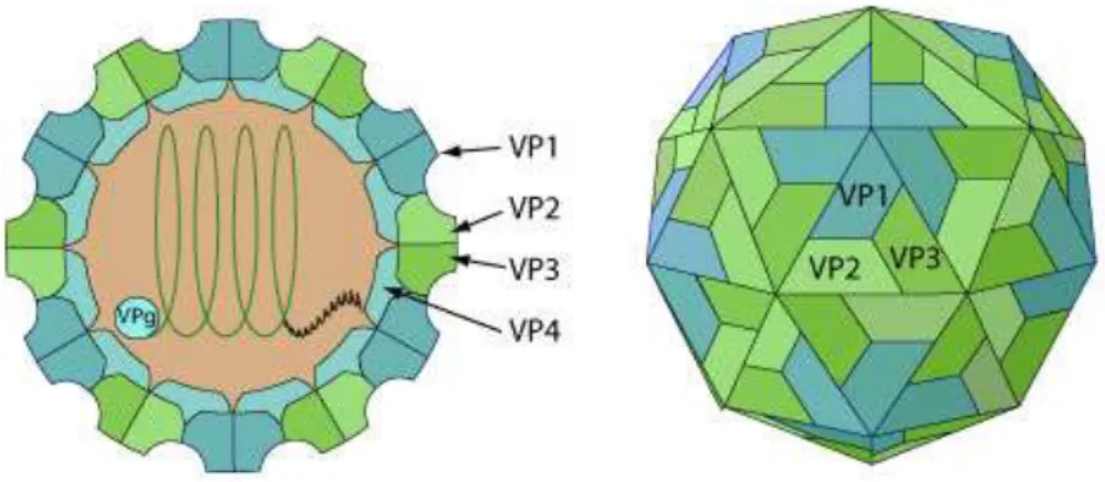
In common with other picornaviruses, FMDV is non-enveloped and has a roughly spherical capsid, exhibiting icosahedral symmetry. The virion has a diameter of 22 -25 nm and it consists of approximately 70 per cent protein and 30 per cent RNA (Cooper et al., 1978; Melnick et al., 1974). It has a molecular mass of about 8.5 × 10⁶ D with a sedimentation constant of 146S (Rueckert,1996).This characteristicsedimentation rate in sucrose gradients is widely
32
used in vaccine production to determine the mass of intact virions present in culture harvests because disintegration of virus particles results in loss of immunogenicity. The capsid consists of 60 capsomers each consisting of four protein (VP1, VP2, VP3 and VP4) (Figure 4). VP1 is the most antigenic protein, is involved in cell attachment and carries an immunological important G-H loop which is one of the most important neutralizing sites of the virus (Logan et
al., 1993).
Figure 4: Diagram of the typical picornavirus icosahedral capsid (Adapted from Arias et al.,
2010)
Source: Viral Zone, 2008 (Swiss Institute of Bioinformatics) Available at http://viralzone.expasy.org/all_by_species/33.html
Legend: Non-enveloped, spherical, about 30 nm in diameter, an icosahedral capsid surrounding
the naked RNA genome. The capsid consists of a densely-packed icosahedral arrangement of 60 protomers, each consisting of 4 polypeptides, VP1, VP2, VP3 and VP4. VP4 is located on the internal side of the capsid.
FMDV exhibits a remarkable resistance to such bactericidal agents as the narcotic solvents (alcohol, ether, chloroform), or such antiseptics as phenol or cresol (Harada et al., 2015; Hong et al., 2015), although, two percent solutions of NaOH or KOH and 4% Na2Co3 are effective disinfectants for FMD contaminated objects (Harada et al., 2015; Hong et al., 2015). On the other hand, in acidic conditions the FMDV particles are disrupted into pentameric subunits composed of five copies each of the virus structural capsid proteins (VP1-3) with the liberation of the internal capsid protein (VP4) and the RNA (Hong et al., 2015; Newman et al., 1973). The most important difference between the physicochemical properties of viruses within the Picornaviridae family is their pH stability (Pereira, 1981). FMDV is stable between pH 7
33
and 9 at 4°C and -20°C. However, in milk and milk products, the virion is protected, and can survive at 70° C for 15 seconds and pH 4.6. In meat, the virus can survive for long periods in chilled or frozen bone marrow and lymph nodes (Mckercher & Callis, 1983). The size of droplet aerosol also plays a role in the survival or drying out of the virus. Indeed, a droplet aerosol size of 0.5 – 0.7 µm is optimal for longer survival of the virus in the air while smaller aerosols dry out. Moreover, in dry conditions the virus also survives longer in proteins, for example in epithelial fragments (Donaldson, 1987; Donaldson et al., 1987; Sellers et al., 1983).
1.1.3.3 Genome organisation
FMDV genome consists of a positive sense single stranded RNA molecule, of approximately 8500 nucleotides in length, and comprises a 5' non-coding region (NCR), a single large open reading frame (ORF) and a short 3' NCR (Belsham, 1993). The 5' NCR is exceptionally long (about 1300 nt) and has a virus encoded protein, 3B, called virus protein genome (VPg) attached to the 5’ end (Figure 5a). The first portion of the 5' NCR is termed the S fragment and is approximately 400 nt long. This is followed by the poly C tract, a homopolymeric tract of predominantly cytidyl residues which is 150-250 nt long and which only occurs in cardioviruses and aphthoviruses within the Picornaviridae family (Rueckert & Wimmer, 1984). The last region of approximately 720 nt contains inverted repeats which are predicted to form pseudo-knots (Clarke et al., 1987). The internal ribosome entry site (IRES) which is immediately upstream of the first AUG initiation codon and is approximately 435 nt in length also occurs within this region (Belsham & Brangwyn, 1990; Ohlmann & Jackson, 1999). The main portion of the virus genome is a single very large open reading frame of 6996 nucleotides encoding a polyprotein of 2332 amino acids (for serotype O) (Forss et al., 1984). Four polyproteins (L1, P1, P2 and P3) are translated and processed into the different structural and non-structural proteins by viral encoded proteases (Lpro, 2A, oligopeptide and 3Cpro) (Rueckert, 1996) (Figure 5b). The L protein represents the leader protein, where two initiations sites (AUG codons) have been identified in FMDV, namely Lab and Lb (Burroughs et al., 1984; Sangar et al., 1988). The P1 gene product is the precursor of the capsid proteins 1A, 1B, 1C and 1D (also known as VP4, VP2, VP3 and VP1 respectively) (Figure 5b). Firstly, the intermediate P1 precursor is processed with the help of viral proteinase 3Cpro to produce VP0, VP1 and VP3 where the products combine to form empty capsid particles. The mature virion is produced after the encapsidation of the virion RNA which is accompanied by the cleavage of VP0 to VP2 and VP4. VP1, VP2 and VP3 are exposed on the capsid surface (Acharya et al.,
34
1989). The P2 (2A, 2B, 2C) and P3 (3A, 3B, 3C, 3D) regions encode for non-structural proteins that are involved in viral RNA replication and protein processing (Belsham, 1993).
Figure 5a: Schematic representation of the FMDV genome (Adapted from Arias et al., 2010)
Figure 5b: Diagram of general structure of picornavirus with cleavage sites of the
35 1.1.3.4 Genetic variation
1.1.3.4.1 Mutations
As mentioned above, FMDV exists in seven distinct serotypes which can be further subdivided into a great number of subtypes. This diversity is expressed mainly in the structural genes leading to more than 30% amino acid exchanges in the capsid proteins between serotypes, whereas the non-structural proteins differ by 2–7% (Domingo et al., 2003). The viruses are subjected to a high genetic drift with a mutation rate of up to 3% base exchanges per year in the structural genes (Beck & Strohmaier, 1987; Beck, 1988). Due to the absence of proofreading-repair activity by the viral replicase (lack of replication error checking mechanisms), FMDV RNA genome replication is highly error-prone (Holland et al., 1982). The high mutation rates result in populations that consist of genetically related but non-identical viruses known as quasispecies. Studies revealed that the rates of mutations of the European serotype FMDV RNA genome can reach 10-2 substitutions per nucleotide site per year (s/n/y) (Gebauer et al., 1988). Similar studies conducted on SAT 1 and SAT 2 FMDV have estimated nucleotide changes of 1.64 % and 1.54 %, respectively per year for the VP1 gene (Vosloo et al., 1996). Moreover, it was estimated that a mutation rate of up to 10-8 – 10-9 nucleotide substitution per year during an epizootiological cycle of FMDV can occur (Domingo et al., 1990). Several in vivo experiments report the generation of highly variable FMD viruses from single animals during infection studies. These observations may have been influenced by molecular host factors and/or selective pressures indirectly incurred from laboratories methodologies (Carrillo et al., 1998; Martinez et al., 1988). Recently, a study conducted during the UK 2001 epidemic demonstrated that nucleotide changes occur throughout the genome at a rate of 2.26 x 10⁻⁵ nucleotide substitutions per site per day. Hence, data obtained from outbreaks like the 2001 epidemic support the experimental observations, demonstrating the role of host-related selective pressures on the variability and evolution of FMDV (Cottam et al., 2006). Comparative genomics studies using full-length sequences representative of all seven serotypes have identified highly conserved genomic regions, indicating functional constraints for variability as well as undefined motifs with likely biological significance (Carrillo et al., 2005). At least 64% of all nt sites within the FMDV genome are susceptible to substitution, including compensatory substitutions. It is important to clarify that most of the “variant” or substitutable residues within the FMDV genome mutate in response to detrimental effects produced by mutations elsewhere in the genome (Carrillo, 2012). Therefore, new variants of FMDV are
36
continuously arising after each replication cycle. The generation of new variants is considered as one of the major problems in the control of FMD by vaccination.
1.1.3.4.2 Recombination
Recombination is another important process driving viral biology and evolution. In RNA viruses, recombination involves the exchange of genetic material between two non-segmented RNA genomes resulting from polymerase ‘jumping’ during RNA synthesis. Consequently, the generation of new antigenic variants may escape immune pressure (King et
al., 1982). Mutations through recombination were first reported in picornaviruses following the
replication of a mixture of mutants in the same cell monolayer (Domingo et al., 2012; Hirst, 1962). Since then, it has been shown that genetic recombination occurs between viruses of the same serotype (King et al., 1985; Pringle, 1965) as well as between serotypes (Chitray et al., 2014; Haydon et al., 2001). For example, recombination has been demonstrated between serotypes O and C (Krebs & Marquardt, 1992), and relatively recent reports document the occurrence of inter-serotypic recombination between serotypes A and Asia 1, resulting in altered antigenic characteristics (Jamal et al., 2011). Intratypic recombination occurs more frequently than intertypic recombination and it appears that recombination events in FMDV occur more readily in the 3' half of the genome, than in the capsid genes of FMDV (Domingo
et al., 1995; King et al., 1985). It was also shown that recombination can involve single or
multiple crossover events when two viruses of the same serotype co-infect cell cultures (King
et al., 1982). Although recombination is not frequent in most RNA viruses, for FMDV, this
phenomenon poses a real threat when attenuated vaccines are used, as reversion to virulence following natural infection of a vaccinated individual is likely given the high recombination frequency in FMDV.
1.1.3.5 Antigenic variation
The concept of antigenic variation derived from the observation of Vallée & Carré in 1922 that an animal that has recovered from FMDV infection can be re-infected and develops clinical signs. The observed genetic variation in the FMD viral genome is the result of a viral evolution process including the replication of viral RNA that is error-prone due to the absence of proofreading in the 3D-encoded RNA-dependent RNA polymerase (Domingo et al., 1990). Hence, antigenic variation can be caused by nucleotide mutations or recombination in the RNA
37
viral genome. One of the consequences of genetic variation through mutation and recombination is that new antigenic variants are constantly being generated as mentioned above. Apart from the non-existence of cross-protection between the 7 FMDV serotypes (Brooksby, 1982) one of the worrying implications of antigenic variation is the fact that vaccination with one antigenic variant of a serotype does not necessarily protect an animal when challenged with a different virus of the same serotype (Cartwright et al., 1982). Among the capsid proteins, VP1 is the most antigenic one and carries the domain mainly responsible for antigenic heterogeneity and cell-virus interaction. The contribution of capsid proteins other than VP1 to the antigenicity of FMDV was demonstrated by many researchers (Barnett et al., 1989; Baxt et al., 1989; Meyer et al., 1997; Meyer et al., 1994; Parry et al., 1989). These independent antigenic sites were identified on the VP2 and VP3 genes. For example, the B-C loop (VP2) was found in serotype A, O and Asia1 (Marquardt et al., 2000; Saiz et al., 1991). However, serological studies and observation in the degree of virulence of the virus in recovered animals have shown that there are significant differences between strains within each serotype (subtypes) (Brooksby, 1982; Grubman & Mason, 2002).
Progress made in the understanding of the genetic differences underlying observed antigenic variation, has played a major role in the epidemiology of FMD. Nowadays, nucleotide sequencing is routinely used to identify the genetic relationships between different isolates and historical strains. However, co-circulation of different types of FMDV is a reality in most parts of the endemic regions which represents a serious complication in the epidemiology of FMDV (Ayelet et al., 2009; Balinda et al., 2010; Ludi et al., 2016; Vosloo et al., 2002a; Wekesa et al., 2015a). Therefore, considering the continual antigenic drift in enzootic situation, vaccine strains selection should be implemented with considerable attention.
1.1.4 Pathogenesis
The pathogenesis of FMD is complex and there is at present many gaps in the level of understanding of this phenomenon (Arzt et al., 2011a; Arzt et al., 2011b). The main route of infection of FMDV in cloven-hoofed animals including ruminants is through the inhalation of droplets, but ingestion of infected feed, inoculation with contaminated vaccines, insemination with contaminated semen, and contact with contaminating clothing, veterinary instruments, etc. can produce FMDV infection (Arzt et al., 2011a; Arzt et al., 2011b; Arzt et al., 2014). However,
38
recent experimental studies have confirmed some aspects of conventional wisdom by demonstrating that pigs are more susceptible to FMDV infection via exposure of the upper gastrointestinal tract (oropharynx) than through inhalation of virus (Stenfeldt et al., 2016a). Three basic phases of FMD pathogenesis in vivo are distinguished: (i) pre-viraemia characterized by infection and replication at the primary replication site(s), (ii) sustained viraemia with generalization and vesiculation at secondary infection sites and (iii) post-viremia/convalescence including resolution of clinical disease that may result in long-term persistent infection.
In cattle, the tissues most consistently infected during the pre-viraemic phase of the disease are the epithelia of the naso-pharynx and larynx (Arzt et al., 2011b). It is therefore likely that this is the primary replication site in ruminants. There is a complex relationship between the tissues of the naso-pharynx and FMDV because not only does initial infection of ruminants take place there but the naso-pharynx is also the site of viral persistence in chronically infected animals (so-called carriers) (Stenfeldt et al., 2016b; Parthiban et al., 2015; Pacheco et al., 2015). Indeed, more than 50% of ruminants that recover from illness and those that are vaccinated and have been exposed to virus can carry virus particles in the naso-pharyngeal region up to 3.5 years in cattle, 9 months in sheep, and more than 5 years in African buffalo (Thomson, 1996).
Vesicle formation, cell lysis and significant inflammation occur at secondary replication sites (oral mucosa, skin of the horn-hoof junction & skin of the teats) but not in the epithelium of the primary replication site. The cells which support viral replication are located in the basal layer of naso-pharyngeal epithelium. However, the mechanism by which viral replication occurs in the naso-pharyngeal epithelium without causing cell lysis is unknown; nor is there an explanation as to why virus can be readily cultured from pharyngeal scrapings (obtained using probing cups) that, in recently infected animals, may contain high levels of antibody (mainly IgA) directed against the infecting virus (Arzt et al., 2011b; Stenfeldt et al., 2015). In pigs, delayed clearance of viral RNA from pharyngeal and lymphoid tissues has been observed but that has not been shown for infectious virus (Arzt et al., 2011a). It is currently concluded that persistent infection of pigs does not occur or at least is not epidemiologically important (Sutmoller & Casas, 2002).
One or two days before the onset of clinical signs, cattle and pigs develop viraemia which may endure for up to 3 days. In summary, at the viraemia stage, FMDV is distributed throughout the body, to reach the best sites of multiplication sites such as the epithelium of
oro-39
pharynx, oral cavity, feet, the udder and heart (Burrows et al., 1981; Zhang & Alexandersen, 2004; Arzt et al., 2010). Virus may also accumulate in the spleen, liver, adrenals, myocardium, pancreas, thyroid and mammary glands. In mammary tissue and myocardium, however, viral replication occurs in secretory epithelial cells of the alveoli and myocytes respectively, resulting in clear microscopic lesions. Development of characteristic vesicular lesions in FMD is dependent on persistent local irritation or friction. In transplantation studies in guinea pigs it was shown that epithelium from predilection sites grafted to other body areas lost that predilection and vice versa (Platt, 1960). This explains why the mouth, feet and teats are predilection sites for the development of lesions and why pigs often develop lesions on the dorsum of the snout, because of “snuffling”.
Viral excretion starts about 24 hours prior to the onset of clinical disease and continues for several days. The acute phase of the disease lasts about one week and viraemia usually declines gradually coinciding with the appearance of strong humoral responses (Murphy et al., 1999). Recovered cattle produce neutralizing antibodies and can resist to re-infection by the same subtype of virus for up to one year. In various parts of the world including South America, East Africa and India/Pakistan, a heat-intolerance syndrome (sometimes referred to as ‘hairy panters’) has been associated with previous infection or ‘chronic FMD’, with a putative endocrine-related pathogenesis. Although, there is still limited information available on this syndrome, Arzt et al., (2011a) have indicated in their review that the extent of the syndrome’s association with FMD remains speculative.
1.2 Clinical signs and pathology 1.2.1 Clinical signs
The incubation period of an infectious disease is defined as the time interval between exposure to an infective dose and first appearance of clinical signs (OIE, 2016). When susceptible animals are in contact with clinically infected animals, clinical signs usually develop in 3 to 5 day (Kitching & Hughes, 2002; Kitching, 2002). However, the incubation period of FMD is variable and depends on the host (age, breed, species and degree of immunity), environment, route of exposure, exposure dose, husbandry conditions and virus strain. Hence, it was estimated that after infection with FMDV, the average incubation period for sheep and goats is 3 to 8 days, at least 2 or more days for pigs, and 2 to 14 days in cattle
40
(Gailiunas & Cottral, 1966; Grubman & Baxt, 2004; Hugh-Jones & Tinline, 1976). The incubation period can be as short as 18 hours for host-adapted strains in pigs, especially under intense direct contact (Kitching & Alexandersen, 2002). The signs can range from a mild or unapparent disease in sheep or goats to a severe one occurring in cattle or pigs (OIE, 2016).
In cattle, following an initial pyrexia around 40°C, lasting one or two days, a variable number of vesicles develop on the tongue, hard palate, dental pad, lips, gums, muzzle, coronary band and interdigital space (Brooksby, 1982; Kitching, 2002; Woodbury, 1995). However, mouth lesions are less common and less pronounced in other species such as sheep and pigs. Vesicles may also be seen on the teats, particularly of lactating cows. Young calves may die before the appearance of vesicles because of the predilection of the virus to invade and destroy cells of the developing heart muscle (Kitching, 2002). Once infection is established within cattle herds, morbidity can approach 100% (Salt et al., 1996; Woodbury, 1995). A chronic panting syndrome characterized by dyspnoea, anaemia, hair overgrowth, and lack of heat tolerance has been reported as a sequela in cattle (Kitching, 2002). Additionally, it has been shown that in cattle, pregnant cows may abort (Radostits et al., 2006).
In sheep and goats, if the clinical signs occur, it tends to be very mild, and may include dullness, fever; and small vesicles or erosions on the dental pad, lips, gums, and tongue. Commonly in sheep and other small ruminant lesions occur where (usually on the dental pad) they may be difficult to detect (Coetzer et al., 1994; Geering, 1967). Mild lameness may be the only sign. In lame animals, there may be vesicles or erosion on the coronary band or in the interdigital space. Infected nursing lambs may die without showing any clinical sign (Kitching & Hughes, 2002).Abortion may result from infection with FMDV and is thought to occur more frequently in sheep than other species (Arzt et al., 2011a).
Infected pigs initially show mild signs of lameness, blanching of the skin around the coronary bands and may develop a fever of up to 42°C but most often, this is in the range of 39°C to 40°C (Kitching & Alexandersen, 2002). The fever is most often associated with anorexia, reluctance to move, and squeal when forced to move. These signs are followed by vesicles on the coronary band, vesicles on the heals, vesicles in the interdigital space (foot involvement is usually severe), and vesicles on the snout. Mouth lesions are not too common and when they occur are smaller and of shorter duration than in cattle and tend to be a
"dry"-41
type lesion; there is no drooling; sows may abort; and piglets may die without showing any clinical sign (Coetzer et al., 1994; Kitching & Alexandersen, 2002; Radostits et al., 2006).
1.2.2 Pathology
FMDV replicates at the site of entry, either in mucosa and lymphoid tissue of the upper respiratory tract or in the dermal and subdermal tissue of a skin abrasion (Kitching, 1992). The virus enters the blood circulation as free virus or associated with mononuclear cells and is distributed around the body to glandular tissue and predilection sites in the stratum spinosum, where secondary replication occurs. The cells of the stratum spinosum undergo ballooning degeneration and as the cells rupture and oedema fluid accumulates, vesicles develop which coalesce to form the aphthae and bullae that characterise FMD (Kitching, 1992). The lesions on the dental pad and tongue appear as reddened areas and progress within a few hours into vesicles. The vesicles are easily ruptured within 24 hours leaving a raw surface and healing occurs within one to two weeks of rupture. Lesions at interdigital areas occur and animals can lose their hooves in severe cases (Donaldson et al., 1984; Geering, 1967). There has also been supportive evidence that FMD virus replicates in the bovine mammary gland and mastitis may occur due to secondary bacterial infection. Moreover, histological studies have revealed the presence of clumps of necrotic secretory epithelial cells in the mammary gland alveolar tissue. A week after the onset of the disease in cattle, an increase in the number of alveoli containing necrotic cells, and luminal exocytosis of all alveoli occurs with concomitant increase in non-secretory areas (Blackwell et al., 1983; Kitching, 1992). In young animals, the virus invades the cells of the myocardium and macroscopic grey areas may be observed, particularly in the wall of the left ventricle, which appears striped (tiger heart). Cells of the skeletal muscle may also undergo hyaline degeneration (Blackwell et al., 1983).
1.3 Epidemiology
Considering the following definition of epidemiology as “study of the frequency and distribution of diseases over time and space, and the role of factors that determine this frequency and distribution within a population at risk” (adapted from Toma et al., 1996), in this section devoted to the epidemiology of FMD, an overview will be given of susceptible hosts, source of infection and mode of transmission, global distribution, serotype diversity and their distribution in Africa. In addition, two important questions related to FMDV transmission will be tentatively
42
clarified. These questions are: (i) what are carriers and how do they contribute to FMDV transmission? and (ii) what is the role of wildlife in FMDV? Lastly, in this section, an overview of epidemiological modelling and statistics used in the thesis, and molecular epidemiology will be briefly presented.
1.3.1 Susceptible hosts
FMDV has a wide host range and can affects over 70 species of both domestic and wild cloven-hoofed animals. Although, not all FMDV have the same host range (Saiz et al., 2002) the most sensitive species belong to the mammalian order of Artiodactyls. Of the domesticated species, cattle, pigs, sheep, goats and water buffalo are susceptible to FMD. The Bactrian camel (two-humped camel) is susceptible to FMD and develops severe lesions, while the dromedary camel (one-humped camel) is apparently resistant to infection. Lamas and alpacas have a high natural resistance to infection. Some will develop mild clinical signs following direct contact with infected cattle, but will not transmit FMD to other camelids under field conditions. Horses are not cloven hoofed and are therefore resistant. Similarly, many species of wildlife, such as African buffalo (Syncerus caffer), bison (Bison spp.), moose (Alces alces), chamois (Rupicapra
rupicapra), giraffe (Giraffa camelopardalis), wildebeest (Connochaetes gnou), blackbuck
(Antilopa cervicapra), warthogs (Phacochoerus aethiopicus), kudu (Tragelaphus
strepsicornis), impala (Aepyceros melampus), and several species of deer, antelopes and
gazelles may become infected with FMDV. Several clinical cases have been reported in captive Asian elephants (Elephas maximus), but there are few reports of FMDV in African elephants (Loxodonta africana), and the latter species is not considered susceptible under natural conditions in southern Africa (Anderson et al., 1993; Ayebazibwe et al., 2010; Bronsvoort et
al., 2008; Bruckner et al., 2002; Thomson, 1995; Thomson et al., 2003; Thomson et al., 2013;
Vosloo et al., 1996; Vosloo et al., 2002; Ward et al., 2007; Weaver et al., 2013).The receptivity of hippopotamus (Hippopotamus amphibious) to FMDV has not yet been reported through seroprevalence in wildlife species (Di Nardo et al., 2015; Thomson et al., 2003).
FMD is not a zoonosis, and only a few possible cases of infection of humans have been described (Bauer, 1997; Berrios, 2007; Capella, 2001; Simmons & Feldman, 2001) and where infection of humans with FMDV does occur the results have only mild and transient consequences (Bauer, 1997). Therefore, human infection does not appear to have any