HAL Id: dumas-02069704
https://dumas.ccsd.cnrs.fr/dumas-02069704
Submitted on 15 Mar 2019Caractérisation de l’atteinte cardiaque de la maladie de
Fabry selon le strain longitudinal, les rehaussements
tardifs et le T1 mapping : relation avec le statut
fonctionnel, et prédiction des évènements
cardiovasculaires
Émilie Testet
To cite this version:
Émilie Testet. Caractérisation de l’atteinte cardiaque de la maladie de Fabry selon le strain longitudi-nal, les rehaussements tardifs et le T1 mapping : relation avec le statut fonctionnel, et prédiction des évènements cardiovasculaires. Médecine humaine et pathologie. 2018. �dumas-02069704�
HAL Id: dumas-02069704
https://dumas.ccsd.cnrs.fr/dumas-02069704
Submitted on 15 Mar 2019HAL is a multi-disciplinary open access
archive for the deposit and dissemination of sci-entific research documents, whether they are pub-lished or not. The documents may come from teaching and research institutions in France or abroad, or from public or private research centers.
L’archive ouverte pluridisciplinaire HAL, est destinée au dépôt et à la diffusion de documents scientifiques de niveau recherche, publiés ou non, émanant des établissements d’enseignement et de recherche français ou étrangers, des laboratoires publics ou privés.
Caractérisation de l’atteinte cardiaque de la maladie de
Fabry selon le strain longitudinal, les rehaussements
tardifs et le T1 mapping : relation avec le statut
fonctionnel, et prédiction des évènements
cardiovasculaires
Émilie Testet
To cite this version:
Émilie Testet. Caractérisation de l’atteinte cardiaque de la maladie de Fabry selon le strain longitudi-nal, les rehaussements tardifs et le T1 mapping : relation avec le statut fonctionnel, et prédiction des évènements cardiovasculaires. Médecine humaine et pathologie. 2018. <dumas-02069704>
UNIVERSITE DE BORDEAUX U.F.R. DES SCIENCES MEDICALES Année 2018 N° de thèse 3070 Thèse pour l’obtention du
DIPLOME D’ETAT DE DOCTEUR EN MEDECINE
Présentée et soutenue publiquement Le 10 septembre 2018 ParEmilie TESTET
Née le 9 septembre 1990 à Agen (47)CARACTERISATION DE L’ATTEINTE CARDIAQUE DE LA
MALADIE DE FABRY SELON LE STRAIN LONGITUDINAL,
LES REHAUSSEMENTS TARDIFS ET LE T1 MAPPING :
RELATION AVEC LE STATUT FONCTIONNEL,
ET PREDICTION DES EVENEMENTS CARDIOVASCULAIRES
Directrice de thèse Madame le Docteur Patricia Réant JURYRESUME
CONTEXTE : La Maladie de Fabry est une maladie de surcharge lipidique liée à l’X dans laquelle
l’atteinte cardiaque est une cause majeure de morbimortalité. La relation entre les paramètres échographiques et d’imagerie par résonance magnétique (IRM) innovants et les paramètres d’épreuve d’effort, ainsi que leur valeur pronostique, n’ont à ce jour pas été évalués.
OBJECTIF : Nous avons évalué la relation entre les paramètres d’imagerie cardiaque (masse
ventriculaire gauche, strain longitudinal (SL), T1 natif et réhaussements tardifs) et d’une part les paramètres de VO2 à l’effort ; d’autre part la survenue d’évènements cardiovasculaires durant le suivi. METHODES : Dans cette étude prospective, monocentrique, tous les patients atteints de Maladie de Fabry référés auprès de notre centre tertiaire ont bénéficié d’un électrocardiogramme (ECG), d’un Holter ECG, d’une échocardiographie, d’une IRM, et d’une épreuve d’effort avec VO2 et ont été comparés à des témoins sains. RESULTATS : De juin 2016 à novembre 2017, nous avons inclus 31 patients atteints de maladie de Fabry (âge moyen 42 ± 16 ans, 39% d’hommes) ayant un degré moyen de sévérité de la maladie (selon le MMSI), que nous avons comparé à 14 témoins appariés. Treize patients (42%) avaient une hypertrophie ventriculaire gauche (>11.0 mm). Le T1 moyen était significativement plus faible chez les patients par rapport aux témoins (934 ± 60 vs. 999 ± 45 ms, p<0.05). Une bonne corrélation était observée entre le pic de VO2 et le SL transmural (r=-0.55) et endocardique (r=-0.53). La pente VE/VCO2 était corrélée à l’épaisseur ventriculaire gauche (r=0.48), et au T1 moyen (r=-0.57). Durant un suivi moyen de 23 mois, 6 patients ont présenté un événement cardiovasculaire. En analyse univariée, le SL transmural global et plus spécifiquement le SL transmural septal basal ; le SL épicardique global et le SL épicardique antérolatéral basal et moyen ; et le T1 moyen étaient significativement prédicteurs d’événement cardiovasculaire (p< 0.05). CONCLUSION : Notre étude suggère que le strain longitudinal et le T1 mapping sont utiles dans la prédiction du statut fonctionnel et des évènements cardiovasculaires chez les patients atteints de Maladie de Fabry. _____________________________________________________________________________________________________________________ DISCIPLINE : CARDIOLOGIE ____________________________________________________________________________________________________________________
MOTS CLES : Maladie de Fabry · Echocardiographie · Strain longitudinal · Strain multi-couches ·
Imagerie par résonnance magnétique · T1 mapping · Réhaussements tardifs · Prognostic
_____________________________________________________________________________________________________________________
Serment d’Hippocrate
Au moment d’être admise à exercer la médecine, je promets et je jure d’être fidèle aux lois de l’honneur et de la probité. Mon premier souci sera de rétablir, de préserver ou de promouvoir la santé dans tous ses éléments, physiques et mentaux, individuels et sociaux.Je respecterai toutes les personnes, leur autonomie et leur volonté, sans aucune discrimination selon leur état ou leurs convictions. J’interviendrai pour les protéger si elles sont affaiblies, vulnérables ou menacées dans leur intégrité ou leur dignité. Même sous la contrainte, je ne ferai pas usage de mes connaissances contre les lois de l’humanité. J’informerai les patients des décisions envisagées, de leurs raisons et de leurs conséquences. Je ne tromperai jamais leur confiance et n’exploiterai pas le pouvoir hérité des circonstances pour forcer les consciences. Je donnerai mes soins à l’indigent et à quiconque me les demandera. Je ne me laisserai pas influencer par la soif du gain ou la recherche de la gloire. Admise dans l’intimité des personnes, je tairai les secrets qui me seront confiés. Reçue à l’intérieur des maisons, je respecterai les secrets des foyers et ma conduite ne servira pas à corrompre les moeurs. Je ferai tout pour soulager les souffrances. Je ne prolongerai pas abusivement les agonies. Je ne provoquerai jamais la mort délibérément.
Je préserverai l’indépendance nécessaire à l’accomplissement de ma mission. Je n’entreprendrai rien qui dépasse mes compétences. Je les entretiendrai et les perfectionnerai pour assurer au mieux les services qui me seront demandés.
Remerciements
Au Professeur Didier LACOMBE
Je vous remercie de me faire l’honneur de présider cette thèse. Veuillez trouver ici le témoignage de ma gratitude et de mon profond respect. Au Professeur Jean-Benoît THAMBO Je vous remercie d’avoir accepté de prendre part à mon jury de thèse, mais également d’avoir été un chef de service aussi bienveillant lors de mes semestres dans votre service. Merci d’avoir eu confiance en moi et pour vos précieux conseils notamment dans mes choix d’avenir. Je suis très honorée de faire partie de votre équipe et d’intégrer un service aussi dynamique et compétent pour le post-internat. Au Docteur Patricia REANT Je te remercie d’avoir accepté de diriger ma thèse. Merci pour tes remarques et tes conseils toujours très avisés. J’admire ta rigueur et ton sens du travail. Au Docteur Xavier IRIART… Je suis ravie que tu aies accepté d’être mon rapporteur de thèse et de prendre part à ce jury. Je te remercie pour tout ce que tu m’as apporté en cardiopathie congénitale notamment en clinique et en imagerie. Merci de m’avoir fait confiance et de m’avoir encouragée à faire ce post-internat, qui j’en suis sûre va me passionner. Je suis enchantée de pouvoir poursuivre ma formation à tes côtés. Au Docteur Caroline ROORYCK-THAMBO C’est un honneur pour moi que vous ayez accepté de prendre part à ce jury, dans une spécialité différente de la vôtre. Veuillez recevoir mes remerciements les plus sincères. Au Docteur Hubert COCHET Je vous remercie d’avoir accepté de participer à mon jury de thèse. Merci de m’avoir montré les techniques d’analyse d’IRM dont j’avais besoin, et pour vos précieux conseils pour mon travail.
A l’équipe médicale de cardiopédiatrie, Zak bien sûr, pour ta bonne humeur et ton humour au quotidien, au KT et autres, pour toutes tes explications sur l’hémodynamique et ta patience quand au début je n’y comprenais pas grand-chose. Merci de m’avoir fait confiance jusque-là, j’espère que je ne te décevrai pas. A PES pour ta patience devant mon ignorance en stats pour le mémoire ; et aussi bien sûr à Jean-Baba et Julie. Je suis ravie de pouvoir travailler à vos côtés. A mes chefs de clinique, Cécile V qui a accompagné nos premiers pas maladroits d’internes, ce bon radio Poustis, la géniale Manu, et la team de rêve de rythmo, Rémi et Nicolas W, mais aussi Nico K et Josselin. Je remercie aussi ce bon vieux Xavier Z, Benjamin, Marina, et les équipes médicales des autres services dans lesquels j’ai travaillé. Merci pour tout ce que vous m’avez apporté. Aux équipes paramédicales, notamment à la super team « soirées » des USIC ; à l’équipe de cardiopéd, notamment Lolo qui m’a déjà supportée 1 an sans presque râler, Mag, Océane, … et aussi aux filles des échos : Lise, Cathy, Aurélie et Nath ; à mes amies Montoises Loren et Manue. A mes co-internes, mes supers co-internes de promo : Marie, Jean, Hugo et Romain ; et bien entendu Sofian, Laure et Aimée, longue vie à notre amitié et à nos week-ends bateau, ski et compagnie. A Margot aussi, une co-interne de folie, que je remercie d’avoir pris le temps de relire ma thèse, et aussi d’avoir contribué à achever plus vite ce bon vieux catamaran (ou pas !). A mes co-internes de cardiopéd, Marie-Chanchan pour nos soirées jusqu’au bout de la nuit, Anaïs pour les journées shopping au lieu de bosser, Camille et Soaz toujours au rendez-vous pour sortir et rigoler. A Esti, Mansour et Reda, ainsi qu’à ma future co-chef Marie-Lou, avec qui je suis ravie de bosser ces deux prochaines années. A Alex aussi bien sûr, co-interne toujours au top dans la difficile épreuve du sous-effectif. A mes vieux co-internes des échos, Cécile, Noumer et Elie, qui sont devenus de supers chefs de clinique. A Coralie bien sûr, à ta patience et ton calme à écouter Marie et moi râler en continu.
Aux amis de la fac, à Guillaume pour ton amitié qui m’est chère et nos retrouvailles à chaque fois que tu reviens de ton grand Nord ; et bien sûr à Alice, Sido, Romain, Fab, Math, Tibo, Inès, Marion, Alex, Olive, Camille, JF, Pauline, Hélène, et puis petit et grand aussi… Merci pour tous nos grands moments d’amitié partagée, nos soirées bordelaises, nos week-ends parfois un peu idiots mais tellement géniaux, nos voyages à l’autre bout du monde, nos mémorables week-end entres filles aussi (notamment ceux dont on n’est pas surs de « redescendre » un jour)… et je sais qu’ils vont être encore nombreux ! A Quentin, Henri, Camille G et Pierre S ; à Maylis qui a été mon super binôme pendant les années d’externat. A mes amis du lycée, surtout Flo, Didie et Brou, la distance nous sépare mais nos retrouvailles sont toujours un grand plaisir. A tous mes amis monos de JEM, avec qui je passais tous mes étés sur l’eau dans l’un des plus beaux endroits du monde en Bretagne, Marie dont l’amitié m’est très chère, et Seb pour la team croisière, mais aussi Jansu, Bruno, Arthur, Polo, Pierrot, Juliette et tous les autres. A ma meilleure amie depuis toujours, Amélie, dont l’amitié dépasse les frontières du bout du
monde, nos retrouvailles sont toujours exceptionnelles. A tout ce que nous avons partagé toutes les deux. Et bien sûr à Marielle, Régis, Alex, Thib et Louis, toujours prêts à m’accueillir pour des vacances de folie à Manteau-Bleu.
A ma famille, Mamiré pour ton soutien sans faille, Papiro et Papijean, qui me manquent
beaucoup évidemment, David, Valérie, Valentine et Nono ; et aussi Dominique et Chloé. A mes parents, sans qui rien de tout ça n’aurait été possible, merci pour le soutien infaillible que vous m’avez apporté depuis le début de ces longues et parfois difficiles études ; et pour tout l’amour dont vous m’avez entourée ; merci d’avoir permis la concrétisation de mon rêve de petite fille ; à mon frère Vincent avec qui je partage beaucoup de choses et notamment la passion de la voile ; et dont je suis fière du parcours. Je vous aime fort.
Table of contents
Abstract ……….………..………....8 Introduction ...………..………....10 Methods.………..………12 Results.
………..………..
16 Baseline participant characteristics ……….………..……….
16 Exercise, electrical and biological status ………………..…...……
16 Echocardiography………...
16 Cardiovascular magnetic resonance imaging ………...……….……...…
21 Correlations……….…
22 Prognosis analysis……….…….………
22 Discussion ……….……..…....……..26 Echocardiographic and CMR characterization of FD patients………..……….……..
26Relation between echocardiographic and CMR parameters and functional status in metabolic exercise test
………...……….………..
27Prognosis factors
……….……….….…………..
28Considerations about localization of LVH and LGE
………..………..
29Considerations about normalized or elevated T1 value in most hypertrophied areas or with fibrosis in FD
………..…..
30Study limitations
………...…………..
31Conclusions………..………..31
Characterization of Fabry Disease cardiac involvement according to
longitudinal strain, late gadolinium enhancement, and T1 mapping:
relation to functional status, and prediction of cardiovascular
outcomes
ABSTRACT BACKGROUNG: Anderson-Fabry disease (FD) is a rare X-linked glycolipid storage disease inwhich cardiac involvement is a leading cause of morbimortality. Neither relation between echocardiographic and cardiovascular magnetic resonance (CMR) innovative parameters, and exercise testing parameters, nor their prognosis value, have been evaluated yet.
AIMS: We sought to evaluate the relation between cardiac imaging parameters (i.e. left
ventricular (LV) mass, multi-layer longitudinal strain (LS), native T1 and late gadolinium enhancements) and on one hand cardiopulmonary exercise capacity in FD patients; and on the other hand, cardiovascular (CV) outcomes during follow-up. METHODS: In this prospective, monocentric study, we evaluated all consecutive FD patients referred in our tertiary center, performing electrocardiogram (ECG), Holter ECG monitoring, echocardiography, CMR, and cardiopulmonary exercise testing, that we compared with healthy controls. RESULTS: From June 2016 to November 2017, we included 31 FD patients (mean age 42 ± 16 years, 39% male) with an overall mild degree of disease severity, that were compared with 14 matched healthy controls. Thirteen FD patients (42%) had left ventricular hypertrophy (LVH) (wall thickness >11.0 mm). Mean T1 in FD patients was significantly lower than in controls (934 ± 60 vs. 999 ± 45 ms, p<0.05). Regarding functional capacity, a good correlation was observed between VO2 peak and global (r=-0.55) and endocardial (-0.53) LS. VE/VCO2 slope was correlated with maximal LV wall thickness (r=0.48), and mean T1 value (r=-0.57). During a median follow-up of 23 months, 6 patients presented CV events. In the Cox univariate analyses, global transmural LS and more specifically basal septal LS; epicardial global LS, and particularly basal and mid anterolateral LS; and anterior T1 value were significantly predictive of CV event (p< 0.05).
CONCLUSIONS: Our study suggests that longitudinal strain and T1 mapping are relevant for prediction of functional status and cardiovascular events in FD patients. Key words: Fabry disease · Echocardiography · Longitudinal strain · Multi-layer longitudinal strain · Cardiac magnetic resonance · T1 mapping · Late gadolinium enhancement · Prognosis Abbreviations list: CMR: Cardiac magnetic resonance CV: Cardiovascular ECG: Electrocardiography ECV: Extracellular volume ERT: Enzyme replacement therapy FD: Fabry disease HR: Hazard ratio LGE: Late gadolinium enhancement LS: Longitudinal strain LV: Left ventricle LVH: Left ventricular hypertrophy MMSI: Mainz Severity Score Index NYHA: New York Heart Association RVH: Right ventricular hypertrophy
Introduction
Anderson-Fabry disease (FD) is a X-linked glycolipid storage disease caused by a deficiency of a-galactosidase A enzyme due to a mutation in the galactosidase alpha gene1,2. The prevalence of FD is historically estimated between 1 in 17,000 to 1 in 117,000 individuals3,4. but it is probably significantly underestimated. The deficiency in the enzymatic activity leads to progressive lysosomal accumulation of complex sphingolipids in vascular endothelial and smooth-muscle cells throughout the body, particularly in the skin, kidney, nervous system, eyes, and heart5,6.
Cardiac involvement in FD represents a leading and increasing cause of morbidity and mortality7,8 and a major determinant for reduction of life-expectancy in this disease9. Accumulation of sphingolipids in cardiac tissues can progressively induce left ventricular hypertrophy (LVH) and myocardial fibrosis, causing a true infiltrative cardiomyopathy. Its complications are various, and can include heart failure with preserved ejection fraction, valvular regurgitation, conduction system disease, atrial and ventricular arrhythmias, and microvascular angina. A specific treatment exists, enzyme replacement therapy (ERT), consisting in administrating the lacking enzyme to restore its role in complex lipids metabolism10-12. It permits a reduction of glycosphingolipids accumulation in cardiac tissue13, demonstrated by significative decrease of left ventricular mass14. Novel chaperone therapy also appeared more recently15. Echocardiography is a substantial non-invasive technique for evaluation and follow-up of patients with FD. Doppler tissue imaging (DTI) and assessment of myocardial deformation by strain in speckle tracking have been shown to allow the early recognition of intrinsic impairment of respectively myocardial relaxation and systolic function, before LVH even develops17-19. However, multi-layer analysis of speckle-tracking strain is a more recently-developed method enabling evaluation of endocardial and epicardial strain20-22, that has not been evaluated yet in FD. Furthermore, neither the prognosis value of these parameters, nor their relation with cardiopulmonary exercise testing parameters, have been assessed yet.
Currently, cardiovascular magnetic resonance (CMR) imaging represents the leading imaging modality for the detection of cardiac involvement in Fabry disease23, because of its valuable capabilities of tissue characterization. Indeed, late gadolinium enhancement (LGE) sign the presence of focal fibrosis in Fabry disease24-26, and its expansion is a major prognostic determinant for ventricular arrhythmias and cardiac outcomes in this disease 27. More recently, it has been shown that native T1 assessment permits early detection of cardiac involvement in
FD as it reflects sphingolipids accumulation28. However, their ability to predict cardiovascular (CV) outcomes and functional status have not been studied.
We hypothesized that these recently-developed cardiac imaging parameters used to detect cardiac involvement in FD can also predict functional status and CV outcomes of FD patients. The aims of this prospective monocentric study were to evaluate the relation between cardiac imaging parameters (i.e. LV mass, longitudinal strain, native T1 and LGE) and cardiopulmonary exercise capacity; and to evaluate the relation between cardiac imaging parameters and CV outcomes during follow-up.
Methods
All patients with genetically confirmed FD, followed at Bordeaux University Hospital, who were referred to the expert center in hereditary cardiomyopathies at the Cardiologic Hospital Haut-Lévêque (Pessac, France) between June 2016 and November 2017, were included and were prospectively evaluated. They were compared with 14 healthy age-matched and gender-matched controls (healthy volunteers). Information concerning the study and data collection was provided to all patients, and the study protocol was approved by the institutional review board. Patients and healthy subjects were included after providing informed consent. For all patients, previous medical histories and clinical characteristics were obtained from the hospital database. The Mainz Severity Score Index (MSSI) was used to reflect the clinical spectrum of FD29; scores were divided into severity bands of mild (<20), moderate (20–40), and severe (>40) affliction. Participants underwent 12-lead electrocardiography (ECG), 24-hour Holter ECG monitoring, blood biomarker (brain natriuretic peptide) measurement, transthoracic echocardiography, cardiopulmonary exercise testing, and, if possible, CMR.
LVH on ECG was assessed using Sokolow-Lyon and Cornell criteria and was considered positive when the sum of voltage in (SV1 + RV5) or (SV3 + RaVL) exceeded 35 and 28 mm, respectively, in men, or 35 and 20 mm, respectively, in women. A 24-hours Holter ECG monitoring was performed to detect conduction abnormalities and atrial or ventricular arrhythmias. Atrial fibrillation was considered significant when lasting more than 30 seconds. Non-sustained ventricular tachycardia was defined as 3 or more consecutive beats at a rate of at least 100 beats/min and lasting less than 30 s.
Standard Echocardiography Echocardiography was performed using M3S probe, Vivid 9 or E95 (GE® Vingmed Ultrasound AS, Horten, Norway). All images were digitally stored for offline analysis using EchoPAC® version 113.1 (GE Vingmed Ultrasound®). Complete 2D and Doppler images were acquired according to standard techniques30,31, 2D measurements were obtained in conventional views over 3 separate cardiac cycles and were averaged. Maximal left atrial volumes were calculated using the biplane area-length formula and indexed to body surface area32. Maximal LV wall thickness was measured on two-dimensional imaging. LVH in echocardiography was defined by a maximal wall thickness > 11.0 mm33. LV end-systolic and end-diastolic volumes were calculated using
subsequently derived. Evaluation of diastolic function included conventional Doppler-based measurements. First, pulsed-wave Doppler velocities were measured from the apical four-chamber view placing a 2-mm sample volume at the tips of the mitral leaflets. Transmitral E and A diastolic velocities and E-wave deceleration time were measured. Tissue Doppler was applied in the pulsed Doppler mode to record E’ mitral annular velocities at the septal and lateral corners, and the measurements were averaged to calculate mean E/E’ ratio to estimate LV filling pressures34. Severity of mitral regurgitation was determined using an integrative approach including semiquantitative and quantitative color Doppler-based parameters as recommended by current guidelines35. Systolic pulmonary artery pressure was calculated as recommended36. Speckle-Tracking Imaging To study longitudinal LV systolic function, two-dimensional recordings of apical four-chamber, two-chamber, and three-chamber views were obtained using 2D-speckle-tracking imaging at a frame rate > 50 Hz. The endocardial border was manually traced, and the region of interest width was adjusted to include the entire myocardium. The software package (EchoPAC® version 113.1) automatically tracks and accepts segments of good tracking quality, and modifications of the region of interest can be manually modified if necessary to ensure optimal tracking of LV hypertrophic endocardium. Individual global strain curves were obtained automatically from each view. We analyzed transmural, endocardial and epicardial strain separately in the 17 segments. Cardiac Magnetic Resonance Imaging CMR imaging was performed on a 1.5-T scanner (Magnetom Avanto®, Syngo MR B15 version; Siemens Medical Solutions®, Erlangen, Germany). LGE sequences were performed, with images acquired 10–20 min following intravenous administration of 0.2 mmol/kg gadolinium-DTPA with breath-hold 2D segmented recovery sequences or phase-sensitive inversion-recovery sequences in identical planes as in cine images. Regional LGE extent was assessed qualitatively. The presence and regional extent of LGE was assessed by two independent observers. Control subjects, and a patient refusing the injection, did not receive gadolinium.
borders from the base to the apex. Total LV mass was defined including papillary muscles and trabeculations in the myocardial mass. Papillary muscle mass was calculated as previously described37. LVH was defined as an increased LV mass index according to published data of normal limits for age and gender38 or as maximum wall thickness > 11.0 mm in adults. Asymmetric wall thickness was defined as a regional wall thickness of ≥ 13.0 mm in ≥ 1 myocardial segment, in accordance with European guidelines on the diagnosis of hypertrophic cardiomyopathy in patients with genetic disorders39, and segmental end-diastolic wall thickness > 1.3-fold the opposing LV segment. Right ventricular hypertrophy (RVH) was defined by maximal wall thickness > 7 mm.
Before contrast administration, T1 mapping was performed using the modified Look-Locker inversion recovery (MOLLI) sequence in the basal and mid–short-axis planes. Sequences were optimized for a heart rate of 60-80/min using a single-breathhold T1 mapping pulse sequence (8.cm slice thickness) before and after administration of an intravenous bolus of gadolinium-based contrast agent (0.2 mmol/kg of gadoterate meglumine). Structures have been acquired orthogonal to the imaging plane to minimize obliquity. To minimize off resonance, the shim has been properly adjusted. Native and post contrast T1 maps have been acquired using the same slice prescription parameters and the same cardiac phase. The resulting pixel-by-pixel color T1 maps were displayed using a customized 12-bit look-up table, where normal myocardium was green, increasing T1 was red, and decreasing T1 was blue. The color map is visible immediately after data acquisition. For the septal T1, a region of interest in the basal and midseptum was drawn with adequate margins of separation from tissue interfaces prone to partial volume averaging (such as between myocardium and blood pool), and the average T1 was obtained, avoiding areas of LGE. Lateral, anterior and inferior T1 were obtained using the same method. Post-contrast T1 mapping was performed 15 min after gadolinium administration for assessment of extra-cellular volume (ECV) fraction quantification, calculated using hematocrit value measured contemporaneously with the CMR study. Metabolic exercise stress test The metabolic exercise stress test was carried out using bicycle ergometry with computerized respiratory gas exchange measurements, (Ergo Card, Medisoft®, Soriennes, Belgium), under the supervision of a cardiologist, in patients whose general health status allowed testing. Patients peddled for 2 min with zero resistance, after which the bicycle ergometer gradually increased the resistance at a ramp rate of 20, 25 or 30 W/min according to sex and previous performance. With each breath, oxygen uptake (VO2), carbon dioxide production (VCO2) and ventilation rate were
recorded, whereas heart rate, ECG and workload were continuously recorded. Exercise was stopped when patients reached maximal workload (exhaustion), at which point, the VO2 max reading was obtained. The anaerobic threshold was reached when there was a rapid increase in CO2 production. The respiratory exchange ratio (VCO2 /VO2) was measured as an indication of the volume of CO2 produced in relation to the volume of O2 consumed. Increased ventilatory drive was reflected by the increase of the slope of ventilation relative to carbon dioxide production (VE/VCO2 slope). O2 pulse was obtained by dividing the VO2 value by the heart rate obtained every 10 seconds of exercise. Predicted maximum watts, VO2 max, O2 pulse and oxygen uptake at anaerobic threshold adjusted for age and sex were calculated according to Wasserman’s equations40. Follow-up and end-points Long-term follow-up was achieved using regular consultation every 1 or 2 years in our hospital and via patients recalls. The follow-up period for each patient was determined using the time from initial consultation to the last data recorded from the patient. Cardiovascular outcomes included CV death, worsening of NYHA functional class, hospitalization for heart failure, onset of atrial fibrillation or ventricular arrhythmia, and stroke.
Statistical Analysis
Continuous variables were presented as mean ± SD. Categorical data were summarized as frequencies and percentages. To compare continuous variables, we used a Mann-Whitney U test. To compare categorical variables, we used a Pearson Chi-square test. Linear correlations were analyzed using Pearson test. Survival without CV event, defined as the end-point, was determined from inclusion until the first end-point or last follow-up and estimated using Cox proportional univariate hazards regression analyses. Survival without event was shown in Kaplan-Meier curves. p values < 0.05 was considered significant. Software package SPSS® for Windows version 17.0 (SPSS Inc, Chicago, IL, USA) was used for all statistical calculations.
Results
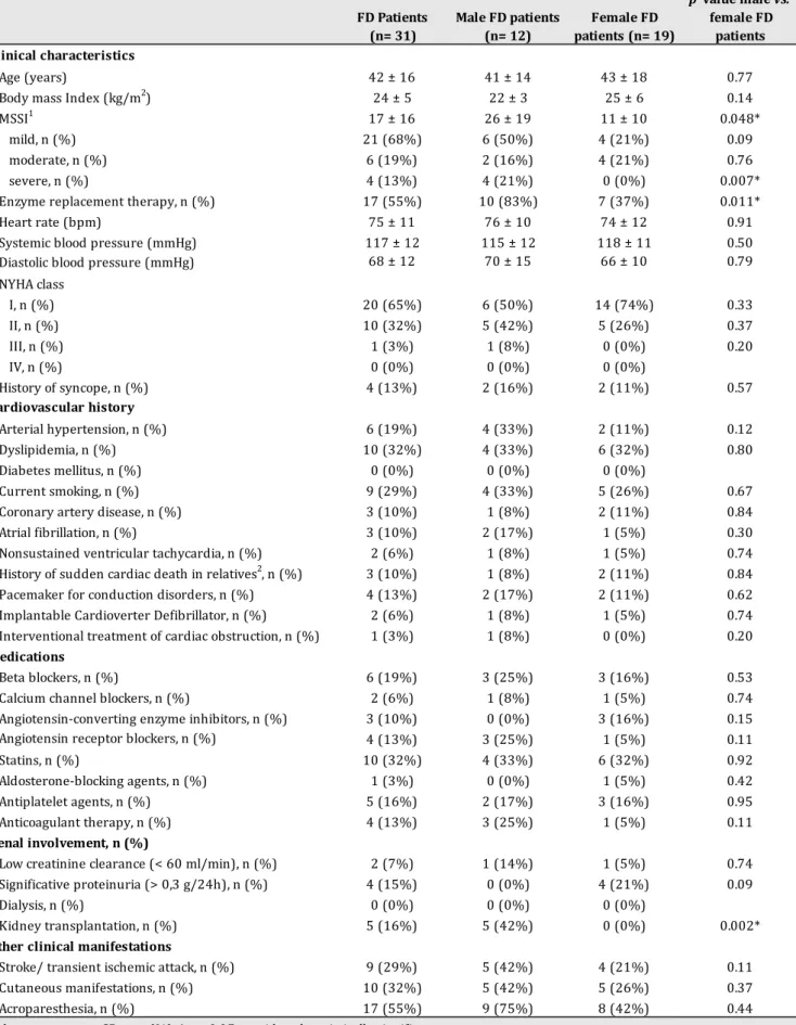
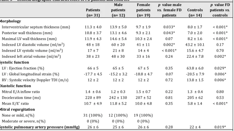
Baseline participant characteristics (Table 1) A total of 31 FD subjects were included in the study (mean age 42 ± 16 years, 39% male), together with 14 matched participants (mean age 42 ± 17 years, p=0.62, 38% male). FD patients had a mean MSSI of 17 ± 16 (range 1-48), reflecting an overall relative mild degree of disease severity. All patients who had a severe degree of disease activity (4 patients) were male. Fifty-five percent of our population was treated by ERT at the time of the inclusion (83% of male patients, 37% of female patients). Six patients had arterial hypertension, well controlled by medical therapy. Three patients had treated coronary artery disease, 3 had known atrial fibrillation. Two patients (1 male, 1 female) had previously had non-sustained ventricular tachycardia, and they both carried an implantable cardioverter defibrillator. Four patients had a pacemaker implanted for conduction disorders. One patient had had an alcohol septal ablation as treatment of cardiac obstruction. Eleven patients (35%) had renal involvement due to the disease, including 5 male patients who underwent a kidney transplantation. Nine patients had a medical history of stroke or transient ischemic attack. Exercise, electrical and biological status (Table 2) Nine patients (32%) had electrical LVH, among which 6 were male. Patients achieving metabolic exercise stress test had a mean performance of 99 ± 27 W (range 50-160), representing 62 ± 24b% of theoretical total wattage. Male performances were significantly poorer than female ones, representing respectively 41 ± 15% vs. 75 ± 20% of theoretical wattage according to sex (p<0.05). Mean VO2 peak was 19.5 ± 4.6 ml/kg/min, which represents respectively 54 ± 10% and 78 ± 28% of theoretical VO2 peak according to sex (p<0.05). Mean oxygen uptake at anaerobic threshold was 13.1 ± 2.7 ml/kg/min, which represents 41 ± 14% of theoretical peak VO2 with a significant difference between male and female patients (32 ± 4 % vs. 47 ± 15 %, p<0.05). Mean CO2 ventilatory efficiency (VE/VCO2 slope) was 34 ± 8. Echocardiography (Table 3 and Figure 1)Mean maximal LV wall thickness at end-diastole was 11.9 ± 4.3 mm in FD patients, and was significantly higher in FD patients than healthy controls (8.0 ± 1.7 mm, p<0.001). Both septal and posterior walls were significantly thicker in male compared to female patients (respectively 13.9 ± 5.0 vs. 9.7 ± 1.9 mm, p<0.05, and 13.1 ± 4.6 vs. 9.3 ± 2.1 mm, p<0.05) and in FD patients compared to controls (p<0.001). Thirteen patients (42%) had echocardiographic LVH. Mean
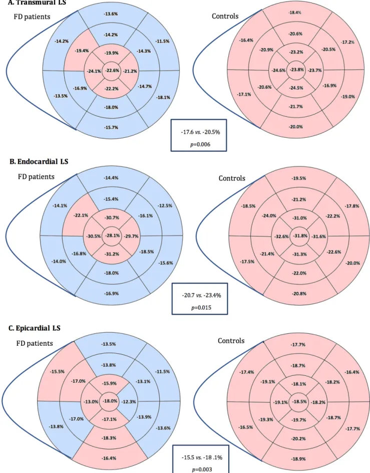
indexed left atrial volume, LV ejection fraction, global longitudinal strain (LS), right ventricle systolic tissue Doppler imaging, E/E’ ratio and systolic pulmonary artery pressure were significantly higher in FD patients than controls (p<0.05). None of our patients encountered impaired LV ejection fraction, mitral regurgitation or elevation of systolic pulmonary artery pressure. Global and regional transmural, endocardial and epicardial LS were represented
Figure 1. Intra- and interobserver mean percentage variability of multi-layer LS analysis had already been assessed: variability was between 3.1 and 6.1 % for global LS, and between 7.8 and 13.0% for regional LS41.
Table 1 Baseline characteristics of FD patients FD Patients (n= 31) Male FD patients (n= 12) Female FD patients (n= 19) p value male vs. female FD patients Clinical characteristics Age (years) 42 ± 16 41 ± 14 43 ± 18 0.77 Body mass Index (kg/m2) 24 ± 5 22 ± 3 25 ± 6 0.14 MSSI1 17 ± 16 26 ± 19 11 ± 10 0.048* mild, n (%) 21 (68%) 6 (50%) 4 (21%) 0.09 moderate, n (%) 6 (19%) 2 (16%) 4 (21%) 0.76 severe, n (%) 4 (13%) 4 (21%) 0 (0%) 0.007* Enzyme replacement therapy, n (%) 17 (55%) 10 (83%) 7 (37%) 0.011* Heart rate (bpm) 75 ± 11 76 ± 10 74 ± 12 0.91 Systemic blood pressure (mmHg) 117 ± 12 115 ± 12 118 ± 11 0.50 Diastolic blood pressure (mmHg) 68 ± 12 70 ± 15 66 ± 10 0.79 NYHA class I, n (%) 20 (65%) 6 (50%) 14 (74%) 0.33 II, n (%) 10 (32%) 5 (42%) 5 (26%) 0.37 III, n (%) 1 (3%) 1 (8%) 0 (0%) 0.20 IV, n (%) 0 (0%) 0 (0%) 0 (0%) History of syncope, n (%) 4 (13%) 2 (16%) 2 (11%) 0.57 Cardiovascular history Arterial hypertension, n (%) 6 (19%) 4 (33%) 2 (11%) 0.12 Dyslipidemia, n (%) 10 (32%) 4 (33%) 6 (32%) 0.80 Diabetes mellitus, n (%) 0 (0%) 0 (0%) 0 (0%) Current smoking, n (%) 9 (29%) 4 (33%) 5 (26%) 0.67 Coronary artery disease, n (%) 3 (10%) 1 (8%) 2 (11%) 0.84 Atrial fibrillation, n (%) 3 (10%) 2 (17%) 1 (5%) 0.30 Nonsustained ventricular tachycardia, n (%) 2 (6%) 1 (8%) 1 (5%) 0.74 History of sudden cardiac death in relatives2, n (%) 3 (10%) 1 (8%) 2 (11%) 0.84 Pacemaker for conduction disorders, n (%) 4 (13%) 2 (17%) 2 (11%) 0.62 Implantable Cardioverter Defibrillator, n (%) 2 (6%) 1 (8%) 1 (5%) 0.74 Interventional treatment of cardiac obstruction, n (%) 1 (3%) 1 (8%) 0 (0%) 0.20 Medications Beta blockers, n (%) 6 (19%) 3 (25%) 3 (16%) 0.53 Calcium channel blockers, n (%) 2 (6%) 1 (8%) 1 (5%) 0.74 Angiotensin-converting enzyme inhibitors, n (%) 3 (10%) 0 (0%) 3 (16%) 0.15 Angiotensin receptor blockers, n (%) 4 (13%) 3 (25%) 1 (5%) 0.11 Statins, n (%) 10 (32%) 4 (33%) 6 (32%) 0.92 Aldosterone-blocking agents, n (%) 1 (3%) 0 (0%) 1 (5%) 0.42 Antiplatelet agents, n (%) 5 (16%) 2 (17%) 3 (16%) 0.95 Anticoagulant therapy, n (%) 4 (13%) 3 (25%) 1 (5%) 0.11 Renal involvement, n (%) Low creatinine clearance (< 60 ml/min), n (%) 2 (7%) 1 (14%) 1 (5%) 0.74 Significative proteinuria (> 0,3 g/24h), n (%) 4 (15%) 0 (0%) 4 (21%) 0.09 Dialysis, n (%) 0 (0%) 0 (0%) 0 (0%) Kidney transplantation, n (%) 5 (16%) 5 (42%) 0 (0%) 0.002* Other clinical manifestations Stroke/ transient ischemic attack, n (%) 9 (29%) 5 (42%) 4 (21%) 0.11 Cutaneous manifestations, n (%) 10 (32%) 5 (42%) 5 (26%) 0.37 Acroparesthesia, n (%) 17 (55%) 9 (75%) 8 (42%) 0.44 Values are mean ± SD or n (%). *p < 0.05 considered statistically significant. 1 scores were divided into severity bands of mild (<20), moderate (20–40), and severe (>40) affliction 2 in ≥ 1 first degree relatives < 40 years or sudden cardiac death in a first degree relative with confirmed hypertrophic cardiomyopathy at any age FD : Fabry disease ; ERT : enzyme replacement therapy ; MMSI : Mainz Severity Score Index ; NYHA : New York Heart Association
Table 2 Electrical, biological and functional status FD Patients (n= 31) Male patients (n= 12) Female patients (n= 19) p value : Males vs. Female patients Electrocardiography First degree atrioventricular block 2 (7%) 1 (10%) 1 (6%) 0.53 Complete Bundle-branch block 2 (7%) 1 (10%) 1 (6%) 0.35 Left ventricular hypertrophy 9 (32%) 6 (60%) 3 (17%) 0.040* 24-Hours holter monitoring Atrial fibrillation 4 (15%) 3 (30%) 1 (6%) 0.09 Nonsustained ventricular tachycardia 3 (11%) 2 (20%) 1 (6%) 0.26 Brain natriuretic peptide (pg/ml) 121 ± 218 209 ± 316 75 ± 135 0.25 Metabolic exercice stress test Total wattage (Watts) 99 ± 27 94 ± 34 102 ± 23 0.44 Total wattage (Mets) 5.5 ± 1.4 5.2 ± 1.6 5.8 ± 1.2 0.22 % Theoretical total wattage 62 ± 24 41 ± 15 75 ± 20 0.002* Systolic blood pressure peak (mmHg) 166 ± 25 167 ± 33 165 ± 19 0.83 Heart rate peak (beats per minute) 146 ± 24 139 ± 21 149 ± 25 0.26 Respiratory exchange ratio (VCO2/VO2) 1.2 ± 0.2 1.2 ± 0.2 1.3 ± 0.1 0.15 VO2 peak (ml/kg/min) 19.5 ± 4.6 19.9 ± 5.5 19.2 ± 4.2 0.70 % Theoretical VO2 peak 68 ± 25 54 ± 10 78 ± 28 0.045* CO2 ventilatory efficiency (VE/VCO2 slope) 34 ± 8 36 ± 10 34 ± 6 0.85 Peak oxygen pulse (VO2/heart rate) (ml/beat/kg) 8.5 ± 1.5 8.9 ± 1.8 8.2 ± 1.2 0.49 % Theoretical peak oxygen pulse 66 ± 27 60 ± 24 70 ± 30 0.57 Oxygen uptake at anaerobic threshold (ml/kg/min) 13.1 ± 2.7 12.6 ± 1.5 13.4 ± 3.3 0.91 % Theoretical oxygen uptake at anaerobic threshold 41 ± 14 32 ± 4 47 ± 15 0.029* Values are mean ± SD or n (%). *p < 0.05 considered statistically significant. FD : Fabry disease Table 3 Echocardiographic characteristics of FD patients and controls Patients (n= 31) Male patients (n= 12) Female patients (n= 19) p value male vs. female FD patients Controls (n= 14) p value FD patients vs. controls Morphology Interventricular septum thickness (mm) 11.3 ± 4.0 13.9 ± 5.0 9.7 ± 1.9 0.033* 8.0 ± 1.7 < 0.001* Posterior wall thickness (mm) 10.8 ± 3.7 13.1 ± 4.6 9.3 ± 2.1 0.043* 7.0 ± 2.0 < 0.001* Maximal LV wall thickness (mm) 11.9 ± 4.3 14.6 ± 5.4 10.3 ± 2.4 0.07 8.2 ± 1.6 < 0.001* Indexed LV diastolic volume (ml/m²) 48 ± 18 60 ± 20 41 ± 11 0.002* 43.2 ± 10.1 0.17 Indexed LV systolic volume (ml/m²) 17 ± 7 21 ± 8 14 ± 4 < 0.001* 15.6 ± 4.7 0.70 Indexed left atrial volume (ml/m²) 38 ± 23 48 ± 30 33 ± 16 0.24 22.4 ± 7.8 0.002* Systolic function LV : Ejection fraction (%) 66 ± 5 65 ± 5 67 ± 5 0.35 63.8 ± 6.0 0.029* LV : Global longitudinal strain (%) -17.7 ± 4.5 -15.2 ± 3.2 -18.8 ± 4.7 0.07 -20.5 ± 7.9 0.006* RV : Systolic velocity Doppler TDI (m/s) 12 ± 2 12 ± 2 12 ± 2 0.72 13.8 ± 1.5 0.006* Diastolic function Mitral E/A inflow ratio 1.4 ± 0.6 1.2 ± 0.3 1.5 ± 0.7 0.22 1.3 ± 0.4 0.80 Deceleration time (ms) 220 ± 89 242 ± 130 207 ± 52 0.81 205 ± 62 0.53
Figure 1 Comparison of transmural, endocardial and epicardial LS between FD patients and controls
Blue segments: significative difference between FD patients and controls LS; Red segments: absence of significative difference between FD patients and controls LS. p value <0.05 was considered significant.
Cardiovascular magnetic resonance imaging (Table 4 and Figure 2) Four patients did not perform CMR, 3 because they carried non CMR compatible pacemaker or defibrillator, and 1 because of claustrophobia; they all had cardiac impairment due to FD. One patient refused gadolinium injection. LV mass index was 94 ± 56 g/m2 and was significantly higher in male than female FD patients (132 ± 73 vs. 71 ± 26 g/m2, p<0.05). Among 9 patients (33%) with LVH, 6 had concentric hypertrophy, 2 had asymmetric septal hypertrophy, 1 had asymmetric inferolateral hypertrophy. Mean septal to lateral ratio in these patients was 1.15. Three patients (11%) had RVH, all were male (p<0.05).
For all patients who had LGE (9 patients, 35%, no significative difference between male and female patients), its localization was mid-wall, and affected basal inferolateral region. Most of the time (6 patients, 67%), it was confined in one segment, and 2 patients had 4 segments with LGE. One male patient had very extensive LGE with 12 affected segments in the anterior, lateral and inferior walls. One female patient had LGE without increased LV mass. Mean native T1 in patients Table 4 CMR characteristics of FD patients and controls FD Patients (n= 27) Male patients (n= 10) Female patients (n= 17) p value male vs. female FD patients Controls (n=11) p value FD patients vs. controls Volumes and function LV end diastolic volume index (ml/m2) 81 ± 5 95 ± 18 72 ± 16 0.005* LV end systolic volume index (ml/m2) 28 ± 10 34 ± 11 24 ± 7 0.004* LV ejection fraction (%) 67 ± 7 68 ± 7 66 ± 7 0.84 Maximal LV diastolic wall diameter 11.8 ± 4.7 15.1 ± 5.9 9.9 ± 2.3 0.024* 8.0 ± 1.1 < 0.001* septal (mm) 11.3 ± 4.7 14.5 ± 6.0 9.4 ± 2.3 0.015* 7.8 ± 0.9 < 0.001* lateral (mm) 10.3 ± 4.0 12.7 ± 5.1 9.0 ± 2.5 0.034* 6.7 ± 1.1 < 0.001* anterior (mm) 9.6 ± 3.8 12.1 ± 4.6 8.1 ± 2.2 0.17 6.9 ± 1.0 < 0.001* posterior (mm) 10.3 ± 4.0 13.2 ± 4.8 8.6 ± 2.0 0.042* 6.4 ± 1.2 < 0.001* LV mass index (g/m2) 94 ± 56 132 ± 73 71 ± 26 0.006* 56 ± 8 < 0.001* LV hypertrophy, n (%) 9 (33%) 6 (60%) 3 (18%) 0.024* 0 (0%) 0.032* Maximal LVH localization, n (% of LVH patients) concentric 6 (67%) 5 (83%) 1 (33%) 0.12 asymmetric septal 2 (22%) 1 (18%) 1 (33%) 0.74 asymmetric inferolateral 1 (11%) 0 (0%) 1 (33%) 0.20 RV hypertrophy, n (%) 3 (11%) 3 (30%) 0 (0%) 0.017* 0 (0%) 0.48 Late gadolinium enhancement, n (%) 9 (35%) 4 (40%) 5 (31%) 0.65 0 (0%) 0.05 Native T1 value (ms) 934 ± 60 899 ± 53 955 ± 53 0.016* 999 ± 45 < 0.001* septal (ms) 931 ± 55 898 ± 53 951 ± 46 0.025* 994 ± 42 < 0.001* lateral (ms) 933 ± 52 903 ± 50 952 ± 45 0.046* 985 ± 40 < 0.001*
with FD was significantly lower than in healthy volunteers (934 ± 60 vs. 999 ± 45 ms, p<0.05). When considering the whole FD group, male mean T1 was significantly lower than female mean T1 (899±53 vs. 955±53 ms, p<0.05) and this significative difference was found separately in the septal, anterior, lateral and inferior walls. Ninety-percent (9/10) of male patients had lower global native T1 than healthy subjects. The only patient with normal T1 was aged seventeen and was the only adolescent of out cohort. Mean T1 was higher in LGE areas than in other zones. LV maximal wall thickness according to mean T1 value is represented Figure 2.
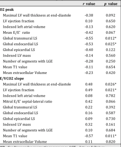
Intra- and interobserver mean percentage variability of native T1 analysis were respectively 3.4% and 2.9% (Appendix 1 and 2). Mean ECV was 24.7 ± 3.0 % in our FD patients, with no significative difference between male and female FD patients (23.9 ± 3.1 % vs. 25.2 ± 2.9 %, p=0.22). Figure 2: LV maximal thickness according to T1 value. LV: Left ventricle Correlations (Table 5) A good inverse correlation was observed between VO2 peak and global (r=-0.55) and endocardial (r=-0.53) LS (p<0.05). A weaker inverse correlation was observed between VO2 peak and epicardial LS (r=-0.40) and mean E/E’ ratio (r=-0.42) as p value was not significant.
VE/VCO2 slope was correlated with maximal LV wall thickness at end-diastole (r=0.48), LV ejection fraction (r=0.49) and mean T1 value (r=-0.57) (p<0.05).
Prognosis analysis (Table 6 and Figure 3)
During a median follow-up of 23 months, six patients (19%) had CV events (2 strokes, 1 non-sustained ventricular tachycardia, 1 atrial fibrillation, 1 appropriate implantable cardiac defibrillator shock, 1 degradation of functional status). 0 5 10 15 20 25 30 800 850 900 950 1000 1050 LV m ax im al wa ll t hi ckn ess ( m m ) Mean T1 value (ms)
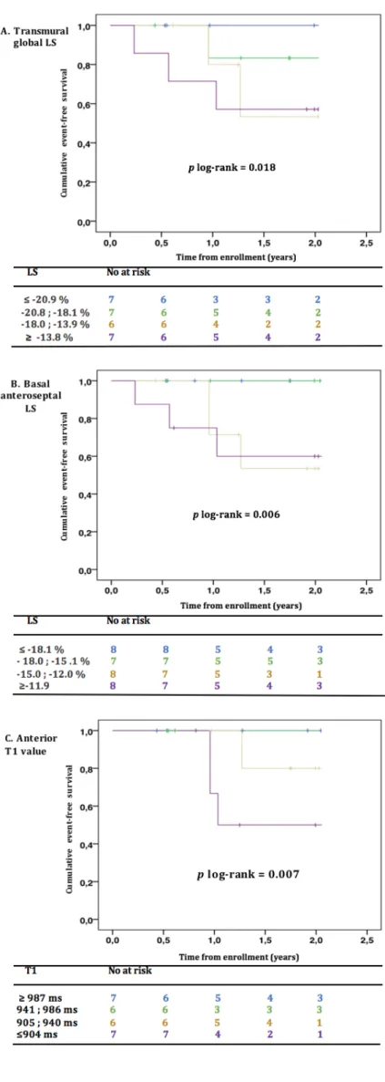
In the Cox univariate analyses, male sex and history of syncope were strongly associated with CV events (HR 5.29, 95%CI 1.02-27.57, p<0.05, and HR 6.72, 95%CI 1.39-32.53, p<0.05). Increased echocardiographic mean E/E’ ratio was significantly predictive of CV outcomes (p< 0.05). Altered transmural global LS, and more specifically basal septal and mid anterolateral LS; and epicardial global LS, and particularly basal and mid anterolateral LS, were significantly predictive of CV outcomes (p< 0.05). Among LS, the most significantly predictive of CV outcomes was basal anteroseptal transmural LS (HR 1.27, CI95% 1.09-1.47, p<0.05). In CMR, maximal wall thickness (HR 1.34, CI95% 1.12-1.61, p<0.05) and LV mass index (HR 1.02, CI95% 1.01-1.04, p<0.05) predicted the upcoming of a CV outcome whereas a high anterior native T1 value was a protective factor (HR 0.98, 95%CI 0.97-1.00, p<0.05). Table 5 Correlations between VO2 peak and VE/VCO2 slope and different echocardiographic and CMR parameters r value p value VO2 peak Maximal LV wall thickness at end-diastole -0.38 0.092 LV ejection fraction 0.10 0.650 Indexed left atrial volume -0.13 0.620 Mean E/E' ratio -0.42 0.067 Global transmural LS -0.55 0.012* Global endocardial LS -0.53 0.025* Global epicardial LS -0.40 0.122 Indexed LV mass -0.14 0.560 Number of segments with LGE -0.28 0.250 Mean T1 value -0.11 0.654 Mean extracellular Volume -0.23 0.420 VE/VCO2 slope Maximal LV wall thickness at end-diastole 0.48 0.026* LV ejection fraction 0.49 0.021* Indexed left atrial volume 0.08 0.782 Mitral E/E' septal-lateral ratio 0.42 0.066 Global transmural LS 0.22 0.392 Global endocardial LS 0.16 0.587 Global epicardial LS 0.09 0.730 Indexed LV mass 0.32 0.161 Number of segments with LGE 0.10 0.684 Mean T1 value -0.57 0.011* Mean extracellular Volume 0.11 0.820 CMR : Cardiovascular magnetic resonnance, LGE : late gadolinium enhancement ; LV : Left ventricle ; LS : Longitudinal strain
Table 6 Univariate Cox analyses for predicting cardiovascular outcomes in FD patients HR CI 95% p Age 1.049 1.00-1.10 0.065 Male sex 5.290 1.02-27.57 0.048* History of syncope 6.720 1.39-32.53 0.018* Ventricular non sustained tachycardia 3.738 0.65-21.38 0.138 Theoretical VO2 peak 0.995 0.97-1.03 0.744 VE/VCO2 slope 1.156 1.03-1.30 0.011* Echocardiographic maximal LV wall thickness 1.341 1.12-1.61 0.002* LV ejection fraction 1.000 0.88-1.13 0.998 Indexed left atrial volume 1.008 0.98-1.03 0.596 Mean E/E' ratio 1.180 1.03-1.35 0.015* CMR maximal LV wall thickness 1.253 1.06-1.48 0.008* Transmural global LS 1.338 1.05-1.71 0.019* Basal inferoseptal transmural LS 1.236 1.06-1.45 0.008* Basal anteroseptal transmural LS 1.267 1.09-1.47 0.002* Basal inferolateral transmural LS 1.083 0.94-1.25 0.265 Mid inferolateral transmural LS 1.027 0.96-1.10 0.462 Basal anterolateral transmural LS 1.066 0.98-1.16 0.156 Mid anterolateral transmural LS 1.127 1.03-1.23 0.010* Endocardial global LS 1.201 0.96-1.50 0.106 Basal inferolateral endocardial LS 1.122 0.97-1.30 0.120 Mid inferolateral endocardial LS 1.075 0.95-1.22 0.250 Basal anterolateral endocardial LS 1.107 1.01-1.21 0.030* Mid anterolateral endocardial LS 1.092 0.99-1.21 0.093 Epicardial global LS 1.344 1.01-1.79 0.045* Basal inferolateral epicardial LS 1.082 0.93-1.27 0.321 Mid epicardial inferolateral epicardial LS 1.100 0.95-1.28 0.219 Basal anterolateral epicardial LS 1.160 1.04-1.30 0.110 Mid anterolateral epicardial LS 1.146 1.02-1.29 0.027* LV mass index 1.020 1.01-1.04 0.010* Late gadolinium enhancement 3.713 0.61-22.43 0.153 Number of segments with LGE 1.255 1.99-1.59 0.063 Global native T1 value 0.986 0.97-1.00 0.074 Septal native T1 value 0.987 0.97-1.00 0.114 Lateral native T1 value 0.985 0.97-1.00 0.104 Anterior native T1 value 0.984 0.97-1.00 0.048* Inferior native T1 value 0.997 0.99-1.01 0.673 Global extracellular volume 1.092 0.76-1.58 0.638 Septal extracellular volume 1.150 0.79-1.66 0.471 Lateral extracellular volume 1.055 0.75-1.49 0.761 Anterior extracellular volume 1.052 0.79-1.40 0.732 Inferior extracellular volume 0.994 0.70-1.41 0.973 *p < 0.05 considered statistically significant. CMR : Cardiovascular magnetic resonnance ; CI : Confidence interval ; HR : Hazard ratio ; LS : Longitudinal strain ; LV : Left ventricle
Figure 3: Kaplan–Meier event-free survival curves for transmural global LS (A), basal anteroseptal LS (B), and anterior T1 value (C) for prediction of CV events in FD.
p log rank < 0.05 was considered
statistically significant.
CV: Cardiovascular; FD: Fabry disease; LS: Longitudinal strain
Discussion
The aim of this study was to investigate the prognosis value of recently-developed echocardiographic and CMR parameters and their relation with functional status in FD. Firstly, we showed a global alteration of basal segments in LS in FD patients compared to healthy subjects; and a lower mean native T1, notably without overlap between adult male patients and healthy controls values. Secondly, we highlighted a significative correlation between VO2 peak and endocardial and transmural LS; as well as between VE/VCO2 slope and mainly T1 value; highlighting the clinical value of these parameters. Thirdly, we demonstrated transmural and epicardial global LS, and regionally septal and lateral LS values, were predictive of CV outcomes.
Echocardiographic and CMR characterization of FD patients
Our study confirms the frequent cardiac involvement in FD patients, as described in other studies16. Our population, though, had an overall mild degree of disease severity according to
MSSI. As expected, male patients were more affected than females, because of the X-linked mutation that causes the disease. Repartition of LVH in our population was consistent with published literature42, although no patient had apical hypertrophy in our study. LGE had always a midwall distribution when present, as previously described43. LV ejection fraction is not impaired in patients with FD; however, LS is significantly altered. In global and endocardial strain, the loss of global deformation is mainly due to basal segments, resulting in an ‘apical sparing’ pattern. This pattern can also be observed in cardiac amyloidosis44.
A slightly different pattern is observed in epicardial strain, with a predominant alteration of anterior, lateral and inferoseptal segments, but with also an ‘apical sparring’ pattern. Kramer et al.45 had compared FD patients with and without LGE, and identified a predominant alteration of
LS in basal posterior and lateral segments which was correlated with the amount of LGE. Our results tend to show a more global basal alteration of LS compared to healthy controls. This may indicate that there is more lipid deposition in basal segments than apical ones, inducing a more important resistance to deformation. This hypothesis is concordant with the predominant hypertrophy in basal segments in our population. With regard to diastolic function, classical mitral E/A inflow ratio was not different between FD patients and healthy controls. However mean E/E’ ratio and indexed atrial volume were significantly increased. Systolic pulmonary artery pressures are also slightly but significantly increased. These parameters confirm the coexistence of diastolic and systolic LV dysfunction in FD.
The myocardial native T1 in patients with FD was significantly lower than in healthy volunteers. All adult male patients had lower mean T1 than healthy subjects. When considering the whole FD group, the male mean T1 was significantly lower than the female mean T1, as observed in previous studies28,46. One male patient had a remarkably a low mean T1, measured at 822 ms, although he didn’t have LVH. It is also interesting to note that this patient had low global LS (-16.4 %), without any other echocardiographic abnormalities. To our knowledge, the effect of specific therapies in patients with cardiac involvement revealed by low LS or low native T1 has not being studied and should be analyzed in future studies. Mean ECV was 24.7 ± 3.0 % in our FD patients, and did not differ from healthy controls. This is concordant with the physiopathology of the disease, which is due to an intracellular (lysosomal) storage disorder. Moreover, we only measured ECV in segments without LGE, to avoid fibrosis. This is consistent with Sado et al. study47 in which ECV measured in septum was not different from healthy subjects.
Relation between echocardiographic and CMR parameters and functional status in metabolic exercise test Global and endocardial LS were significantly correlated to VO2 peak. Epicardial LS and mean E/E’ ratio also seemed correlated to VO2 peak, but without any significance. Peak VO2, reflecting the maximal aerobic metabolism capacity, provides objective assessment of functional capacity in patients with heart failure, and has been shown to be of great importance in predicting survival from heart failure48-50. LS provides a good evaluation of functional status and seem superior to classical systolic function parameters; however multi-layer analysis doesn’t seem to be superior to transmural LS. Furthermore, T1 value had a strong correlation with the VE/VCO2 slope. LV wall thickness and LV ejection fraction were also significantly correlated to VE/VCO2 slope. On the contrary, mean E/E’, LV mass, LGE, and ECV failed to predict functional status. VE/VCO2 slope is a recently developed parameter that reflects respiratory efficiency; it has been shown to be significantly
is the first study that demonstrates the usefulness of echocardiographic systolic function parameters, and CMR T1 value, in predicting functional status in FD. However, multi-layer LS failed to show a superiority compared to transmural LS in predicting exercise capacity in FD.
Also, we found a major alteration of exercise capacity in our patients compared to normative population data, reflected by low theoretical VO2 peak, particularly in males. This is partly explained by deconditioning, as % theoretical mean oxygen uptake at anaerobic threshold is <.50% in our patients. Prognosis factors In our population, 19.3% of FD patients presented a CV event during a median follow-up of 23 months. All of them were treated by ERT. The reduction of glycosphingolipid accumulation under ERT has been demonstrated in various tissues11-13,53,54; particularly in the myocardium55. However, despite the relatively low degree of disease severity (reflected by MSSI) in our population and the relatively good results of the therapy, CV outcomes are frequent. As expected, male sex is a strong prognosis factor in our study (HR=5.29). History of syncope is also a strong predictive factor of CV event in univariate analysis. In cardiopulmonary exercise testing, only VE/VCO2 slope was significantly predictive of CV event, suggesting its prognosis value in heart failure can probably be extended in FD cardiomyopathy. We tried to find interesting LS pattern, studying global, multi-layer and regional strain. On the one hand, global transmural and epicardial LS were significantly predictive of CV outcomes in univariate analysis. On the other hand, basal inferoseptal and anteroseptal transmural LS, and basal and mid anterolateral epicardial LS were significantly predictive of CV outcomes in univariate analysis. The absence of clear interesting multi-layer pattern in FD is consistent with predominant involvement of mesomyocardial areas in CMR. Anyhow, in addition to the ability of speckle-tracking imaging to early detect myocardial dysfunction18, to predict functional status56, to reflect myocardial fibrosis44, and to assess treatment efficiency57 in FD, global and regional strain have a good prognosis value. Global transmural LS and basal anteroseptal transmural LS seem to have the strongest prognosis value and should be used to identify the most severe patients.
Kaplan Meier’s analysis of event-free survival show a significant impact on prognosis when global LS value is higher than -18.0%; and when basal anteroseptal LS value is higher than -15.0%.
All patients who encountered a cardiac event during follow-up had LGE areas, signing local myocardial fibrosis. Presence of LGE failed to predict CV outcomes, it can probably be explained by the small sample size of our study; but the number of LGE segments tend towards significance in predicting CV outcomes in univariate analysis. A relationship between LGE and malignant ventricular arrhythmias has been shown by Kramer et al.27 in a 73-patients’ study, in which 18% of patients with LGE vs. 0% of patients without LGE experienced ventricular arrhythmias. ERT did not seem to reduce the risk of progression of fibrosis in this study, and the only independent predictive factor of ventricular arrhythmias was the annual increase in fibrosis during follow-up. Furthermore, Weideman et al.58 showed that under ERT, reduction of LVH was significant in patients without fibrosis and accompanied with improvement of myocardial function, whereas patients with mild or severe fibrosis showed a minor reduction in LVH and no improvement in myocardial function. Moreover, Pieroni et al.59 presented the case of a 55-year old man that developed a quick and significant progression of cardiac involvement of his disease despite ERT, while cutaneous and renal involvement remained stable. These studies suggest that the effect of ERT depends on the stage of the disease at baseline. Patients with evidence of fibrosis in CMR may have limited or even no benefits at all from ERT, and fibrotic remodeling seems to be even worse at a late stage of the disease. Therefore, substitutive treatment should probably be introduced early in disease progression, before the development of myocardial fibrosis, to achieve at least a stabilization of cardiac involvement. Reduction of progression of myocardial fibrosis would probably prevent global CV events as well as ventricular arrhythmias. Considerations about localization of LVH and LGE In our study, LVH predominate in septal segments in FD patients, whereas LGE, predominant in inferolateral basal area, never affects the septal wall. Frustaci et al.60 showed in a FD family that patients with most LVH had larger and more vacuoled cells, with marked necrosis and apoptosis. Glycolipid accumulation was shown to induce oxidative damage of proteins in cardiomyocytes, leading to degradation of contractile elements, reduced ATP synthesis, and cell death61,62. Therefore, fibrosis seems to be caused by apoptosis of enlarged, lipid-engorged cardiomyocytes, and should be found in the most hypertrophic areas. However, in our study, LVH does not match
cellular apoptosis. Moreover, Kawano et al.64 showed that LV basal posterior thinning, defined as septum/posterior wall thickness ratio > 1.3, was a prognosis factor in FD. Development of fibrosis would occur in most hypertrophic areas as a progression of myocardial involvement. However, in our study, 2 females had myocardial fibrosis without LVH, and all females who had LVH had fibrosis. In contrast, all males who had fibrosis had invariably LVH. These observations have already been made on a larger scale in Niemann et al. study65. In females, progression toward severe LVH is delayed, probably because of residual activity of alpha-galactosidase, and development of fibrosis does not necessarily require LVH. Furthermore, Moon et al.25 studied myocardial histological sections of a deceased patient who had massive concentric LVH: he had a thinned inferolateral region, concordant with location of LGE and collagen, but without increased lipid accumulation. Evolution towards fibrosis in the inferolateral wall doesn’t seem to depend on early LVH of this zone, and the fact that maximal fibrosis has a peculiar predilection for the inferolateral wall is thereby unexplained. Moon et al.24 hypothesized that cardiac involvement in FD could impair resistance to physical stress within the myocardium, that might be most prominent in the inferolateral region, where significant shear forces combine with a watershed vascular territory. Indeed, several other cardiomyopathies have predilection for lateral wall involvement66,67.
In conclusion, the physiopathology of development of cardiac fibrosis still remains unclear in FD.
Considerations about normalized or elevated T1 value in most hypertrophied areas or with fibrosis in FD We noticed a higher native T1 value in LGE areas compared to other segments. Sado et al.46 had highlighted normal or even elevated T1 in the inferolateral wall in FD patients, where is found myocardial fibrosis. As a reminder, T1 mapping was at first studied for assessment of diffuse interstitial fibrosis, revealed by elevated T1 value68. In FD, however, the lipid storage yields low T1, making it a useful biomarker for early detection of cardiac involvement. Sado et al.46 hypothesized that the inferolateral wall myocardium in FD underwent a 4-phase transition from normal (pre-detectable storage) to low (storage) to pseudonormalization (storage and fibrosis) to elevation (fibrosis). As fibrosis distorts T1 value, it has to be measured in areas without LGE. In our study, patients with the highest LV maximal wall thickness did not have the lowest mean T1 value (Figure 2). Nordin et al. 69 even recently showed that from a certain degree of LVH, in men, the correlation between T1 and LVH reverses and T1 increases (toward normal) with LVH, whereas ECV is not impacted. They hypothesized that, in males, storage is triggering sarcomeric
protein expression via myocyte hypertrophy70,71 and usual LVH rather than storage LVH, which are diluting the T1 lowering of sphingolipid. This hypertrophic and inflammation phase precede the diffuse fibrosis phase. Study limitations Limitations of this study include no histological validation of the presence of storage with low native T1 and the presence of fibrosis with LGE. The relatively small number of patients, leading to a low power study, could explain the lack of statistical difference. This is inherent in a monocentric study, carried out on a rare disease. Furthermore, there is a large heterogeneity in our population, as we know that male and female patients do not have the same phenotype, and two phenotypes exists in male patients: classical and late-onset phenotypes72. Cardiovascular outcomes covered a broad spectrum of CV events in our study. Number of events was insufficient to permit multivariate analysis. Among our patients, some of them could not perform MRI, unfortunately reducing the percentage of patients with cardiac involvement performing MRI.
Conclusions
Our study suggests that longitudinal strain and T1 mapping are reproductible, enables characterization of cardiac involvement, and predicts functional status and cardiovascular outcomes in FD patients. These data should be confirmed in larger studies.
Fundings:
from Shire
® France
References
1. Brady RO, Gal AE, Bradley RM, et al. Enzymatic defect in Fabry’s disease. Ceramidetrihexosidase deficiency. N Engl J Med. 1967;276:1163-7.
2. Zarate YA, Hopkin RJ. Fabry’s disease. Lancet. 2008;372:1427–35.
3. Meikle PJ, Hopwood JJ, Clague AE, et al. Prevalence of lysosomal storage disorders. JAMA. 1999, 281:249-254. 4. Houge G, Skarbovik AJ. Fabry disease - a diagnostic and therapeutic challenge. Tidsskr Nor Laegeforen. 2005;125:1004-1006 5. Germain DP. Fabry disease. Orphanet J Rare Dis. 2010;5:30. 6. Mehta A, Ricci R, Widmer U, et al. Fabry disease defined: baseline clinical manifestations of 366 patients in the Fabry outcome survey. Eur J Clin Invest. 2004;34:236-42. 7. Patel MR, Cecchi F, Cizmarik M, et al. Cardiovascular events in patients with Fabry disease natural history data from the fabry registry. J Am Coll Cardiol. 2011;57:1093–9. 8. Mehta A, Clarke JT, Giugliani R, et al. Natural course of Fabry disease: changing pattern of causes of death in fos - Fabry outcome survey. J Med Genet. 2009;46:548-52. 9. Waldek S, Patel MR, Banikazemi M, et al. Life expectancy and cause of death in males and females with Fabry disease: findings from the Fabry Registry. Genet Med. 2009;11:790–796. 10. Schiffmann R, Murray GJ, Treco D, et al. Infusion of alpha- galactosidase a reduces tissue globotriaosylceramide storage in patients with Fabry disease. Proc Natl Acad Sci USA. 2000; 97:365-70.
11. Schiffmann R, Kopp JB, Austin HA 3rd, et al. Enzyme replacement therapy in Fabry disease: a randomized controlled trial. JAMA. 2001;285:2743-9. 65.
12. Eng CM, Guffon N, Wilcox WR, et al. Safety and efficacy of recombinant human alpha-galactosidase a replacement therapy in Fabry’s disease. N Engl J Med. 2001;345:9-16. 13. Thurberg BL, Fallon JT, Mitchell R, et al. Cardiac microvascular pathology in Fabry disease:
evaluation of endomyocardial biopsies before and after enzyme replacement therapy. Circulation. 2009;119:2561-7.
14. Hughes DA, Elliott PM, Shah J. Effects of enzyme replacement therapy on the cardiomyopathy of Anderson-Fabry disease: a randomised, double-blind, placebo controlled clinical trial of agalsidase alfa. Heart. 2008;94: 153–158.
15. Hughes DA, Nicholls K, Shankar SP, et al. Oral pharmacological chaperone migalastat compared with enzyme replacement therapy in Fabry disease: 18-month results from the randomised phase III ATTRACT study, J Med Genet. 2017 Apr; 54(4): 288–296.
16. Yeung DF, Sirrs S, Tsang MYC, et al. Echocardiographic Assessment of Patients with Fabry Disease. J Am Soc Echocardiogr. 2018;31:639–649.
17. Pieroni M, Chimenti C, Ricci R, et al. Early detection of Fabry cardiomyopathy by tissue Doppler imaging. Circulation. 2003;107:1978-84.
18. Shanks M, Thompson RB, Paterson ID, et al. Systolic and diastolic function assessment in fabry disease patients using speckle-tracking imaging and comparison with conventional echocardiographic measurements. J Am Soc Echocardiogr. 2013;26:1407–1414.
19. Saccheri MC, Cianciulli TF, Lax JA, et al.. Two-dimensional speckle tracking echocardiography for early detection of myocardial damage in young patients with Fabry disease. Echocardiography. 2013;30:1069–1077.
20. Ozawa K, Funabashi N, Takaoka H, et al. Characteristic myocardial strain identified in hypertrophic cardiomyopathy subjects with preserved left ventricular ejection fraction using a novel multi-layer transthoracic echocardiography technique. Int J Cardiol. 2015;184C:237– 243.
21. Ozawa K, Funabashi N, Kamata T, et al. Inter- and intraobserver consistency in LV myocardial strain measurement using a novel multi-layer technique in patients with severe aortic stenosis and preserved LV ejection fraction. Int J Cardiol. 2017;228:687–693
22. Sebastian SI, Haugaa KH, Zahid W, et al. Layer-specific quantification of myocardial deformation by strain echocardiography may reveal significant CAD in patients with non–ST-segment elevation acute coronary syndrome. JACC Cardiovasc Imag. 2013;6:535–544