Percutaneous stereotactic en bloc excision of nonpalpable breast
carcinoma: a step in the direction of supraconservative surgery
E. Lifrange,1 R. F. Dondelinger,2 J. M. Foidart,3 J. Bradfer,4 P. Quatresooz5 and C. Colin11
Breast Department, 2Department of Medical Imaging, 3Department of Obstetrics and Gynecology, 4Department of Radiotherapy and
5Department of Pathology, University Hospital Sart Tilman, 4000 Liège, Belgium
SUMMARY
Recently, the advanced breast biopsy instrumentation (ABBI) system has been introduced as an alternative to conventional breast biopsy techniques. This study was prospectively conducted to evaluate the potential of the ABBI method in locoregional management of a consecutive series of patients with nonpalpable
mammographically detected breast carcinomas. Sixty-one consecutive patients underwent an ABBI procedure as a first step before possible surgery for nonpalpable breast lesions that would in any case require complete excision. For the 27 patients in whom the ABBI biopsy revealed malignancy further surgery was recommended, including re-excision of the biopsy site and axillary dissection in cases of infiltrating carcinoma. We calculated the probabilities that the ABBI specimen would have tumor-free margins and that a definitely complete excision had been achieved as a function of the mammographic or pathological diameter of the cancer. For cancer with a pathological diameter less than 10 mm measured on the ABBI specimen, the probability (92%) of obtaining complete resection was significantly better than for larger lesions (P = 0.01, Fisher's exact test). Although the therapeutic perspectives for the ABBI method are limited at present, we suggest that this approach is a first step in the direction of a surgical strategy that is better adapted to the pathological characteristics peculiar to these small tumors, whose incidence is increasing.
INTRODUCTION
Over the past few years, breast surgeons have been confronted with an increasing number of nonpalpable mammographically detected lesions. Mammograms performed in asymptomatic patients detect 0.5-2% of nonpalpable breast lesions (NPBL), 10-30% of which are cancers.1 At present, 30% of the breast lesions that we operate on in our institution are NPBL. Pathological examination of surgically excised lesions confirms an underlying cancer in 50% of these cases and a high-risk lesion, such as a lobular carcinoma in situ, atypical hyperplasia, or radial scar, in 15% of them.
Classically, excision is guided by a hook wire placed prior to the surgical intervention. Despite the preoperative localization, the volume of tissue excised often exceeds the dimensions of the lesion by far still with no
guarantee that the resection margins are tumor-free, as frozen-section evaluation of these borders is usually not possible.2 A few targeted lesions are not even found in the biopsy specimens.3,4
Progress in the field of endoscopic surgery suggested a way of improving the quality of the diagnostic specimen and also of achieving total excision of the smallest NPBL through a small incision. At the end of 1994, we felt it should be possible to remove a small lesion as a single tissue specimen through a large electroinsulated cannula inserted percutaneously and perpendicular to the targeted lesion under stereotactic control, with the patient lying prone on a dedicated table.5 A system coupling distal section-coagulation and traction would allow the
subsequent en bloc excision of the lesion. The advanced breast biopsy instrumentation (ABBI) system (United States Surgical Corporation, Norwalk, CT, USA) incorporated this principle. This device, in conjunction with a stereotactic biopsy table, allows single-pass excision of cylindrical specimens of variable diameter (0.5-2 cm) from the subcutaneous tissue up to and beyond the targeted lesion.6 Since the FDA approval was granted for the ABBI as a diagnostic tool in early 1996, its feasibility, safety, accuracy and cost-benefits for the diagnostic management of nonpalpable lesions have been assessed.619
In a previous study, we evaluated the proportion of our patients in whom NPBL would have unequivocally required complete excision but could be adequately removed with ABBI.20 The present prospective nonrandomized study was conducted to evaluate the potential of the ABBI method for the locoregional management of a consecutive series of nonpalpable breast carcinomas.
MATERIALS AND METHODS
From May 1998 to December 2000, 448 consecutive patients aged 22-80 years (mean 54) were referred for management of NPBL. Patients underwent either image-guided needle biopsy or an ABBI procedure. When feasible, an ABBI procedure was offered to patients as a first step before possible surgery for lesions which would definitely require complete excision: mammographic features highly suggestive of malignancy (BI-RADS category 5),21 suspicious abnormality (BI-RADS category 4) in the presence of high-risk factors (i.e., family or personal history of breast cancer, recent or growing lesion), or the patient's request for complete excision in the presence of a probably benign or suspicious lesion (BI-RADS category 3 or 4). Exclusion criteria for an ABBI procedure were: multiple lesions, mammographic diameter of the lesion larger than 15 mm, breast compressible to less than 25 mm, hemorrhagic diathesis, body weight over 135 kg (3001b) and inability of the patient to lie still for 1 h in a prone position.
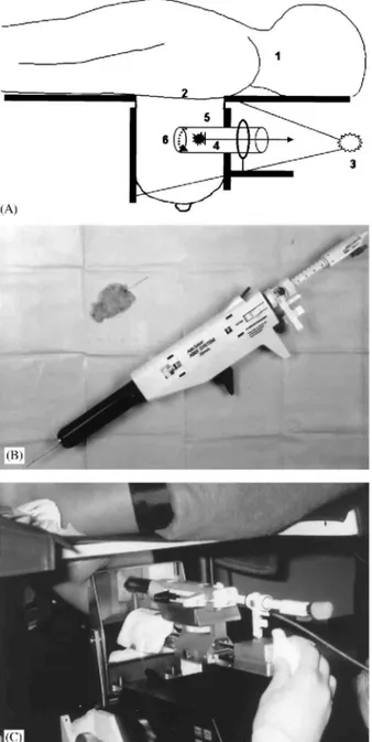
Fig. 1: (A) Principles of removing a small nonpalpable breast lesion as a single tissue specimen (E.L., 1994). The patient lies in the prone position (1). The breast is protruding through a central aperture in the stereotactic table (2). Each step of the procedure is radiologically checked (3). With the breast compressed as for mammography, a hook-wire is inserted as far as the suspicious lesion (4). The skin is incised and a plastic cannula is put in place beyond the targeted lesion (5). The bottom of the specimen is sectioned and the cannula containing the specimen is then removed from the breast (6). (B) The ABBI device allows percutaneous en bloc excision of a cylinder of breast tissue up to 20 mm in diameter. (C) The patient is positioned prone on a dedicated stereotactic table, and the biopsy device is targeted using digital stereomammography.
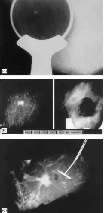
Fig. 2: (A) Spot compression view of the right breast of this 66-year-old woman confirmed a nonpalpable 6-mm
spiculated mass. (B) Partial digital radiographs (same magnification) show the pre-biopsy view of the targeted lesion (left) and the biopsy cavity after the ABBI procedure using a 20-mm cannula (right). (C) Digital radiograph of the ABBI specimen shows apparently complete excision. Histopathological analysis confirmed a 6-mm well-differentiated infiltrating ductal carcinoma resected in sano. Surgical re-excision of the biopsy site confirmed the residual tumor-free status.
All patients gave informed consent and agreed to potential subsequent surgery. Overall, 61 ABBI procedures were completed. Patient selection, the ABBI procedure and surgery were performed by the same investigator (E.L.). The ABBI procedure was performed in an outpatient setting, as previously described.6 The ABBI device is targeted by the dedicated stereotactic imaging unit of a prone table (United States Surgical Corporation and Lorad, Danbury, CT, USA) (Fig. 1A-C). Patients were each given one 0.5-mg tablet of alprazolam (Xanax, Pharmacia-Upjohn, Puurs, Belgium) before the procedure. The patient was positioned prone with the breast protruding through an opening and ECG monitoring and electrocautery contact were established. The lesion was targeted using digital stereomammography. Each step of the procedure was documented with digital imaging. The biopsy device consisted of a plastic cannula of variable diameter (5, 10, 15, 20 mm) with a rotating distal circular blade. We usually choose the largest diameter to obtain clear margins. The breast was surgically
prepared. Once deep local anesthesia (15 ml of 1% lidocaine with epinephrine) was achieved, a guide wire was inserted proximal to the targeted lesion, after which the cannula was introduced through a skin incision 5 mm larger than the cannula diameter and advanced 10-15 mm beyond the center of the lesion. Correct positioning was confirmed by stereomammography. An electrocautery snare at the distal end of the cannula cut and cauterized the deep end of the specimen. The device was removed together with the enclosed specimen. Digital radiographs of the breast and the excised specimen were taken to confirm that excision was complete (Fig. 2A-C). The patient was then placed in a supine position. Bleeding was stopped by electrocauterization when necessary. Small metallic clips were placed at the site of the lesion and the wound was closed with absorbable sutures. A pressure dressing was applied for 24 h. The average total procedure time was 70min and the patient was discharged after an observation period of 2h.
When cancer was present, the pathologist reported on pathological type, size, grade and resection margins. Ink margins were considered negative when free of tumor. The presence of an extensive intraductal component (EIC) and/or lymphovascular invasion (LVI) and the results of immunohistochemical (IHC) analyses (estrogen-progesterone receptors, c-erbB-2, Ki67, p53, hsp27) were also reported.
For patients found to have a malignant lesion, further surgery was recommended. Open surgery was scheduled within the month following the ABBI procedure and involved complete re-excision of the biopsy site, even when the ABBI specimen-resection margins were negative for tumor, and axillary lymph-node dissection for invasive carcinoma. Radiation therapy was given after conservative surgery for invasive carcinoma or according to the Van Nuys prognostic index for ductal carcinoma in situ (DCIS).22 Adjuvant therapy was administered according to the recommendations of the 6th St Gallen International Consensus Conference.23 Twelve to 42 months of follow-up were available for the 61 ABBI patients.
RESULTS
Malignancy was confirmed in 27 (44%) of the 61 ABBI specimens (Table 1). The 27 mammographic lesions ranged in size from 5 to 15 mm (mean 8.5): 16 were masses, 9 had the appearance of microcalcifications, and 2 were masses containing microcalcifications. Lesions were excised with a 15-mm cannula in 5 (19%) of the 27 cases and a 20-mm cannula in 22 (81%). All 27 ABBI procedures were successful. In all cases, specimen and wound radiographs suggested complete excision of the lesion and the histological findings were consistent with the mammographic features. However, the mammographic size of the lesion was not predictive of the
pathological size: for 16 (59%) of the 27 ABBI specimens, the size of the excised lesion differed by more than 2 mm from the mammographic size. The 27 en block specimens enabled accurate histopathological
characterization. Type, size, grade, resection margins, presence of EIC and/or LVI, and IHC test results were determined for all specimens. Clinically mild ecchymoses were common, but none of the patients experienced hematoma. One mild wound infection occurred, which was treated with oral antibiotics.
Two patients whose en bloc biopsy specimens demonstrated carcinoma measuring < 1 cm and with negative margins more than 2 mm wide refused subsequent surgery and underwent radiotherapy: one was a 75-year-old patient whose ABBI specimen demonstrated low-grade 9-mm DCIS with negative margin widths larger than 2 mm and the other, a 58-year-old patient whose ABBI specimen demonstrated low-grade 4-mm invasive ductal carcinoma (IDC) with negative margin more than 4 mm wide. One 80-year-old patient accepted re-excision, but did not undergo axillary dissection. Overall, 25 (93%) of the 27 biopsy sites were re-excised and 18 (95%) of the 19 patients with invasive cancer underwent axillary lymph-node dissection. For all patients with carcinoma in situ in the ABBI biopsies, noninvasive disease was confirmed at surgery. The ABBI specimen margins were tumor free in 11 (41%) of the 27 patients diagnosed with carcinoma. None of the patients with tumor-free margins in the ABBI specimen was found to have residual disease at surgery. Finally, surgical re-excision confirmed residual tumor-free status in 64% (16/25) of the patients with malignant ABBI specimens. These 16 patients were confirmed to be node-negative for invasive carcinoma, except one elderly patient who did not undergo axillary dissection.
We calculated the probabilities that an ABBI specimen would have tumor-free margins and that complete excision would be achieved as a function of the mammographic or histological diameters of the cancer measured directly in the ABBI specimen (Table 2). Indeed, for nonpalpable breast cancer with a histological diameter less than 10 mm, the probability (92%) of obtaining complete resection was significantly better than the
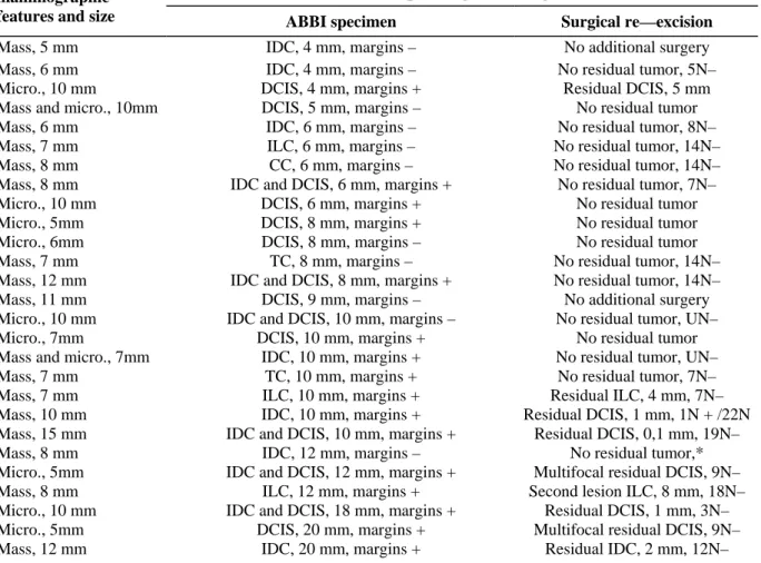
Table 1: mammographic features and histopathological findings in the ABBI specimen and at re-excision for the
27 confirmed breast cancers
Histopathological findings mammographic
features and size ABBI specimen Surgical re—excision
Mass, 5 mm IDC, 4 mm, margins – No additional surgery
Mass, 6 mm IDC, 4 mm, margins – No residual tumor, 5N–
Micro., 10 mm DCIS, 4 mm, margins + Residual DCIS, 5 mm
Mass and micro., 10mm DCIS, 5 mm, margins – No residual tumor
Mass, 6 mm IDC, 6 mm, margins – No residual tumor, 8N–
Mass, 7 mm ILC, 6 mm, margins – No residual tumor, 14N–
Mass, 8 mm CC, 6 mm, margins – No residual tumor, 14N–
Mass, 8 mm IDC and DCIS, 6 mm, margins + No residual tumor, 7N–
Micro., 10 mm DCIS, 6 mm, margins + No residual tumor
Micro., 5mm DCIS, 8 mm, margins + No residual tumor
Micro., 6mm DCIS, 8 mm, margins – No residual tumor
Mass, 7 mm TC, 8 mm, margins – No residual tumor, 14N–
Mass, 12 mm IDC and DCIS, 8 mm, margins + No residual tumor, 14N–
Mass, 11 mm DCIS, 9 mm, margins – No additional surgery
Micro., 10 mm IDC and DCIS, 10 mm, margins – No residual tumor, UN–
Micro., 7mm DCIS, 10 mm, margins + No residual tumor
Mass and micro., 7mm IDC, 10 mm, margins + No residual tumor, UN–
Mass, 7 mm TC, 10 mm, margins + No residual tumor, 7N–
Mass, 7 mm ILC, 10 mm, margins + Residual ILC, 4 mm, 7N–
Mass, 10 mm IDC, 10 mm, margins + Residual DCIS, 1 mm, 1N + /22N
Mass, 15 mm IDC and DCIS, 10 mm, margins + Residual DCIS, 0,1 mm, 19N–
Mass, 8 mm IDC, 12 mm, margins – No residual tumor,*
Micro., 5mm IDC and DCIS, 12 mm, margins + Multifocal residual DCIS, 9N–
Mass, 8 mm ILC, 12 mm, margins + Second lesion ILC, 8 mm, 18N–
Micro., 10 mm IDC and DCIS, 18 mm, margins + Residual DCIS, 1 mm, 3N–
Micro., 5mm DCIS, 20 mm, margins + Multifocal residual DCIS, 9N–
Mass, 12 mm IDC, 20 mm, margins + Residual IDC, 2 mm, 12N–
Abbreviations: Micro./micro., microcalcifications; DCIS, ductal carcinoma in situ; IDC, invasive ductal carcinoma; ILC, invasive lobular carcinoma; TC, tubular carcinoma; CC, colloid carcinoma; N, axillary lymph node; —, negative; +, positive for tumor; margins —, negative margins for tumor; margins +, positive margins for tumor.
*An 80-year-old patient, treated with lumpectomy and tamoxifen but no axillary dissection or radiotherapy.
Table 2: Probabilities of the ABBI specimen having tumor-free margins and of complete excision as a function
of the lesion diameter measured on mammography and in the pathological specimen
Lesion diameter (mm) (n = 27)
ABBI specimen with tumor-free margins
(n = 27)
No residual disease at re-excision (n = 25)* Mammography <10(n = 17) 47% (8/17) 75% (12/16) 10-15 (n = 10) 30% (3/10) 44% (4/9) Pathological specimen <10(n = 14) 64% (9/14) 92% (11/12) 10 or >10(n = 13) 15% (2/13) 38% (5/13)
Two patients refused re-excision.
Of the eight patients with DCIS alone, six (75%) underwent conservative surgery and radiation therapy. One (13%) of eight patients with extensive DCIS involving the margins of the re-excision tissue subsequently underwent mastectomy without axillary dissection and one (13%) of eight patients with recurrent disease underwent mastectomy rather than re-excision. Of the 19 patients with invasive disease, 17 (89%) underwent conservative surgery and radiation therapy. The two (11%) remaining patients with residual DCIS involving the margins of the re-excision tissue subsequently underwent mastectomy. One (6%) of the 18 patients who underwent axillary dissection had nodal metastases.
Overall, 23 (88%) of the 26 patients with primary breast cancer were treated with conservative surgery and three (12%) underwent mastectomy after re-excision of the ABBI biopsy site at which the attempt to obtain tumor-free margins had failed.
A posteriori, the ABBI procedure was adequate (no residual disease at biopsy site, no axillary involvement) for locoregional surgical management in 63% (15/ 24) of the patients with malignant ABBI specimens and 92% (11/12) of the breast cancers measuring < 1cm found in this series of consecutive patients. The 12-42 months of follow-up available have been unremarkable for the 61 patients who underwent an ABBI procedure.
DISCUSSION
The basic principles of breast cancer surgery have evolved over the course of the 20th century. A certain number of experimental and clinical observations have invalidated Halsted's concept, according to which breast cancer necessarily progresses in successive steps from a local stage through an intermediary phase of regional extension to systemic involvement.24 The studies by Tubiana and Koscielny showed that metastatic potential developed when a tumor size of 1 mm was reached and that distant dissemination was not derived from the lymph nodes.25
27
These findings have been confirmed by observations of metastases developing far distant from diagnosed cancers measuring less than 1 cm (10% at 10 years) or from cancers with tumor-negative axillary lymph nodes (30% at 10 years). An early diagnosis and tumor responsiveness to adjuvant therapies (hormonal and/or chemotherapy) are currently considered decisive elements in the prognosis for survival.23
Since the 1950s, extended radical mastectomy has gradually given way to modified radical mastectomy.28-30 At present, mastectomy accounts for only 40-50% of the surgical interventions for breast cancer. So-called conservative breast surgery, i.e. quadrantectomy according to Veronesi et al.31 or lumpectomy according to Fischer et al.,32 combined with radiation therapy is recommended as first-line therapy for more than half the patients, provided that the margins of the excised tissue are tumor free. The main objectives of local treatment are complete excision with tumor-free margins and characterization of the prognostic and predictive factors that will guide the choices of systemic therapy. Axillary node surgery has also evolved in parallel with breast surgery, moving towards the concept of prognostic evaluation; for this the quality of the specimen takes priority over the number of nodes removed.3335
The diagnostic characterization of NPBL has progressed markedly over the past years with the advent of stereoguided needle biopsy techniques that eliminate unnecessary surgical resection of benign lesions while carrying a low rate of missed cancers among the lesions not excised.5,36-40 Surgical management of these lesions should evolve with the primary objectives of improving localization and limiting tissue trauma. In an earlier study we confirmed that the ABBI method was a safe and accurate biopsy procedure.20 We established that an ABBI excision was feasible and adequate for less than 40% (17/43) of the benign lesions that would otherwise have been surgically excised in our department, and for only 21% (11/53) of the nonpalpable mammograhically detected carcinomas. Many (62%) of the excised NPBL in that study were not amenable to an ABBI procedure, primarily due to technical limitations.
In this study, we calculated the probabilities of an excision having tumor-free margins and of a complete excision as a function of the lesion's diameter measured on mammography and in the ABBI specimen. To the best of our knowledge, no other investigation has examined these parameters. The probability of an ABBI procedure having excised a lesion completely, i.e., with no residual disease in the biopsy cavity, is approximately 75% for malignant NPBL with a diameter of less than 10 mm on mammography. In the case of lesions with a radiographic diameter of 10-15 mm the probability of an adequate excision is less than 50%. Based on these estimations, we conclude that the complete-excision rate with an ABBI procedure for subclinical cancers can be improved by limiting the indications to suspicious lesions whose largest mammographic diameter is smaller than 10 mm. When the largest diameter of the cancerous lesion measures less than 10 mm on the ABBI specimen, the probability of complete excision is higher than 90%. Above this threshold of 10 mm, the probability falls to about 40%. In light of these findings, to increase the chance of complete excision with tumor-free margins at the time of ABBI biopsy, we decided to immediately re-excise the biopsy bed before suturing the wound whenever the mammographic or macroscopic appearance of the specimen was highly suggestive of malignancy.
In this larger series of subclinical breast cancers, we confirmed that complete excision of cancers smaller than 1cm was possible with the ABBI procedure in around 90% of the selected cases.20 However, for invasive lesions we need to develop less invasive alternatives to lymph node dissection for prognostic axillary evaluation. The benefit of systematic lymph node dissection for all invasive lesions is now being questioned.41-44 The percentages of patients with axillary lymph node invasion by subclinical invasive lesions measuring less than 1 cm are low,
and the morbidity attributable to axillary node dissection is considerable.45,46 In addition, the current trend is to prescribe adjuvant therapy for the majority of patients, even when the axillary lymph nodes are free of metastatic disease.23 We think that this trend will become more widespread. It can be hoped that, in the future, further refinement of the histological prognostic characterizations of subclinical cancers less than 1 cm in size and DNA microarray analyses will definitively guide the therapeutic choices independently of axillary lymph node staging.47-52 In this context, the ABBI procedure performed under local anesthesia and radiological control could be envisaged as a supracon-servative alternative surgical means of achieving complete resection with tumor-free margins. While we await this hypothetical evolution, the technique of identifying the sentinel lymph node seems to be the most appropriate way of limiting the surgical trauma caused by axillary lymph node staging.33-35 Recently, Haigh et al. demonstrated that a previous excisional biopsy did not compromise successful identification of the sentinel lymph node.53
Although the therapeutic perspectives for the ABBI method are limited at present by the small number of potential indications and the need for axillary lymph node staging for invasive lesions, this procedure may have a contributory role within the framework of the management of subclinical cancers. Stereotactic large-core breast biopsy, which is currently used to diagnose NPBL, provide fragmented tissue specimens and almost total disappearance of the tumor, when it is small.54-56 The mammographic disappearance of the lesion poses several problems when the lesion is malignant. The surgical intervention will not always be optimally guided by a clip at the center of the biopsy bed.54 Although the different IHC factors (homone receptors, proliferation markers, etc.) can be characterized in core biopsies, the architectural structure of the lesions may be lost to pathological analysis and thereby compromises the conclusions drawn (e.g., diagnosis of a small tubular carcinoma within a radial scar). In addition, the pathological diameters of small cancerous lesions, which are an important prognostic factor, cannot be correctly determined on the basis of percutaneous core biopsies.56 In contrast, examination of an ABBI specimen allows a precise pathological diagnosis, since the quality of the material is comparable to that obtained during surgical excision. Notably, among the 27 cancers identified in this series, only in 3 patients (12%) was a two-step surgical procedure performed. No patient with a lesion smaller than 1 cm underwent mastectomy, and no patient with in situ carcinoma was unnecessarily subjected to axillary lymph node dissection. For subclinical cancers measuring less than 1 cm, analysis of the results obtained with our classic surgical experience, without reference to our experience with the ABBI procedure, showed that two-step
interventions were performed in 24% of cases, mastectomy in 10% and axillary lymph node dissection in 38% of those with DCIS, during the period from May 1994 to May 1998. This comparison suggests that surgical management of subclinical breast cancers under 1 cm in size is now better after an ABBI procedure.
We suggest that the present approach represents a first step towards a surgical strategy that is better adapted to the pathological characteristics particular to these small tumors whose incidence is increasing and whose prognosis is generally excellent.57 The concept of supraconservative surgery will only be defensible as long as it respects the therapeutic requirements of conventional conservative surgery: that is to say, comparable
percentages for locoregional recurrences, disease-free survival and overall survival, combined with better functional and esthetic results.
Acknowledgements
This work was supported by a grant from the Ministry of Education and Research of the French Community of Belgium.
References
1. Hall F M, Storella J M, Silverstone D Z, Wyshak G. Nonpalpable breast lesions: recommendations for biopsy based on suspicion of carcinoma at mammography. Radiology 1988; 167: 353-358.
2. Chinyama C N, Davies J D, Rayter Z, Farndon J R. Factors affecting surgical margin clearance in screen-detected breast cancer and the effect of cavity biopsies on residual disease. Eur J Surg Oncol 1997; 23: 123-127.
3. Jackman R J, Marzoni FA. Needle-localized breast biopsy: why do we fail? Radiology 1997; 204: 677-684. 4. Israel P Z. The revolution in breast biopsy: where is the surgeon? Am Surg 1997; 62: 93-95.
5. Lifrange E, Kridelka F, Colin C. Stereotaxic needle-core biopsy and fine-needle aspiration biopsy in the diagnosis of nonpalpable breast lesions: controversies and future prospects. Eur J Radiol 1997; 24: 39-47.
6. D'Angelo P C, Galliano D E, Rosemurgy A S. Stereotactic excisional breast biopsies utilizing the advanced breast biopsy instrumentation system. Am J Surg 1997; 174: 297-302.
7. Kelley W E, Schwartzberg B S, Uddo J F. Letter to the editor. Advanced breast biopsy instrumentation. J Am Coll Surg 1997; 185: 604-605.
8. Ferzli G S, Hurwitz J B, Puza T, Van Vorst-Bilotti S. Advanced breast biopsy instrumentation: a critique. J Am Coll Surg 1997; 185: 145-151.
9. Kelley W E, Bailey R, Bertelsen C et al. Stereotactic automated surgical biopsy using the ABBI biopsy device: a multicenter study. Breast J 1998; 4: 302-306.
10. Damascelli B, Frigerio L F, Lanocita R et al. Stereotactic
excisional breast biopsy performed by interventional radiologists using the advanced breast biopsy instrumentation system. Br J Radiol 1998; 71: 1003-1011.
11. Matthews B D, Williams G B. Initial experience with the advanced breast biopsy instrumentation system. Am J Surg 1999; 177: 97-101. 12. Bloomston M, D'Angelo P, Galliano D, Butlert J, Dean R, Rosemurgy A S. One hundred consecutive advanced breast biopsy
instrumentation procedures: complications, cost and outcome. Ann Surg Oncol 1999; 6: 195-199.
13. Damascelli B, Frigerio L F, Patelli G et al. Stereotactic breast biopsy: en bloc excision of microcalcifications with a large-bore cannula device. AJR Am J Roentgenol 1999; 173: 895-900.
14. Sheth D, Wesn C A, Schroder D, Boccaccio J E, Lloyd L R. The advanced breast biopsy instrumentation (ABBI) experience at a community hospital. Am Surg 1999; 65: 726-730.
15. LaRaja R D, Saber A A, Sickles A. Early experience in the use of the advanced breast biopsy instrumentation: a report of one hundred twenty-seven patients. Surgery 1999; 125: 380-384.
16. Ferzli G S, Puza T, Van Vorst-Bilotti S, Waters R. Breast biopsy with ABBI: experience with 183 attempted biopsies. Breast J 1999; 5: 26-28.
17. Rebner M, Chesbrough R, Gregory N. Initial experience with the advanced breast biopsy instrumentation device. AJR Am J Roentgenol 1999; 173: 221-226.
18. Leibman A J, Frager D, Choi P. Experience with breast biopsies using the advanced breast biopsy instrumentation system. AJR Am J Roentgenol 1999; 172: 1409-1412.
19. Jacobs A, Chevinsky AH, Diehl W, Smith TJ. Advanced breast biopsy instrumentation (ABBI) and management of nonpalpable breast abnormalities: a community hospital experience. Breast 2001; 10: 421-426
20. Lifrange E, Dondelinger R F, Fridman V, Colin C. En bloc excision of nonpalpable breast lesions using the advanced breast biopsy instrumentation system: an alternative to needle guided surgery? Eur Radiol 2001; 11: 796-801.
21. American College of Radiology. Breast Imaging Reporting and Data System (BI-RADS™), 2nd edition. Reston, VA: American College of Radiology, 1995.
22. Silverstein M J, Lagios M D, Craig P H et al. A prognostic index for ductal carcinoma in situ of the breast. Cancer 1996; 77: 2267-2274. 23. Goldhirsch A, Glick J H, Gelber R D, Senn H J. Meeting highlights: international consensus panel on the treatment of primary breast cancer. J Natl Cancer Inst 1998; 90: 1601-1608.
24. Halsted W S. The results of radical operations for the cure of cancer of the breast. Ann Surg 1907; 46: 1-19.
25. Tubiana M, Koscielny S. Histoire naturelle des cancers humains et facteurs pronostiques. L'exemple du cancer du sein. Bull Cancer 1987; 74: 43-57.
26. Tubiana M, Koscielny S. Natural history of human breast cancer: recent data and clinical implications. Breast Cancer Res Treat 1991; 18: 125-140.
27. Koscielny S, Le M G, Tubiana M. The natural history of human breast cancer. The relationship between involvement of axillary lymph nodes and the initiation of distant metastases. Br J Cancer 1989; 59: 775-782.
28. Patey D H, Dyson W H. The prognosis of carcinoma of the breast in relation to the type of operation performed. Br J Cancer 1948; 2: 7-13.
29. Auchincloss H. Significance of location and number of axillary metastases in carcinoma of the breast: a justification for a conservative operation. Ann Surg 1963; 158: 37-46.
30. Madden J L. Modified radical mastectomy. Surg Gynecol Obstet 1965; 121: 1221-1230.
31. Veronesi U, Salvadori B, Luini A, et al. Breast conservation is a safe method in patients with small cancer of the breast. Long term results of three randomized trials on 1973 patients. Eur J Cancer 1995; 31: 1574-1579.
32. Fisher B, Anderson S, Redmond C K, Wolmark N, Wickerham D L, Cronin W M. Reanalysis and results after 12 years of follow-up in a randomised clinical trial comparing total mastectomy with lumpectomy with or without irradiation in the treatment of breast cancer. N Engl J Med 1995; 333: 1456-1461.
33. Krag D N, Weaver D L, Alex J C, Fairbank J T. Surgical resection and radiolocalization of the sentinel lymph node in breast cancer using a gamma probe. Surg Oncol 1993; 2: 335-340.
34. Giuliano A E, Kirgan D M, Guenther J M, Morton D L. Lymphatic mapping and sentinel lymphadenectomy for breast cancer. Ann Surg 1994; 220: 391-401.
35. Giuliano A E, Haigh P I, Brennan M B et al. Prospective observational study of sentinel lymphadenectomy without further axillary dissection in patients with sentinel node-negative breast cancer. J Clin Oncol 2000; 18: 2553-2559.
36. Azavedo E, Svane G, Auer G. Stereotactic fine-needle biopsy in 2594 mammographically detected non-palpable lesions. Lancet 1989; 1: 1033-1035.
37. Ciatto S, Rosselli del Turco M, Bravetti P. Nonpalpable breast lesions: stereotaxic fine needle aspiration cytology. Radiology 1989; 173: 57-59.
38. Parker S H, Burbank F, Jackman R J et al. Percutaneous large-core breast biopsy: a multi-institutional study. Radiology 1994; 193: 359-364.
39. Liberman L, Fahs M C, Dershaw D D et al. Impact of stereotaxic core breast biopsy on cost of diagnosis. Radiology 1995; 195: 633-637.
40. Liberman L, Sama M P. Cost-effectiveness of stereotactic 11-gauge directional vacuum-assisted breast biopsy. AJR Am J Roentgenol 2000; 175: 53-58.
41. Fentiman I S, Epstein R, Barr L. Is routine axillary nodal dissection necessary in the treatment of breast cancer? Eur J Cancer 1996; 32: 1460-1463.
42. Haffty B G, Ward B, Pathare P et al. Reappraisal of the role of axillary lymph node dissection in the conservative treatment of breast cancer. J Clin Oncol 1997; 15: 691-700.
43. Maibenco D C, Weiss L K, Pawlish K S, Severson R K. Axillary lymph node metastases associated with small invasive breast carcinomas. Cancer 1999; 85: 1530-1536.
44. Yiangou C, Shousha S, Sinnett H D. Primary tumor characteristics and axillary lymph node status in breast cancer. Br J Cancer 1999; 80: 1974-1978.
45. Warmuth M A, Bowen G, Prosnitz L R et al. Complication of axillary lymph node dissection for carcinoma of the breast: a report based on a patient survey. Cancer 1998; 83: 1362-1368.
46. Hack T F, Cohen L, Katz J, Robson L S, Goss P. Physical and psychological morbidity after axillary lymph node dissection for breast cancer. J Clin Oncol 1999; 17: 143-149.
47. Fein D A, Fowble B L, Hanlon A L et al. Identification of women with T1-T2 breast cancer at low risk of positive axillary nodes. J Surg Oncol 1997; 65: 34-39.
48. Velanovich V. Biologic tumor markers, lymph node status and decision about adjuvant chemotherapy for breast cancer. Am Surg 1997; 63: 330-333.
49. Olivotto I A, Jackson J S, Mates D et al. Prediction of axillary lymph node involvement of women with invasive breast carcinoma: a multivariate analysis. Cancer 1998; 83: 948-955.
50. Orr R K, Col N F, Kuntz K M. A cost-effectiveness analysis of axillary node dissection in postmenopausal women with estrogen receptor-positive breast cancer and clinically negative axillary nodes. Surgery 1999; 126: 568-576.
51. Parmigiani G, Berry D A, Winer E P, Tebaldi C, Iglehart J D, Prosnitz L R. Is axillary lymph node dissection indicated for early-stage breast cancer? A decision analysis. J Clin Oncol 1999; 17: 1465-1473.
52. van't Veer L J, Dai H, van de Vijver M J et al. Gene expression profiling predicts clinical outcome of breast cancer. Nature 2002; 415: 530-536.
53. Haigh P I, Hansen N M, Qi K, Giuliano A E. Biopsy method and excision volume do not affect success rate of subsequent sentinel lymph node dissection in breast cancer. Ann Surg Oncol 2000; 7: 21-27.
54. Burbank F, Forcier N. Tissue marking clip for stereotactic breast biopsy: initial placement accuracy, long term stability and usefulness as a guide for wire localization. Radiology 1997; 205: 407-415.
55. Liberman L, Dershaw D D, Morris E A, Abramson A F, Thornton C M, Rosen PP. Clip placement after stereotactic vacuum-assisted breast biopsy. Radiology 1997; 205: 417-422.
56. Liberman L, Zakowski M F, Avery S et al. Complete percutaneous excision of infiltrating carcinoma at stereotactic breast biopsy: how can tumor size be assessed? AJR Am J Roentgenol 1999; 173: 1315-1322.
57. Tabar L, Duffy SW, Vitak B, Hsiu-His Chen, Prevost T C. The natural history of breast carcinoma. What have we learnt from screening? Cancer 1999; 86: 449-462.