HAL Id: tel-03131258
https://tel.archives-ouvertes.fr/tel-03131258
Submitted on 4 Feb 2021
HAL is a multi-disciplinary open access
archive for the deposit and dissemination of sci-entific research documents, whether they are pub-lished or not. The documents may come from teaching and research institutions in France or abroad, or from public or private research centers.
L’archive ouverte pluridisciplinaire HAL, est destinée au dépôt et à la diffusion de documents scientifiques de niveau recherche, publiés ou non, émanant des établissements d’enseignement et de recherche français ou étrangers, des laboratoires publics ou privés.
Mutations in V-ATPase assembly factors cause
Congenital Disorder of Glycosylation (CDG) with
autophagic liver disease
Magda Cannata Serio
To cite this version:
Magda Cannata Serio. Mutations in V-ATPase assembly factors cause Congenital Disorder of Glyco-sylation (CDG) with autophagic liver disease. Human genetics. Université Sorbonne Paris Cité, 2019. English. �NNT : 2019USPCB038�. �tel-03131258�
UNIVERSITÉ PARIS DESCARTES
École Doctorale Bio Sorbonne Paris Cité
Mutations in V-ATPase assembly factors cause
Congenital Disorder of Glycosylation (CDG)
with autophagic liver disease
Magda CANNATA SERIO
Defense date - 7th May 2019
Jury panel:
Prof. Thomas BRAULKE - UKE Hamburg Dr. Frank LAFONT - University of Lille Dr. Arnaud BRUNEEL - University Paris Sud Dr. Fabienne FOUFELLE - University Paris Descartes
Dr. Etienne MOREL - University Paris Descartes Dr. Matias SIMONS - University Paris Descartes
In life one must never resign, never surrender to mediocrity, but instead get out of the grey zone in which everything is habit and passive resignation. One must cultivate the courage to rebel.
Contents
Contents 7 List of Figures 11 List of Tables 15 List of Abbreviations 17 Abstract 21 Introduction 25 1 The V-ATPase 271.1 V-ATPase structure and subunits . . . 27
1.2 V-ATPase function . . . 31
1.2.1 Role of the V-ATPase in the Golgi: regulation of pH and glycosylation 34 1.2.2 Role of the V-ATPase in endolysosomes and lysosomes acidification 36 1.2.3 Role of the V-ATPase in fusion of vesicles . . . 36
1.2.4 Role of the V-ATPase in autophagy . . . 38
1.2.5 Role of the V-ATPase in mTOR regulation . . . 43
1.2.6 Role of the V-ATPase on the plasma membrane . . . 44
1.3 Regulation of the V-ATPase . . . 44
1.4 V-ATPase and disease . . . 45
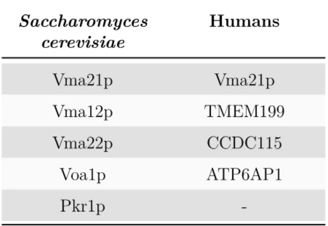
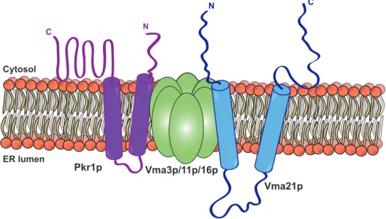
2 V-ATPase assembly and assembly factors 49 2.1 ER assembly factors in yeast . . . 50
2.1.1 Vma21p . . . 50
2.1.2 Vma12p and Vma22p . . . 52
2.1.3 Pkr1p . . . 54
2.1.4 Voa1p . . . 55
2.2 Regulation of V0 assembly in yeast . . . 56
2.3 ER assembly factors in humans . . . 56
2.3.1 VMA21 . . . 56
3 V-ATPase accessory subunits 69
3.1 ATP6AP1 . . . 69
3.1.1 ATP6AP1-CDG . . . 70
3.2 ATP6AP2 . . . 73
3.2.1 ATP6AP2 as a signalling receptor . . . 77
3.2.2 ATP6AP2 and the V-ATPase . . . 80
3.2.3 ATP6AP2 in canonical and non-canonical Wnt pathways . . . 80
3.2.4 ATP6AP2 as soluble protein: s(P)RR/ATP6AP2 . . . 82
3.2.5 ATP6AP2 and pathology in humans . . . 83
Aim of the Study 85 Results 89 Part I - Mutations in the V-ATPase assembly factor ATP6AP2 cause a glycosylation disorder with autophagic defects 91 Part II - Mutations in the V-ATPase assembly factor VMA21 are associated with a glycosylation disorder with autophagic liver disease 125 Discussion 173 Summary of the Findings and Discussion 175 Mutations in the ER assembly factors form a new subgroup of metabolic diseases with autophagic defects, impaired glycosylation and liver steatosis: solving the function of the enigmatic ATP6AP2 protein . . . 175
Mutations in the ER assembly factors form a new subgroup of metabolic diseases with autophagic defects, impaired glycosylation and liver steatosis: insights from VMA21 . . . 182
Mutations in the ER assembly factors ATP6AP2 and VMA21 are associated with impaired glycosylation . . . 186
Are mutations in the ER assembly factors ATP6AP2 and VMA21 a novel form of non-alcoholic fatty liver disease (NAFLD)? . . . 188
Conclusion 189
Bibliography 191
Bibliography 193
List of Figures
1 Structure of the V-ATPase . . . 28
2 Subunits and domains of the V-ATPase . . . 30
3 Rotary mechanism of the V-ATPase . . . 32
4 The pH of the secretory pathway and intracellular compartments in the mammalian cell . . . 33
5 V-ATPase is required to regulate the Golgi pH . . . 34
6 Distinct endocytic compartments with progressively decreasing pH 37 7 Signalling pathways regulating autophagy . . . 40
8 Regulated assembly of V-ATPases in response to nutrient avail-ability . . . 46
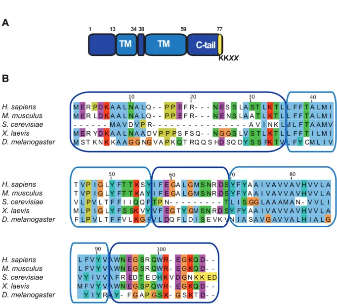
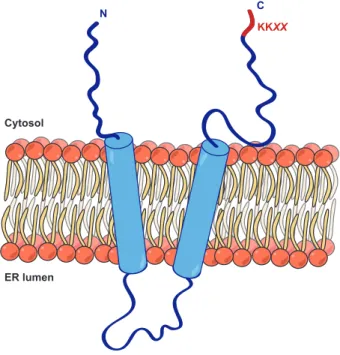
9 Structure of Vma21p and sequence alignment . . . 51
10 Structure and orientation of Vma21p in the ER . . . 52
11 The complex Vma12p/Vma22p binds Vph1p in the ER . . . 53
12 Model of Pkr1p and its function in the V-ATPase assembly . . . . 54
13 Schematic model of Voa1p . . . 55
14 Assembly of the V-ATPase complex in yeast . . . 57
15 Schematic drawing of TMEM199 and CCDC115 . . . 63
16 Schematic model of ATP6AP2 . . . 74
17 Structure of ATP6AP2 and sequence alignment . . . 75
18 Sequential processing of ATP6AP2 . . . 76
19 Functions of ATP6AP2 . . . 79
List of Tables
1 V-ATPase ER assembly factors in Saccharomyces cerevisiae and
humans . . . 49
2 VMA21 deficient patients: genetic, clinical and laboratory data . 61
3 Laboratory tests and normal values . . . 62
4 TMEM199 deficient patients: genetic, clinical and laboratory data 66
4 CCDC115 deficient patients: genetic, clinical and laboratory data 67
List of Abbreviations
4EBP1 4E-binding protein 1
ALP Alkaline phosphatase
ALT Alanine aminotransferase
AP-MS Affinity-purification mass spectrometry
AST Aspartate aminotransferase
Atg Autophagy-related genes
ATP6AP1 ATPase H+ transporting accessory protein 1
ATP6AP2 ATPase H+ transporting accessory protein 2
CCDC115 Coiled-coil domain containing 115
CDG Congenital disorder of glycosylation
CK Creatine kinase
COP I/II Coat protein I/II
CTSB/CTSD Cathepsin proteases B and D
CTF C-terminal fragment
DGAT Diacylglycerol acyltransferase
ECD Extracellular domain
ER Endoplasmic reticulum
ERAD ER-associated degradation
ERGIC ER-Golgi intermediate compartment
ERK Extracellular signal-regulated kinases
FL Full-length
Fz Frizzled
HDL High-density lipoprotein
HPLC High performance liquid chromatography
HRP Handle region peptide
HSP Heat shock protein
IEF Isoelectric focusing
LAMP Lysosome associated membrane protein
LC3 Microtubule-associated protein light chain 3
LDH Lactate dehydrogenase
LDL-C Low density lipoprotein (LDL) cholesterol
LDs Lipid droplets
LRP5/6 Low-density lipoprotein receptor-related protein 5/6 MALDI Matrix-assisted laser desorption/ionization
MAPK Mitogen-activated protein kinases
mTORC1 Mammalian target of rapamycin complex 1
NAFLD Non alcoholic fatty liver disease
NTF N-terminal fragment
p62 Ubiquitin-binding protein p62
PCP Planar cell polarity pathway
PERK Protein kinase R (PKR)-like endoplasmic reticulum kinase Pkr1p Pichia farnosiakiller toxin resistance
PLZF Promyelocytic leukaemia zinc finger protein 1
PMD Psychomotor disease
PRR Prorenin receptor
RAAS Renin angiotensin-aldosterone system
RAS Renin angiotensin system
RAVE Regulator of ATPase of vacuoles and endosomes
s(P)RR Soluble prorenin receptor
S6K1 S6 kinase 1
S1P Site-1 protease
SNARE Soluble N-ethylmaleimide-sensitive factor attachment protein receptors
SP Signal peptide
SREBP Sterol response-element binding protein
TGF-β1 Transforming growth factor-β1
TGN Trans-Golgi network
TM Transmembrane
TMEM199 Transmembrane protein 199
UPR Unfolded protein response
UTR Untranslated region
V-ATPase Vacuolar H+-ATPase
vma Vacuolar membrane ATPase
Voa1p V0 assembly protein 1
WD Wilson’s disease
XMEA X-linked myopathy with excessive autophagy
Abstract
The V-ATPase is a large complex involved in the acidification of intracellular or-ganelles. It is formed by a proton pore (V0 domain) and an ATP hydrolysis domain
(V1 domain). Pioneering studies in Saccharomyces cerevisiae have shown that the
as-sembly of the V0 domain occurs in the endoplasmic reticulum (ER) under the guidance
of five assembly factors: Vma21p, Vma12p, Vma22p, Pkr1p and Voa1p. The newly as-sembled V0 sector is then escorted by Vma21p to the cis-Golgi, where it will bind the
preassembled V1 domain to constitute a functional holoenzyme. How the assembly works
in mammalian systems is currently still unclear. Yet, it was recently shown that all the assembly factors, except Pkr1p, are conserved in mammals and that mutations in three of them, ATP6AP1, TMEM199 and CCDC115, were identified to cause a new subgroup of congenital disorders of glycosylation (CDG) in humans. Using human genetics and functional validation, I identify in the first part of my thesis a novel mammalian as-sembly factor called ATP6AP2 that causes a similar CDG-like disease when mutated. Apart from glycosylation defects, patients with missense mutations in ATP6AP2 show steatotic liver disease, cognitive defects and immune defects. Previously exon-skipping mutations in ATP6AP2 had been associated with a late onset cerebral disease, including Parkinsonism and epilepsy. Our work revealed that ATP6AP2 missense mutations lead to defective V-ATPase activity, subsequently causing reduced organellar acidification, lysoso-mal degradation and autophagic flux. Because of this, clearance of lipid droplets cannot take place in the autolysosomes, giving rise to the steatotic phenotype in the patients hepatocytes. Consistent with the similar clinical phenotype, we found that ATP6AP2 in-teracts with V0 assembly factors, while the missense mutations reduced this interaction,
suggesting a compromised V-ATPase assembly in the patients. By contrast, in patients with exon-skipping mutation we found normal glycosylation of serum proteins as well no effect on the interaction between ATP6AP2 and ATP6AP1, suggesting that the missense mutations have a stronger impact on overall ATP6AP2 function than the exon-skipping ones. These results shed light on the V-ATPase assembly in the ER and suggest that ATP6AP2 is an additional mammalian member of the assembly factors group.
In the second part of my thesis, I demonstrate that mutations in VMA21 also cause CDG with liver disease. Previously, mutations of VMA21 had been associated with a X-linked myopathy with excessive autophagy (XMEA), characterized by progressive vac-uolation and atrophy of skeletal muscle. Yet, we were able to identify VMA21 mutations
with a similar clinical phenotype compared to the other V-ATPase assembly factors defi-ciencies. Using patient fibroblasts, we tested the functional impact of the newly identified
VMA21 mutations on V-ATPase assembly and function, with the attempt to highlight
the differences with the XMEA ones. First, we could show that VMA21 mutations are hypomorphic and reduce both VMA21 mRNA and protein expression. Second, VMA21 mutations cause autophagic defects with decreased lipid droplet degradation, similar to those observed in ATP6AP2-deficient cells. Finally, patients fibroblasts showed an ac-cumulation of unesterified cholesterol in vesicular structures, similar to what has been reported for the lysosomal storage disease Niemann-Pick type C (NPC). The sequestra-tion of cholesterol in lysosomes triggers lipogenic pathways mediated by the sterol response element-binding protein (SREBP), most likely leading to elevated serum cholesterol lev-els in the patients. Altogether, our results show that V-ATPase deficiencies are a novel group of metabolic syndromes that affect lysosomal/autophagic homeostasis. Studying these rare V-ATPase assembly disorders, that are featured by liver steatosis as a unifying pathology, may lead to a better understanding of the pathogenesis of non-alcoholic fatty liver disease (NAFLD), which is a common problem in metabolic syndrome.
Chapter 1
The V-ATPase
The vacuolar (H+)-ATPases, or V-ATPases, are a family of ATP-dependent proton pumps, present in almost all the eukaryotic cells, involved in the acidification of intracellu-lar compartments and in the transport of protons from the cytoplasm to the extracelluintracellu-lar space (Nelson, 2003; Forgac, 2007; Kane, 2012; Breton et al., 2013; Sun-Wada et al., 2013; Marshansky et al., 2014; Cotter et al., 2015). The V-ATPases are multisubunit complexes and are composed of two different domains: the V1 domain, a
peripheral complex of 650 kDa involved in the binding and hydrolysis of the ATP, and the V0 domain, a membrane-embedded complex of 260 kDa implicated in the translocations of
protons (Forgac, 2007; Roh et al., 2018). Both domains work together as a rotary ma-chinery to drive proton transport across cell membranes (Forgac, 2007). The V-ATPases play a pivotal role in many intracellular processes that range from membrane trafficking, lysosomal protein degradation, post-translational processing and glycosylation, protein sorting as well as nutrient signalling and facilitation of ion transport (Forgac, 2007) (Figure 1).
1.1 V-ATPase structure and subunits
The V-ATPases are formed by fourteen different subunits, which are organized with different stoichiometry (Oot et al., 2017). The standard nomenclature, used to identify the V-ATPase subunits and the V-ATPase associated genes, has been approved by the HUGO Gene Nomenclature Committee (HGNC). The symbol used is the root “ATP6 ”, it is followed by the indication of the specific domain (V1 or V0) and then followed by the
letter indicating the subunit name. To avoid any confusion, the subunits in the V1 and
the V0 domains are named always with a capital letter, especially since the Guidelines for
Human Gene Nomenclature (http://www.gene.ucl.ac.uk/nomenclature/ guidelines.html) have suggested not to use names containing lowercase letters as symbols for human genes (Smith et al., 2003). The current nomenclature system has been implemented in 2010 to include more than forty V-ATPase variants of the fourteen subunits plus the two accessory proteins (Miranda et al., 2010). The catalytic V1 domain is made of eight
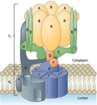
Figure 1: Structure of the V-ATPase
The V-ATPase is formed by two domains: the ATP hydrolytic domain V1 (A-H colored
yellow, orange and green) and the proton-translocator V0 domain (a, c, c’, c” and e,
colored blue and grey). From Forgac et al., 2007.
different subunits (A-H) and the membrane embedded V0 proton channel is composed of
six-seven subunits (a, d, e, f, plus two to three proteolipid isoforms referred to as subunits
c, (c’) and c”) (Forgac, 2007). The core V-ATPase complex is highly conserved from
simple eukaryotes to higher animals, including humans (Oot et al., 2017). Interestingly, biochemical purification from bovine cells has suggested that there are two subunits, which cannot be found in yeast. These two proteins do not belong to the core subunits and thus have been defined accessory subunits of the V-ATPase, ATP6AP1 and ATP6AP2 (Getlawi et al., 1994; Supek et al., 1994; Ludwig et al., 1998) (see Chapter 3 – V-ATPase accessory subunits).
The V-ATPase structure has been characterized extensively in the budding yeast
Sac-charomyces cerevisiae. These unicellular organisms require the proton pump to survive
under specific environmental conditions, for example in alkaline conditions or toxic levels of metals (Graham et al., 2003; Eide et al., 2005; Kane, 2006). Moreover, the formation of a proton gradient generated by the V-ATPase leads to the sequestration of Ca2+ and Zn2+ ions inside the vacuole (Klionsky et al., 1990). The deletion of any of
Introduction - Chapter 1
the V-ATPase subunits results in a number of specific growth and cellular phenotypes, which include the “vma- phenotype”, a phenotype characterized by a failure to grow on media buffered to pH 7.5 (Graham et al., 2003; Kane et al., 2006), sensitivity to a variety of metals (including Ca2+ and Zn2+ ions) (Eide et al., 2005; Kane, 2007)
and inability to acidify the vacuoles (Weisman et al., 1987). In Saccharomyces
cere-visiae, the subunit a is expressed as two isoforms, Vph1p and Stv1p. Both have specific
localizations, with Vph1p being found in the vacuole while Stv1p is present in the Golgi (Manolson et al., 2004). In higher organisms, most of the subunits are expressed as multiple isoforms, sometimes in a tissue-specific fashion (Toei et al., 2010), In humans, for example, there are four isoforms of the a subunit (a1, a2, a3 and a4 ) with distinct vesicular and cell type distribution: V0A1 is expressed on the synaptic vesicles, V0A2 on intracellular vesicles like Golgi and early endosomes, V0A3 is on the plasma membrane of osteoclasts, whereas V0A4 is expressed on the plasma membrane of renal intercalated cells (Nishi et al., 2002; Pamarthy et al., 2018).
The V1 domain is formed by three subdomains: the A3B3 cylinder, the central stalk
and the peripheral stalks. The A3B3 subdomain is a hexamer shaped cylinder, made
up of alternating A and B subunits, performing the ATP hydrolysis. There are three ATP hydrolytic sites located each one at the interface between A/B subunits (Forgac,
2007; Maher et al., 2009). The energy derived from ATP hydrolysis is coupled to the
movement of the central stalk, which acts as a rotor for the complex (Cotter et al.,
2015). The central stalk consists of single copies of subunits D, F and d. The last one,
localized on top of the V0 domain, provides the connection between the proteolipid ring
and the central stalk in V1 (Forgac, 2007). It has been shown that subunit H is essential
to inhibit the ATPase activity of the V1 domain when it is not yet attached to the V0
domain (Diab et al., 2009). The peripheral stalks consist of a core of EG heterodimers, connected to single copies of the subunits C, H and a (Zhang et al., 2008; Benlekbir et
al., 2012). The peripheral stalks tether the A3B3 hexamer to the subunit a, preventing
the rotation of the stator during ATP hydrolysis (Zhang et al., 2008; Benlekbir et
al., 2012) (Figure 2).
The structure of the V0 domain differs between yeast and humans. In Saccharomyces
cerevisiae, it consists of six subunits: a, c, c’, c”, d and e (Smith et al., 2003; Forgac,
2007). The mammalian V0 domains are composed of five subunits, since the c’ subunit
is not conserved: a (a1, a2, a3 and a4 isoforms), c, c”, d (d1 and d2 isoforms) and e (e1 and e2 isoforms), respectively (Marshansky et al., 2014). The current structural models describe the V0 domain as composed of a proteolipid ring of c subunits, with
adjacent a single copy of the a and e subunits (Smith et al., 2003; Forgac, 2007). The proteolipid subunits are small highly hydrophobic proteins, containing from four to five transmembrane helices (TMs) (Flannery et al., 2004; Wang et al., 2007). Each c subunit contains an essential buried acidic residue that undergoes to reversible protonation during proton transport (Hirata et al., 1997; Toei et al., 2010). The d
A E C B B’ D F V1V0 v v v DF A A B B V1 V0 TtC aNT(prox) aNT(dist) Cfoot Chead HCT D G E EG2 EG1 EG3 G E A3B3 ADP ATP ATP ATP ADP ADP ADP ATP A A A B B B B B B B B B A A A A A A ATP ADP ATP ADP EG1 EG2 EG3 A3B3DF V1V0 HCT HNT aNT aCT d c8c’c” C EG1 A
EG1 EG2 EG3
HNT H+ out in aCT 90° ATP 90° H+ G E E E G A G ADP + Pi B F D C d EG3 d c8c’c” aNT Ch Cf HCT aNT c’,c’’ c, H+ Glu 137 (c) Glu 108 (c'') H+ Arg735 B A c c c c c c c c c’ c'' 2 2 2 2 2 2 2 2 2 4 4 4 4 4 4 4 4 4 5 3 1 1 1 1 1 1 1 1 1 2 3 3 3 3 3 3 3 3 3 4 aCT EG2 HNT F
Figure 2: Subunits and domains of the V-ATPase
(A) The V-ATPase is formed by two domains: the ATP hydrolytic domain V1 and the
proton-translocator domain V0. (B) The ATP hydrolysis occurs at three of the six AB
interfaces and drives counterclockwise rotation of the central DF rotor. The A3B3 cylinder
seen towards the cytosol. (B’) Crystal structure of A3B3DF. The arrows in (B, B’) show
the site of the ATP hydrolysis. (C) The rotation of the central DF rotor drives the rotation of the proteolipid ring past the essential arginine residue in a. Protons enter a cytoplasmic half-channel and, after being carried 360o on lipid exposed glutamate residues
on the c subunits, are released into the luminal half-channel. (D) Crystal structures of the DF subunits, stalk EGC in two conformations, H subunit and C subunit, all from S.
cerevisiae. (E) CryoEM map of V1-V0coordinated with the models of individual subunits
and domains. (F) Crystal structures of the a subunit from M. ruber, d subunit from T.
Introduction - Chapter 1
subunit is present on top of the proteolipid ring, thus providing the connection between the ring and the central stalk in V1 (Forgac, 2007). The a subunit is a 100 kDa protein
with eight transmembrane domain, with the N-terminal domain in close proximity to the H subunit (Diepholz et al., 2008; Zhang et al., 2008; Benlekbir et al., 2012). Moreover, the subunit a possesses two hemi-channels and a crucial buried Arg residue (R735), which are required for proton translocation across the membrane (Forgac, 2007) (Figure 2).
The V-ATPase holoenzyme operates by a rotary mechanism (Hirata et al., 2003;
Imamura et al., 2003). The ATP hydrolysis, occurring in the V1 domain, causes
a rotation of the entire rotary assembly, formed by subunits D, F, d and the ring of proteolipid subunits. Subunit a and the A3B3 hexamer are kept in fixed positions by
the peripheral stators. Rotation of the proteolipid ring relative to subunit a leads to active transport of protons (Forgac, 2007). The subunit a provides for entry and exit of protons to the protonatable carboxyl groups of the glutamic acid (Glu) residues of the proteolipid ring. This occurs through the two aqueous hemi-channels of the subunit
a and induces the rotation of the protonatable sites of the proteolipid ring. Following
rotation, these sites are placed in contact with the hydrophobic environment and become protonated (Forgac, 2007). The essential TMs in both subunit a and in the proteolipid subunits of the V-ATPase undergo helical swivelling, meaning that they rotate about an axis through the center of the helix, relative to each other, which is possibly coupled to catalysis (Kawasaki-Nishi et al., 2003: Wang et al., 2004). Both inhibitors Bafilomycin A1 and Concanamycin A inhibit the V-ATPase activity by blocking this helical swivelling and then the proton transport (Bowman and Bowman, 2002; Huss et al., 2002). As there are three catalytic nucleotide-binding sites in the V1 domain and
six–ten protonatable sites on the proteolipid ring of V0 domain, the predicted H+/ATP
stoichiometry for the V-ATPases is 2–3.3 (Kettner et al., 2003) (Figure 3).
For intracellular pH regulation, the excess of positive charges across the organellar membrane due to increasing of ions H+ is counterbalanced by efflux of ions mediated by
exchangers or cations efflux (Steinberg et al., 2010; Weinert et al., 2010; Cang et al., 2015). Moreover, the intracellular acidity generated by the V-ATPase proton gradient is counteracted by proton back-flux called intrinsic H+ leakage that prevents the
over-acidification of the lumen of organelles (Paroutis et al., 2004).
1.2 V-ATPase function
The V-ATPases are ubiquitously expressed in the cell and are involved in the acidifi-cation of membrane-bounded compartments, thus playing a crucial role in many intracel-lular processes, including membrane trafficking, protein glycosylation and sorting, protein degradation in the lysosomes or nutrient signalling.
A B D C F H A A A B B B D C G GE E e a c d c c" c" c H+ F D e a c d c c" c" c’’ F D e c c c’ c" c F D a c d c c" c’ c H C H C E108 E789 R799 Cytoplasm Lumen a e H+ H+ H+ R735 H74 E145 H C d E137 E789 R799 R743 R735 H+ E108 H+ H+ H+ ADP + Pi ATP E137 R735H+ H+ E108 R799 E789 H743 H+ E145 H+ E108 R799 E789 H743 H+
Figure 3: Rotary mechanism of the V-ATPase
(A) Protons enter at the cytoplasmic side of the hemi-channel in subunit a. (B) Proto-nation of buried Glu residues in the proteolipid ring (E137 in subunit c, E145 in subunit
c’ and E108 in subunit c”). (C) Forced exposure of these residues into the
hydropho-bic phase of the bilayer keep protonated the Glu residues. (D) Rotation is facilitated by ATP hydrolysis that occurs at the catalytic sites in the interface of subunits A and B. The ATP hydrolysis drives rotation of a central rotor that includes subunits D and F (colored orange) of V1 connected to subunits d and the proteolipid ring of V0 (colored blue). The
subunits a and e of V0 (colored grey) are kept fixed relative to the A3B3 hexamer thank to
the peripheral stalks. Following rotation, protons are displaced from the proteolipid sub-units into a luminal hemi-channel of subunit a as a result of the interaction between the glutamic acid side chains and a crucial buried arginine residue (R735) in transmembrane helix-7 (TM7) of subunit a. Other buried charged residues in the C-terminal domain of subunit a (H743, E789, R799) line the luminal hemi-channel through which protons exit the membrane. From Forgac et al., 2007.
Introduction - Chapter 1
Indeed, the luminal pH is not homogeneous through the secretory pathway (Paroutis et
al., 2004; Casey et al., 2010; Pamarthy, et al., 2018). Since the assembly and the
consequent activity of the V-ATPase start only in the Golgi, the pH of the endoplasmic reticulum (ER) is near neutral and similar to that of the cytosol, while downstream compartments, where the V-ATPase is active, become progressively more acidic. The pH of the cis-Golgi drops down significantly with a more acidic pH of 6.7 and the acidification becomes more marked in subsequent cisternae of the Golgi complex, reaching pH 6.0 in the
trans-Golgi network (TGN). Furthermore, the pH of secretory granules has been reported
to be as low as 5.2 (Anderson and Pathak, 1985; Wu et al., 2000; Paroutis et
al., 2004). It has been proposed that the progressive reduction of pH along the secretory
pathway depends on different isoforms of the V-ATPases, on the variation of density of V-ATPases and on the assembly of the V-ATPases (Casey et al., 2010) (Figure 4).
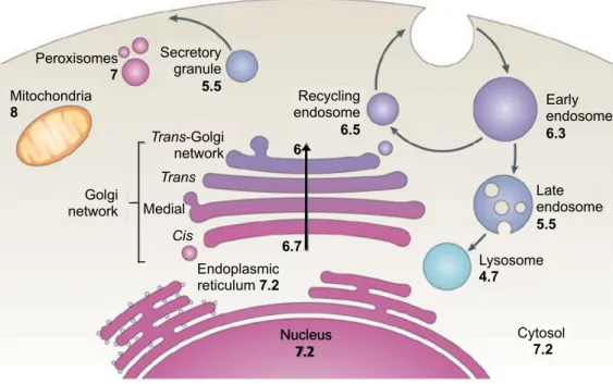
Early endosome 6.3 Peroxisomes 7 Secretory granule 5.5 Mitochondria 8 Late endosome 5.5 Recycling endosome 6.5 Lysosome 4.7 Cytosol 7.2 Golgi network Trans-Golgi network Trans Medial Cis Endoplasmic reticulum 7.2 6.7 6 Nucleus 7.2
Figure 4: The pH of the secretory pathway and intracellular compartments in the mammalian cell
pH of each organelle is indicated by the numbers. Mitochondrial pH refers to the matrix. Early endosomes refer to the sorting endosomal compartment. pH of the multivesicular late endosomes refers to the bulk luminal fluid; pH of the fluid contained by the internal vesicles might differ. From Casey et al., 2010.
1.2.1 Role of the V-ATPase in the Golgi: regulation of pH and
glycosylation
The Golgi pH plays a pivotal role in membrane trafficking (Griffiths et al., 1983;
Palokangas et al., 1998), protein sorting (Matlin, 1986; Caplan et al., 1987; Ellis and Weisz, 2006), and glycosylation of proteins and lipids (Kuismanen et al., 1985; Thorens and Vassalli, 1986; Gawlitzek et al., 2000; Axelsson et al., 2001; Campbell et al., 2001, Kellokumpu et al., 2002). The decrease of the
luminal pH through the secretory pathway is dictated by a simultaneous increase of proton import from the luminal side and a decrease of proton export to the cytosol (Kim et
al., 1996; Demaurex et al., 1998; Farinas and Verkman, 1999; Schapiro and
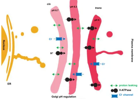
Grinstein, 2000; Wu et al., 2001). Also in the Golgi, the steady-state pH is dictated
by a balance of three components: (1) V-ATPase, involved in the import of protons, (2) proton export/elimination (also called proton leaking), (3) counter-ion conductance, performed by Cl- channel families, such as the Golgi pH regulator (GPHR) and Golgi
anion channel (GOLAC1 and 2) (Kim et al., 1996; Demaurex et al., 1998; Farinas
and Verkman, 1999; Grabe and Oster, 2001; Wu et al., 2001; Paroutis et al., 2004; Thompson et al., 2006; Maeda et al., 2008) (Figure 5).
pH 6.7 pH 6.3 pH 6 proton leaking V-ATPase Cl-channel Pl a s m a m e m b ra n e ER cis trans Golgi pH regulation Nu c le u s Cl -H+ Cl
-Figure 5: V-ATPase is required to regulate the Golgi pH
Processing and sorting of glycoproteins at the Golgi require a pH that becomes progres-sively more acidic from cis- to trans-Golgi (indicated by the color change). The gradient of pH from cis- to trans-Golgi is guaranteed by the increase in the density of V-ATPases con-comitant with a decrease in the density of H+ leaks. The chloride channels are present at
low and uniform density to keep the membrane potential near zero by providing counter-ion movement to compensate for H+ influx. The numbers indicate the pH. Adapted from
Introduction - Chapter 1
For the regulation of glycosylation by the Golgi pH, two different hypotheses can be proposed. One is that some glycosylation enzymes, such as sialidases, have a specific pH optimum (Gawlitzek et al., 2000). The other is that an increased pH causes incorrect localization of glycosyltransferases within the Golgi and post-Golgi organelles (Axelsson et al., 2001), leading to a loss of ability to execute glycosylation reactions in the correct order (Maeda and Kinoshita, 2010). The catalytic activity of most of the glycosyltransferases depends on the luminal pH and on the distribution of divalent cation, such as Ca2+, Mg2+and/or Mn2+. The last ones are important to modify the conformation
of the substrates that have to enter in the catalytic pocket of the glycosyltransferases (Petrová et al., 2001). Moreover, it has been shown that already an increase in Golgi pH of only 0.2 is sufficient to inhibit a terminal sialylation of N -glycans and to induce selective mislocalization of the corresponding sialyltransferase. The higher pH does not affect the other glycosylation enzymes or Golgi markers, indicating that mislocalization of certain Golgi glycosyltransferases is primarily responsible for the pH-induced glycosylation defects (Rivinoja et al., 2009a, b; Maeda and Kinoshita, 2010). Perturbation in the Golgi pH not only impairs glycosylation but also membrane trafficking and protein sorting (Rivinoja et al., 2009a, b). Interestingly, it has been demonstrated that some glycosyltransferases can form aggregates in the more acidic environment (pH 6.3) of the
trans-Golgi and TGN. The formation of aggregates enables their rapid movement through
1.2.2 Role of the V-ATPase in endolysosomes and lysosomes
acidification
The progressive luminal acidification is important in various aspects of endocytosis and intracellular membrane trafficking. Indeed, the acidic pH in the endocytic compartment allows the dissociation of the ligands from their receptors complexes and the recycling of the receptors (Forgac, 2007). For instance, the endocytic acidification is important for the continued uptake of low density lipoprotein (LDL), a major carrier of plasma cholesterol, as well as termination of signalling from receptors such as epidermal growth factor receptor (EGFR) (McGuire et al., 2017). Moreover, even the normal trafficking of membranes between endocytic compartments seems to be controlled by luminal acidi-fication (Weisz, 2003). It has been shown that the acidiacidi-fication of early endosomes, due to the proton accumulation driven by the V-ATPase, recruits the small GTPase ADP-ribosylation factor 6 (Arf6) and the ARF nucleotide-binding site opener (ARNO): the two proteins are involved in endocytosis and have been implicated in vesicle-coat formation, remodelling of the actin-cytoskeleton and in lipid modification (Hurtado-Lorenzo et
al., 2006). Arf6 and ARNO interact with the V0 subunits a2 and c of the V-ATPase
(Hurtado-Lorenzo et al., 2006; Recchi and Chavrier, 2006). These observations indicate that the V-ATPase, through the a2 subunit, can regulate membrane trafficking (Hurtado-Lorenzo et al., 2006).
In the lysosomes, the V-ATPases provide the essential acidic environment for protein degradation, for example by promoting the activity and the proteolytic activation of acid-dependent proteases such as cathepsin B and D (CTSB and CTSD), and for the recovery of the resultant amino acids via proton-coupled amino acid transporters. Within secretory vesicles, V-ATPases both activate processing of precursor molecules and drive uptake of small molecules, such as neurotransmitters (Breton and Brown, 2013) (Figure 6).
1.2.3 Role of the V-ATPase in fusion of vesicles
The V-ATPases have also been proposed to play an important role modulating the fusion of vesicles. Pioneering studies performed in yeast uncovered a pivotal role of two V0 subunits, the subunit c and a, in the fusion of vacuolar membranes downstream of
SNARE complex assembly (Peters et al., 2001; Bayer et al., 2003). Indeed, a first evidence is suggested by the fact that deletion of the V0 proteolipid subunit Vma3 could
inhibit the traffic along the secretory pathway more severely than mutations in V1subunits
(Yamashiro et al., 1990; Morano and Klionsky, 1994), even if mutations in both abolish the proton pump activity (Stevens and Forgac, 1997; Peters et al., 2001). Furthermore, Peters and Bayer suggested the formation of trans-complexes formed by V0 domain from apposed vacuolar membranes, which may form a proteolipid channel
spanning both membranes (Peters et al., 2001; Bayer et al., 2003). Both groups proposed that the formation of these trans-complexes follows the docking and pairing of
Introduction - Chapter 1 Plasma membrane Recycling endosome Early endosome pH ~ 6.0 ECV ARF6 ARF6 ADP ADP H+ eCOPI BFA/Baf ARF1GTP pH-dependent ATP Baf pH-dependent ATP H+ ADP Late endosome pH 5.0 - 6.0 Baf Lysosome pH 5.0 - 6.0 ADP H+ H+ ADP ATP ATP ARF6 ARF6 ARF6 ARF6 ARNO ARNO
Figure 6: Distinct endocytic compartments with progressively decreasing pH
The V-ATPase (colored orange) pumps protons into the lumen of the different endosomal compartments, contributing to intra-endosomal acidification. Inhibition of V-ATPase activity by Bafilomycin A1 (Baf) or Brefeldin A (BFA) blocks endocytic trafficking at the level of early endosomes. This occurs with ARF1-dependent recruitment of the endosomal COPI coat (COPI, dashed line) and the formation of endosomal carrier vesicles (ECVs). The recruitment of ARNO and ARF6 to the early endosomal membrane is pH-dependent. The two proteins interact with the V-ATPase a2 and c subunits, respectively. From Recchi and Chavrier, 2006.
the trans-SNARE (t-SNARE) and is independent of the proton gradient, suggesting a role of the V0 subunits in the direct fusion of vacuoles (Bayer et al., 2003). Moreover, Bayer
et al. demonstrated the importance of the subunit a (Vph1p) on both fusion partners,
suggesting a symmetric requirement of the integral multisubunit cylinders to connect the two vacuolar membranes before fusion (Lindau and Almers, 1995; Zimmerberg,
2001). Subsequently, a genetic screen for synaptic malfunction in Drosophila confirmed
the role of the subunit a1 of the V0 domain as an essential component for synaptic
transmission, also acting downstream of SNAREs (Hiesinger et al., 2005). For this specific role, the interaction of Ca2+/calmodulin with the subunit a1 was critical and leads
to spontaneous release of neurotransmitters at the neuromuscular junction (Wang et al.,
2014). Additional studies have proven the role of the subunit a in the regulation of vesicles
fusion: the subunit a3 regulates the secretion of insulin in pancreatic beta cells
(Sun-Wada et al., 2006), the subunit a1 mediates the fusion lysosome-phagosome during
phagocytosis in zebrafish (Peri and Nüsslein-Volhard, 2008), while the subunit a is involved in apical secretion of multivesicular bodies and in the exosome-mediated apical secretion of Hedgehog-related proteins in Caenorhabditis elegans (Liégeois et al., 2006;
Rama et al., 2018). Although biochemical studies propose a direct interaction of the
subunit c with several SNAREs present on synaptic vesicles (Bennet et al., 1992;
O’Connor et al., 1993; Galli et al., 1996; Shiff et al., 1996), only in 2010 Di
Giovanni et al. provide the first evidence of a direct interaction between a V0 subunit
and SNAREs. Indeed, the same group found in yeast two-hybrid (Y2H) that the subunit
c binds directly the t-SNARE synaptobrevin (VAMP2) through its C-terminal. In vivo,
they confirmed that, when this interaction is disrupted, there is a significant decrease of neurotransmission, pinpointing the importance of the interaction between V-ATPase subunits and SNARE in the vesicles fusion (Di Giovanni et al., 2010). Conversely, subsequent studies have correlated the function of the V-ATPase with fusion of vesicles by proposing that lack of V-ATPase subunits leads to impaired vesicles acidification and thus to impaired exocytosis (Coonrod et al., 2013; Poëa-Guyon et al., 2013). Intriguingly, mutations in both V0 (Kornak et al., 2008; Jansen et al., 2016a, b, c)
and V1 subunits (Van Damme et al., 2017; Zhao et al., 2018) have been reported
to disrupt vesicular trafficking, Golgi homeostasis and post-Golgi anterograde transport.
1.2.4 Role of the V-ATPase in autophagy
Lysosomal degradation is closely related to autophagy, a catabolic pathway by which cells remove misfolded or aggregated proteins and damaged organelles. Autophagy pro-vides the energy in response to nutrient and environmental stress and recycles macro-molecules providing molecular building blocks (Mizushima and Komatsu, 2011; Kim
and Guan, 2015). The name autophagy derives from the Greek and means “eating of
self”. It was used by Christian de Duve more than 40 years ago, with the observation of vesicles containing amorphous materials, such as mitochondria, and partially degraded
Introduction - Chapter 1
cytoplasmic organelles by electron microscopy (Clark et al., 1957; De Duve et al.,
1966). In the late 1970s, it was observed that amino acid starvation was able to induce
autophagy in cultured mammalian cells and in perfused rat livers (Mitchener et al.,
1960; Mortimore and Schworer, 1977). So far, more than thirty autophagy-related
genes (Atg) have been described and most of them have been identified by genetic screen-ing in yeast (Glick et al., 2010). Many of these genes are conserved in mammals, highlighting the fundamental nature of the autophagic process (Nakatogawa et al.,
2009). There are three different types of autophagy: macroautophagy, microautophagy
and chaperone-mediated autophagy (CMA). Each type of autophagy promotes the prote-olytic degradation of cytosolic components inside the lysosomes. The cell uses macroau-tophagy to deliver cytoplasmic cargos to the lysosome through the autophagophore, a double membrane-bound vesicle that fuses with the lysosome to form an autolysosome. The cytosolic components are degraded through microautophagy: they are directly taken up by the lysosomes through invagination of the lysosomal membrane. In the last type of autophagy, the CMA, the proteins are conjugated with chaperones, such as members of the heat shock proteins, and then transported across the lysosomal membrane by the lysosomal-associated membrane protein 2A (LAMP2A) (Saftig et al., 2008; Glick et
al., 2010) (Figure 7).
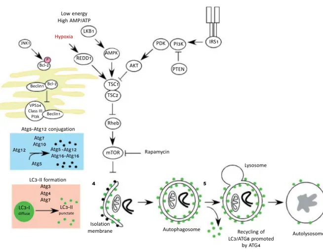
At the molecular level, autophagy occurs in five key stages: (1) formation of the phagophore (also called nucleation); (2) Atg5–Atg12 conjugation, followed by interaction with Atg16L and multimerization with other Atg at the phagophore; (3) processing of LC3 and insertion into the phagophore membrane; (4) capture of the targets for degradation; (5) formation of the autolysosome, derived by the fusion of the autophagosome with the lysosome and degradation of the cargos by the lysosomal proteases (Glick et al., 2010) (Figure 7).
1. Phagophore formation. In mammalian cells, the formation of the phagophore
initiates with membranes derived from the ER (the so-called omegasome)
(Hayashi-Nishino et al., 2009; Yla-Anttila et al., 2009), but also from other cytosolic
mem-branes organelles, such as the Golgi, the trans-Golgi, the mitochondria, the late endosomes (Mizushima, 2007; Mizushima and Klionsky, 2007; Axe et al., 2008) and, possi-bly, even from the nuclear envelope under restricted conditions (English et al., 2009). In yeast, the formation of the phagophore requires the activity of the Atg1 kinase in complex with Atg13 and Atg17. It has been proposed that the formation of this complex recruits the transmembrane protein Atg9 that promotes lipid recruitment to the expand-ing phagophore (Kundu and Thompson, 2005; Klionsky, 2007; Simonsen and
Tooze, 2009). mTOR can inhibit this process phosphorylating Atg13 and preventing it
from interacting with Atg1 (Díaz-Troya et al., 2008). In mammalian cells, exist two homologues of Atg1, Ulk-1 and Ulk-2, but how they work in promoting autophagy is still not well understood (Kundu and Thompson, 2005). The vesicular protein sorting 34 (Vps34) class III phosphoinositide 3-kinases (PI3K) uses the phosphatidylinositol (PI)
Low energy High AMP/ATP 4 3 1 2 Hypoxia Lysosome Autolysosome Autophagosome Rapamycin Recycling of LC3/ATG8 promoted by ATG4 Atg5-Atg12 conjugation LC3-II formation Isolation membrane Atg12 Atg5 Atg7 Atg10 Atg5 -Atg12 Atg16-Atg16 mTOR Rheb TSC1 TSC2 Atg3 Atg4 Atg7 LC3-II punctate LC3-I diffuse IRS1 PTEN PI3K PDK AKT AMPK LKB1 REDD1 JNK1 Bcl-2 Beclin1 Beclin1 P Bcl-2 5 VPS34 Class III PI3k
Figure 7: Signalling pathways regulating autophagy
Autophagy is a complex process that involves the following key steps: (1) nucleation of the phagophore in the ER initiated by Beclin-1/VPS34; (2) Atg5–Atg12 conjugation, interaction with Atg16L and multimerization of the Atg proteins; (3) processing of LC3 and insertion in the extending phagophore membrane; (4) capture of the targets for degradation, recycling of some LC3-II; (5) fusion of the autophagosome with the lysosome, proteolytic degradation by lysosomal proteases. The autophagy pathway is regulated by several signalling pathways, including JNK-1, a stress-signalling kinase that promotes autophagy by activating Bcl-2 and promoting the interaction of Beclin-1 with VPS34. Autophagy is regulated also by the mTOR kinase, via the inhibition of the ATG1/Ulk-1/-2 complexes at the earliest stages in phagophore formation. mTOR integrates metabolic, growth factors and energy signalling into autophagy. Autophagy can be induced by hypoxia and low cytosolic ATP levels. Inhibition of the autophagy can occur by the insulin receptor and its adaptor, IRS1. mTOR activity is promoted through inhibition of TSC1/TSC2 and increased Rheb GTPase activity. Adapted from Glick et al., 2010.
Introduction - Chapter 1
as substrate to generate phosphatidyl inositol triphosphate (PI3P), a lipid important for phagophore elongation and involved in the recruitment of other Atg proteins (Xie
and Klionsky, 2007). Vps34 interacts with Beclin-1 and promotes its catalytic activity
increasing the levels of PI3P. Additional regulatory proteins complex can regulate posi-tively or negaposi-tively the phagophore formation and then the autophagy induction in the ER: UVRAG, BIF-1, Atg14L and Ambra activates the autophagy (Liang et al., 2006;
Fimia et al., 2007; Takahashi et al., 2007), while Rubicon and Bcl-2 can inhibit it
(Pattingre et al., 2005; Matsunaga et al., 2009; Zhong et al., 2009). Indeed, in starvation, the interaction of Beclin-1 with Bcl-2 (and Bcl-XL) in the ER inhibits Beclin-1 activity, disrupting its interaction with Vps34 (Pattingre et al., 2005; Maiuri et al.,
2007; Wei et al., 2008).
2. Atg5–Atg12 conjugation. Atg7 acts as an E1 ubiquitin enzyme and activates Atg12
by binding to its carboxyterminal glycine residue, in an ATP-dependent manner. Atg12 interacts with Atg10, an E2-like ubiquitin carrier protein that potentiates covalent linkage of Atg12 to lysine 130 of Atg5 (Glick et al., 2010). The Atg5–Atg12 complexes and dimers of Atg16L interact together to form a big complex (Atg5–Atg12–Atg16L). This complex associates with the phagophore, induces a curvature into the growing phagophore through asymmetric recruitment of LC3B-II (Glick et al., 2010). Once the autophago-some is formed, the complex Atg5–Atg12–Atg16L dissociates from the phagophore. For that reason, the conjugated Atg5–Atg12 is a relatively poor marker of autophagy (Barth et al., 2010; Zhang et al., 2016).
3. LC3 processing. LC3B, Atg8 in yeast, is expressed in most cell types as a
full-length cytosolic protein. Under autophagy induction, LC3B is proteolytically cleaved by the cysteine protease Atg4 to generate LC3B-I. After the cleavage, LC3B-I exposes a car-boxyterminal glycine that is then activated by the E1-like Atg7. Activated LC3B-I is then transferred to a different E2-like carrier protein (Atg3) and the phosphatidylethanolamine (PE) is conjugated to the carboxyl glycine to generate LC3B-II. Atg5–Atg12 recruit and integrate LC3B-II in the growing phagophore. LC3B-II is found on both the internal and external surfaces of the autophagosome, where it plays a role in both hemifusion of membranes and in selecting cargos for degradation (Glick et al., 2010). During the autophagy, the synthesis and processing of LC3 are increased, thus LC3 is the golden standard marker of autophagy (Barth et al., 2010). During autophagy, the LC3-related protein GABARAP [γ-aminobutyric type A (GABAA)-receptor associated protein] local-izes at the autophagosomes with LC3-II and undergoes to similar processing (Kirkin et al., 2009). The role of the LC3-related molecules in autophagy is not clear, but their interactions may determine the selection of cargos engulfed in the autophagosome (Schwarten et al., 2009; Glick et al., 2010; Zhang et al., 2016).
4. Selection of cargos for degradation. In general, autophagy is considered as a
ran-dom process engulfing indiscriminately cytosol organelles, including mitochondria, ER and Golgi membranes (Eskelinen, 2008). However, there are evidence that LC3B-II
could act as a receptor on the growing phagophore membrane interacting with adaptor proteins on the target (e. g. protein aggregates, mitochondria) promoting their selective uptake and degradation. In this context, the best characterized protein is the ubiquitin-binding protein p62/sequestosome-1 (SQSTM1), an adaptor that promotes turnover of poly-ubiquitinated protein aggregates through interaction with LC3 at the autophago-some. p62 has largely used as marker for defective autophagic degradation (Glick et al.,
2010; Zhang et al., 2016).
5. Fusion with the lysosome. The last step in the maturation of the autophagosome
is the fusion with early and late endosomes. This happens prior to fusion with the lysosome to form the final autolysosome (Mizushima, 2007). When autophagosomes and endosomes fuse, the pH starts to lower acidifying the autophagic vesicle before delivery of lysosomal acid proteases (Eskelinen, 2005). This process requires the small G protein Ras-related protein (Rab7) bounded to GTP (Gutierrez et al., 2004; Jägeret al.,
2004) and also the Presenilin protein (Eskelinen, 2005). The cytoskeleton is involved
in the autolysosome formation since drugs against the microtubules block fusion of the autophagosome with the lysosome (Webb et al., 2004). The CTSB and CTSD, present inside the lysosomes, are required for turnover of autophagosomes and for the maturation of the autolysosome, and can be both used as marker of lysosomal homeostasis (Koike et
al., 2005; Zhang et al., 2016). LAMP1 and LAMP2 are involved in the autolysosome
maturation and in the fusion of the autophagosome with the lysosomes. For instance, inactivation of LAMP2 is associated with the Danon disease in humans, an X-linked disease that leads to a pathogenic accumulation of autophagosomes in the cardiac muscle (Tanaka et al., 2000).
As the V-ATPase is important in the acidification of the lysosomes and is necessary to activate the lysosomal acid hydrolases, it is of great importance in autolysosomal degradation (Kissing et al., 2015; Pamarthy et al., 2018). Thus, many studies point to the requirement of functional V-ATPase for autophagy (Mijaljica et al., 2011) and Bafilomycin A1, the V-ATPase inhibitor, is also used as a classical inhibitor of autophagy (Carr et al., 2008). However, the precise role of V-ATPase in regulating the membrane dynamics of the autophagic flux is not completely clear (Pamarthy et al., 2018). It has been shown that the inhibition of the activity of both V-ATPase and Ca2+ pump
SERCA via the treatment with Bafilomycin A1 leads to a block in autophagic flux, while genetic inactivation of the V-ATPase still allows fusion of lysosomes with autophagosomes (Mauvezin et al., 2015). These results pinpoint to the involvement of V-ATPase in the degradation of autophagic cargos in lysosomes more than a function in regulating the autophagic flux (Pamarthy et al., 2018).
Introduction - Chapter 1
1.2.5 Role of the V-ATPase in mTOR regulation
Another very exciting link between autophagy and the V-ATPase is the involvement of the V-ATPase in signalling by the master growth regulator mTORC1 activity (Zoncu et al., 2011). The activation of mTOR occurs in presence of nutrients and is mediated by Akt kinase, PI3K and growth factor receptor signalling (Kim and Guan, 2015). When the amino acids are present in adequate levels, the V-ATPase interacts with the Ragulator, a scaffolding complex that anchors the Rag GTPases to the lysosomes (Zoncu et al., 2011). Rag GTPases promote the translocation of mTORC1 to the lysosomal surface, where mTORC1 can come in contact with Rheb, an important mTOR activator (Zoncu et al., 2011; Dibble and Manning, 2013). In Drosophila epithelial cells, V-ATPase/mTOR signalling is required for Rheb-stimulated protein uptake, suggesting a positive feedback loop between endocytosis and mTOR activation (Gleixner et al.,
2014). When the V-ATPase function is disrupted either genetically or pharmacologically,
mTORC1 remain inactive, even when amino acids are abundant (Zoncu et al., 2011) (Figure 7).
The main downstream effectors of mTORC1 are the ribosomal protein S6 kinase 1 (S6K1), the eukaryotic translation initiation factor 4E-binding protein 1 (4EBP1) and UNC-51-like kinase 1 (ULK1) (Yoon and Choi, 2016). When mTORC1 is activated, it phosphorylates the hydrophobic motif of S6K1 and leads to the activation of S6 inducing ribosome biogenesis (Shimobayashi and Hall, 2014). Instead, mTORC1 phosphory-lates 4EBP1 on multiples sites inhibiting 4EBP1 and leading to translational initiation (Shimobayashi and Hall, 2014). In presence of nutrients, mTORC1 is activated and binds the ULK1 complex. This complex is formed by ULK1, the homolog of Atg1 in yeast, the mammalian homolog of Atg13, Atg101 and FIP2000. The binding of mTORC1 to the ULK1 complex leads to the phosphorylation of both Atg13 and ULK1, inhibiting ULK1 activity and leading to an overall inhibition of autophagosome formation (Ueno
and Komatsu, 2017). However, in starvation, mTORC1 is inactivated and dissociates
from the ULK1 complex. In this way, mTORC1 can initiate the autophagosome formation (Rabinowitz and White, 2010).
mTORC1 is repressed by nutrient starvation, reduced growth factors and by the drug Rapamycin (Kim and Guan, 2015). Moreover, mTORC1 is initially inactivated during autophagy, to ensure reduced cell growth, but the generation of energy supplied by the degradation of autolysosomal products at the end of the autophagic flux leads to the reactivation of the kinase complex. This reactivation is required for the formation of new lysosomes and highlight the important role of mTORC1 in the completion of the autophagic flux (Kim and Guan, 2015).
At the transcriptional level, autophagy is regulated by the transcription factor EB (TFEB) (Settembre et al., 2011, 2012, 2013; Settembre and Ballabio, 2014). mTORC1 directly phosphorylates TFEB at Ser142 and Ser211, leading to its cytoplasmic sequestration and cytoplasm-to-nucleus shuttling (Martina et al., 2012; Settembre
et al., 2012). TFEB’s activity is inhibited by the Rag GTPases in presence of amino acids. Indeed, the Rag GTPases bind and sequester TFEB in the lysosome (Martina
and Puertollano, 2013). The absence of Rag in RagA and RagB deficient cells leads
to a constitutively activation of TFEB, regardless of nutrient availability (Kim et al.,
2014).
1.2.6 Role of the V-ATPase on the plasma membrane
A small number of cells, including renal epithelial cells, epididymal clear cells, phago-cytes and osteoclasts, possess plasma membrane V-ATPases extruding protons into the extracellular space (Nishi and Forgac, 2002; Maxson and Grinstein, 2014). The plasma membrane ATPases are involved in many cellular processes: in renal cells, V-ATPases are important in the maintenance of the plasma pH homeostasis and in the secretion of acid into the urine, thus regulating the acid-base balance (Brown and
Bre-ton, 2000; Maxson and Grinstein, 2014). In the epididymal clear cells, the V-ATPase
pumps across the membrane the H+ to provide the required low pH for sperm
matura-tion and storage (Brown and Breton, 2000; Maxson and Grinstein, 2014). In the osteoclasts, the plasma membrane V-ATPases are involved in the bone resorption, pumping H+ in the extracellular resorption lacuna, and are required for the activity of
secreted acid hydrolases involved in the degradation of the organic bone matrix (Maxson
and Grinstein, 2014). By modifying tissue pH and extracellular matrix composition,
plasma membrane V-ATPases are also important in the survival and invasiveness of tu-mor cells (Hinton et al., 2009; Capecci and Forgac, 2013; von Schwarzenberg et al., 2013; Cotter et al., 2015).
1.3 Regulation of the V-ATPase
Since so many essential intracellular processes are regulated by pH, the regulation of the V-ATPase activity must be tightly controlled (Collins and Forgac, 2018).
One level of regulation involves a separate, and even simultaneous, assembly of the V0
and V1 domains. It is known that the assembly of the V0 subunits takes place in the ER
(see Chapter 2 – V-ATPase assembly and assembly factors). Only later in the Golgi, the V0 domain binds to either the almost assembled V1 domain plus the subunit
C, or to individual V1 subunits (Forgac, 2007).
Another level of regulation is provided by the reversible dissociation of the V1 and V0
domains from each other and is thought to preserve cellular ATP stores (Forgac, 2007;
Kane, 2012). In yeast, V-ATPase disassembly occurs upon glucose starvation. This
process is rapid, reversible and does not require new protein synthesis (Kane, 1995). Only V-ATPases localized to the vacuole undergo disassembly in response to glucose withdrawal, while this phenomenon does not occur for the Golgi localized V-ATPases
Introduction - Chapter 1
(Kawasaki-Nishi et al., 2001). The presence of glucose allows the reassembly of the V-ATPase and requires two glycolytic enzymes, the aldolase and the phosphofructokinase, and the presence of the heterotrimeric regulator of ATPase of vacuoles and endosomes (RAVE) complex, which binds directly the V-ATPase in a glucose-dependent fashion (Smardon et al., 2002; Lu et al., 2004; Chan and Parra, 2014). Moreover, Forgac and his collaborators could show that regulated assembly of the yeast V-ATPase is controlled by the Ras/cAMP/protein kinase A (PKA) pathway in response to changes in glucose availability (Bond and Forgac, 2008; Collins and Forgac, 2018).
Also in humans, the V-ATPase assembly/disassembly is a finely regulated process that responds to a plethora of stimuli. The mammalian V-ATPases respond to glucose availability and, in kidney, elevated glucose levels (20 mM; normal physiological range 3.5–5.5 mM) cause increased V-ATPase assembly and acidification of intracellular vesicles. Probably, this occurs to maintain the neutral cytosolic pH during times of increased glycolysis (Sautin et al., 2005). The glucose-mediated assembly in mammalian cells depends on PI3K (Sautin et al., 2005) which also controls mTORC1 (Lemmon and
Schlessinger, 2010). It has been recently reported that, in mammalian cells, the
V-ATPase assembly and activity on lysosomes is increased also during glucose starvation (McGuire and Forgac, 2018). This is in contrast to the yeast model, where the glucose starvation causes dissociation of the V-ATPase, probably to store the ATP for nutrient-limiting conditions (Kane, 1995; McGuire and Forgac, 2018). The level of amino acids inside the cell can directly regulate the V-ATPase, indeed amino acid starvation leads to a rapid and reversible increase in V-ATPase assembly and activity in lysosomes, in a PI3K and mTORC1 independent fashion (Stransky and Forgac,
2015). The increased V-ATPase assembly lowers the lysosomal pH values, increasing
protein turnover and amino acid levels, suggesting that a regulated assembly of the V-ATPase contributes to amino acid homeostasis (Stransky and Forgac, 2015; Collins
and Forgac, 2018) (Figure 8).
1.4 V-ATPase and disease
Genetic defects in V-ATPase genes have been associated with several hereditary human diseases.
Loss-of-function mutations of the ATP6V0A3 subunit (MIM: 604592) have been found to be causative for autosomal recessive osteopetrosis (ARO) (Frattini et al., 2000;
Kornak et al., 2000; Bhargava et al., 2012). ARO is a rare infantile malignant
osteopetrosis characterized by high bone density due to a failure of bone resorption. The clinical features comprise abnormal bone remodelling, deficient hematopoiesis and neurologic impairment (Kornak et al., 2000; Balemans et al., 2005).
Mutations in ATP6V0A4 were identified in patients with recessive distal renal tubu-lar acidosis (dRTA) (Smith et al., 2000). The gene encodes subunit a4 of the
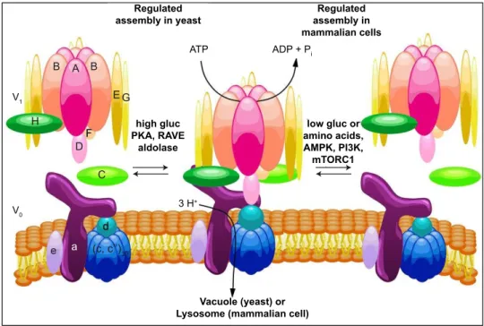
V-Regulated assembly in yeast Regulated assembly in mammalian cells ATP ADP + P i Vacuole (yeast) or Lysosome (mammalian cell)
V1 V 0 3 H+ a low gluc or amino acids, AMPK, PI3K, mTORC1 high gluc PKA, RAVE aldolase d e B A B D F E G H (c, c’’)10 C
Figure 8: Regulated assembly of V-ATPases in response to nutrient availabil-ity
The assembly of the V-ATPase is a major form of V-ATPase regulation. It is a reversible process that involves the dissociation of the catalytic V1 domain and the proton
translo-cator V0 domain in response to nutrient levels. On the left side of the figure, the regulated
assembly in yeast. In this model, the assembly increases in high glucose conditions and is promoted by PKA, the two glycolytic enzymes aldolase and phosphofructokinase and the assembly factor RAVE. On the right side of the figure, the regulated assembly in mam-malian cells, where in contrast, glucose starvation leads to increased V-ATPase assembly. This process is controlled by both the AMPK and PI3K/AKT pathways, but not PKA. Increased assembly in mammalian cells also occurs in response to amino acid starvation, and this effect is not controlled by PI3K or mTORC1. From Collins and Forgac, 2018.
Introduction - Chapter 1
ATPase, which was found to be exclusively expressed in fetal and adult kidney. Moreover, ATP6V0A4 mutations lead either to late-onset sensorineural hearing loss (SNHL) or to normal hearing (Guillard, 2012). Few patients developed severe hearing loss at young age (Stover et al., 2002; Guillard et al., 2009).
Mutations in ATP6V1B1 are associated with recessive dRTA, a rare genetic disease caused by failure of intercalated cells in the collecting duct to secrete the required protons for urinary acid excretion (Guillard et al., 2009; Guillard, 2012). When untreated, this acidosis may lead to osteomalacia and rickets, with the dissolution of bones. The ATP6V1B1 mutations cause also a SNHL (Karet et al., 1999; Vargas-Poussou et
al., 2006).
Mutations in both V-ATPase subunits and ER assembly factors have been associated with congenital disorders of glycosylation (CDG) type II. CDG are a group of genetic diseases with impaired synthesis/attachment of glycans to glycoproteins and glycolipids and synthesis of glycosylphosphatidylinositol (GPI) (Freeze et al., 2014). Originally, the CDG have been classified into two groups, based on the profile of the transferrin glycoforms obtained by isoelectric focusing (IEF). This classification was mainly developed to classify deficiencies in the N -glycosylation pathway: (1) CDG-I covering the defects in the assembly of the N -glycan or its subsequent transfer to the protein, (2) CDG-II was referred to defects in the glycosylation of the proteins, either in the late ER or in the Golgi (Aebi et al., 1999). However, the growing group of CDG caused by defects in protein
O-glycosylation, glycolipid biosynthesis, vesicular trafficking and pH homeostasis made
necessary a revision of the existing nomenclature. Nowadays, each disorder is named by the official gene symbol (not in italics), followed by the suffix “-CDG” (Jaeken et al.,
2009). The V-ATPase mutations associated with CDG can be divide into two groups:
(1) CDG with cutis laxa, caused by mutations in the core subunits (ATP6V0A2, ATP6V1A and ATP6V1E1);
(2) CDG with liver involvement, caused by mutations in the ER assembly factors (this group will be discussed in Chapter 2 – V-ATPase assembly and assembly factors and the Results part).
Mutations in ATP6V0A2 have been shown to cause an autosomal recessive form of
cutis laxa type II (ARCLA2, MIM: 611716) and wrinkly skin syndrome (WSS), in
combi-nation with glycosylation defects (Kornak et al., 2008; Morava et al., 2008). Cutis
laxa is a rare genetic disorder of the connective tissue characterized by excessive sagging
of skin folds and decreased elasticity of the skin (Guillard, 2012). ARCLA2 is a syn-dromic disease, resulting in the involvement of other organs, such as the skeletal system and the central nervous system, the last one with a variable degree, either with mental retardation or without (Morava et al., 2008; Guillard et al., 2009). All the patients with ATP6V0A2 mutations show a CDG type II on transferrin IEF and abnormal IEF of apoC-III. Moreover, mass spectrometry reveals abnormal glycosylation of total serum proteins, with combined defect in N - and mucin-type O-linked glycans (Kornak et al.,
2008; Morava et al., 2008). It has been suggested that the defects leading to cutis
laxa could be due to V-ATPase related defects (Kornak et al., 2008; Guillard et
al., 2009) and to abnormal function/transport of glycosyltransferases inside the Golgi
(Kornak et al., 2008; Hucthagowder et al., 2009). Indeed, the most severe cases show abnormal folding/processing of the elastin in the elastic fibers (Guillard, 2012).
Mutations in the V-ATPase subunits ATP6V1E1 (MIM: 108746) and ATP6V1A (MIM: 607027) have been recently discovered as causative of a metabolic syndrome character-ized by cutis laxa. Similarly to what has been shown for mutations in ATP6V0A2, these mutations affect the V-ATPase structure and assembly, leading to impaired protein glyco-sylation, Golgi trafficking and lysosomal function (Van Damme et al., 2017). The pa-tients show a variable CDG type II pattern in the transferrin IEF and abnormal transferrin glycosylation on mass spectrometry analysis. However, N -glycosylation is less affected than in individuals with ATP6V0A2 mutations. Even if both mutations do not affect the protein expression, they lead to reduced fully assembled V-ATPase complexes. As a consequence, the mutations lead to Golgi homeostasis defects, with abnormal swelling and Golgi fragmentation, and to compromised glycosylation of the collagen fibers. More-over, defects in the V-ATPase lead to an accumulation of autolysosomes in the patients fibroblasts, suggesting impaired autophagy and lysosomal function (Van Damme et al.,
2017). The cutis laxa phenotype is less severe than the one caused by ATP6V0A2, with
a generalized skin wrinkling in ATP6V1E1 mutations, and a phenotype characterized by large skin folds and abnormal fat distribution improving over time, in ATP6V1A muta-tions. In addition to cutis laxa, the seven described patients also display a multisystemic involvement, with facial features, hypotonia, joint contractures, congenital hip dysplasia and cardiopulmonary features (cardiomyopathy, congenital heart defects) (Van Damme et al., 2017).