University cf Montreal
Regulation of lectin-like
oxidized Iow-density lipoprotein receptor-1 (LOX-1). Relevance te diabetic vasculopathy.
By Ling Li
Department cf Biomedical Sciences Faculty of medicine
This thesis was submitted to the Graduate School in partial fulfiilment cf the requirements for
the degree of PhiIosophi doctot (Ph.D) in Biomedical Sciences.
April 2004 /Gradeconfêrô\
oui ULI. 07
W
o
CL1
Université
dI
de Montréal
Direction des bibliothèques
AVIS
L’auteur a autorisé l’Université de Montréal à reproduite et diffuser, en totalité ou en partie, par quelque moyen que ce soit et sur quelque support que ce soit, et exclusivement à des fins non lucratives d’enseignement et de recherche, des copies de ce mémoire ou de celle thèse.
L’auteur et les coauteurs le cas échéant conservent la propriété du droit d’auteur et des droits moraux qui protègent ce document. Ni fa thèse ou le mémoire, ni des extraits substantiels de ce document, ne doivent être imprimés ou autrement reproduits sans l’autorisation de l’auteur.
Afin de se conformer à la Loi canadienne sur la protection des renseignements personnels, quelques formulaires secondaires, coordonnées ou signatures intégrées au texte ont pu être enlevés de ce document. Bien que cela ait pu affecter la pagination, il n’y a aucun contenu manquant. NOTICE
The author of this thesis or dissertation has granted a nonexclusive license allowing Université de Montréal to reproduce and publish the document, in part or in whole, and in any format, solely for noncommercial educational and research purposes.
The author and co-authors if applicable retain copyright ownership and moral rights in this document. Neither the whole thesis or dissertation, nor substantial extracts from it, may be printed or otherwise reproduced without the author’s permission.
In compliance with the Canadian Privacy Act some supporting forms, contact information or signatures may have been removed from the document. While this may affect the document page count, t does not represent any loss of content from the document.
University of Montreal Facufty of graduate studies
This thesis entitled
“Regulation of lectin-like oxidized low-density lipoprotein receptor-1 (LOX-1). Relevance to diabetic vasculopathy.”
Presented by Ling Li
Has been evaluated by a jury made up of the following members
Dr. Omar Serti, President
Dt. Geneviève Renier, Director of teseatch Dt. Alain Rivard, Member
Dr. Pierre Julien, External member
Dt. Eugenio Rasio, Representative of doyen of FES
April 2004
RÉSUMÉ
L’athérosclérose est la principale cause de décès et la complication majeure du diabète de type 2. Le récepteur-1 “lectin-like oxidized low-density lipoprotein” (LOX-1) est un récepteur vasculaire nouvellement identifié pour les [DL oxydées (LDL0x) et les produits de glycation (AGEs). LOX-1 est surexprimé dans les lésions athérosclérotiques humaines et dans l’aorte de rats diabétiques, ce qui suggère un rôle de ce récepteur dans la pathogenèse de l’athérosclérose associée au diabète. Dans la présente étude, nous avons étudié la régulation vasculaire de LOX-1 par le glucose et la CRP et le rôle de LOX-1 dans l’adhésion des monocytes à l’endothélium et la formation de cellules spumeuses. Nos résultats démontrent que: 1) l’incubation de cellules endothéliales aortiques humaines (HAECs) et de macrophages dérivés de monocytes humains (hMDM) avec des concentrations de glucose élevées augmente l’expression génique et protéique de LOX-1 de manière dose- et temps-dépendant; 2) l’augmentation de l’expression protéique de LOX-1, induite par des concentrations élevées de glucose, est abolie par des agents antioxydants et des inhibiteurs de la protéine kinase C (PKC), des protéines kinases activées par les mitogènes (MAPK), du facteur nucléaire-kappaB (NF-icB) et de la protéine activée-1 (AP-1). Des MDM humains incubés en présence de concentrations élevées de glucose démontrent une augmentation de l’expression de PKC32 et une phosphorylation des protéines kinases régulées par des signaux extracellulaires (ERK) 1/2. L’activation de ces kinases est inhibée par les antioxydants et par l’inhibiteur spécifique de PKCj32, le LY379196. Des concentrations élevées de glucose augmentent également la liaison des protéines nucléaires extraites de MDM
humains aux séquences régulatrices NE-KB et AP-1 présentes dans le promoteur du gène de LOX-1; 3) l’incubation de HAECs et de MDM avec des concentrations élevées de glucose augmente l’adhésion des monocytes à l’endothélium et la formation de cellules spumeuses, respectivement, par l’entremise de mécanismes de signalisation LOX-1 -dépendant; 4) l’incubation de HAECs avec la CRP augmente l’expression génique et protéique de LOX-1 de façon dose- et temps-dépendant. Cet effet est attenué pat une préincubation des cellules avec des anticorps anti-CD32 et anti-CD64. Enfin, le traitement des HAECs par la CRP induit une augmentation de l’adhésion des monocytes à l’endothelium. Cette augmentation est dépendante de LOX-1. Globalement, ces résultats démontrent que des concentrations élevées de glucose augmentent in vitro l’expression endothéliale et macrophagique de LOX-1 et que ces effets sont associés à une adhésion accrue des monocytes à l’endothélium et à la formation de cellules spumeuses. Nous avons également demontré que la CRP augmente, in vitro, l’expression endothéliale de LOX-1 et que cet effet est associé à une augmentation de l’adhésion des monocytes à I’endothélium. En résumé, ces données suggèrent un rôle clé de divers facteurs métaboliques et inflammatoires dans la régulation vasculaire de LOX-1 et suggèrent un rôle de LOX-1 comme médiateur de l’athérogenèse associée au diabète humain. Une meilleure compréhension du rôle des facteurs métaboliques et inflammatoires dans la régulation vasculaire de LOX-1 dans le diabète pourrait permettre le développement de nouvelles stratégies dans le traitement de l’athérosclérose associée au diabète.
SUMMARY
Atherosclerosis is the leading cause of death and the major complication cf type 2 diabetes. Lectin-like oxidized low-density lipoprotein receptor-1 (LOX-1) is a newly identified vascular receptor for oxidized LDL (0xLDL) and advanced glycation end products (AGEs). LOX-1 is highly exptessed in human atherosclerotic lesions and is upregulated in the aorta cf diabetic rats. In the present study, we sought to determine the regulation of vascular LOX-1 in diabetes. Our resuits demonstrated that: 1) incubation of human aortic endothelial cells (HAECs) and human monocyte-derived macrophages (hMDM) with elevated glucose concentration enhanced, in a dose- and time-dependent manner, LOX-1 gene and protein expression; 2) high glucose-induced macrophage LOX-1 protein expression was abo!ished by antioxidants, protein kinase C (PKC), mitogen-activated protein kinase (MAPK), nuclear factor-kappaB (NE-KB) and activated protein-1 (AP-1) inhibitors. n human MDM cultured with high glucose, increased expression of PKCE32 and enhanced phosphorylation of extracellular signal-regulated protein kinase (ERK) 1/2 were also observed. Activation cf these kinases was inhibited by the antioxidant and by the PKCf32 inhibitor, LY379196. High glucose also enhanced the binding of nuclear proteins extracted from human MDM te the NE-KB and AP-1 regulatory elements cf the LOX-1 gene promoter. High glucose-induced endothelial LOX-1 protein expression was abolished by antioxidants, PKC, MAPK, and NE-KB inhibitors. High glucose enhanced the binding cf nuclear protein extracted from HAECs to
endothelium and foam cell formation, respectively, through LOX-1-dependent signaling mechanism; 4) incubation of HAECs with CRP enhanced, in a dose and time-dependent manner, LOX-1 mRNA and protein levels. This effect was reduced by anti-CD32 and -CD64 antibodies, whereas it was unaffected by hyperglycemia, tumor necrosis factor Œ (TNFŒ) and interleukin-6 (IL-6). In CRP treated HAECs, a LOX-1-dependent induction of monocyte adhesion to
endothelium was observed. Overall, these resuits demonstrate that high glucose enhances human endothelial and macrophage LOX-1 expression in vitro and that this effect is associated with enhanced monocyte adhesion to endothelium and foam cell formation. We also found that CRP increases human endothelial LOX-1 expression in vitro and that this effect is associated with increased monocyte adhesion to endothelium. Taken collectively, these data indicate a key role for metabolic and inflammatory factors in the regulation of vascular LOX-1 in diabetes. They identify LOX-1 as one potential important mediator in the pathogenesis of atherosclerosis in human diabetes. Better understanding of the
role of metabolic and inflammatory factors in the regulation of vascular LOX-1 in
diabetes might provide new strategies in the treatment cf diabetic atherosclerosis.
CONTENTS
Summary iH
List cf figures xvi
List cf tables xix
List of abbreviations xx Acknowledgements xxiv Dedication xxv I. Introduction 1 1.1. Atherosclerosis 1 1.1.1. General 1
1.1.2. Development and progression of the atherosclerotic lesion 1
1.1.2.1. Endothelial dysfunction in atherosclerosis 1
1.1.2.1.1. Endothelial function 1
1.1.2.1.2. Endothelial dysfunction 2
1.1.2.2. Fatty-streak 3
1.1.2.3. Advanced, complicated lesion cf atherosclerosis 4
1.1.2.4. Unstable fibrous plaques in atherosclerosis 4
1.1.2.4.1. Local factors 4
1.1.2.4.2. Systemicfactors 5
1.1.3. Riskfactors 6
11.3.1.1. Hypercholesterolemia and dyslipoproteinemia .6
1.1.3.1.2. Hypertension 8
1.1.3.1.2.1. Renin-angiotensin system (RAS) 8
1.1.3.1.2.2. Endothelin 9
1.1.3.1.3. Diabetes 10
1.1.3.1.4. Family history, age and gender 10
1.1.3.1.5. Obesity and smoking habit 11
1.1.3.2. Newriskfactors 12 1.1.3.2.1. CRP 12 1.1.3.2.2. Lp(a) 13 1.1.3.2.3. Fibrinogen 15 1.1.3.2.4. Homocysteine 16 1.1.3.2.5. Infection 12 1.1.4. Pathogenesis of atherosclerosis 17
1.1.4.1. Endothelial dysfunction hypothesis 17
1.1.4.1 .1. Risk factors for endothelial dysfunction 18
1.1.4.1.2. Mechanisms cf endothelial dysfunction in
o
atherosclerosis 191.1.4.3. Oxidative stress hypothesis .21 1.1.4.4. Hypothesis of infection 24 1.1.4.5. Hypothesis of inflammation 24 1.11. Diabetes mellitus (DM) 28 1.11.1. Definition 28 1.11.2. Classification 29 1.11.2.1. Type 1 diabetes 29 1.11.2.2. Type 2 diabetes 34 1.11.2.2.1. Prevalence 35 1.11.2.2.2. Etiology 35 1.11.2.2.3. Geneticfactors 36 1.11.2.2.3.1 Gene candidates 38
1.11.2.2.3.2. Mutations in genes involved in
insulin resistance 38
1.11.2.2.3.3. Mutation in genes encoding 13-celI proteins
involved in the quality and quantity of secreted
insulin (figure 3) 42
1.11.2.2.3.4. Mutations in genes involved in lipid
metabolism and obesity 44
Q
1.11.2.2.4.1 Obesity.471.11.2.2.4.2. Diet 48
1.11.2.2.4.3. Physical activity 48
1.11.2.2.4.4. Stress 49
1.11.2.3. Pathophysiology of type 2 diabetes 49
1.11.2.3.1. Action of insulin 50
1.11.2.3.2. Insulin resistance 51
1.11.2.3.2.1. Genetics and environment 52
1.11.2.3.2.2. Hepatic insulin resistance 53 1.11.2.3.2.3. Insulin resistance in skeletal muscle 53 1.11.2.3.2.4. Insulin resistance in adipocyte 54 1.11.2.3.3. Hyperinsulinemia and glucose intolerance 57
1.11.2.4. Complications associated with type 2 diabetes 58
1.11.2.4.1. Macrovascular complications 61
1.11.2.4.1 .1. Traditional risk factors 60
1.11.2.4.1.1.1. Dyslipidemia 60
1.11.2.4.1.1.2. Hypertension 61
1.11.2.4.1.1.3. Smoking 61
1.11.2.4.1.1.4. Obesity 62
1.11.2.4.1.2. Novel riskfactors .70
1.11.2.4.1.2.1. Insulin resistance 70
1.11.2.4.1.2.2. Homocysteine 71
1.11.2.4.1.2.3. Microalbuminuria 71
1.11.2.4.1.2.4. Abnormal fibrinolysis and
hypercoagulation 72
1.11.2.4.1.2.5. Inflammation 72
1.11.2.4.1.2.6. Endothelial dysfunction 73 1.11.2.4.1.3. Inflammation: the linkbetween obesity, insulin
resistance, type 2 diabetes and atherosclerosis 74 1.11.2.4.1.3.1. Inflammation and obesity 75 1.11.2.4.1.3.2. Inflammation and insulin resistance 77 1.11.2.4.1.3.3. Inflammation and type 2 diabetes 78 1.11.2.4.1.3.4. Inflammation in endothelial dysfunction
and atherosclerosis 79 1.11.2.4.2. Mictovascular complications 80 1.11.2.4.2.1. Retinopathy 81 1.11.2.4.2.2. Nephropathy 81 1.11.2.4.2.3. Neuropathy 82
0
1.11.2.5. Diagnosis 821.11.2.6. Ireatment .83
1.11.2.6.1. Diet and physical activity 83
1.11.2.6.1.1. Weight loss 83
1.11.2.6.1.2. Life style changes 84
1.11.2.6.2. Oral hypoglycemants 85 1.11.2.6.2.1. Sulfonylureas 85 1.11.2.6.2.2. Meglitinides 87 1.11.2.6.2.3. Metformin 87 1.11.2.6.2.4. Thiazolidinediones 89 1.11.2.6.2.5. Acarbose 90
1.11.2.6.2.6. Other medications that may reduce
the risk cf type 2 diabetes 91
1.11.2.6.3. Insulin therapy 91
1.11.3. Gestational diabetes (GDM) 92
1.11.4. Other type cf diabetes 92
1.111. Lectin-like oxidized low density lipoprotein receptor-1 (LOX-1) 93
1.111.1. General 93
1.111.2. LOX-1 properties 93
1.111.2.1. Structure cf LOX-1 gene 93
J
1.111.2.1.2. 5-flanking region of the LOX-1 gene.94
1.111.2.1.3. Human LOX-1 mRNA structural organization 95
1.111.2.2. Structure of LOX-1 protein 96
1.111.2.2.1. The domain structures and functions of LOX-1 96
1.111.2.2.2. Post-translational modification of LOX-1 97
1.111.2.2.3. Soluble forms of LOX-1 98
1.111.2.3. LOX-1 tissue distributions 98
1.111.3. LOX-1 binding ligands 98
1.111.4. Regulation of LOX-1 expression 100
1.111.4.1. Inducing Factors 100 1.111.4.1 .1. Pro-inflammatory cytokines 100 1.111.4.1 .2. Pro-atherosclerotic stimuli 100 1.111.4.1.3. Oxidative stress 101 1.111.4.1.4. Pathological conditions 102 1.111.4.2. Inhibitory factors 103
1.111.5. Role of LOX-1 in atherosclerosis 104
1.111.5.1. Role of LOX-1 in endothelial dysfunction 104
1.111.5.2. Role of LOX-1 in the progression
1.111.6. Role of LOX-1 in type 2 diabetes .106
1.1V. CRP 107
I.IV.l. CRP and atherosclerosïs 108
I.IV.l .1. CRP as a predictive risk marker of cardiovascular events...109
I.IV.1 .2. Role of CRP in atherosclerosis 111
I.IV.1.2.1. Role of CRP on monocyte adhesion/recruitment...112
I.IV.1.2.2. Role of CRP on foam celI formation 113
I.IV.1.3. Comparison of CRP to other risk factors 114
I.IV.1.4. Goal of screening and therapeutic options 115
I.IV.2. CRP and Diabetes 115
I.V. Hypothesis and objectives 117
II. Results 119
11.1. Glucose enhances endothelial LOX-1 expression. Role for LOX-1 in glucose-induced human
monocyte adhesion to endothelium 120
11.2. Glucose enhances human macrophage LOX-1 expression. Role for LOX-1 in glucose-induced
macrophage foam celI formation 155
11.3. C-reactive protein fCRP) enhances lectin-like oxidized low-density lipoprotein receptor-1 (LOX-1) expression in human
Q
to CRP-induced endothelial dysfunction .192III. Discussion 222
IV.
Conclusions and perspectives 248IV.l. Conclusion 249
IV.2. Perspectives 250
LIST 0F FIGURES Introduction:
Figure 1. Modulation of cellular function by ROS in cardiovascular
diseases 22
Figure 2. Downstream effects resulting from insulin/insulin
receptor interaction 40
Figure 3. Genetic defects in beta-celis in type 2 diabetes 43
Figure 4. Pathogenesis cf type 2 diabetes 52
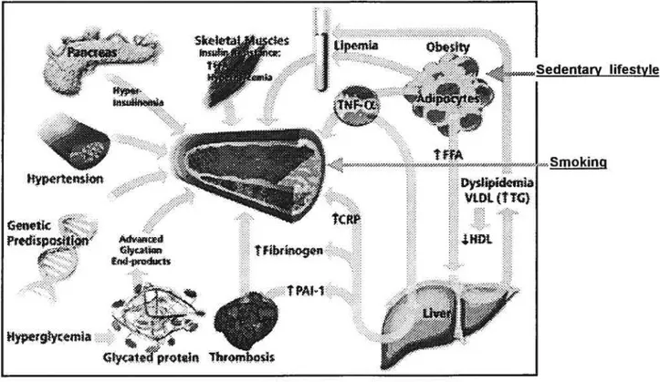
Figure 5. Model of development of diabetic vascular complications 58 Figure 6. Multiple cardiovascular risk factors in diabetes 59
Figure 7. The formation of AGEs 63
Figure 8. Hyperglycemia-induced activation cf
The diacylglycerol-PKC pathway 66
Figure 9. Exogenous and endogenous stimuli leading
to reactive oxygen species generation 68 Figure 10. Inflammation : the link between obesity, insulin resistance,
diabetes, and atherosclerosis 75
Figure 11. Hypothetical model of chronic inflammation and adipocyte
insulin resistance 76
Figure 12. Diagrammatic representation cf the
LOX-1 gene and protein 95
Figure 13. Role of LOX-1 atherosclerosis 106
Figure 14. CRP level and cardiovascular risk 111 Figure 15. Direct comparison cf CRP to several other lipid and non-lipid
o
First article:Figure.1. Time- and dose-dependent effect cf high glucose on LOX-1
mRNA levels in HAECs 146
Figure.2. Time- and dose-dependent effect cf high glucose on LOX-1
protein expression in HAECs 148
Figure.3. Effect cf high glucose and TNFΠon endothelial LOX-1
protein expression 150
Figure.4. Effect of PKC, MAPK, NE-KB inhibitors and antioxidants on
gluccse-induced LOX-1 mRNA levels 151
Figure.5. Effect cf high glucose on the binding cf nuclear proteins extracted from HAECs to the NF-KB sequence cf the
LOX-1 gene promoter 152
Eigure.6. Effect cf high glucose on human monocyte adhesion
te endcthelial cells 154
Second article:
Figure.1. Time- and dose-dependent effect cf high glucose on LOX-1
mRNA levels in human MDM 180
Figure.2. lime- and dose-dependent effect cf high glucose on LOX-1
protein expression in human MDM 181
Figure.3. Effect cf high glucose on TNF-Œ-induced macrophage
on glucose-induced LOX-1 mRNA levels .183 Figure.5. Effect of high glucose on PKC and MAPK activation
in 1H P-1 MDM. Modulatory effect of NAC
and PKC/MAPK inhibitors 185
Figure.6. Effect cf high glucose on the binding of nuclear proteins extracted from 1H P-1 MDM to the NE-KB
sequence of the LOX-1 gene promoter 186 Figure.7. Effect of high glucose on the binding of nuclear proteins
extracted from 1H P-1 MDM to the AP-1 sequence
of the LOX-1 gene promoter 188
Figure.8. Effect of high glucose on 0xLDL uptake by human
MDM. Role for LOX-1 190
Third article:
Fïgure.1. lime- and dose-dependent effect cf CRP on LOX-1 mRNA
levels in HAECs 216
Eigure.2. lime- and dose-dependent effect of CRP on endothelial
LOX-1 protein expression 217
Figure.3. Dose-dependent effect of IL-6 on endothelial
LOX-1 protein expression 219
Figure.4. Effect of CRP on human monocyte adhesion
to endothelial cells 220
LIST 0F TABLES
Introduction:
Table 1. Favorable and atheroprotective effects of the healthy
endothelium 2
Table 2. Novel risk factors for atherosclerotic vascular disease 14 Table 3. The effects cf oxidized LDL and/or its products-oxidized
phospholipids and/or oxysterols 21
Table 4. Classification of the types of diabetes 31 Table 5. Etiologic classification cf diabetes mellitus 32
Table 6. Diabetic risk factors 36
Table 7. Genetic Classification of type 2 diabetes 41 Table 8. Criteria for the diagnosis 0f diabetes mellitus 83
LIST 0F ABREVIATIONS
ACE : angiotensin converting enzyme AGEs : advanced glycation end products AP-1 : activator protein-1
APP acute phase protein AT1 : angiotensin II type 1
BAECs bovine aortic endothelial cells BMI : body mass index
CD4OL : CD4O Hgand
CHD coronary heart disease CML $ carboxyl methyl lysine
C. pneumoniae: Chlamydia pneumoniae
CRD carbohydrate-recognition domain CRP : C-reactive protein
CS : cystathionine synthase CVD : cardiovascular disease DAG : diacylglycerol
DCCT : diabetes control and complications trial DCs : dendritic cells
DM $ diabetes mellitus
DPP $ diabetes prevention program
EBT : electron beam computed tomography eNOS : endothelial NO synthase
ET-1 : endothelin-1
FDPS : finish diabetes prevention study FFAs free fatty acids
FGF-2 fibroblast growth factor -2 FPG fasting plasma glucose GDM gestational diabetes
HAECs : human aortic endothelial celis
HCAECs : human coronary artery endothelial ceils HDL high-density lipoprotein
HE : high fat
HNF : hepatocyte nuclear factor H. pylori : Helicobacter pylori
ICAM-1 intercellular adhesion molecule-1 IDDM : insulin-dependent diabetes mellitus IFG : impaired fasting glucose
lGT : impaired glucose tolerance IFN-y : interferonj’
IL-1 : interleukin-1 IL-6 : ïnterleukin-6 IL-8 : interleukin-8 IL-12 : interleukin-12
IRS-1 : insulin receptor substrate-1
IRAS insulin resistance atherosclerosis study 8-iso-PGF-2Œ: 8-iso-prostaglandin F2Œ
LFA-1 : lymphocyte function-related antigen-1
LOX-1 : lectin-like oxidized low-density lipoprotein receptor-1 MAPK : mitogen-activated protein kinase
MCP-1 monocyte chemotactic protein-1 M-CSF : macrophage-colony stimulating factor MODY : maturity-onset diabetes of the young MR-FIT : multiple risk factor intervention trial MTH FR : methylenetetrahydrofolate reductase NAC : N-acetylcysteine
NDDG national diabetes data group
NIDDM : non-insulin-dependent diabetes mellitus NE-KB : nuclear factor-kappa B
NO : nitric oxide
NOS nitric oxide synthase OGIT : oral glucose tolerance test 0xLDL $ oxidized low-density lipoprotein PAl-1 : plasminogen activator inhibitor-1 PC : phosphocholine
PDGF platelet-derived growth factor PKC : protein kinase C
PI $ phosphatidylinositol PLC $ phospholipase C
PPAR : peroxisome proliferator-activated receptors P5 : phosphatidylserine
RAS renin-angiotensin system ROS : reactive oxygen species SMCs : smooth muscle celis SSA : serum amyloid A
SSRE shear-stress responsive etement
TE tissue factor
TEPI : TF pathway inhibitor
TGF-f3 : transforming growth factor f3
TNF-Πtumor necrosis factot a
IRE tetradecanoylphorbol 13-acetate-responsive element TRIPOD : troglitazone in prevention of diabetes
tRNAs : transfet RNA
UKPDS : united kingdom prospective diabetes study
VCAM-1 vascular ceIl adhesion molecule-1 VDCs vascular DCs
VLDL very low-density lipoproteins
WHHL watanabe hereditary hyperlipidemic
WHO : world health organization
C
I would like to express my most sincere appreciation to my supervisor Dr. Geneviève Renier, for her solid and skillful guidance, as well as her encouragement throughout this long dissertation. I feel honored to have been her student, and value greatly the wisdom she has shared with me. Under her supervision I experienced the most wonderful learning experience of my life, and met the challenges of achieving professional and personal milestones. Dr. Renier has indeed become a mentor, a sister, and a friend. Furthermore, I acknowledge my colleagues, Dr. Jean-Claude Mamputu, Marie-Claude Beauchamp, and Fritz Maingrette for their enthusiastic support of my efforts.
Q
I dedicate my wotk to my loving and caring parents, who have always been there to encourage and support me. You both taught me the importance cf friendship, respect, and love, for ail aspects cf life. I dedicate my work te my husband and loveiy daughter, whose love and strength have sustained my enetgy to complete this work. In addition, I dedicate this work to my brother, as well as my best friends, who through their goodness of nature have given me, as the song goes, “the wind beneath my wings.”
Q
I.
INTRODUCTION
I.
IntroductionLI.
Atherosclerosis1.1.1. General
Atherosclerosis is a chronic, multifactorial and progressive disease characterized by the accumulation of lipids and fibrous elements in the large and medium arteries. Epidemiological studies over the past 50 years have identified numerous risk factors for atherosclerosis. These can be grouped into factors with an important genetic component and those that are largely environmental. Among the primary events in atherosclerosis are the accumulation of low-density lipoproteins (LDL) in the subendothelial space [Guyton et al. 1996, Van De Graif et al. 19921 and the adhesion of inflammatory cells, such as monocytes and T lymphocytes, to the endothelium. The notion that atherosclerosis is an immune mediated inflammatory disease is now widely accepted and provides the basis for the development of new strategies in the diagnosis and management of this disease.
1.1.2. Development and progression of the atherosclerotic lesion 1.1.2.1. Endothelial dysfunction in atherosclerosis
1.1.2.1.1. Endothelial function
The vascular endothelium is considered to be a monolayer acting as a selectively permeable barrier between blood and tissues. It is a dynamic and heterogeneous organ. It has also become evident that the vascular endothelium
is an active paracrine, endocrine, and autocrine organ that is indispensable for the regulation cf vascular tone and maintenance cf vascular homeostasis as shown in Table I [Bonetti et al. 2003].
Table J. Favorable and Atheroprotective Effects of the Healthy Endothelium. Promotion of vasodilation
Antioxidant effects Antiinflammatory effects
Inhibition cf leukocyte adhesion and migration
Inhibition cf smooth muscle celI proliferation and migration Inhibition cf platelet aggregaticn and adhesicn
Anticoagulant effects Profibrinolytic effects
Endothelium can generate effector molecules, such as nitric oxide (NO), prostacyclin, platelet-derived growth factor (PDGF), angiotensin Il, and endothelin [Ross 1999]. These molecules control and regulate thrombosis, inflammation, vascular tone, and vascular remodeling. Furthermore, the endothelium has important anti-coagulant and fibrinolytic functions.
LL2.1 .2. Endothelial dysfunction
Endothelial dysfuncticn is characterized by an imbalance between the production cf relaxing and contracting factors, anticoagulant and procoagulant mediators, or growth-inhibiting and promoting factors. Endothelial dysfunction is closely linked to the occurrence cf vascular diseases such as hypertension,
diabetes, inflammation, and aging and preceeds the development of atherosclerosis. It is characterized by a reduction of the bioavailability of vasodilators such as NO and an increased production of endothelium-derived contracting factors [Bonetti et al. 2003]. This imba lance leads to an impairment 0f endothelium-dependent vasodilation, which represents the functional characteristic of endothelial dysfunction. Aside from denoting the impaired endothelium-dependent vasodilation, endothelial dysfunction also comprises a specific state of endothelial activation, which is characterized by a proinflammatory, proliferative, and procoagulatory milieu that favors ail stages of atherogenesis [Anderson. 19991. This results in increased permeability of endothelium to macromolecules such as LDL and enhanced migration of inflammatory cells to the subintimal space through the effect of oxidized LDL (0xLDL), monocyte chemotactic protein 1 (MCP-1), interleukin-8 (IL-8), PDGF, macrophage-colony stimulating factor (M-CSF), and osteopontin, leading to the formation of early atherosclerotic lesions or fatty streaks [Kita et al. 2001].
1.1.2.2. Fafty-streak lesion
As mentioned above, early atherosclerotic lesions, or fatty streaks are characterized by the accumulation of LDL and the migration of monocytes and lymphocytes into the subendotheliai space of the arterial wall. Fatty streaks initiaily consist of lipid-laden monocyte-derived macrophages (foam cells) mixed together with variable numbers of T lymphocytes [Ross 1999]. The formation of foam cells is mediated mainly by M-CSF, tumor necrosis factor a (TNF-a), and
C
interleukin-1 (IL-1) [Rosenfeid et al. 1990, Hamilton et al. 1999]. Later, these ceNs are joined by various numbers of smooth muscle cells (SMCs). SMC migration is stimuiated by PDGF, flbroblast growth factor-2 (FGF-2) and transforming growth factor 3 (TGF-J3) [Boring et al. 1997, Boisvert et al. 1998]. Platelet adherence and aggregation are aiso observed and are stimulated by integrins, P-selectin, fibrin, thromboxane A2, and tissue factor (TF) [Ross 19991.
1.1.2.3. Advanced lesion of atherosclerosis
As fatty streaks progress to intermediate and advanced lesions, they tend to form a fibrous cap that walls off the lesion from the lumen. This represents a type of healing or fibrous response to the injury [Ross 1999]. Advanced atherosclerotic lesions result from a variety of pathogenetic processes, including macrophage foam cell formation and death, accumulation of extracellular lipid, reduction of structural intercellular matrix and SMCs, generation of minerai deposits, chronic inflammation, and neovascularization [Stary 2000]. Coronary artery calcification is rather a marker for atherosclerotic lesion. Ectopic vascular calcification occurs commonly in atherosclerosis. Its presence and location in cotonary arteries have been documented by ultrafast computed tomography scanning [Fallavallita et al. 1994, Janowitz 2001]. Electron beam computed tomography (EBT) can also rapidiy and noninvasively detect and quantify caicified atherosclerotic plaque in the coronary arteries [O’Rourke et al. 2000]. A higher quantity of cotonary artery calcium is associated with Iikelihood of obstructive lesions [O’Rourke et al. 2000], and with an increased risk of future
cardiovascular disease (CVD) [Arad et al. 2000]. The recent finding that TNF-Πpromotes, through the cycc adenosine monophosphate (cAMP) pathway, osteoblastic differentiation of vascular celis [Tintut et al. 2000] suggests a role of pro-inflammatory cytokines in the vascular calcification process.
1.1.2.4. Unstable fibrous plaques in atherosclerosis
1.1.2.4.1. Local factors
The influx of inflammatory celis, macrophages, and T lymphocytes in atherosclerotic plaques increases with plaque progression and these cells preferentially accumulate at sites of plaque rupture [Ross 1999, Hosono et al. 2003]. Inflammatory mediators and proteases released by these cells and expressed in human atherosclerotic lesions contribute to plaque rupture. For example, it is well known that macrophages release metalloproteinases and other proteolytic enzymes at sites of thinning of the fibrous cap. These enzymes cause degradation of the matrix, which leads to hemorrhage from the vasa vasorum or from the arterial lumen, resulting in thrombus formation and arterial occlusion [Ross 1999]. Apoptosis of vascular ceils such as SMCs and macrophages are associated with plaque instability as well [Von det Thusen et al. 2002, Kockx et al. 2000, Geng et al. 1995, Libby et al. 1996]. Local modulators of plaque rupture also include factors involved in the coagulation cascade [Khrenov et al. 2002]. For example, both TE and TF pathway inhibitor (TFPI) are expressed in advanced human atherosclerotic lesions and are abundant at sites of plaque rupture [Kaikita et al. 1999]. Studies also
Ç
demonstrate that plasminogen activator inhibitor (PAl) and prothrombin are increased in advanced human plaques [Falkenberg et al. 1996, Smith et al. 1981]. Recent studies on gene profiling have identified a differential expression of various genes between stable and ruptured human atherosclerotic plaques including perilipin and cathepsin K. In addition to suggest an important role for these genes in plaque stability, this study also supports a role for many other unknown genes such as vasculin [Faber et al. 2001 Bijnens et al. 2003, Lutgens et al. 20031.1.1.2.4.2. Systemïc factors
It has been stated that plaque rupture does not occur as an isolated phenomenon but rather as a systemic disease [Lutgens et al. 2003]. Rupture of the fibrous cap leads to the exposure of the thrombogenic parts of the atherosclerotic plaque with subsequent activation of the coagulation cascade and platelet aggregation. This leads to thrombosis and acute ischemia resulting from abrupt luminal compromise. Histomorphological features of vulnerable plaques include a) positive remodeling producing less luminal stenosis; b) large lipid cote ( 40% plaque volume); C) large number of inflammatory cells; d) a thin cap
depleted of SMCs and collagen and finally increased neovascularity. Recently, the term ‘cardiovascular vulnerable patient” has been ptoposed to define subjects susceptible to an acute cotonary syndrome or sudden cardiac death based on plaque, blood, or myocardial vulnerability [Naghavi et al. 2003]. Vulnerable patients often present with multiple ruptured plaques. Systemic
(J
factors that are correlated with plaque rupture are altered blood rheology, increased coagulability, increased systemic inflammation, and recurrent infections. These unfavorable systemic changes often interact synergistically withrisk factors of atherosclerosis, such as hyperlipidemia, smoking, and diabetes
[Fuster et al. 1999, Corti et al. 2003, Sambola et al. 2003].
1.1.3. Risk factors
113.1. Traditional riskfactors
1.1.3.1.1. Hypercholesterolemia and dyslipoproteinemia
High plasma levels of cholesterol and cf pro-atherogenic lipoproteins
represent major risk factors for cardiovascular diseases. In particular, increased levels cf circulating LDL is considered as a major risk factor of coronary heart disease and one of the earliest events in athetogenesis is the accumulation cf LDL in the vessel wall. Retention and modification cf LDL are key factors in atherosclerotic lesion formation. LDL can be modified by several mechanisms,
including oxidation, glycation, aggregation, association with proteog lycans, or
incorporation into immune complexes [Steinberg et al. 1997, Khoo et al. 1998,
Khoo et al. 1992, Navab et al. 1996]. Progressive oxidation of LDL takes place
during the process cf LDL particle trapping in arteries. OxLDL is internalized by macrophages through surface scavenger receptors [Steinberg et al. 1997, Khoo et al. 1992, Navab et al. 1996, Griendling et al. 1997, Han et al. 1997]. The uptake of oxLDL by macrophages results in intracellular cholesterol ester accumulation and foam cell formation. Macrophage-derived foam cells are
present in ail stages cf atherogenesis and play a key role in the development and progression cf atherosclerosis. Lipoprotein fa) (Lp(a)) closely resembles LDL in its content cf cholesterci and apolipoprotein B-100 but differs by the presence cf an attached glycoprotein, known as apoprotein fa). The involvement cf Lp(a) in the pathogenesis cf atherosclerosis is strongly suggested by the presence of Lp(a) in the human atherosclerotic lesions [Rath et al. 1989, Jutgen et aI. 1993]. Numerous cross-sectional and prospective studies have also revealed associations between high plasma levels cf Lp(a) and atherosclerotic vascular diseases, such as ccrcnary heart disease and stroke [Utermann 1995, Djurovic et al. 1997]. The effect cf high plasma levels cf Lp(a) in the develcpment cf athercsclercsis has aise been well-studied in transgenic animal mcdels [Lawn et aI. 1992, Fan et ai. 2001]. Although the importance cf high LDL chclestercl as a risk factcr for ccrcnary artery disease is well estabiished, the rcle cf hypertriglyceridemia as an independent risk factcr for the develcpment cf vascular diseases is stiii ccntrcversial. Varicus factors asscciated with hypertriglyceridemia, including dyslipcprcteinemia, aiteraticns cf hemcstatic processes, cbesity and hypertension may acccunt for the deletericus effect cf high triglyceride ieveis on the atherosclerotic prccess [Malaguarnera et ai. 2000]. Althcugh, the meta-analysis cf Austin and Hckanscn, 1998 has recentiy suggested that hypertriglyceridemia is an independent risk factor for cardiovascular diseases, many investigatcrs stiil believe that triglyceride-rich iipcproteins cannct migrate thrcugh the vascular endcthelium [Faegerman, 1998]. Recently it has been suggested that postprandial hypertriglyceridemia
(
may cause endothelial dysfunction via enhanced oxidative stress [Bae et aL20011. In addition, a lcw level of HDL-choiesterol as an important risk factor foc
cardiovascular disease has been weII established thtough epidemiological and
clinical studies [Gordon et al. 1989]. A growing body cf evidence suggests that HDL exerts part cf its antiatherogenic effect by counteracting LDL oxidation
[Assmann et al. 2004].
1.1.3.1.2. Hypertension
1.1.3.1.2.1. Renin-angiotensin system (RAS)
The RAS plays an important role in the pathogenesis cf cardiovascular disease. Angiotensin II, the principal product of the RAS, increases blood pressure through increasing vascular resistance, stimulating aldosterone synthesis and release, increasing renal tubular sodium reabsorption, augmenting the release cf antidiuretic hormone, and enhancing sympathetic outflow from the brain. Nctably, angiotensin II induces cardiac and vascular cell hypertrophy and hyperplasia directly by activating the angiotensin II type 1 (AT1) receptor and indirectly by stimulating the release cf several growth factors and cytokines [McConnaughey et al. 1999]. The mechanisms thrcugh which angiotensin II increases blood pressure include binding cf angiotensin te specific receptors on
SMC, eliciting the activation cf phospholipase C (PLC) and leading to increased
intracellular calcium concentrations and 5MO hypertrophy [Gibbons et al. 1992]. Angiotensin II also decreases NO production, increases cxidative stress [Yanagitani et al. 1999] and induces the activation cf nuclear factor-icB (NF-KB)
[Kranzhôfer et al. 1999], protein kinase C (PKC) [Henrion et al. 1996] and mitogen activated protein kinases (MAPK) [Haendeler and Berk, 2000].
1.1.3.1.2.2. Endothelin
Endothelin is a potent vasoactive peptide, produced by endothelial oeIls. Endothelin is secreted in an abluminal direction by endothelial ceils and acts in a paracrine fashion on underlying SMCs to cause vasoconstriction and elevate blood pressure [Oparil et aI. 2003]. Circulating endothelin levels are increased in some hypertensive patients, particularly African Americans and persons with transplant hypertension, endothelial tumors, and vasculitis [Ergul et al. 1996]. Da
Silva et al. [2004] recently reported that endothelin-1 (ET-1) contributes to
increased arterial pressure in a model of visceral obesity produced by feeding Sprague-Dawley rats with a high-fat (HE) diet.
LL3.1.3. Diabetes
Patients with diabetes are at high risk of cardiovascular diseases [Resnick et al. 2002]. Several risk factors associated with diabetes including dyslipidemia, obesity, hypertension, hyperglycemia and hyperinsulinemia contribute to the accelerated atherosclerosis in human diabetes. (See section 1.11. for details)
1.1.3.1.4. Family history, age and gender
It is clear that positive family history, advanced age and male gender
Numerous studies have shown that individuals with a family history cf coronary heart disease (CHD) are at higher risk 0f atherosclerosis. For example, Pankow et al. [1997] and Li et aI. [2000] investigated family CHD risk scores and found higher traditional CHD risk factors in individuals with a family history of heart disease.
Because of protection by female hormones, the onset of CHD tends to be delayed in women by about 10 years with some catch-up after menopause [Matthews et al. 1989].
1.1.3.1.5. Obesity and smoking habit
Obesity, especially visceral obesity, is associated with a cluster of metabolic complications increasing the risk of type 2 diabetes and CHD. For example, obese patients with visceral obesity show increased glycemic and insulinemic responses to an oral glucose load compared to normal weight individuals or compared to obese individuals with low visceral adiposity [Kothari et al. 1998]. Viscerally obese patients are also characterized by an unfavorable plasma lipid profile which includes elevated triglyceride and apolipoprotein B concentrations, reduced high density lipoproteins (HDL)-cholesterol levels as well as increased proportion of small, dense LDL particles [Despres et al. 2000].
Despite the epidemiological evidence linking cigarette smoking with cardiovascular disease, the precise components of cigarette smoke responsible for this relationship and the mechanisms by which they exert their deleterious effects have not yet been fully elucidated. Cigarette smoke is a complex mixture
and only a few components have been extensively studied. Nicotine and carbon monoxide are much less damaging than is whole smoke. There is considerable evidence that cigarette smoking can result in both morphological and biochemical endothelial disturbances both in vivo and in vitro [Michael Pittilo 2000].
1.1.3.2. New risk factors
In recent years, a number of new candidate risk factors or markers have been proposed as significant predictors of atherosclerosis and its complications. These risk factors and markers are summarized in Table 2 [Hackam et al. 2003]. Among these factors, four of them will be discussed emphatically due to their
unique advantages. 1). There is substantial evidence on their predictive abilities.
2). A genetic basis for premature disease involves these factors. 3). Modifying
treatments of these factors are available. 4). These factors are the subject cf ongoing or completed clinical trials. Ihese factors are C-reactive protein (CRP),
Lp (a), fibrinogen, homocysteine, and infection.
1.1.3.2.1. CRP
CRP is a circulating acute-phase reactant that is increased many-fold during the inflammatory response to tissue injury or infection [Du Clos. 2000]. This protein has received substantial attention in recent years as a promising
biological predictor of atherosclerotic disease [Pearson et al. 2003]. Studies have
provided evidence that CRP is flot only a risk marker for atherosclerosis but also promotes atherosclerosis. (See section
I.VL
for details).1.1.3.2.2. Lpfa)
Lp(a) is an LDL-Iike particle in which an apolipoprotein fa) moiety is Iinked via a disulfide bond to apoB-100 of LDL [Milionis et al. 20001. Concentrations of Lp fa) are Iargely under genetic control and vary substantially between individuals depending on the size of the apo fa) isoform present. On the other hand, Lpfa) levels change littie with diet or exercise, unlike other lipoproteins such as LDL and HDL [Kraft et al. 1996]. Cross-sectional and many prospective studies have revealed the association between high plasma levels of Lp(a) and atherosclerotic vascular diseases [Djurovic et al. 1997, Danesh et al. 2000]. These epidemiological evidence largely support the hypothesis that Lp fa) is a highly atherothrombotic lipoprotein. Lpfa) is an acute-phase reactant [Min et al. 1997], more than doubling in concentration in response to the proinflammatory cytokine interleukin 6 (IL-6) [Craig et al. 1992]. The mechanisms through which Lpfa) triggers atherosclerosis have been identified. Lp(a) binds avidly to endothelial cells, macrophages, fibroblasts, and platelets, as well as to the subendothelial matrix [Pillarisetti et al. 1997, Poon et al. 1997]. After binding, it promotes proliferation of vascular SMCs and chemotaxis of human monocytes [Pillarisetti et al. 1997, Grainger et al. 1994, Poon et al. 1997]. However, its most important putative role in atherothrombosis is to inhibit dot fibrinolysis at sites of tissue injury. As Lp(a) has unique structural homology to plasminogen, it is thought to compete with plasminogen for binding to plasminogen receptors, fibrinogen, and fibrin [Loscalzo 1990]. Lpfa) may also induce the production of PAl-1 and may inhibit the secretion of tissue-plasminogen activator by
endotheliaf celis [Li et al. 1997, Levin et al. 1994]. Thus, Lp(a) is considered as an emerging cardiovascular risk factor due to its properties as an atherogenic and prothrombotic molecule [Lippi et aI [2003].
Table 2. Novel risk factors for atherosclerotic vascular disease. lnflammatory Mackers
CRP
Interleukins (e.g. IL-6) Serum amyloid A
Vascular and cellular adhesion molecules Soluble CD4O ligand
Leukocyte count
Homeostasis/Thrombosis Markers Fibrinogen
Von Willebrand factor antigen PAl-1 Tissue-plasminogen activator Factors V, VII D-dimer Fibrinopeptide A Prothrombin fragment 1+2 Platelet-Related Factors Platelet aggregation Platelet activity
Platelet size and volume Lipid-Related Factors SmaIl dense LDL Lp fa) Remnant lipoproteins Apolipoprotien Al and B HDL subtypes OxLDL Other Factors Homocysteine Lipoprotein-associated phospholipase A (2) Microalbuminuria Insulin resistance PAl-1 genotype
Angiotensin-converting enzyme genotype
ApoE genotype
I nfectious agents: Cytomegalovirus, Chlamydia pneumonia, Helicobacter
Pylori, Herpes simplex virus
1.1.3.2.3. Fibrinogen
Fibrinogen is a circulating gtycoprotein, which acts at the final step in the coagulation pathway [Herrick et al.1999J. Epidemiological data support an independent association between elevated levels of fibrinogen and cardio vascular morbidity [Danesh et al. 1998, Matesca et al. 1999, Orem et al. 2003]. A recent study reports the effect of genetic variant of platelet fibrinogen receptor on the risk of cardiovascular event associated with elevated fibrinogen plasma levels [Boekholdt et al. 2004]. Ihe potential pathophysiological mechanisms by which elevated fibrinogen levels mediate cardiovascular risk include the following aspects [Koenig et al. 2003]. 1). It forms the substrate for thrombin and represents the final step in the coagulation cascade. 2). It is essential for platelet aggregation and modulates endothelial function. 3). It promotes SMC proliferation and migration. 4). It interacts with the binding of plasmin with its receptor. 5). It represents a major acute phase protein.
1.1.3.2.4. Homocysteine
Homocysteine is a non-protein-forming sulfhydryl amino acid derived from the methionine metabolism [Mangoni et al. 2002]. Cells remetabolize homocysteine by a number of possible pathways involving several different enzymes. These enzymes variously use B vitamins as substrates or cofactors, namely folate, cobalamin, and pyridoxine [De Bree et al. 2002]. Hyperhomocysteinemia is considered to be a novel risk factor for coronary, cerebral, and peripheral atherosclerotic disease. A common gene mutation
encoding one cf the enzymes that metabolizes homocysteine, namely 5, 10-methylenetetrahydrofolate teductase [MTHFRJ leads to moderate increase in homocysteine levels, particularly in the presence of 10w folate intake [Frosst et al.
1995]. Hyperhomocysteinemia seen in general adult populations may be associated with cystathionine synthase (CS) and MTHFR deficiencies [McCully 1996, Nehler et al. 1997, Nyard et al. 1997, Malinow 1995]. Mild homocysteine increase is observed in 20-30% cf patients with atherosclerotic disease. Treatment with folic acid returns plasma homocysteine concentrations to normal
[Yarnell 1991J. There is also considerable epidemiological evidence that the
homocysteine concentration is a risk factot for venous thrombosis [den Heijer et
al. 1998, Ray 1998].
Several mechanisms have been proposed to explain the atherogenic properties ot homocysteine. They include endothelial dysfunction, oxidative damage, SMC proliferation, activation of the PKC/c-fos signaling pathway, platelet aggregation, and activation of the coagulation pathway [Dalton et al. 1997, Welch et al. 1997, Durand et al. 1997, Mangoni et al. 2002, De Bree et al. 2002, Werstuck et al. 2001].
1.1.3.2.5. Infection
There is increasing evidence that infectious pathogens, such as Helicobacter pylori, cytomegalovirus, and Chlamydia pneumoniae, can promote the atherosclerotic cascade. Proposed mechanisms include macrophage transformation, endothelial injury, chronic inflammation, and thrombosis.
Herpes viruses and Chlamydia pneumoniae are two main types cf infecticus micrccrganisms that have been shcwn te correlate with the incidence cf atherosclerosis [Leinonen et al. 2002]. Bcth organisms have been found in coronary atheromatous lesions [Hendrix et al. 1990, Jackson et al. 19971. A possible role cf hepatitis A in the pathogenesis cf atherosclerosis has also been proposed [Zhu et al. 2000]. The role cf infectious agents in the pathcgenesis cf athercsclerosis will further be discussed in section
1.1.4.4..
1.1.4. Pathogenesis of atherosclerosis
Athercsclercsis has been reccgnized for over a century, and the understanding cf its pathcgenesis has undergone many changes. We discuss some cf the hypothesis asscciated with the develcpment cf athercscleorsis as fol lows.
1.1.4.1. Endothelial dysfunction hypothesis
The endothelium retains a reduced vascmctor tone, prevents leukccyte and platelet adhesicn, and inhibits the prcliferaticn cf vascular SMCs under physiclcgical ccnditions. Conversely, endcthelial dysfuncticn refers te several pathological conditions, including altered anticoagulant and anti-inflammatory prcperties cf the endothelium, impaired modulation cf vascular grcwth, and dysregulaticn cf vascular remodeling. Endothelial dysfunction plays a pathcgenic rcle in the initial development cf atherosclerosis [Ross 1999, Poredos 2002].
Abundant evidence has proven that endothelial dysfunction also preceeds the development of atherosclerotic lesions [Celermajer 1997, Poredos 2001].
I.I.4.tl. Riskfactors for endothelial dysfunction
Endothelial dysfunction has been demonstrated in subjects with different risk factors of atherosclerosis, including hypercholesterolemia, diabetes, hypertension, smoking, obesity, and others [Celermajer et al. 1992, Libby et al. 2002]. Treatment of these risk factors results in improvement of endothelial dysfunction. Over the past few years, it has been documented that chronic inflammation is a key pathogenic factor in endothelial dysfunction [Verma et al. 2003]. Inflammatory activity and endothelial dysfunction are thought to be strongly interrelated [Ross 1999]. Inflammatory cytokines such as TNFa and IL-6 can cause endothelial dysfunction either directly or indirectly [Ross 1999, Stehouwer et al. 1997, Yudkin et al. 1999]. CRP is induced by cytokines such as IL-6 [Xing et al. 1998] under inflammatory conditions and has been shown to induce endothelial dysfunction [Verma et al. 2003]. For example, CRP potently downregulates eNOS transcription in endothelial cells and destabilized eNOS mRNA, with resultant decrease in both basal and stimulated NO release [Verma et al. 2002]. Induction of adhesion molecule expression has also been documented in CRP-treated endothelial cells in vitro. Importantly, investigators have shown a direct correlation between endothelial dysfunction and plasma levels of CRP has been demonstrated [Fichtlscherer et al. 2000a, Fichtlscherer et al. 2000b].
LL4.1.2. Mechanisms of endothelial dysfunction in atherosclerosis
Endothelial dysfunction implies diminished production or availability of NO.
NO is generated by the conversion of the amino acid L-arginine to NO and L citrulline by the enzyme NO synthase (NOS). it is the key endothelium-derived
relaxing factor and plays a pivotai role in the regulation of vascular tone and vasomotor function [Albrecht et aI. 2003]. Diminished NO production leads to decreased endothelium-dependent vasodilation, increased platelet aggregation and adhesion of monocytes to the endothelium, and enhanced vascular SMC proliferation. Reduction of NO production/bioactivity mediates oxLDL-induced endothelial dysfunction/activation [Cominacini et al. 2001]. NO released from the endothelium is decreased in patients with coronary atherosclerosis [Ludmer et al. 1986, Vita et al. 1990J. Endothelial dysfunction is elicited from imbalance
between the release of endothelium-derived relaxing and contracting factors, such as ET-1 and angiotensin [Verma et al. 2002]. High plasma ET-1
concentrations have been reported in myocardial infarction, cardiogenic shock, unstable angina pectoris, coronary artery disease in general, cardiac failure, and essential hypertension [Poch et al. 1995, Agapitov et aI. 2002]. Endothelium dysfunction/activation is associated with increased expression of endothetial cell adhesion molecules including selectins, vascular ceil adhesion molecule (VCAM 1), and intercellular adhesion molecule-1 (ICAM-1), which promote adherence of monocytes to endothelium. This process
can
be triggered by proinflammatory cytokines, CRP, 0xLDL, and CD4O/CD4O ligand (CD4OL and CD154) interactionSchonbeck et al. 2001]. Endothelial dysfunction is associated with major risk factors cf atherosclerosis including hyperlipidemia, hypertension, smoking, diabetes, and aging.
114.2. Lipid infiltration hypothesis
In the early l9th century, von Rokitansky [1852] found that the main characteristic feature of atherosclerosis is cholesterol accumulation in the intima. Much later, the lipid infiltration hypothesis was supported by the studies of Deng et al. [1993] demonstrating the presence of atherosclerotic lesions in rabbits fed a cholesterol-enriched diet. The lipid infiltration hypothesis postulates that high plasma LDL levels associated with several risk factots including inappropriate diet, obesity, physical inactivity, cigarette smoking and low estrogen levels, resuits in the penetration of LDL into the arterial wall. This leads te subendothelial lipid accumulation, which attracts macrophages and leads to their conversion to foam cells. LDL also causes migration and multiplication of SMCs in the subendothelial region. This hypothesis has been reinforced over the past years by clinical studies showing a direct correlation between plasma cholesterol levels and CHD and aIse demonstrating a reduction of cardiovascular events by normalization of plasma lipid levels [Ito et al. 2001, Koizumi et al. 2002, Sasaki et al. 2003].
The lipid infiltration theory alone does net explain everything about the atherosclerotic process. Indeed, high cholesterol levels and inflammation appear to be two essential components in the pathogenesis of atherosclerosis and both
components seem to play a key role in the formation of the atherosclerotic lesion. In fact, both processes interact, with oxLDL and/or its products having proinflammatory activities [Witztum et al. 2001] (See Table 3).
Table 3. The effects 0f oxidized LDL and/or its products-oxidized phospholipids andlor oxysterols. Mimic biological effects of platelet-activating factor.
Induce monocyte binding to endothelial ceils.
Increase tissue factor activity in endothelial celis (minimally modifïed-LDL). Increase expression of M-CSF, monocyte chemotactic protein-1.
Increase expression 0f VCAM-1 (LPC and 1 3-HOO stea rate). Induce apoptosis.
Inhibit nitric oxide release or function.
Induce expression of intedeukin-1 and interleukin-8. Increase collagen synthesis in smooth muscle ceils. lnccease intracellular calcium.
Inhibit lipooolvsaccharide-induced exDression of NFKB.
1.1.4.3.
Oxidative stress hypothesisThis hypothesis proposes that many pathogenic mechanisms in atherogenesis may resuit from increased oxidative stress. Oxidative stress is a state in which excess reactive oxygen species (ROS) overwhelm endogenous antioxidant system. ROS are reactive chemical entities that can be classified into two categories: free radicals and non-radical derivatives [Kukreja et al. 1994]. Free radicals are more reactive than the corresponding non-radicals. While ROS in low concentrations serve as signaling molecules, the excessive induction of
C
ROS elicits harmfulShear stros TNF-a
Ang II
VEG F [Cellutar response to ROS
J
Hypertension
t
AtheroscterossI
f 0M Vasculopathyf
Restenosis VSMCs Activated ceils TNF-cz Mechanicai stretch High gucoso CellularlnjuryFigure f. Modulation of cellular function by ROS in cardiovascular diseases. [Taniyama et al. 2003)
As shown in figure 1, ROS ptay major roles in the initiation and progression of cardiovascular dysfunction associated with diseases such as hyperlipidemia, diabetes mettitus (DM), hypertension, ischemic heart disease, and chronic heart failure. ROS affect many functions of the endothelium. The most weII known ïs endothelium-dependent vasoretaxation, which is ïmpaired by a Ioss of NO bioactivity in the vessel watt [Mugge et aI. 1991]. ROS atso cause endothefial apoptosis [Dimmeter et aL 2000, Li et aI. 1999], increase monocyte adhesion [Marui et al. 1993, Khan et al. 1996, Chappell et al. 1998], and play a
rote in angiogenesis fMaulik et aI. 2002, Kurokï et aI. 1996, Arbiser et al. 2002].
ROS can cause vascular SMCs growth [Rao et aI. 1992, Sundaresan et aI. 1995, Patterson et al. 1999J and migration [Weber et al. 2002], as well as induce the
ECs ceils Hypertrophy Proilferation Migration Matrix regulatlon Cytokirie production
(
expression of matrix components [Rajagopalan et al. 1996] and inflammatory mediators such as NFKB [Li et aI. 2002].Several studies have demonstrated the ability of various antioxidant compounds to prevent atherogenesis in animais. For example, it has been shown that probucol inhibited the formation of atherosclerosic lesions independently of
its choiesterol-iowering properties in Watanabe hereditary hyper! ipidemic (WHHL) rabbits [Carew et al. 1987]. This agent aiso reduces the formation of
atherosclerotic lesions in cholesterol-fed monkeys [Sasahara et al. 1994]. Moreover, vitamin E has been shown to reduce atherosclerosis both in WHHL rabbits [Williams et al. 19921 and cholesterol-fed hamsters [Parkec et ai. 1995].
While these resuits suggest that antioxidants may protect against atherosclerosis, human studies are less convincing. Aithough in epidemiological studies a higher intake of antioxidant vitamins or a higher plasma concentrations
of vitamin E seem to protect patients to some extent [Riemersma et al. 1989], clinical intervention studies in patients at high risk of cardiovascular events have
been inconciusive [Steinberg et al. 2002]. This has led to a general consensus that antioxidant supplements are of no value in the prevention of CVD in subjects at high risk. For examples, several large-scale, double-blind, placebo-controlled trials have shown convincingly that neither E3-carotene [Omenn et aI. 1996, Hennekens et al. 1996, Alpha-Tocopherol, Beta-Carotene Cancer Prevention Study Group 1994] nor vitamin E alone in the Heart Outcomes Prevention Evaluation (HOPE) study and the Heart Protection Study (HPS) [Yusuf et al.
Ç
2000, Heart Protection Study Collaborative Group 2002] or in combination withother antioxidant vitamins [Brown et al. 2001], reduces the risk of fatal or nonfatal
infarction in an unselected population of people with established CHD or at high risk of CHD.
1.1.4.4. Hypothesis of infection
There is reasonable evidence suggesting that infection may contribute ta the pathogenesis of atherosclerosis. Several studies have addressed the possible raie 0f infectious agents in the development cf atherosclerosis. One interesting candidate is Chlamydia pneumoniae (C.pneumoniae), a human respiratcry pathogen, which has been linked in bath sero-epidemiological and immunohistochemical studies with CHD [Saikku et aI, 1998, Elkind et aI, 2000, Schmidt et aI, 2000]. Results of animal experiments and preliminary intervention trials with antibiotics [Gurfinkel et al 1999] further suggest a raIe of this pathogen in atherogenesis. A second infecticus agent is Helicobacter pylori (H. pylori), a pathogen invoived in peptic ulcer [Danesh et al. 1997]. H. pylori DNA is found in atherosclerotic plaques, but is absent in healthy vascular walls [Farsak et al. 2000].
1.1.4.5. Hypothesis of inflammation
Several studies have firmly established that immune mechanisms play a
key raie in the pathogenesis cf atheroscierosis. in fact, recent advances in basic
science have demcnstrated a fundamental raIe for inflammation in mediating ail stages cf atherosclerosis from initiation thrcugh progression, and ultimately
thrombotic complications. Whilst the inflammatory response and cell-mediated immunity may be initially protective in atherogenesis, persistent and excessive inflammation will fayot the progression of the disease.
Signs of inflammation accompany the eatliest accumulation of lipid within the arterial wall. Blood leukocytes adhere poorly to the normal endothelium. When the endothelial monolayer becomes inflamed, t expresses adhesion moiecules that bind cognate ligands on circulating leukocytes. Once adherent to the endothelium, the leukocytes contribute to the local inflammatory response and simultaneously penetrate into the intima. MCP-1 is responsible for transmigration of monocytes into the intima and a family of T-cell chemoattractants is in charge for transmigration of lymphocytes. M-CSF is most essential for monocyte/macrophage differentiation in the atherosclerotic lesions. Monocyte-derived macrophages are scavenging and antigen-presenting cells. They secrete cytokines, chemokines, growth-regulatory molecules, metalloproteinases and other hydrolytic enzymes. These secretory products may play a critical role in converting fibrous plaque from stable to unstable ones. Proinflammatory cytokines such as IL-1, IL-6, IL-8, interleukin-lO (IL-10), interleukin-12 (IL-12), and TNFΠproduced by macrophages in the arterial wall provide chemotactic stimuli ta adherent leukocytes directing their migration into the intima. Activated macrophages express class II histocompatibility antigens such as HLA-DR that a!low them to present antigens ta T lymphocytes [Raines et al. 1996]. Both CD4 and CD8 T cells are present in the lesions at aIl stages of the atherosclerotic process, particularly CD4 subtype [Zhou et al. 1996, Hansson et
al. 1989]. T ceils are activated when they bind antigen processed and presented by antigen presenting ceHs including SMCs [Hansson et al. 1989], dendritic ceits (DCs), and macrophages. Activated T ceils elaborate inflammatory cytokines
such as TNFŒ and interferoni’ (IFN-y). IFN-y is the predominant cytokine produced by intraplaque T ceils. It activates macrophages and induces inflammatory responses. Several in vitro and in vivo data indicate that IFN-y is proatherogenic. Among these, it has been shown that IFN-y downregulates ABCI a protein that regulates cholesterol effiux from macrophages [Wang et al. 2002], antagonizes the production of collagen [Billiau et al. 1998], which is widely believed to stabilize plaque structure and that blockage of IFN1’ resuits in an approximately 60% reduction in atherosclerosis [Gupta et al. 1997]. Thus, in human lesions, T ceils may play s role in both lesion development and instability.
OxLDL and heat-shock proteins are candidate antigens initiating adaptive immunity in atherosclerosis. A third proposed autoantigen is 32-glycoprotein lb.
Some evidence also implicates microbial pathogens in atherogenesis and bacteria may induce innate immunity and autoimmunity.
DCs arise trom a common CD34+ progenitor in the bone marrow and
constitute a family of celis that are able to induce primary immune responses [Banchereau et al. 1998, Austyn 1998, Palucka et al. 1999]. DCs express high levels of both class I and class Il MHC molecules and co-stimulatory moiecules, and this thereby relates to their unique ability for activating naïve T ceils [Banchereau et al. 1998, Austyn 1998, Palucka et al. 1999]. In the arterial wall, DCs are present in their immature forms and become activated during
atherogenesis [Bobryshev et al. 1995a, Bobtyshev et al. 1995b, Bobryshev et al. 1998, Bobryshev et al. 1999]. For example, immunohistochemical and ultrastructural studies have shown that the number of DCs increased in atherosclerotic arteries [Bobryshev et al. 1995, Bobryshev et al. 1996a, Bobryshev et al. 1996b, Bobryshev et al. 1998]. Also, DCs have been identified in atherosclerotic lesions in rats with experimental hypercholesterolemia and in apoE deficient mice [Ozmen et al. 2002, Bobryshev et al. 1999a, Bobryshev et
al. 1999b]. Although vascular DCs become activated and differentiate at early
stage of atherosclerosis, advanced atherosclerotic plaques are enriched either by adventitial vascular DCs invading directly [Bobryshev et al. 1998] or by blood DCs invading via inflamed neovessels [Bobryshev et al. 1998, Bobryshev et al.
1999]. Although the functional significance of DCs in atherosclerotic lesions
requires clarification, it is reasonable to postulate that DCs in the arterial watt may be involved in antigen capture and antigen processing [Bobryshev et al. 1998]. Bobryshev et al [1998] observed in the atherosclerotic lesions. DCs frequently located in areas enriched with T cells, particularly within inflammatory infiltrates. DCs clustering with T ceNs display ICAM-1 and VCAM-1 [Bobryshev et
al. 1998], which interact with LFA-1 and VLA-4, respectively and this co Iocalization is thought to be essential for T celi activation. It has also been found that inflammatory cytokines as well as heat-shock proteins [Springer 1994] are responsible for DC activation, while chemokines control DC migration [Banchereau et al. 1998, Lane et aI. 1999, Palucka et al. 1999, Dieu et al. 1998,
C
G
Arnold-Schild et aI. 1999, Todryk et al. 1999]. Taken together, DCs may play a crucial role in T ceil activation in atherosclerosis.Recent studies have provided evidence that mast celis are present in various stages cf atherosclerotic lesions. Mast celis can assist in the recruitment cf monocytes and lymphocytes into vascular tissues, theteby propagating the
inflammatory response.
1.11. Dïabetes mellitus (0M)
1.11.1. Definition
DM is a group of metabolic syndromes characterized by chronic
hyperglycemia, the hallmark of the disease [Olefsky et al. 1992] and disturbances
of carbohydrate, fat and protein metabolism resulting from defects in insulin
secretion, insulin action or both. The effect of chronic hyperglycemia 0f diabetes is associated with long-term damage, dysfunction and failure cf various organs, especially the eyes, kidneys, nerves, heart, and blood vessels. Symptoms cf marked hyperglycemia include polyuria, polydipsia, weight loss, polyphagia, and blurred vision. In its most severe forms, ketoacidosis or a non-ketotic hyperosmolar state may develop and lead to stupor, coma and death in the absence of effective treatment.
1.11.2. Classification
The classification cf DM was brought into order by the National Diabetes Data Group cf the USA and the second World Health Organization (WHO) Expert
Committee on Diabetes MeHitus in 1979 and 1980. Apart from minor modifications by WHO in 1985, little has been changed since that time. This classification cf DM is shown in Table 4. In light cf the new knowledge regarding the etiology of different forms of diabetes, an etiologic classification of diabetes is also presented in Table 5.
1.11.2.1. Type I diabetes
Type 1 diabetes is a chtonic disease that occurs when the pancreas ptoduces too little insulin to regulate blood sugar levels appropriately. Type I diabetes have weak link with Class I antigens (B8 and Bis), but have stronger link with Class Il antigens (DR3 and DR4) and subtypes cf DR4 antigens (Dw4, 10, and 14) [Braun 1992]. Approximately 95% cf white patients with type 1 diabetes have either DR3 or DR4 antigens, and around 55 to 60% have bcth antigens. The HLA-DR3 and DR4 genes are believed to be the primary susceptibility genes for type I diabetes. The observation that heterozygosity for DR3/DR4 increases the risk for diabetes compared with homozygosity for other high-risk alleles suggests a polygenic mode of inheritance [Bertrams 1984]. Although the genetic contribution is important for developing type 1 diabetes, it is
insufficient and requires an environmental factor to trigger its initiation.
Type 1 diabetes is classifled as classic autoimmune (type lA) and idiopathic (type 1 B) diabetes [Report cf the Expert Committee on the Diagnosis and Classification cf Diabetes Mellitus 1997, Alberti et al. 1998]. Type lA diabetes, results from a cellular-mediated autoimmune destruction of the beta
ceils of the pancreas. It accounts for only 5-10% of this type of diabetes. It can become manifest with hyperglycemia presenting in the first days of life or in adults over the age of 60. Several evidences support that type 1 diabetes is an immune-mediated disease [Rewers et al. 1996, Wucherpfennig et al. 2001, Atkinson et al. 2001]: First, it is Iinked with class II HLA (D-region) antigens known to be associated with autoimmune disease. Second, it may occur with other forms of immune endocrinopathies and coupling of diabetes with immune endocrinopathy may cluster in families. Third, an early lesion in experimental and human type 1 diabetes consists of lymphocytic infiltration of the islets Langechans (insulitis or isletitis) resembling lymphocytic infiltrations in other autoimmune diseases. Fourth, antibodies directed against both cytoplasmic and celi-surface determinants on islet ceils are present in many type 1 diabetic patients at diagnosis. Finally, immunotherapy prevents overt diabetes in experimental models of diabetes.
Markers of the immune destruction of the 3-cell are detectable in type 1 diabetes. These antibodies include autoantibodies to islet celis, insulin, glutamic acid decarboxylase (GAD65), and tyrosine phophatases lA-2 and IA-2f3. Several
of these autoantibodies are present in 85-90% of individuals with type 1 diabetes
having impaired fasting hyperglycemia levels. In this form of diabetes, the rate of 13-cell destruction is quite variable, being rapid in infants and children and slow in ad u Its.
Table 4. Classification of the Types of Diabetes. [World Health Organization 1985]
Class name Characteristcs
lnsulin-dependent diabetes Low or absent levels0fcirculating endogenous insulin and dependent on mellitus (IDDM) injected insulin to prevent ketosis and sustain life.
Onset predominantly in youth but can occur at any age. Associated with certain HLA and GAD antigens.
Abnormal immune response and islet ceil antibodies are frequently present at diagnosis.
Etiology probably only partially genetic, as only —35% of monozygotic twins are concordant for IDDM.
Non-insulin-dependent diabetes lnsulin levels may be normal, elevated, or depressed; hyperinsulinemia mellitus (NIDDM) and insulin resistance characterize most patients; insulinopenia may
develop as the disease progresses.
Not insulin-dependent or ketosis-prone under normal circumstances, but may use insulin for treatment of hyperqlycemia.
Onset predominantly after the age of 40 years but can occur at any age. Approximately 50% 0f men and 70% 0fwomen are obese.
Etiology probably strongly genetic as 60%—90% 0fmonozygotic twins are concordant for NIDDM.
Gestational diabetes (GDM) Glucose intolerance that has its onset or recognition during pregnancy. Associated with older age, obesity, family history of diabetes.
Conveys increased risk for the woman for subsequent progression to NIDDM.
Associated with increased risk 0f macrosomia.
Other types 0f diabetes, including In addition to the presence 0f the specific condition, hyperglycemia at a diabetes secondary to or level diagnostic 0f diabetes is also present.
associated with:
Pancreatic disease Causes of hyperglycemia are known for some conditions, e.g., Hormonal disease pancreatic disease; in other cases an etiologic relationship between Drug or chemical exposure diabetes and the other condition is suspected.
Insu lin receptor abnormalities Certain genetic syndromes
![Figure 2. Downstream effects resulting from ïnsulin/insulin receptor interaction. [Kahn 1994]](https://thumb-eu.123doks.com/thumbv2/123doknet/1981802.1259/68.918.196.753.153.571/figure-downstream-effects-resulting-insulin-insulin-receptor-interaction.webp)
![Figure 3. Genetic Uefects in Beta-Ceils in type 2 diabetes. [Kahn et al. 1996].](https://thumb-eu.123doks.com/thumbv2/123doknet/1981802.1259/71.918.151.737.380.747/figure-genetic-uefects-beta-ceils-type-diabetes-kahn.webp)
![Figure 7. The formation of AGEs. [Aronson et aI. 2002]](https://thumb-eu.123doks.com/thumbv2/123doknet/1981802.1259/91.918.134.796.352.766/figure-formation-ages-aronson-et-ai.webp)
![Figure 8. Hyperglycemia-induced activation of the DAG-PKC pathway. [Koya et aI. 1998]](https://thumb-eu.123doks.com/thumbv2/123doknet/1981802.1259/94.918.150.828.398.777/figure-hyperglycemia-induced-activation-dag-pkc-pathway-koya.webp)
![Figure 9. Exogenous and endogenous stimuli leading to ROS generation. [Evans et al. 2002]](https://thumb-eu.123doks.com/thumbv2/123doknet/1981802.1259/96.918.143.814.411.760/figure-exogenous-endogenous-stimuli-leading-ros-generation-evans.webp)
