Université de Montréal
REGULATION OF CHOLESTEROL INTAKE BY
THE CORPUS LUTEUM
par
Leonor Miranda Jiménez
Centre de recherche en reproduction animale Département de biomédecine vétérinaire
Faculté de médecine vétérinaire
Thèse présentée à la Faculté de médecine vétérinaire en vue de l’obtention du grade de
philosophiæ doctor (Ph.D.) en sciences vétérinaires
option reproduction
Avril 2009
Université de Montréal Faculté de médecine vétérinaire
Cette thèse intitulée
REGULATION OF CHOLESTEROL INTAKE BY
THE CORPUS LUTEUM
présentée par Leonor Miranda Jiménez
a été évaluée par un jury composé des personnes suivantes
Alan K. Goff, président-rapporteur Bruce D. Murphy, directeur de recherche
Lawrence C. Smith, membre du jury Benjamin Tsang, examinateur externe Paul D. Carrière, représentant du doyen
Résumé
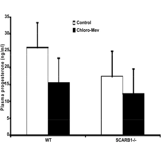
L’approvisionnement en cholestérol est un facteur limitant la stéroïdogenèse ovarienne. Pour cette raison, la majorité du cholestérol requis pour la synthèse des stéroïdes est importé de la circulation via les récepteurs des lipoprotéines de haute (HDL) et de basse densité (LDL) nommés scavenger receptor (SR-BI) et low-density lipoprotein receptor (LDLr). L’ARN messager de SR-BI est exprimé dans les ovaires de porcs durant toutes les étapes de la folliculogenèse ainsi que dans le corps jaune (CL). L’expression de la protéine SR-BI a également été détectée dans les follicules de souris lors du cycle œstral. Chez les deux espèces, l’expression est concentrée dans le cytoplasme et en périphérie des cellules du follicule. Les gonadotrophines induisent l'expression de SR-BI dans les cellules de la granulosa porcines, avec une expression cytoplasmique qui augmente durant la période périovulatoire, et avec une migration aux périphéries cellulaires durant la maturation du CL. Une conformation de 82 kDa de SR-BI est fortement exprimée dans le CL porcin, avec une conformation moins abondante de 57 kDa. Les différences entre les conformations sont attribuables à la glycosylation. La culture in vitro de follicules porcins avec des gonatrophines chorioniques humaines (hCG) a induit une hausse de régulation dépendante du temps du SR-BI de 82 kDa dans les cellules du granulosa. SR-BI et LDLr ont été exprimés réciproquement, avec LDLr étant le plus élévé dans les cellules folliculaires du granulosa et diminuant précipitamment avec la formation du CL. Pour explorer plus en détail les mécanismes d’approvisionnement en cholestérol de la stéroïdogenèse ovarienne, nous avons examiné des souris soumis à un traitement de désaccouplement de l'ovulation, et des souris portant la mutation nulle du gène Scarb1 (SR-BI-/-). Les résultats ont démontré que des ovocytes enfermés dans des structures lutéinisées expriment SR-BI. Les souris SR-BI-/ - présentaient de petits CLs, et de large follicules avec des cellules de thèque hypertrophiées et des kystes folliculaires avec des cavités remplies de sang et une diminution de 50% du niveau de progestérone dans le sérum. Les souris SR-BI-/ - traitées avec une combinaison de 20 μg / g de mevinoline et 100 μ g / g de chloroquine ont démontré une diminution de 43% du niveau de progestérone sérique chez le type sauvage et de 30% chez les souris SR-BI-/ -. L’expression protéique de l’enzyme limitant pour la synthèse du cholestérol, 3-hydroxy-3-methyl-glutaryl-CoA reductase (HMGR), a augmenté chez les souris SR-BI-/-. Nous avons présenté des preuves démontrant que les cellules des follicules expriment le SR-BI durant la
stéroïdogenèse et que la lutéinisation augmente l’expression de SR-BI. La maturation post-transcriptionelle est caractérisée par la glycosylation. Sous des conditions normales, l’expression de LDLr est arrêtée durant la lutéinisation. Ainsi SR-BI devient le facteur principal pour l’importation du cholestérol extracellulaire. En plus, la perturbation extracellulaire du cholestérol synthétisé de novo et l’absorption par les LDLr chez les souris SR-BI-/- diminuent la fonction lutéal. L’homéostasie du cholestérol ovarien est très importante pour une lutéinisation adéquate et sa perturbation mène à une réduction, mais non à un blocage complet, de la fonction lutéal. En conclusion, l’expression de SR-BI est un facteur important, mais non essentiel, pour maintenir l’homéostasie du cholestérol ovarien et la synthèse des stéroïdes, et la lutéinisation. Un réseau de mécanismes complémentaires et compensatoires d’approvisionnement en cholestérol agit en concert pour assurer la synthèse des stéroïdes ovariens.
Mots-clés:
Ovaire, follicule, corps jaune, cholestérol, SR-BI, Récepteurs de lipoprotéines.
Abstract
Ovarian cholesterol supply is rate limiting to ovarian steroidogenesis. For this reason, the majority of cholesterol required for steroid synthesis is imported via scavenger receptor-BI (SR-BI) and the low-density lipoprotein (LDL) receptor from circulating HDL and LDL. SR-BI mRNA is expressed in pig ovaries at all stagesof folliculogenesis and in the corpus luteum (CL). SR-BI protein expression in mouse ovary during estrous cycle was also detected. In both species, expression is concentrated in cytoplasm and periphery of follicular cells. Gonadotropins induce SR-BI expression in pig granulosa cells, with cytoplasmic expression increasing through the periovulatory period, with migration to the cell peripheryas the CL matured. An 82-kDaform of SR-BI is strongly expressed in the pig CL, with the less abundant 57-kDa form, differences between forms are attributable to glycosylation. In vitro culture of pig follicles with human chorionic gonadotropin (hCG) induced time-dependent upregulation of 82-kDa SR-BI in granulosa cells. SR-BI and LDL receptor werereciprocally expressed, with the latter highest in follicular granulosa cells, declining precipitously with CL formation. To further explore mechanisms of cholesterol supply to ovarian steroidogenesis, we examined mice treated to uncouple ovulation and mice bearing null mutation of the Scarb1 gene (SR-BI-/-). Results show entrapped oocytes in luteinized structures expressed SR-BI. SR-BI-/- mice displayed small corpora lutea, large follicles with theca cells hypertrophied, follicular cysts with blood filled cavities and 50% decreased in plasma progesterone. In SR-BI-/- mice, treatment with a combination of 20 μg/g of mevinolin and 100 μg/g of chloroquine (CHLORO) was employed to disturbed cholesterol sources. Serum progesterone was reduced by 43% in wild type and 30% in SR-BI-/- mice. The protein expression of the rate-limiting enzyme for cholesterol synthesis, 3-hydroxy-3-methyl-glutaryl-CoA reductase (HMGR) increased in SR-BI-/- mice. It was concluded that follicular cells express SR-BI during follicle development and luteinization causes upregulation of SR-BI expression. Posttranslational maturation is characterized by glycosylation. Under normal conditions expression of the LDLr (low density lipoprotein recepors) is extinguished during luteinization such that SR-BI becomes the principal means of importation of extracellular cholesterol. Further, perturbation of cholesterol de novo synthesis and uptake from LDLr in SR-BI-/- mice leads to a reduction of luteal function. Ovarian cholesterol homeostasis is central to adequate luteinization, and its perturbation leads to
reduction, but not to complete impairment, of luteal function. We conclude that SR-BI expression is an important but not essential factor in maintaining ovarian cholesterol homeostasis, steroid synthesis and luteinization. A network of complementary and compensatory cholesterol supply mechanisms act in concert to assure ovarian steroid synthesis.
Table of contents
RÉSUMÉ iii
ABSTRACT v
TABLE OF CONTENTS vii
LIST OF TABLES xi
LIST OF FIGURES xii
DEDICATION xiv
ACKNOWLEDGEMENTS xv
LIST OF ABBREVIATIONS xvi
1. INTRODUCTION 1 2. LITERATURE REVIEW 3 2.1 FOLLICLE 3 2.1.1 Follicular structure 3 2.1.2 Granulosa cells 4 2.1.3 Theca cells 5 2.1.4 Follicular development 6 3. OVULATION 12
3.1 Role of the granulosa layer during ovulation 13
3.2 Role of the theca layer during ovulation 18
4. PROLIFERATION AND DIFFERENTIATION 21
4.1 Hormonal aspects of proliferation versus differentiation 21
4.2 Cell cycle control 22
5. LUTEINIZATION 24
5.1 Corpus luteum structure 24
5.2 Corpus luteum classification 26
5.4 Gene expression in the corpus luteum 32 6. CHOLESTEROL IMPORTATION INTO STEROIDOGENIC
TISSUE
33
6.1 LDL cholesterol importation 33
6.2 HDL cholesterol importation 35
6.3 Generalities about SR-BI 36
6.4 SR-BI intracellular location 38
6.5 Tissue and cells type-specific expression of SR-BI 38
6.6 SR-BI hormonal regulation 39
6.7 SR-BI molecular regulation 40
6.8 Other regulators of SR-BI 42
6.9 SR-BI deficiency 43
6.10 Alternate splicing SR-BI and cholesterol importation 44
7. STEROIDOGENESIS 46
7.1 Follicular steroidogenesis 46
7.2 Key enzymes in steroidogenesis 48
7.3 Luteal steroidogenesis 50
8 HYPOTHESIS AND OBJETIVES 51
8.1 Experimental goals 51
CHAPTER ONE
LIPOPROTEIN RECEPTOR EXPRESSION EVOLUTION DURING THE
LUTEINIZATION OF THE OVARIAN FOLLICLE 52
ABSTRACT
53 INTRODUCTION 54
MATERIALS AND METHODS 56
Pig SR-BI cloning, sequencing, and RT-PCR 57
Western blot analysis 58
Immunofluorescence 59
Deglycosylation of SR-BI 59
Statistical analyses 60
RESULTS 60 Expression of SR-BI in granulosa cells from ovarian
follicles and corpora lutea
60
Changes of cholesterol importation during the peri- ovulatory and luteinization periods
62
Gonadotropin induction of the pattern of cholesterol importation in vitro 63 DISCUSSION 65 ACKNOWLEDGEMENTS 68 REFERENCES 68 FIGURE LEGENDS 71 CHAPTER TWO
SCAVENGER RECEPTOR B-I AND LUTEAL FUNCTION IN THE MOUSE
84
ABSTRACT 85 INTRODUCTION 86
Experiment 1 87 Experiment 2 88 Experiment 3 90 RESULTS 92 Experiment 1 92 Experiment 2 92 Experiment 3 94 DISCUSSION 95 ACKNOWLEDGEMENTS 98 REFERENCES 99 FIGURE LEGENDS 103 GENERAL DISCUSSION 113 BIBLIOGRAPHY 118
List of tables
Table 1 Hydroxysteroid dehydrogenase and P450, enzymes involved in the active ovarian steroid hormone biosynthesis
List of figures
FIGURES: LITERATURE REVIEW
Figure 1 Principal candidate genes involved in ovulation 19 Figure 2 Principal sources of cholesterol, and key enzymes involved in
cholesterol metabolism by steroidogenic tissue for steroid biosynthesis
34
Figure 3 Protein domain and configuration of SR-BI inserted in the plasma membrane
37
FIGURES: CHAPTER ONE
Figure 1 Follicules and corpora lutea express SR-BI transcripts 75 Figure 2 Developmental sequence SR-BI of protein abundance in
ovarian tissues
76
Figure 3 Effects of Endo-H and PGNase declycosylation on SR-BI from corpus luteum as revealed by immunoblot
77
Figure 4 Immunocytochemistry demonstrating the divergent cellular localization of SR-BI in cells from porcine ovary
compartments
78
Figure 5 Evolution of SR-BI cellular localization during periovulatory and luteal phases of the estrous cycle in the pig. Follicular and luteal SR-BI membrane and cytoplasmic expression
79
Figure 6 Comparison of LDLr and SR-BI abundance in follicles and corpora lutea by immunoblot
80
Figure 7 Reciprocal LDL receptor and SR-BI expression induction within granulosa cells, from porcine follicles in culture
81
Figure 8 Induction of SR-BI expression in isolated luteinized porcine granulosa cells with 100 ng/ml luteinizing hormone (LH) and transcriptional blockade with Act-D
82
Figure 9 Effects of lipoproteins in culture medium on expression of SR-BI by luteinized porcine granulosa cells
FIGURES: CHAPTER TWO
Figure 1 Protein expression of SR-BI in mouse follicles during proestrus, estrus and in the corpus luteum of mature mice undergoing normal estrous cycles
105
Figure 2 Protein expression of SR-BI in the ovarian theca, granulosa, cumulus oophorus and luteal cells of mature mice undergoing normal estrous cycles
106
Figure 3 Bright field microscopy images of hematoxylin-eosin stained sections of ovaries from immature mice stimulated with eCG, hCG and meloxicam
107
Figure 4 Protein expression of SR-BI in the corpus luteum and in follicles of immature mice stimulated with eCG, hCG and meloxicam
108
Figure 5 Plasma progesterone concentrations from wild type or SR-BI -/-mature mice stimulated with eCG and hCG
109
Figure 6 Bright field microscopy images of hematoxylin-eosin stained sections of ovaries from wild type mice or SR-BI-/- mice stimulated with eCG and hCG and mevinolin+ chloroquine
110
Figure 7 Bright field microscopy images of atypical structures in hematoxylin-eosin or periodic acid-Schiff (PAS) stained sections of ovaries from SR-BI-/- mice stimulated with eCG and hCG
111
Figure 8 Protein expression of HMGR in corpus luteum from wild type or SR-BI-/- mice stimulated with eCG and hCG and/or
mevinolin + chloroquine
Dedication
Este trabajo se lo dedico con amor y cariño a:A Rosa†, mi madre, por su grandeza.
A mi padre y hermanos por la red de amor y cobijo que han fabricado y conservado para dar bienestar y confianza a cada uno de los miembros de la familia.
A mi esposo, por su ayuda y apoyo en el logro de mis objetivos.
A mi inseparable amiga y colega, mi hija Irene Adriana, quien a su tierna edad compartió los momentos de felicidad pero también de desesperanza que se sucedieron durante la realización del doctorado. -Pollue, espero que cuando tú estés en la misma situación yo pueda estar a tu lado-
Acknowledgements
Thanks to Dr. Murphy for the valuable guidance through my PhD program. Studies were funded by Colegio de Postgraduados, México. CONACyT, México. SRE, México. Ministère de l’Éducation du Québec, University of Montreal and an Operating Grant to B.D. Murphy from the Canadian Institutes of Health Research.
Thanks to researchers and professors at the CRRA.
Thanks to my classmates for their contributions, expertise and support throughout my studies.
To Mira Dobias for the support in the laboratory
Sincere thanks to Joëlle Desmarais for the honest and moral help.
To the people that contributed before and during my studies.
I cannot end this, without thanks to my friends, who formed a nucleus of moral support. Thanks to Estefan and Luisa, Estrella and Kasi, Samira, Karim, Gaby and husband, Jorge and Cecilia, Martha and Alex, Anamaría, Adolfo, Mary Cruz, Adriana, Norma, Lucille, and Francee.
List of abbreviations
ACTH adrenocorticotropic hormone ADAMTS-1 disintegrin and
metalloproteinase with thrombospondin motifs
AC Adenylyl cylase ACAT Cholesterol acyl transferase
Ahr Aryl hydrocarbon receptor ALAS 5-Aminolevulinate synthase
Ang-1 Angiopoietin 1 Ang-2 Angiopoietin 2
ANOVA Analysis of variance Apo-A apolipoprotein A
Apo-B apolipoprotein B Apo-E apolipoprotein E
Apo-1 apolipoprotein 1 bp base pares
BSA Bovine serum albumin cAMP cyclic adenosine monophosphate
Ca2 + Calcium ion CBR carbonyl reductase
CCAAT CCAAT box of about minus 70
bp CDB-2924 Synthetic Receptors antagonist progesterone Cdk2 cyclin dependent kinase 2 Cdk4 Cyclin dependent kinase 4 Cdk6 Cyclin dependent kinase 6 CD36 cluster determinant 36 a
leukocyte differentiation antigen
Chloro-mevi Chloroquine-mevinolin C/EBPβ CCAAT/enhancer binding proteins
CE cholesteryl ester CRHBP corticotrophin releasing hormone binding protein
CL corpus luteum CL-I postovulatory CL
CL-II developing CL CL-III mid-luteal CL
COX-1 Cyclo-oxygenase-1 COX-2 Cyclo-oxygenase-2
CP45011A cytochrome p450 cholesterol
side-chain cleavage CRE cAMP response element CREB cAMP response element binding
protein CycE Cyclin E
CYP11A cytochome p450 cholesterol
List of abbreviations
(continued)Dazla DAZ Like autosomal DAPI 4’diamino-2-phenyindole dilactate
DAX-1 dosage-sensitive sex reversal-1 DNA deoxyribonucleic acid
DHT dehydrotestosterone EGF epidermal growth factor
EG-VEGF endocrine gland VEGF ET-2 endothelin-2
ET-I endothelin-I ERα estrogen receptor alpha
Erg-1 early growth response protein-1 E2 17ß-estradiol
eCG Equine chorionic gonadotropin E2F transcription factor E2F FBS fetal bovine serum Figla the factor in the germline
alpha
FC free cholesterol Foxl2–/– forkhead box
1/2 knockout mice
FSH follicle-stimulating hormone Foxo3a forkhead transcription factors 3a
GAPDH glyceraldehyde-3-phosphate dehydrogenase
GCS g-Glutamylcysteine synthetase
GDP guanosine diphosphate GH growth hormone
GITC guanidine isothiocyanate buffer GnRH gonadotropin releasing hormone
GTPase Guanosine triphosphate hydrolase GTP guanosine triphosphate
GST glutathione S-Transferase H hours
hCG human chorionic gonadotropin HB2 HDL-binding proteins HDL high-density lipoprotein HMG-CoA-
reductase 3-hydroxy-3-methylglutaryl coenzyme A-reductase HDL-CE high-density lipoprotein
cholesteryl ester
3α-HSD 3α-hydroxysteroid dehydrogenase HPS hematoxylin, phloxine, saffron 17beta
HSD-7
17-beta-hydroxysteroid dehydrogenase type 7
List of abbreviations
(continued)3ß-HSD 3ß-hydroxysteroid dehydrogenase IGF-I insulin growth factor-1 20α-HSD 20α-hydroxysteroid
dehydrogenase IgG Immunoglobulin G
IL-Ib Interleukin-1b IU International units
kDa kilo Daltons Kg kilogram
KO knock-out LCAT lecithin cholesterol
acyltransferase
LDL low-density lipoprotein LDLr low density lipoprotein receptors
LH luteinizing hormone LH-r Luteinizing hormone
receptor LIM essential nuclearregulator of
myogenic differentiation
LIMPII lysosomal integral membrane protein II
LMO7 LIM domain only 7 MAPK mitogen-activated protein
kinase MDP glycosyl
Phosphotidylinositol
anchored membrane dipeptidase
min minute
mg Milligrams ml mililiters
mM millimolar mm Millimeters
mm3 millimeter cubic MMP1 matrix metalloproteinase 1 MMP-2 matrix metalloproteinase 2 MMP-9 matrix metalloproteinase 9 MMP-14 matrix metalloproteinase 14 MMP-19 matrix metalloproteinase 19 mRNA messenger ribonucleic acid MT-1 metallothionin-1
µl Microliter M molarity
NCI-H295R human adrenocortical cells Ngf nerve growth factor
Nm Nano moles P probability
PAP-III pancreatitis-associated protein-III PBR peripheral benzodiazepine receptor
List of abbreviations
(continued)PBS phosphate buffered saline PCR polymerase chain reaction PDZK1 PDZdomain-containing-1 protein PDZ PDZ domain, associated with
plasma membrane
PG prostaglandin PGE2 prostagandin E2
PGFα prostaglandin F-2alpha PGH2 prostaglandin H2
PGI2 prostaglandin I2 PGHS enzyme PG endoperoxide
G/H synthase
PGG2 prostaglandin G2 pH potential of hydrogen
PKA protein kinase A PKC protein kinase C
P450scc cytochrome 450side chain cleavage enzyme
P450arom cytochome P450 aromatase PMSG pregnant mare serum
gonadotropin PR progesterone receptor pRB phosphorylated retinoblastoma protein RGS2 regulator of G-protein signaling protein-2
RNA ribonucleic acid RIA radioimmunoassay
RT reverse transcriptase RT-PCR reverse transcription-polymerase chain reaction RU486 mifepristone, progesterone
receptor inhibitor SCP2 sterol carrier protein-2 SCARB1 scavenger receptor class B type1 SDS-PAGE sodium dodecyl sulphate
polyacrylamide gel electrophoresis
SF-1 steroidogenic factor-1 SOD superoxide dismutase
SREBP sterol regulatory element binding
protein SR-BI scavenger receptor class B type1 SR-BII scavenger receptor class B typeII SR-BI +/+ scavenger receptor class B
type1, wild type mice SR-BI -/- scavenger receptor class B type1,
List of abbreviations
(continued)SRE1 sterol regulatory element 1 StAR steroidogenic-acute regulatory protein
T testosterone Taq DNA-
polymerase
Thermus aquaticus DNA polymerase
TBS Tris buffer saline TGF-ß transforming growth factor-ß TIMP-1 metalloproteinase-1 inhibitor TNFα tumor necrosis factor-α TSG-6 tumor necrosis factor-stimulated
gene-6
TTBS tweenTris-buffered saline
VEGF vascular endothelial growth factor
VitE vitamin E
vLDL very low density lipoproteins WT wild type
XL-1 blue XL Blue competent cells Y1-BS1 mouse adrenocortical cell line
ZK98299 onapristone, progesterone
receptor inhibitor YTPLL dileucine base motif YXXZ tyrosine-based motif ZP1 zona pellucida protein 1 ZP2 zona pellucida protein 2 ZP3 zona pellucida protein 3 ZP -/- ZP knockout mice
1. INTRODUCTION
In females, the ovaries are the basis for gamete production and hormone synthesis in support of reproductive behavior and gestation. The events of follicular development, ovulation and luteinization are governed by endocrine, paracrine and autocrine factors. In folliculogenesis, some follicles leave their quiescent state to grow, then to ovulate, or die as atretic follicles. The ovulatory follicles collapse to form an ephemeral gland, known as corpus luteum (CL) whose primary function is progesterone production. This hormone regulates the estrous cycle and maintains pregnancy. In humans, the CL synthesizes upwards of 40 mg/day of progesterone (Murphy 2000). The steroidogenic luteal cells are provided with highly efficient machinery to maintain luteal progesterone production. Steroid production by the CL varies remarkably among mammalian species. In humans, monkeys and ruminants, the CL is maintained by pituitary derived luteinizing hormone (LH) acting principally through the cAMP/protein kinase A pathway (Niswender, Juengel et al. 2000). In contrast, it is well established that prolactin and estradiol are critical luteotrophic hormones in rodents and rabbits acting through their respective receptors (Stormshak, Zelinski-Wooten et al. 1987). The substrate for steroid hormone synthesis is cholesterol, derived from multiple sources. Luteal cells can produce cholesterol de novo, this method plays a minor role in the normal functioning tissue as evidenced by the low levels of 3-hydroxy-3-methylglutaryl coenzyme A-reductase (HMG-CoA reductase), the rate-limiting enzyme in the cholesterol biosynthetic pathway (Gwynne and Strauss 1982). The major
mechanisms for cholesterol delivery to steroidogenic cells are the endocytotic pathway involving low density lipoprotein (LDL) and the selective uptake of cholesterol esters from high-density lipoprotein (HDL) (Brannian and Stouffer 1993). SR-BI is the receptor that mediates the
selective uptake of cholesterol esters from HDL (Acton, Rigotti et al. 1996) and its deletion produces infertility and reduced lipid levels in the CL, as measured by oil red-O staining, suggesting a reduction in cholesterol ester storage (Trigatti, Rayburn et al. 1999). The authors report that progesterone profiles were within the normal range in these animals, suggesting that other sources of cholesterol may have increased in these animals to correct for the absence of HDL delivery.
Recent exploration of the influence of hepatic SR-BI on female fertility by inducing hepatic SR-BI expression in mice with null mutation of the SR-BI-/- gene by adenovirus transduction or stable transgenesis indicated that these treatments restore female fertility by increasing hepatic metabolism of lipids (Yesilaltay, Morales et al. 2006). Given the apparent compensation by alternate sources, the goal of this work was to explore the role SR-BI in ovarian steroidogenesis and to examine possible alternatives sources of cholesterol that maintain normal luteal steroidogenesis in SR-BI -/- mice.
2. LITERATURE REVIEW
In this section the physiological and molecular bases of the reproductive phenomena regulating the presence of reproductive cyclicity in mammals are described, focusing on follicular development, ovulation and corpus luteum formation, events that were studied in this investigation.
2.1. FOLLICLE
2.1.1 Follicular structure
Follicles function to provide suitable conditions to maintain oocyte development, as well as the source of steroid hormones. Follicles produce other substances that act as both autocrine and paracrine modulators of cellular function and include peptides, proteins, glycoproteins, proteoglycans and growth factors whose expression varies according to reproductive and follicular development conditions (Murdoch, Dailey et al. 1981; Lumsden, Kelly et al. 1986; Richards, Hickey et al. 1987; Wise 1987; Prevost, Belanger et al. 1989).
The follicle consists of two cellular compartments. The compartment within the basement membrane of the follicle is known as the granulosa layer, and components of this layer are in direct contact with the oocyte. This layer varies in the number and size of the cells according to the hormonal characteristics (Richards, Jahnsen et al. 1987; Burke, Cardenas et al. 2005) and with the stage of follicular development. For example granulosa cells are flattened in primordial follicles, and this form changes to polyhedral or cubical in primary follicles (Braw- Tal 2002). During early phases of follicular development, the surrounding stromal cells begin to
aggregate concentrically around the follicular basal lamina and form the theca layer (Motta, Makabe et al. 1994). Communication between the two types of follicular cells has been observed in numerous in vivo and in vitro experimental paradigms (Tajima, Orisaka et al. 2006). Despite of the joint function of the follicular cells, for this review they are described separately.
2.1.2 Granulosa cells
As mentioned before, granulosa cells form a continuous layer surrounding the oocyte and can be divided into different subpopulations, based on morphology and response to hormonal stimuli (Redmer, Kirsch et al. 1991). These cells display high proliferative capacity. The corona radiata cells maintain an intimate contact and provide nutrients to the oocyte. The cumulus oophorus cells support and integrate the oocyte within the follicle, and contribute during the final stage to oocyte maturation, and serve as a barrier to sperm penetration prior to fertilization. The majority of the granulosa cells are known as mural cells, and these populate the follicular cavity with their most basal layer maintaining close contact with the basal membrane (Motlik, Fulka et al. 1986; McGregor, Flaherty et al. 1989).
Granulosa cell proliferation varies according to their localization, to the estrous cycle stage and to the follicular development phase. Proliferation in granulosa cells evaluated by thymidine incorporation showed more elevated proliferative activity in cumulus oophorus cells relative to mural granulosa cells (Khamsi and Roberge 2001). Some data suggest that cumulus granulosa cells continue to proliferate for up to 10 h after an ovulatory stimulus, with evidence that cyclin E protein increases after hCG, and there was a tendency of initial decline followed by a transient increase in Cdk2 protein activity 8 h after hCG (Cannon, Cherian-Shaw et al. 2005).
Proliferation is maintained in granulosa cells in the proximity of the oocyte during ovulation of the rat follicle (Cannon, Cherian-Shaw et al. 2005). Observations with electronic microscopy of granulosa cells in co-culture have allowed observation of the morphology and content of cellular elements characteristic of steroidogenic and mitotically active cells (Nottola, Heyn et al. 2006).
2.1.3 Theca cells
The theca layer consists of an internal and external theca. In species as rodents, a special group of cells named interstitial cells capable of steroid production differentiate from the internal theca (Brook and Clarke 1989). This differentiation of theca is an important physiological event occurring during early follicular development and, apparently the theca cell layer develops from stromal cells and is regulated by granulosa cells (Orisaka, Tajima et al. 2006).
The internal theca cell layer consists of undifferentiated fibroblast cells, blood vessels, and cholinergic as well as adrenergic nerve fibers. The structure of these cells is characterized by their elevated concentration of smooth endoplasmic reticulum as well as tubular crest mitochondria, characteristic of active steroidogenic cells (Motta, Nottola et al. 1995). The
vascularization of these cells increases as the follicle develops (Macchiarelli, Vizza et al. 1992). Studies on follicular vasculature showed that large vessels are located in the outer theca layer, while smaller vessels are observed neighboring the inner layer, close to the basal membrane (Augustin, Braun et al. 1995; Redmer, Doraiswamy et al. 2001; Feranil, Isobe et al. 2004). The theca is the source of vascularization of the avascular granulosa cells during luteinization. The increase in blood vessels is a response to angiogenic factors including vascular endothelial growth factor (VEGF), angiopoietins 1 and 2 (Ang-1 and Ang-2) and endocrine gland VEGF
(EG-VEGF) originating from the granulosa cells, and regulated by LH (Phan, Rakenius et al. 2006, for review see Fraser 2006).
Within growing follicles, theca cells are stimulated by LH to synthesize androgens that serve as substrate for estrogen synthesis in granulosa cells. There is evidence in ruminants that, after ovulation, theca cells are transformed into the small luteal cells of the corpus luteum (Alila and Hansel 1984; O'Shea 1987) and granulosa cells are believed to be transformed in large luteal cells (Alila and Hansel 1984) but this issue has not been definitively resolved.
2.1.4 Follicular development
The ovary is a primary functional organ of the female reproductive system, and it plays two major physiological roles. First, the ovary is responsible for the differentiation and release of mature oocytes for fertilization (McGee and Hsueh 2000). Second, it is responsible for synthesizing and secreting hormones that are essential for follicle development, menstrual and estrous cyclicity and maintenance of the reproductive tract and its function (Hirshfield 1991).
Ovarian follicle development is a complex process that begins with the establishment of primordial follicle, after birth, depending on the species in a process that involves apoptosis of some of the oocytes and initiation of meiotic division of some others (Gomperts, Garcia-Castro et al. 1994; Pepling and Spradling 1998). The surviving oocytes become enveloped by somatic cells, thereby producing primordial follicles considered to be in a ‘‘quiescent’’ state, since no major morphological changes take place until they enter the pool of growing follicles (Zamboni, Thompson et al. 1972). The resting follicle is a round structure with a diplotene oocyte
surrounded by a single layer of flattened follicular cells (Pepling and Spradling 2001). The primordial follicle population present at birth has long been believed to be predetermined, following a log-linear rate of decline during the reproductive life (Miller, Charleston et al. 1999). Although this idea has recently been challenged (Johnson, Canning et al. 2004; Johnson, Bagley et al. 2005), the weight of the evidence argues against postnatal oogenesis in mammals. Several studies of gene mutation, particularly knockin and knockout mice, have elucidated the importance of genes that control differentiation of germ cells that culminate in alteration of primordial follicular development. Examples of these are the Dazla gene (Ruggiu, Speed et al. 1997; McNeilly, Saunders et al. 2000), the factor in the germline alpha (Figla) (Soyal, Amleh et al. 2000), the nerve growth factor (Ngf ) and others (Dissen, Romero et al. 2001).
Recruitment of primordial follicles into the growing pool begins the coordinated and interdependent development of the somatic and germ cell components of the ovarian follicle. The end of reproductive life, or ovarian senescence, occurs when this pool of primordial follicles is depleted by death or through growth, followed by subsequent ovulation and/or atresia (Hirshfield 1991). Although some primordial follicles will be stimulated to grow immediately, the majority will remain dormant, perhaps because of autocrine or paracrine inhibition, until they receive signals to enter the growing pool (McGee and Hsueh 2000). The follicle that is recruitedfrom the pool of resting follicles to enter the growth phase is termed primary follicle. The primary follicle
enlarges because of an increase in size of the oocyte (Baca and Zamboni 1967; Zamboni 1974; Motta, Makabe et al. 1994), and conversion of the squamous or flattened granulosa cells into cuboidal granulosa cells (Hirshfield 1991). Another characteristic of the primary follicle is the formation of a zona pellucida, which surrounds the oocyte and is maintained throughout growth
until the oocyte is ovulated (Rankin, Familari et al. 1996; Rankin, Talbot et al. 1999; Rankin, O'Brien et al. 2001). The follicular cells are still arranged in a single layer to surround the oocyte
and still changing shape to polyhedral or cubical. In parallel, beginning with the early phases of follicular development, the surrounding stromal cells start to aggregate concentrically around the follicular basal lamina and on time form the theca cells layer (Zamboni 1974; Motta, Makabe et al. 1994).
Conversion of primordial into primary follicles involves participation of genes and proteins that regulate organization of zona pellucida by synthesis of the extracellular zona pellucida proteins ZP1, ZP2 and ZP3 (Rankin, Talbot et al. 1999). Mice Zp1–/– have loosely organized zonae pellucidae and reduced litter sizes. Zp2–/– and Zp3–/– mice are sterile (Rankin, Familari et al. 1996; Rankin, O'Brien et al. 2001).
Forkhead genes have also been shown to play a role in folliculogenesis at the primary follicle stage. In Foxl2–/– ovaries, granulosa cell differentiation is blocked at the squamous to the cuboidal transition (Schmidt, Ovitt et al. 2004). Foxo3a ovaries have a tremendous increase in the number of early growing follicles with enlarged oocytes (Castrillon, Miao et al. 2003).
By the time that the follicle reaches the pre-antral stage, granulosa cells have proliferated to form multiple layers. In addition, the pre-antral follicle acquires an outer layer of thecal cells separated from the follicle by the basal lamina (van den Hurk and Zhao 2005). At the end of this stage, the pre-antral follicle has several layers of granulosa cells and has acquired an extensive network of gap junctions (Hirshfield 1991). These junctions are intercellular membrane channels that allow nutrients, inorganic ions, second messengers and small metabolites to pass from cell to
cell (Kidder and Mhawi 2002). The next step in the developmental sequence is formation of the antral cavity with fluid rich in water, electrolytes, serum proteins and with high concentrations of steroid hormones secreted by the granulosa cells (Hirshfield 1991). In some species, antral cavity formation is not accompanied by notable changes in oocyte morphology, while in others, the oocyte continues to expand during the early antral phase of development (van den Hurk and Zhao 2005). At the antral stage, most of the follicles will undergo atresia, whereas the remaining antral follicles will survive under the influence of FSH and grow to the pre-ovulatory stage (Hirshfield 1991). Factors such as the aryl hydrocarbon receptor (Ahr) and superoxide dismutase (SOD) have been shown to regulate follicular growth to the antral stage (Gupta RK 2006 ). The Ahr is a ligand-activated transcription factor that functions in mediating the toxicity of various contaminants (Pocar, Fischer et al. 2005). Without this gene, there is a decrease in the number of antral follicles and reduced fertility (Benedict, Lin et al. 2000; Benedict, Miller et al. 2003).
The follicles that survive the massive atresia visited on the preantral and early antral population acquire the capacity to produce estrogen and grow to the pre-ovulatory stage (Hirshfield 1991). They exhibit elevated estradiol concentration, higher granulosa cell number, and elevated LH binding capacity in comparison to non-estrogenic follicles (Ireland and Roche 1983). The pre-ovulatory follicle is capable of releasing the oocyte for fertilization, upon stimulation by LH (McGee and Hsueh 2000). Rupture of follicle basement membrane during ovulation is controlled by several different genes and proteins described in ovulation section (below). After the oocyte is released for fertilization, the remaining theca and granulosa cells differentiate into the CL (Hirshfield 1991; Elvin and Matzuk 1998; McGee and Hsueh 2000)
Folliculogenesis consists of two phases: The first, considered independent of gonadotropin influence, also called basal folliculogenesis, includes the passage from primordial follicle to primary follicle, and is regulated by growth factors. The second, gonadotropin dependent phase, includes follicle development to ovulation, and is also known as tonic folliculogenesis (Peluso, Luttmer et al. 1984; Hillier 1994). Follicular development is highly heterogeneous, due to the fact that follicles of the same size develop at different rates throughout the estrous cycle (Rajakoski 1960; Matton, Adelakoun et al. 1981; Driancourt, Thatcher et al. 1991).
In the complexity of follicular development, recruitment, selection and dominance also need to be considered (Matton, Adelakoun et al. 1981; Hodgen 1982; Goodman and Hodgen 1983). Recruitment considered as independent of gonadotropins, in which a group of follicles initiates growth and acquires the capacity to respond to gonadotropins. The follicles then depend on gonadotrophin support to continue its development; until reaching a preovulatory size (Fortune 1994). At this stage of follicular development not only is gonadotropic action (mainly FSH) necessary but there are also autocrine or paracrine influences, e.g growth factors such as epidermal growth factor (EGF; Dorrington, Bendell et al. 1987; Richards, Russell et al. 2002; Kaczmarek, Schams et al. 2005). Selection is the process by which few follicles, from the total recruited population, escape atresia and continue their development. Dominance is when one or more follicles are selected to undergo ovulation. Dominance may involve interference with follicles in less advanced development in the cohort of the recruited follicles, reducing their FSH support (Thatcher, Driancourt et al. 1991). This effect has been attributed to the secretion of estradiol and inhibin A, coming from the more developed follicles (Andersen and Byskoy 2006)
Specifically, inhibin and estradiol block FSH and favor the increase of LH receptor first in theca cells of tertiary follicles (McNatty, Fidler et al. 2000), an action that is inhibited by progesterone administration. Apparently, the inhibin-estrogen synergism favors LH receptors in granulosa cells and reduce FSH receptors in a feedback process (Lisk 1964; Ramirez, Abrams et al. 1964; Williams and Lipner 1981; Kaneko, Noguchi et al. 2002; Martin, Fogwell et al. 1991; Medan, Nambo et al. 2004; Pak, Chung et al. 2006) It is possible that the selection also occurs in response to the reduced response of small follicles to the FSH stimuli (Driancourt, Thatcher et al. 1991; Driancourt, Webb et al. 1991). Dominance is the mechanism by which one or two follicles (several in mice and pigs) are rescued from atresia (Hodgen 1982). In this process it is considered that the “dominant” follicle becomes hypersensitive to FSH action, which is regulated by autocrine and paracrine factors, such as insulin like growth factor-I (IGF-I) action, (Eden, Jones et al. 1988; Behl and Kaul 2002). Recently Murayama and co-workers suggested that insulin itself may support the maturation of preovulatory follicles and the insulin-induced increase in growth hormone receptor (GHR) in dominant follicles may be a turning point for follicles to enter the preovulatory phase during final follicular development (Shimizu, Murayama et al. 2008).
Differences among species exist. In most domestic animals, two or three waves of follicular activity occur during the estrous cycle (Rajakoski 1960; Savio, Keenan et al. 1988; Fortune 1994). In cattle the first wave has been identified at day 0 (ovulation day) and the second at day 10. During the first wave, the recruitment of several follicles has been observed, but only one reaches the dominant stage, initiating its regression at day 11 of the estrous cycle, this wave is known as non-ovulatory. As in the first wave, second wave is characterized by a varied
number of follicles that initiated to grow, nevertheless, only one becomes dominant and proceeds to ovulation, (for review Ireland, Mihm et al. 2000).
3. OVULATION
Ovulation is the rupture of the ovarian follicle to liberate the oocyte (Espey 1980). Initially, this process was considered to be regulated exclusively by increased follicular pressure; later, the enzymatic action for collagen lysis was added (Espey 1980). More modern concepts consider ovulation as a complex process whereby ovarian follicles reactivate oocyte meiosis, create a rupture pore in the apical follicle wall and initiate tissue restructuring and differentiation to form the CL (Russell and Robker 2007). This complex process is controlled by synchronous participation of endocrine hormones, as well as autocrine, paracrine and intracrine mechanisms from the oocyte, theca, mural and cumulus granulosa cells.
Preovulatory follicles contain two distinct sublineages of granulosa cells with highly different responses to gonadotropin action. Mural granulosa cells respond to LH surge due to greater receptor levels abundance relative to cumulus cells (Peng, Hsueh et al. 1991). Comparing follicle cell contents of LH receptors, theca cells express the highest quantity, while in mural and cumulus granulosa cells LH receptors increase during and after ovulation (Bukovsky, Chen et al. 1993; Goudet, Belin et al. 1999; Robert, Gagne et al. 2003).
The LH surge stimulates G protein-coupled receptors within the plasma membrane of cells in the ovarian follicles, and signals of adenylyl cyclase appear, increasing cyclic adenosine monophosphate (cAMP) which in turns regulates protein kinase A (PKA) (Richards 2001). After the PKA catalytic unit is activated its moves to the nucleus, where it phosphorylates a number of transcription factors such as CREB (Mukherjee, Park-Sarge et al. 1996; Yazawa, Mizutani et al. 2003). The interaction of phosphorylated CREB with the coactivator CREB-binding protein stimulates the expression of gonadotropin/ CRE-regulated ovarian genes (Mukherjee, Park-Sarge et al. 1996). This process is temporally associated with immediate transcriptional regulation of numerous genes, and is presumed to be involved in the synthesis and/or activation of specific proteases that degrade the follicle wall (Ohnishi, Ohnishi et al. 2005).
In recent years, molecular and transgenic techniques have allowed the integration of gonadotropin-regulated effects, steroids and prostanoids that regulate the complex gene cascade that produces ovulation (Robker, Russell et al. 2000; Richards, Russell et al. 2002).
3.1 Role of the granulosa layer during ovulation
There is extensive information about the changes occurring in the granulosa and theca cells during ovulation as well as the role of other follicular tissues, the oocyte and related epithelial, endothelial and connective tissues. Although some changes have their origin within the theca cell layer, or simultaneously within the theca and granulosa layers, the granulosa cell layer undergoes the greater alteration during ovulation, displaying changes as soon as the LH surge occurs, some of these initial changes include activation of the genes taking part in the inflammation, ovulation and cell remodeling processes (Espey 1980).
Within follicular tissue, one enzyme for which transcription is upregulated during the early stages of the ovulatory process is 5-aminolevulinate synthase (ALAS). ALAS increases within the granulosa layer as soon as 30 min after hCG, and declines once the mature ovarian follicles start to rupture (Espey 2006). ALAS expression is involved during the acute inflammatory phase response, one of the characteristics during ovulation, which is an abundance of overlapping pathways activated by tissue damage (Ferreira and Gong 1995; Lentsch and Ward 2000). ALAS has been associated with cytochrome P450scc during steroid metabolism. Also ALAS expression within the corpora lutea as it develops from the ruptured follicles is significant.
Initial changes during the first minutes after LH include the induction of genes for early growth response protein-1 (Egr-1) (Russell, Doyle et al. 2003). Egr-1 is a transcription factor that promotes expression of numerous genes important in inflammation, vascular hyperpermeability, hypoxia, coagulation, and other events associated with tissue damage (Espey, Ujioka et al. 2000; Yan, Fujita et al. 2000; Yan, Lu et al. 2000; Silverman, De Sanctis et al. 2001). In the mouse, Egr-1 mRNA increases in the granulosa layer within 30 min after hCG application, reaches a peak from 2-4 h, remains moderately elevated through 12 h after application, and declines 24 after hCG application (Espey, Ujioka et al. 2000; Russell, Doyle et al. 2003). On the other hand, epiregulin, a member of the epidermal growth factor (EGF) family, has been associated with Egr-1 mRNA expression, as well as with increases of cyclooxygenase 2 (COX-2) mRNA and protein expression (Sasaki, Pai et al. 1998). It has been observed that epiregulin expression presents irregular and temporal expression patterns (Sasaki, Pai et al. 1998; Shirakata, Komurasaki et al. 2000).
COX-2, the key enzyme for prostaglandin synthesis is a component of the ovulatory process (Richards, Russell et al. 1998). COX-2 plays a functional role in cumulus cell expansion and it is induced during the inflammation process, (Sirois and Richards 1993; Williams and DuBois 1996; Song, Sirois et al. 1998). COX-2 null mice do not ovulate (Morham, Langenbach et al. 1995); and cumulus cells do not expand, (Davis, Lennard et al. 1999). In the mouse, COX-2 mRNA, confined to the granulosa layer of the mature follicles, increases 4 h, and declines 8 h after hCG application, (Richards, Russell et al. 1998; Davis, Lennard et al. 1999; Joyce, Pendola et al. 2001). In vitro granulosa cells express COX-2 and VEGF (vascular endothelial growth factor) after stimulation with recombinant adiponectin involving MAPK pathway, indicating that these molecules act to produce tissue remodeling (Ledoux, Campos et al. 2006).
The inflammatory process characterizing ovulation is followed by the imminent elevation of toxic products, which must be neutralized (Buetler 1998). Hence, the granulosa cells trigger the expression of genes such as: g-glutamylcysteine synthetase (GCS), GCS mRNA is distributed within the mature follicle granulosa layer, even before hCG administration (Clague, Sevcik et al. 1992; Jarrell, Sevcik et al. 1992). GCS expression increases with hCG administration in mural granulosa cells, also, hCG permits the expression in theca interna that surrounding the granulosa cells. GCS expression is elevated between 4 and 8 h after hCG application, specifically in the theca tissue as well as in some localized areas of the ovarian stroma. GCS is a metalloprotein that serves as the rate limiting enzyme for synthesis of glutathione. Glutathione is a tripeptide that protects cells against oxidative stress during the acute inflammatory reactions and other conditions promoting cellular damage (Clague, Sevcik et al. 1992; Buetler 1998; Soltaninassab, Sekhar et al. 2000).
Also in mice at 4-8 h after hCG, tumor necrosis factor-stimulated gene-6 (TSG-6) is up-regulated in granulosa cells and goes throughout granulosa cumulus cells 12h after hCG (Yoshioka, Ochsner et al. 2000). TSG-6 is a common component of inflammatory reactions, and may exert a negative feedback action by neutralizing hyaluronan-induced inflammatory stress on the extracellular matrix of the cumulus cell-oocyte complex, during the ovulatory process (Fulop, Kamath et al. 1997; Yoshioka, Ochsner et al. 2000). At the same time, but in different location, the regulator of G-protein signaling protein-2 (RGS2) is expressed, along the antral border of the mural granulosa as well as within the cumulus mass surrounding the oocyte (Ujioka, Russell et al. 2000). RGS2 has been characterized as a GTPase-activating protein, thought to attenuate cell signaling by hydrolyzing GTP to GDP (Ujioka, Russell et al. 2000).
Follicular preparation for oocyte release is accompanied by changes from estradiol synthesis to acute progesterone synthesis, requiring the presence of genes involved in the steroidogenesic process as progesterone progesterone receptors (PR) presence and activity, (Robker, Russell et al. 2000). Progesterone action in ovary regulates follicular granulosa cells during ovulation by increasing its levels and by decreasing in estradiol levels, regulating mitotic or apoptotic activities. Other progesterone actions are regulated by membrane progesterone receptors that decrease cAMP with subsequent inhibition of mitosis (Peluso 2006). Progesterone action during ovulation has been clarified using progesterone receptor null mice that display CL formation with no follicular rupture (Lydon, DeMayo et al. 1995). In addition, anovulation in progesterone receptor null mice has been related with decrease in ADAMTS and cathepsin L, both regulated by FSH and LH in a PR-dependent manner (Robker, Russell et al. 2000). ADAMTS is a protein with strong proteolytic effects and believed to act in follicular wall
degradation, while cathepsin L is a papain family also with proteolytic effects (Robker, Russell et al. 2000). Another element is endothelin which is expressed in granulosa cells and decreases in animals with blockage in progesterone receptors (Palanisamy, Cheon et al. 2006).
A rate limiting factor in steroidogenic activity is maintained is transport of cholesterol into mitochondria by StAR (Ronen-Fuhrmann, Timberg et al. 1998; Chaffin, Dissen et al. 2000). It is coexpressed with adrenodoxin, whose transcript, increases significantly in granulosa cells 4 h after hCG administration, and continues in postovulatory luteinized tissue (Espey and Richards 2002). Adrenodoxin and StAR are also are co-expressed with cytochrome P450scc (Rodgers, Lavranos et al. 1995; Grinberg, Hannemann et al. 2000; Muller, Lapko et al. 2001). StAR mRNA has an initial peak after hCG and a second higher peak once the corpora lutea are synthesizing substantial amounts of progesterone (Ronen-Fuhrmann, Timberg et al. 1998; Chaffin, Dissen et al. 2000).
The presence of 3α-hydroxysteroid dehydrogenase (3α-HSD) is necessary to catalyze the oxydoreductive processes during steroid production (Penning, Pawlowski et al. 1996; Ma and Penning 1999). In rats, 3α-HSD mRNA is detectable within theca interna cells of mature follicles before hCG stimulation, and the protein is translocated to the granulosa layer 2 h after hCG application and continues to be elevated until 8 h after hCG application, declining thereafter, although a limited amount still remains within luteal tissue (Penning, Pawlowski et al. 1996; Espey, Yoshioka et al. 2001; Espey and Richards 2002).
Ovulation requires degradation of follicular wall by the action of proteolytic enzymes that include disintegrin and metalloproteinase with thrombospondin motifs (ADAMTS-1) and cathepsin L for proteolysis of extracellular matrix (Robker, Russell et al. 2000). ADAMTS-1 which increase significantly in the granulosa layer by 4 h after hCG application and reaches a peak at12 h after hCG application (Espey, Yoshioka et al. 2000), at the time when rat follicles begin to rupture. ADAMTS-1 transcription depends on the usual increase in ovarian progesterone synthesis and on the up-regulation of ovarian progesterone receptor during ovulation (Doyle, Russell et al. 2004; Porter, Clark et al. 2005; Shozu, Minami et al. 2005). Cathepsin L is induced in granulosa cells of growing follicle by FSH, but the highest levels of cathepsin L mRNA occur in preovulatory follicles in response to LH (Robker, Russell et al. 2000).
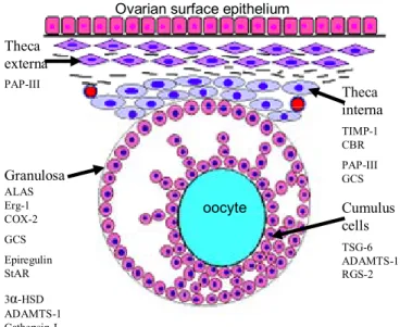
3.2 Role of theca the layer during ovulation
As mentioned previously, the expression of genes at the moment of the ovulation is not exclusive to the granulosa cell layer, as many changes occur simultaneously in theca and granulosa layers (Fig.1).
oocyte Theca interna Theca externa Granulosa
Ovarian surface epithelium
TIMP-1 CBR PAP-III GCS PAP-III ALAS Erg-1 COX-2 GCS Epiregulin StAR 3α-HSD ADAMTS-1 Cathepsin-L TSG-6 ADAMTS-1 RGS-2 Cumulus cells
Figure 1. Principal candidate genes involved in ovulation: They are listed according to the follicular compartment; ALAS= amino levulinic acid synthetase, Erg-1= , CoX-2= Cyclooxygenase-2, GCS= γ-glutamilcystein synthetase, epiregulin, StAR (steroidogenic acute regulatory protein), 3a-HSD (3-alpha-hydoxysteroid-dehydrogenase) ADAMST-1(Disintegrin and metalloproteinase with trombospondin motifs), Cathepsin-L, TIMP-1 (Tissue inhibitor of metalloproteinase-1), CBR (Carbonyl reductase), PAP-III (Pancreatitis-associated protein-III), TSG-6 (Tumor necrosis factor-stimulated gene-6), RGS-2 (Regulator of G-protein signaling protein-2) (Adapted from Ohnishi, Ohnishi et al. 2005).
During ovulation, theca cells express multiple matrix metalloproteinases (MMPs) including MMP-1, MMP2, MMP9, MMP14 AND MMP19 (Hagglund, Ny et al. 1999). At least one metalloproteinase-1 inhibitor (tissue inhibitor of metalloproteinase-1 or TIMP-1) mRNA is expressed on a temporal pattern basis. Similar to ADAMTS-1 mRNA, it is present within both thecal and stromal ovarian tissue and increases at the time of ovulation (Simpson, Byers et al. 2001). During ovulation, TIMP-1 regulates ADAMTS-1 degradation as well as other ovarian metalloproteinases (Espey, Ujioka et al. 2003). In the mouse, TIMP-1’s protective mechanism results in the presence of carbonyl reductase (CBR) mRNA, detectable before hCG, and with an increase over 2 h after hCG, reaching a peak at 8 h, and declining 24 h after hCG treatment (Espey, Yoshioka et al. 2000). Its spatial distribution is mainly within thecal and stromal regions; however, the granulosa layer shows a little expression (Espey, Yoshioka et al. 2000). CBR increases during ovulation, it might function as a local protective response to the substantial steroid and prostanoid increases within the interstitial tissue of the ovary (Inazu and Fujii 1998; Espey, Yoshioka et al. 2000).
During ovulation, blood vessels degradation may be controlled by pancreatitis-associated protein-III mRNA (PAP-III) that is expressed 8 h after hCG administration in mice (Yoshioka, Fujii et al. 2002), and limited to the hilar region of the ovarian stroma. It is principally found within endothelial cells limiting the inner walls of blood vessels. PAP-III is a macrophage chemoattractant induced in and released from injured tissue (Heller, Fiedler et al. 1999). Ovarian PAP-III mRNA might be expressed in conjunction with a protective response to the hyperemia, exudation, proteolysis, and inflammation characterizing the ovulation event (Yoshioka, Fujii et
al. 2002; Namikawa, Okamoto et al. 2006). As previously noted, theca cell action at ovulation, has not been elucidated; however, Richards et al., have proposed that the theca cell layer may serve as protective anti-inflammatory barrier to prevent inappropriate release of oocytes (Richards, Russell et al. 2002).
4. PROLIFERATION AND DIFFERENTIATION
During and following ovulation, changes at cell cycle activity occur and culminate with final cell differentiation, characterized by arrest in cellular proliferation. These changes include several molecular cascades within the luteal cells. For such a reason, this section attempts to briefly describe the aspects related to these changes, focused on cell proliferation versus cell differentiation.
4.1 Hormonal aspects in proliferation versus differentiation
It has been well documented that FSH and estradiol are mitogenic factors, whereas the LH surge terminates granulosa cell proliferation and initiates differentiation by inverting the action of estradiol and FSH over the positive regulators of cell cycle progression in follicular cell types (Richards JS 1980; Rao MC 1978). This action depends on hormones binding to specific receptors and activation of the cAMP and PKA pathway (Jonassen and Richards 1980). Estradiol regulates cell proliferation by inducing Cyclin D1 expression as well as decreasing cdk inhibitor levels (Foster and Wimalasena 1996; Altucci, Addeo et al. 1997). In granulosa cells, estradiol induces cyclin D2 and cyclin E expression, with concurrent p27 reduction (Robker and Richards 1998). Other molecules regulating cell cycle include: a) activin, produced in high levels within preovulatory granulosa cells, has been shown to stimulate DNA synthesis (Miro and
Hillier 1996) b) Insulin-like growth factor-1 (IGF-1) is also implicated in granulosa cell proliferation (Zhou, Refuerzo et al. 1995). Studies in IGF-1 null mice showed that large antral follicles are present within ovaries, indicating that IGF-1 may be more important for differentiation than for proliferation (Baker, Hardy et al. 1996; Chaffin, Schwinof et al. 2001).
4.2 Cell cycle control
During follicular development follicular cells are highly proliferative (Rao, Midgley et al. 1978; Hirshfield 1991), a characteristic that is diminished or lost toward luteinization (Richards, Hedin et al. 1986; Richards 1994). As mentioned before, estradiol activates events that favor proliferation (Hampl, Pachernik et al. 2000; Cannon, Cherian-Shaw et al. 2005). Early, during the G1 phase, D-type cyclin activates cyclin dependent kinase 4 and 6 (cdk4 or cdk6); later, within G1 phase, cyclin E accumulation activates cdk2 (Koepp, et al 2001). Progression through S phase is regulated by cooperation of cdk4/6 and A/D-type cyclins with cdk2/cyclin E complexes (Koff, Cross et al. 1991; Lew, Dulic et al. 1991) followed by the initiation of M phase by cyclin B-cdk2 complexes (Lew, et al. 1996). The S-phase is regulated by cyclin/cdk binding, resulting in a complex requiring phosphorylation to activate, in turn, cellular substrate phosphorylation such as retinoblastoma protein (pRb) which further releases the transcription factor E2F, regulating DNA synthesis and gene expression (Inaba, Matsushime et al. 1992; Xiong, Menninger et al. 1992; Xiong, Zhang et al. 1992; Stevaux and Dyson 2002). Cyclin D2-null mice display impaired granulosa cell proliferation, underdeveloped ovarian follicles, and lack of ovulation (Sicinski, Donaher et al. 1996).
Arrest of the cell cycle in G1 phase with the subsequent CL formation is the result of inhibitors of cell cycle kinases, such as Cip/Kip (p21Cip1, p27Kip1, p57Kip2), as well as Ink4 family, another family of cdk inhibitors (Sherr and Roberts 1999). The Cip/Kips have relatively broad specificity and are able to bind cdk4/6 and cdk2, enabling them to inhibit the activity of several kinase cascades, thereby blocking the cell cycle progression at multiple points, in particular at the G1 phase (Polyak, Lee et al. 1994). Both p27 and p21Cip1, are highly expressed in the corpus luteum, showing slight different patterns of induction (Robker and Richards 1998). Loss of p27 results in impaired differentiation, as has been observed by the inability of granulosa cells to luteinize normally, and produce enough progesterone in order to support pregnancy (Fero, Rivkin et al. 1996).
Analysis of the cell cycle regulators after application of hCG or the LH surge showed that cyclin D2 and p27 are regulated inversely (Robker and Richards 1998). During luteinization, there is down-regulation of cyclin D2 and while p27 is up-regulated, leading to the cell cycle arrest at the G1 phase and at 24h of hCG addition or LH surge (Fero, Rivkin et al. 1996; Nakayama, Ishida et al. 1996). Using in situ hybridization in hormone-treated-hypophysectomized rats, it has been shown that cyclin D2 mRNA expression increases and p27 mRNA expression decreases in granulosa cells (Uilenbroek and Richards 1979; Richards 1980). Specifically, in rats treated with estradiol and FSH, cyclin D2 mRNA and protein are expressed at high levels within granulosa cells of preovulatory follicles (Robker and Richards 1998). However, different types of granulosa cells display different proliferative responses: the cells next to the oocyte maintain their mitotic capacity beyond the beginning of luteinization (24h after PMSG), in comparison with mural granulosa cells (Cannon, Cherian-Shaw et al. 2005).
In summary, the major change observed in granulosa cells during the peri-ovulatory transition after gonadotropin action is cdk2 down-regulation, produced by cell cycle inhibitors with a consequent arrest in proliferation.
5. LUTEINIZATION
As described above, follicular cells undergo morphological and biochemical changes in response to the preovulatory surge of LH, resulting in formation of a transitory endocrine ovarian gland, the CL (Zeleznik and Somers 1999). The CL results from a final follicular cell differentiation, termed luteinization, with a shift in the secretion of estradiol to progesterone, an important regulator of the length of reproductive cycle and base for pregnancy maintenance (Uilenbroek 1985; Keyes and Wiltbank 1988; Zeleznik and Somers 1999; Niswender, Juengel et al. 2000; Murphy, Gevry et al. 2001).
During CL formation, one of the major changes is the vasculization of the granulosa cells via elements that invade the follicle from the theca compartment (Bruce and Moor 1976). This invasion also carries the theca cells in a centripetal fashion, and subsequently throughout the CL by lateral branching of centripetal veins and arteries. This is the mechanism by which the steroidogenic theca cells are dispersed throughout the corpus luteum and is a process facilitated by angiogenic factors (Murphy, Gevry et al. 2001).
5.1 Corpus luteum structure
The CL is a heterogeneous gland composed of small and large steroidogenic luteal cells, fibroblasts, endothelial, pericytes, and immune cells (Channing 1969; Channing 1969). These
cells have different morphological, endocrine, and biochemical features. Interactions between the corpus luteum cell components are essential for the maintenance of health and steroidogenic function of the CL (Nelson, McLean et al. 1992; Davis, Rueda et al. 2003; Townson and Liptak 2003). Once luteinization takes place, a defined cell population undergoes extensive hypertrophy and differentiates into large steroidogenic luteal cells, whereas another cell population remains much smaller and comprises small steroidogenic luteal cells (Fitz, Mayan et al. 1982). The primate corpus luteum consists of luteinized granulosa cells, which may correspond to the large steroidogenic luteal cell population in other species (Murphy 2000) and comprise from 25% to 35% of the CL’s total volume. The primate luteinized theca cells make up from 12% to 18%. The rest of the CL includes blood vessels 11%, connective tissue 22% to 29%, and fibroblasts 7 to 11% (Gillim, Christensen et al. 1969; Niswender, Juengel et al. 2000; Wulff, Dickson et al. 2001; Davis, Rueda et al. 2003).
The large luteinized cells and the small luteinized cells not only differ in cell population number but also in structure, function and response to different hormonal stimuli (Hoyer and Niswender 1985). Large luteal cells possess abundant mitochondria, rough endoplasmic reticulum, lipid droplets, as well as secretory granules. Large luteal cells contain oxytocin (cow and ewes) and relaxin (rat, pig and cow Taylor and Clark 1993; Bathgate, Moniac et al. 1999). In the mouse, these cells are characterized by a small round nucleus, spherical shape, and abundant cytoplasm (Galosy and Talamantes 1995). In the rat, large luteal cells undergo a dramatic increase in protein content with luteal development, a result of hypertrophy (Gillim, Christensen et al. 1969; Wulff, Dickson et al. 2001) . There is a concomitant increased capacity in the large cells to produce steroids with direct result of the enhanced expression of sterol carrier
protein 2 (SCP2), P450scc, adrenodoxin and adrenodoxin reductase proteins, specifically required to transport and process cholesterol for steroid production (McLean, Nelson et al. 1992).
In contrast to large luteal cells, small luteal cells show less cytoplasm and large oval nuclei (Galosy and Talamantes 1995), moderate mitochondria number, greater amounts of smooth endoplasmic reticulum and ribosomes, minor amounts of Golgi apparatus and no lipid droplets and secretory grains (O'Shea, Wright et al. 1987; Kenny, Farin et al. 1989). These cells produce approximately 15% of the basal progesterone secreted by the CL and have LH receptors (O'Shea, Wright et al. 1987).
In ruminants and rodents, small and large luteal cells differ in many aspects: (a) basal rates of progesterone secretion, with large cells producing more than 80% of the progesterone, (b) an absence of LH receptors, but presence of growth hormone in large luteal cells (Lucy, Collier et al. 1993; Webb, Woad et al. 2002) and PGF2α receptors (Meidan, Girsh et al. 1990), (c) large luteal cells express cycloxygenase 2 (COX-2) in response of PGF2α and participate in luteolysis (Tsai and Wiltbank 1997).
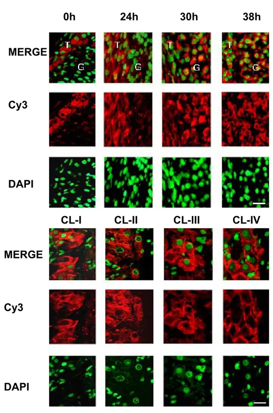
5.2 Corpus luteum classification
Classification of CL is based on a) the female reproductive condition, b) duration of CL or life span and c) progesterone secretion. In the first classification four CL types have been reported in reproductive mammals: 1) normal cycle CL, 2) pregnancy CL, 3) lactation CL, and 4) pseudopregnancy CL. According to its life span, CL development is divided in three phases. In the growing phase, there is hypertrophy of luteal cells. Between estrous cycle days 4-7 in the
cow, the corpus luteum increases in weight and size, acquiring a great capacity to react to LH, causing increases in progesterone secretion. (Kesler, Weston et al. 1981). During the maintenance phase, progesterone is synthesized and secreted by steroidogenic cells. During the regressive phase, there is morpho-physiological death of the endocrine gland (Rothchild 1981). Subnormal corpus luteum function has been associated with infertility and incapacity to maintain gestation. A subnormal corpus luteum function can display short duration of persistence, or of normal duration but low progesterone production (Garverick, Parfet et al. 1988). In ewes, both normal and abnormal CL development are similar until day 4 post-ovulation; nevertheless, at day 5, abnormal CL diminishes in weight and progesterone secretion and regresses during day 6 (Hunter, Southee et al. 1988) .
Abnormal CL may result from a deficiency during the final phases of follicular maturation. Braden et al., (Braden, King et al. 1989) demonstrated that abnormal CL originate from follicles showing more granulosa cells, fewer LH and FSH receptors, and less 17β-estradiol (E2) concentration, in comparison with follicles forming normal corpora lutea (Niswender, Juengel et al. 2000).
5.3 Corpus luteum regulation
It has been known for some time that maintenance of CL depends of luteotropic hormones such as LH (Kaltenbach, Graber et al. 1968; Denamur, Martinet et al. 1973), hCG, and others such as prolactin, E2, prostaglandin, estrogens and hormones derived from maternal or fetal tissues. The importance of these substances depends on the species. LH is considered as the most important luteotropin (Keyes and Wiltbank 1988; Berisha and Schams 2005). One effect of LH as luteotropic hormone is through the activation of protein kinases to produce cyclic AMP
response element binding (CREB) phosphorylation (Mukherjee, Park-Sarge et al. 1996; Yazawa, Mizutani et al. 2003). The interaction of phosphorylated CREB with the coactivator CREB-binding protein (CBP) stimulates the expression of ovarian genes, but its cAMP dependence is lost when luteinization is initiated (Wu and Wiltbank, 2002). Nevertheless, phosphorylation of CREB is higher in luteal cells than in granulosa cells, suggesting that other kinases such as mitogen-activated protein kinase (MAPK) could be implicated (Gonzalez-Robayna, Alliston et al. 1999; Alliston, Gonzalez-Robayna et al. 2000; Hazzalin and Mahadevan 2002).
In some species, such as the rat, rabbit, and pig, estrogens are luteotropic (Stormshak, Zelinski-Wooten et al. 1987). In the rabbit, estradiol has been considered the primary luteotropic hormone (Robson 1937) and LH plays a secondary role. In the rat, estradiol and prolactin appear to be directly involved in stimulation of progesterone secretion. Similarly prolactin is essential to maintain expression of estradiol and LH receptors, and LH stimulates synthesis of estradiol from the corpus luteum. In the pig, the luteotropic role of estrogen is less defined, but the CL growth and increase in progesterone secretion are attributed to the estradiol action (Conley and Ford 1989). Other luteotropic hormones reported are growth hormone (GH) (Liebermann and Schams 1994) and IGF-I (Constantino, Keyes et al. 1991; Parmer, Roberts et al. 1991; Sauerwein, Miyamoto et al. 1992) that increased secretion of progesterone from luteal tissue. GH receptor mRNA has been identified in ovine, bovine, and rat luteal tissue (Carlsson, Nilsson et al. 1993; Lucy, Collier et al. 1993; Juengel, Nett et al. 1997). Growth hormone could have a direct effect on luteal function by increasing secretion of progesterone and oxytocin (Liebermann and Schams 1994). In addition, GH may influence luteal function indirectly by increasing expression of IGF-I (Parmer, Roberts et al. 1991; Obasiolu, Khan-Dawood et al. 1992; Juengel, Nett et al. 1997).


![Table 1. Hydroxysteroid deshydrogenase and P450enzymes involved in active ovarian steroid hormone biosynthesis [adapted from Payne and Hales 2004]](https://thumb-eu.123doks.com/thumbv2/123doknet/12320293.325485/69.918.102.778.146.906/hydroxysteroid-deshydrogenase-enzymes-involved-ovarian-steroid-hormone-biosynthesis.webp)