Open Archive Toulouse Archive Ouverte (OATAO)
OATAO is an open access repository that collects the work of some Toulouse researchers and makes it freely available over the web where possible.This is
an author'sversion published in:
https://oatao.univ-toulouse.fr/23130Official URL :
https://doi.org/10.1007/s00586-017-5318-xTo cite this version :
Any correspondence concerning this service should be sent to the repository administrator:
Juricic, Mélodie and Pinnagoda, Kalitha and Lakhal, Walid and Sales de Gauzy, Jérôme and Abbo, Olivier Pancreatic fracture: a rare complication following scoliosis surgery. (2018) European Spine Journal, 27 (9). 2095-2099. ISSN 0940-6719
OATAO
Pancreatic fracture: a rare complication following scoliosis
surgery
Mélodie Juricic Jr.1 • Kalitha Pinnagoda2 • Walid Lakhal1 • Jérome Sales De Gauzy1 • Olivier Abbo2
Abstract
Study design Grand Round case report.
Objective We report a pancreatic fracture associated with
Wirsung duct disruption, following a scoliosis surgery in a cerebral palsy adolescent.
Summary of background data Spinal fusion surgery is the
standard treatment for severe neuromuscular scoliosis. Many complications such as digestive ones account for its com plexity. Postoperative acute pancreatitis is well described, although its pathophysiology remains unclear. To our knowl edge, pancreatic fracture following scoliosis correction has
igj Olivier Abbo
Pediatric Orthopedic Department, Hôpital des Enfants de Toulouse, CHU Toulouse, 330, Avenue de Grande Bretagne, 31059 Toulouse Cedex 9, France
2 General Pediatric Surgery Department, Hôpital des Enfants de Toulouse, CHU Toulouse, 330, Avenue de Grande Bretagne, 31059 Toulouse Cedex 9, France
never been described to date. Clinical presentation is not specific, and management is not consensual.
Case report A 14-year-old adolescent had posterior spi nal fusion for neuromuscular scoliosis due to cerebral palsy. During the postoperative course, she developed progressive nonspecific abdominal symptoms. The abdominal CT scan demonstrated a pancreatic fracture and a surgical explora tion was decided as perforations of the bowel were highly suspected. Drains were placed around the pancreatic area as the retrogastric region was out of reach due to local inflam mation. Conservative management led to the occurrence of a pseudocyst in the following weeks as the pancreatic leakage progressively dropped.
Discussion Two hypotheses have been proposed: direct iatrogenic trauma from lumbar pedicle screws and pancre atic rupture related to the correction of the spinal deformity. As the latter seems the most likely, spinal surgeons should be aware of this occurrence following severe scoliosis correction.
Conclusion Spinal fusion for severe neuromuscular sco
liosis is a difficult procedure, with a high rate of complica tions. Among them, pancreatic fracture should be considered when abdominal pain persists in the postoperative period. Conservative management is advocated especially in case of a poor general condition.
Keywords Scoliosis • Posterior spinal fusion • Pancreatic
fracture · Pseudocyst • Cerebral palsy
Case presentation
A 14-years-old girl was referred to the spine clinic at our institution for severe neuromuscular scoliosis as a conse quence of cerebral palsy following a great prematurity
(30 weeks of gestation). She had a 120° right thoracolumbar curve on preoperative radiographs and was Risser Stage 1 (Fig. 1).
A gastric tube had been inserted months before to improve enteral intake ahead of the spinal procedure. We performed a posterior instrumentation, correction and fusion from T2 to S1 using hybrid implants with screw-hook and sublaminar bands (Fig. 2). Cranial and caudal rods were connected using domi-noes to improve the reduction, and achieve spinal balance. The surgical procedure was uneventful as well as the immedi-ate postoperative course. During the first week after the pro-cedure, the patient gradually developed abdominal symptoms including mild abdominal pain, vomiting and abdominal dis-tension despite ceasing any enteral intake. As fever appeared at day 7, biological workup showed an increased lipase blood level (272 U/ml) associated with 27,000/ml white blood cells and C protein-reactive level of 105 mg/l.
Diagnostic imaging section
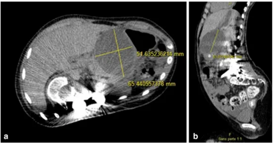
A CT scan then showed a major intra-peritoneal effusion associated with a suspected jejunal perforation and a corpo-real fracture of the pancreas (Fig. 3).
Historical review of the condition, epidemiology, diagnosis, pathology, differential diagnosis
Scoliosis is frequent in cerebral palsy patient with a reported incidence from 4 to 65% [1]. The aims of surgery are to stop the progression and re-balance the spine, therefore, allowing a seated position and facilitated nursing. Surgical correction of neuromuscular scoliosis allows a major benefit from a respiratory and autonomic point of view.
In cerebral palsy, several comorbidities and a major thora-columbar deformity are often associated, increasing the risk of major complications [2, 3]. In these patients, surgery bears a higher risk of mortality and morbidity, according to data from the scoliosis research society (SRS), with 0.3 and 17.9%, respectively [4].
Not only septic, neurological but also digestive compli-cations are classically described [5]. Among the latter cat-egory, some are nonspecific, such as reflex ileus, but others are inherent to the biomechanics of spinal deformity cor-rection (Fig. 3).
The superior mesenteric artery syndrome is the most reported digestive complication of scoliosis surgery, but creatic injuries are scarcely described. Studies on acute pan-creatitis found prevalence rates ranging between 8.5 and 31% [6, 7]. Pathophysiology remains unclear, but the hypothesis
of a pancreatic compression against the spinal block, associ-ated with an induced hypo-perfusion is the most plausible [8]. Clinical picture may include mild specific abdominal pain associated with an increased blood lipase and amylase up to three times the normal levels. Abdomino-pelvic CT scan usually confirms the diagnosis, although it is not sys-tematically ordered [9, 10].
Unlike induced pancreatitis, we report here the case of a pancreatic fracture discovered early postoperative of poste-rior spinal fusion for thoracolumbar scoliosis with a major
Cobb angle of 120° in an adolescent with neonatal cerebral palsy.
Pancreatic trauma is rare, accounting for less than 1% of all abdominal traumas [11]. The deep position of this organ can explain the injury mechanism, with lesions occurring as it is crushed against the fulcrum of the spine during an often indirect trauma. Clinical diagnosis is difficult and often delayed, in an exclusive indirect or direct traumatic context [12, 13].
This diagnosis is confirmed by abdomino-pelvic CT scan, leading to a staging of severity described by Moore et al. [14]. Abnormal pancreatic function tests may frequently be absent in an early stage. This confirmation may also be delayed and subtle due to underestimated lesions according to the imaging specificity and sensitivity (around 80%) [15].
Thus, in the current case, after an initial diagnosis wan-dering, linked to the nonspecific picture, repeated imaging demonstrated the pancreatic fracture, with a peri-pancreatic and abdominal effusion strongly evocative of a pancreatic fistula, which finally confirms the rare diagnosis of a pancre-atic fracture with pancrepancre-atic ductal disruption [16] (Fig. 3). Hypotheses concerning the underlying mechanism have been difficult to confirm, most likely, a massive pancreatic distension occurred during the reduction of the major thora-columbar deformity. However, the possibility of a direct iatrogenic injury related to the pedicle screws cannot be ruled out. Indeed, a hematoma of the small intestine wall
Fig. 2 Postoperative radiograph showing T2–S1 fusion with hybrid instrumentation and a satisfactory curve correction
Fig. 3 Abdomino-pelvic CT scan coronal view showing a grade III
was diagnosed on the initial radiological workup. Moreo-ver, abdominal exploration confirmed proud pedicle screws, although far away from the injured area.
Nevertheless, the modified anatomical reports could explain this late hypothesis. Another similar case has recently been reported by Al-Binali et al. [17] with delayed diagnosis of small intestine lesion due to spinal instrumenta-tion (Fig. 4).
Rationale for treatment and evidence‑based literature
Pancreatic fractures are severe, with morbidity and mor-tality rates estimated at 26.5 and 5.3%, respectively. Iso-lated pancreatic fracture prognosis is directly reIso-lated to the presence of pancreatic duct disruption. The reported com-plications are the development of pancreatic pseudocysts, chronic pancreatitis, and rarely diabetes in serious pancreas decay [18].
High severity stages (Moore III–V), high elevation of pancreatic enzymes 15 days later are the most common pre-disposing factors in pseudocyst occurrence.
Therapeutic management of isolated pancreatic fractures should be exclusively conservative in low-stage lesions (I and II) [19]. The management of high grade fractures (III IV) has long been debated; latest published data tend to underline the value of conservative treatment, even if a pan-creatic duct rupture is associated. Reports in favor of early emergency surgery management with ductal rupture argue that this attitude considerably reduces the occurrence of recurrences [20–22] which has been estimated to be around 40%.
Given the high morbidity rate of pancreatic resection (about 60%), the conservative attitude remains preferable in case of isolated pancreatic fracture in hemodynamically stable patients.
When secondary pseudocyst occurs, it seems also advisable to favor a conservative management, which is effective in half of the cases. Expansive pseudocyst, symp-tomatic or compressive ones, may be managed with either endoscopic or image-guided drainage [23].
Procedure section
A surgical abdominal exploration confirmed the suspected diagnosis. The jejunal lesion was resected with direct anas-tomosis. The duodeno-pancreatic region was out of reach due to the local inflammation. Drains were placed in the retrogastric region. During the exploration, the extremities of some lumbar pedicle screws were observed, a few mil-limeters proud in the lumbar region in the retroperitoneal area but 10 cm away from the pancreatic area. The post-operative course confirmed the pancreatic lesion involv-ing the Wirsung duct as proved by the important leakage with increased lipase level (initially around 500 ml/day). A conservative management was decided with nil per os and initial antibiotics due to the jejunal perforation.
In spite of the pancreatic duct rupture, we favored the conservative approach for our patient, given the morbid-ity and mortalmorbid-ity rates of proximal pancreatic surgery in a patient with respiratory and nutritional deficiencies. Fur-thermore, delayed diagnosis of pancreatic duct rupture also influenced the therapeutic management.
Outcome and follow‑up
During the further weeks, the amount of fluid dropped pro-gressively to disappear whereas a pseudocyst grew from 2 to 6 cm in diameter (Fig. 4). Despite this evolution, it was decided to manage it conservatively because of the absence of induced symptoms. From an orthopedic point of view, the scoliosis correction was satisfactory with a 50% correction and a good spinal balance. Several weeks later, abdominal symptoms reappeared along with the pseudocyst growth and another biological pancreatitis. Endoscopic drainage (gastrocystostomy) allowed improving clinical and biologi-cal parameters. After 6 months of follow-up, the patient remained pain-free and lipase level has eventually dropped to normal. Lately an acute digestive peritonitis without evi-dent cause led to the patient death.
Conclusion
Pancreatic fracture in children and adolescents is rare and the diagnosis complex, as its presentation is often nonspe-cific. To our knowledge, its occurrence in a postoperative context of scoliosis surgery has never been described. If iatrogenic acute pancreatitis is a relatively common entity, pancreatic fracture should also be considered. Conservative management generally allows complete healing after a few weeks. Secondary pancreatic pseudocyst, which is the most frequent complication, can require an endoscopic or image-guided drainage if symptomatic.
Compliance with ethical standards
Conflict of interest The authors declare no Grant or conflict of
in-terest. References
1. Miller F et al (2005) Cerebral palsy, 1st edn. Springer, New York, pp 433–522
2. Lipton GE et al (1999) Factors predicting postoperative compli-cations following spinal fusions in children with cerebral palsy. J Spinal Disord 12(3):197–205
3. Guigui P et al (2005) Complications of surgical treatment of spinal deformities: a prospective multicentric study of 3311 patients. Revus de chirurgie orthopédique 91:314–327
4. Reames DL et al (2011) Complications in the surgical treatment of 19,360 cases of pediatric scoliosis: a review of the Scolio-sis Research Society Morbidity and Mortality database. Spine 36:1484–1491
5. Weissa HR, Goodall D (2008) Rate of complications in scoliosis surgery—a systematic review of the Pub Med literature. Scoliosis 3:9
6. Battugs B et al (2009) Prevalence and risk factors in postoperative pancreatitis after spine fusion in patients with cerebral palsy. J Pediatr Orthop 29:256–262
7. Laplaza FJ et al (2002) Pancreatitis after surgery in adolescent idiopathic scoliosis: incidence and risk factors. J Pediatr Orthop 22(1):80–83
8. El Bouyousfi M et al (2016) Acute pancreatitis following scoliosis surgery: description and clinical course in 14 adolescents. Eur Spine J 25(10):3316–3323
9. Abousamra O et al (2016) Risk factors for pancreatitis after poste-rior spinal fusion in children with cerebral palsy. J Pediatr Orthop B. doi:10.1097/BPB.0000000000000376 (Epub ahead of print)
10. Malka D, Rosa-Hézode I (2001) Positive and etiological diag-nosis of acute pancreatitis. Gastroenterol Clin Biol 25(Suppl 1):S153–S168
11. Sutherland I et al (2010) Pancreatic trauma in children. Pediatr Surg Int 26(12):1201–1206
12. Englum BR et al (2016) Management of blunt pancreatic trauma in children: review of the National Trauma Data Bank. J Pediatr Surg 51:1526–1531
13. Aekovutz MS et al (1997) Pancreatic trauma in children: mecha-nisms of injury. J Trauma 42:49–53
14. Moore EE et al (1990) Organ injury scaling, II: Pancreas, duode-num, small bowel, colon, and rectum. J Trauma 30(11):1427–1429 15. Kshirsagar AY et al (2015) Isolated pancreatic tail injury: a rare
presentation. Ann Med Surg 4:230–232
16. Debi U et al (2013) Pancreatic trauma: a concise review. World J Gastroenterol 19(47):9003–9011
17. Al-Binali AM et al (2001) Acute lower gastrointestinal bleeding as a late complication of spinal instrumentation. J Pediatr Surg 36(3):498–500
18. Aydogbu B et al (2016) Predicting pseudocyst formation fol-lowing pancreatic trauma in pediatric patients. Pediatr Surg Int 32(6):559–563
19. Abbo O et al (2013) Conservative management of blunt pancreatic trauma in children. Eur J Pediatr Surg 23:470–473
20. de Blauuw I et al (2008) Pancreatic injury in children: good out-come of nonoperative treatment. J Pediatr Surg 43:1640–1643 21. Wood JH et al (2010) Operative vs non operative management of
blunt pancreatic trauma in children. J Pediatr Surg 45(2):401–406 22. Koganti SB et al (2016) Predictors of successful non-operative management of grade III & IV blunt pancreatic trauma. Ann Med Surg 10:103–109
23. Mattix KD et al (2007) Pediatric pancreatic trauma: predictors of nonoperative management failure and associated outcomes. J Pediatr Surg 42:340–344