Université de Montréal
3
Synthesis and Characterizatïon of Cationic
Polymers Derived from Cholic Acid
Par
Shanshan Chen
Département de chimie
Faculté des arts et des sciences
Mémoire présenté à la Faculté des études supérieures
en vue de l’obtention du grade de
Maître ès sciences (M.$c.) en chimie
Octobre 2006
Université
de Montréal
Direction des bibliiothèqties
AVIS
L’auteur a autorisé l’Université de Montréal à reproduire et diffuser, en totalité ou en partie, par quelque moyen que ce soit et sur quelque support que ce soit, et exclusivement à des fins non lucratives d’enseignement et de recherche, des copies de ce mémoire ou de cette thèse.
L’auteur et les coauteurs le cas échéant conservent la propriété du droit d’auteur et des droits moraux qui protègent ce document. Ni la thèse ou le mémoire, ni des extraits substantiels de ce document, ne doivent être imprimés ou autrement reproduits sans l’autorisation de l’auteur.
Afin de se conformer à ta Loi canadienne sur la protection des renseignements personnels, quelques formulaires secondaires, coordonnées ou signatures intégrées au texte ont pu être enlevés de ce document. Bien que cela ait pu affecter ta pagïnation, il n’y a aucun contenu manquant.
NOTICE
The author of this thesis or dissertation has granted a nonexclusive license allowing Université de Montréat to reproduce and publish the document, in part or in whole, and in any format, solely for noncommercial educational and research purposes.
The author and co-authors if applicable retain copyright ownership and moral rights in this document. Neither the whole thesis or dissertation, flot substantial extracts from it, may be printed or otherwise reproduced without the author’s permission.
In compliance with the Canadian Privacy Act some supporting forms, contact information or signatures may have been removed from the document. While this may affect the document page count, it does flot represent any Ioss of content from the document.
Université de Montréal
Faculté des études supérieures
Ce mémoire intitulé
Synthesis and Characterization of Cationic
Polymers Derived from Cholïc Acid
Présentée par
Shanshan Chen
a été évalué jar un jury composé des personnes suivantes
Présidente-rapporteuse :
Francoise Winnik
Directeur de recherche :
Julian Zhu
Membre du jury:
William Skene
SOMMAIRE
L’idée de cette recherche est inspirée par les interactions entre espèces
chargées dans la nature. Les molécules d’ADN ainsi que ta surface des cellules sont normalement chargées négativement et par conséquent des polymères cationiques ont le potentiel d’interagir avec celles-ci. Des interactions de ce genre ont déjà été étudiées pour des applications bio-médicales, incluant le relargage de médicaments, la thérapie génique, et en génie tissulaire. Les polymères contenant des composants endogènes tels que les acides biliaires, peuvent être utilisés pour ce genre d’applications.
Cette étude se concentre sctr la synthèse de polymères chargés positivement, dérivés de l’acide cholique (un des acides biliaires). [(2’-Diméthyl amino)éthylène]-3a-methacryloyl-7ci, l2Œ-dihydroxy-5 f3-cholanoaminde (mo nomère A) et [(2 ‘-tert-butyloxycarboxamido )éthylnèj-3 a-methaciyloyl-7a, Ï 2a-dihydroxy-5f3-cholanoamide (monomère B), ont été synthétisés et serviront pour préparer des polymères cationiqLles. Cependant comme de nombreux problèmes de synthèse ont été rencontrés avec le monomère A, seulement monomère B a été utilisé. Des homopolymères et copolymères ont été synthétisés par polymérisation radicalaire. L’homopolymère est sensible au pH et a été copolymérisé avec le N isopropylaciylamide qcti est connu pour générer des polymères thermosensibles. Les différents ratios de ces deux composants (monomère B et le N isopropylacrylamide) affectent la sensibilité au pi-I et à la température. Les
propriétés physiques et chimiques de tous les homopolymères et copolymères ont été caractérisées par les techniques physico-chimiques telles que la chromatographie d’ exclusion stérique, la spectroscopie de résonance nucléaire magnétique, la spectroscopie infra-rouge à transformée de Fourier, la spectrométrie de masse, l’analyse élémentaire, le titrage potentiométrique, l’analyse thermogravimétrique, et la spectrophotometrie UV-visible.
Mots clés : acide cholique; vecteur non-viral; charge positive; polymère thermosensible; polymère sensible au pH
ABSTRACT
The idea of this research stems from interactions observed between charged species in nature. DNA molecules and the surfaces of celis are normally negatively charged. Positively charged polymers can potentially interact with ceils and DNA molecules, and have been considered for bio-medical applications including drug deÏivery, gene therapy and tissue scaffolds. Polymers containing endogenous compounds such as bile acids may be used for such applications.
The focus of this study relates to the synthesis of positively charged polymers derived from cholic acid ta bile acid). [(2’ -Dimethylamino)ethylene]
3a-methacryloyl-7a, I 2a-dihydroxy-5 f3-cholanoamide (monomer A) and [(2’-tert butyloxycarboxamido)ethylene]-3 a-methacryloyl-7a, l2Œ-dihydroxy-5 f3- chol anoamide (monomer B) were synthesized from cholic acid and served as monomers for preparing cationic polymers. However, because there are lots of synthetic problems with monomer A, only monomer B was used for polymerization. Homopolymers and copolymers were synthesized from monomer B by free radical polymerization. The homopolymer displayed pH sensitivity. Monomer B derived from cholic acid was copolymerized with N isopropylacrylaminde, which is known to generate thermosensitive polymers. The different ratios ofthese two components affect the thermo- and pH-sensitivities of the copolymers. The chemical and physical properties of ail the homopolymers and copolymers were characterized by the use of various physical-chemical techniques such as size exclusion chromatography, nuclear magnetic resonance, Fourier transform infrared spectroscopy, mass spectrometry, elernental analysis, potentiometric titration, thermogravimetry and UV-visibl e spectrophotometry. Keywords: cholic acid; non-viral vector; positive charge; thernio sensitive polymer; pH sensitive polymer;
ACKNOWLEDGEMENT$
I would like to thank my director, Prof. Julian Zhu, who gave me kind help, instruction and encouragement dctring my graduate study and research. His knowledge, diligence and meticulous scientific approacli affected me greatly. t should be very beneficial for me in my chemistry research career in the future.
I also want to thank my colleagues, Marc Gauthier, Yilong Chen in the lab for the many discussions, exchange of ideas and their help with my experiments.
Finally, I would like to thank my parents, the Gauthier family and Julien Devaud. They gave me spiritual encouragement and support so that I could continue and finish my studies at the Université de Montréal even when I had diffi culti es.
TABLE 0F CONTENTS
SOMMARI RE ABSTRACT ACKNOWLEDGEMENTS TABLE 0F CONTENTS LIST 0F FIGURES LIST 0F TABLESLIST 0F SYMBOLS AND ABBREVIATIONS
1. INTRODUCTION 1
1.1 Background 1
1 .2 Vectors for gene therapy - Concept
1.2.1 Viral Vectors 3
1 .2.2 Non-viral vectors 4
1 .3 Current research in cationic polymers 9
1.3.1 Cationic polymers in gene therapy 10
1.3.2 pH sensitivity of cationic polymer in drug release 15
1 .4 Thermo sensitive polymers 16
1.5 Background on bile acids and their polyrners 18 1 .6 Design of cationic polymers containing bile acid derivatives 21 References
2. EXPERIMENTS 30
2.2 Synthesis ofmonomers .30 2.2. 1 [(2‘-di methylamino)ethylene]-3 u-rnethacryloyl,7a,I
2a-dihydroxy-5f3-cholanoamide 4 30
2.2.2 [(2’ -tert-butyloxycarboxamido)ethylene]-3 a-methacryloyl
7a,1 2u-dihydroxy-5 f3-cholanoamide 7 33
2.3 Synthesis ofpolymers 35
2.3.1 Homopolymers 35
2.3.2 Copolymers 36
2.4 Methods ofphysical characterization 3$
2.4.1 Nuclear magnetic resonance 3$
2.4.2 Elemental analysis 3$
2.4.3 Mass spectrometry 3$
2.4.4 Thermogravimetric Analysis 39
2.4.5 Size exclusion chromatography 39
2.4.6 Ultraviolet-visible spectrophotometry 39
2.4.7 Fourier transform infrared spectroscopy 40
2.4.$ pH mete 40
2.5 Characterization ofpH- orthermo-pH sensitivities 40
2.5.1 pH sensitivity ofthe homopolyrne 40
2.5.2 pH sensitivities ofthe copolymer 40
References
3. RESULTS AND DISCUSSION 43
3. 1 Synthesis of a monomer bearing a tertiaiy amine group 43
3. 1.1 Synthetic procedures 43
3.1.2 NMR spectroscopy and mass spectrometry 45
3.1.3 Aspects ofthe synthesis 4$
3.2 Synthesis ofa monomer bearing a prirnary amine group 51
3.2. 1 Synthetic procedures 5 1
3.2.2 NMR spectroscopy and mass spectnirn 54
3.2.3 Aspects ofthe synthesis 57
3.3 Preparation ofthe polymers .60
3.3.1 pH-sensitive polymer 60
3.3.2 Thenrio and pH-sensitive copolyrners 62 3.4 Characterization of pH-sensitive polyrners 65
3.4.1 NMRspectroscopy 65
3.4.2 Fourier transform infrared spectroscopy 66
3.4.3 Thermogravimetric analysis 68
3.4.4 pH sensitivity 70
3.5 Characterization ofcopolymers 71
3.5.1 NMR spectroscopy 71
3.5.2 Thermogravimetric analysis 75
3.5.3 pH- and thermo sensitivity 7$
Reference
4. CONCLUSIONS 84
4. 1 Synthesis 84
LIST 0F FIGURES
Figure 1.1 Figure 1.2 Figure 1.3 Figure 1.4 Figure 1.5 Figure 1.6 Figure 1.7 Figure 1.8 Figure 1.9 Figure 1.10 figure 3.1 Figure 3.2 Figure 3.3 Figure 3.4 Figure 3.5 Figure 3.6 Figure 3.7 Figure 3.8Generalized representation ofthe delivery ofa DNA-based
therapeutic using a viral or non-viral DNA delivery vector 2
The general strctcture of a cationic lipid 5
Formation oflipoplexes 6
Schematic ofthe substrate-mediated delivery strategy, CP represents
the cationic polymer used for condensation 7
pH dependent ionization ofpolyelectrolytes 10 Schematic generalized representation ofthe deliveiy ofa DNA based therapeutic using a non-viral vector (cationic polymer) 11
Structures of the commonly used cationic polymers for gene therapy 12 The chemical structure of PNIPAM and the dehydration of PNIPAM
above its LCST 17
The chemical strctctures ofselected bile acids 19
The structure ofcholic acid 19
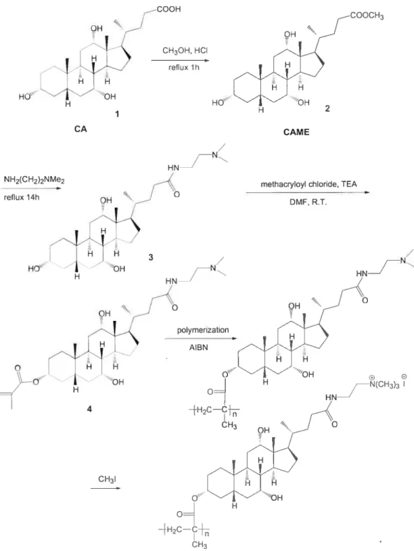
Scheme for synthesis ofpolymers containing cholic acid derivatives
and bearing quarternary amino groups 44
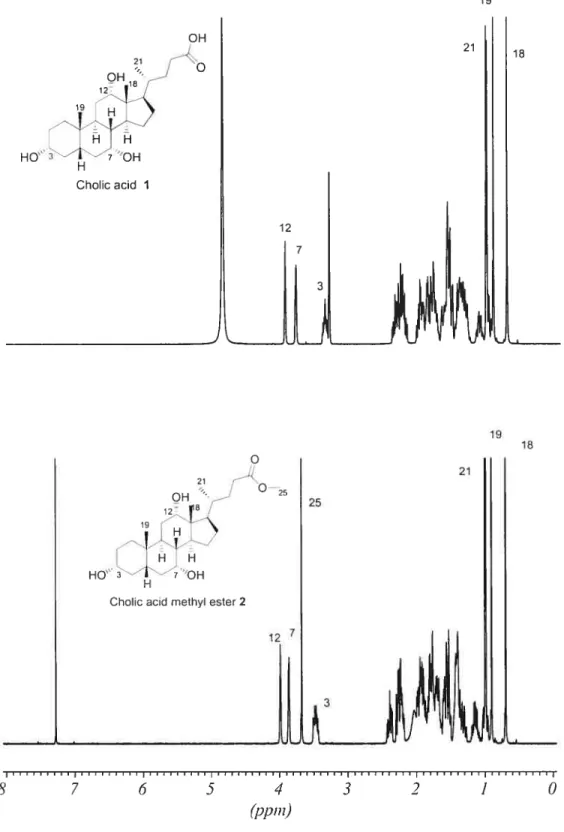
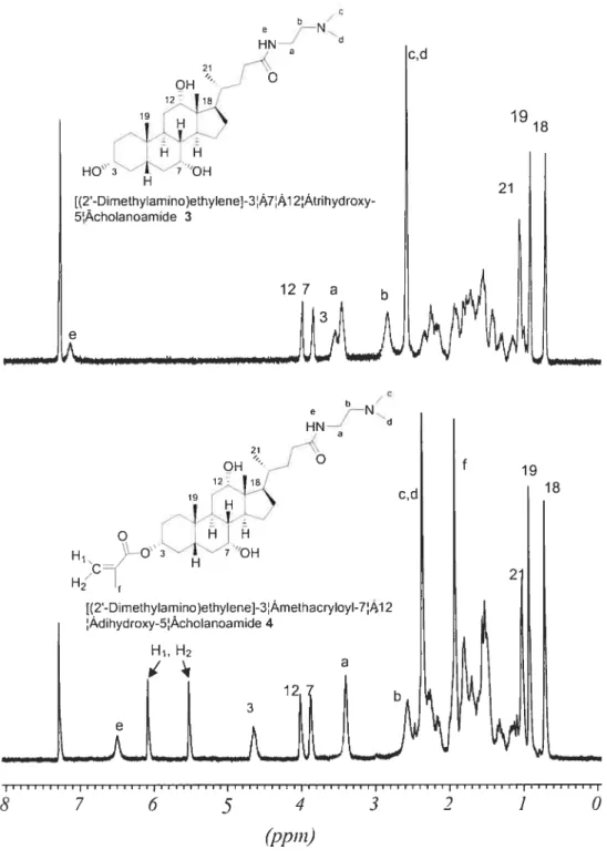
‘H NMR spectra ofcompound 1 in CD3OD and 2 in CDC13 46 ‘H NMR spectra of compound 3 in CDC13 and 4 in CDC13 47
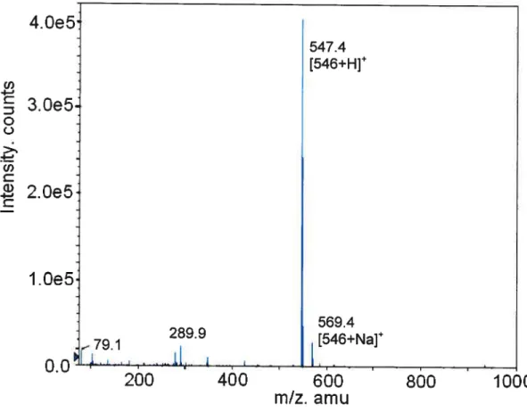
Mass spectrum ofmonomer 4 48
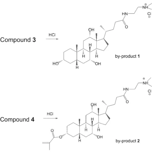
Activation ofan acid chloride by a ternary amine 50 Representation ofthe ionization ofthe ternary amino group on 3 and
4 duringthe methacrylation 50
Scheme ofprimary amine polymer 53
‘H NMR spectra of compound 5 in CD3OD, compound 6 in CDC13
and monomer 7 in CDC13 55
queou compartn)ent Double layer 0f Cationic liposanw phospholipids + -L+ + + + NeqaUuely—charjeU DN plasPia + + + + + + + Positiuel-cFarged double laJer 0f phospholiplds Condensation of OHA in presence of lipasones + Hegatiuely-charged DHA plasna in an aqucous conpattnent Condensed DHA plasna in an F aqueous conpartnent
figure 1.3 formation of lipoplexes. (This figure is posted on web site
http://www. unilim.fr/theses/ 2003/sante/200311mo01 OOc/these_body. html).
Po5itiuely—charqed double layer 0f _________ phospholipid5
÷
+ --4../
LipoplexesIn vivo, an overail net positive charge of the lipoplexes helps to associate the complex with the negatively charged ceil membrane. Entiy of the lipoplexes into the ce!! may occur by the process of endocytosis via the lipid moieties of the liposome. 23 Following cellular intetnalization, the complexes appear in the endosomes and later in the nucleus.
(2) Cationic polyrners
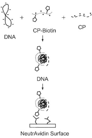
In general, the structures of cationic polymers (polycations) for non-viral vectors are divided into two parts: cationic branched polymer and hydrophilic linear polymers with ligands. Cationic polymers (i.e., proton-sponge polymers) can easily complex with anionic DNA molecules. The complexes formed between DNA and cationic polymers are called polyplexes as shown in figure 1 .4.
-b -b cP CP-Biotin DNA
q...
6”’
DNAq..
6’
II NeutrAvidin SurfaceFigure 1.4 Schematic of the substrate-mediated delivery strategy, CP represents the cationic polymer used for condensation.26
Polyplexes are commonly used as non-viral vectors.24 The general mechanisrn of action of polyplexes is based on the generation of positively charged complexes which can interact with the negatively charged celi surface. When cationic polymers are mixed with DNA, they readily self-assemble together. The cationic bral1ched polymers trap DNAs by electrostatic interaction, and then the complex is sctrrounded by a hydrophilic linear polymer which may possess a terminal ligand (such as biotin). The result is the formation of small toroidal or spherical structures. When polypiexes are added to ceils in culture, the polyplexes are taken up by the cells after the ligand is detected by the targeting ccli. These complexes often take advantage of the pH change in the endosome to trigger an endosomal rupture and improve DNA uptake.25 Similar to that of cationic lipids, transfection activity of cationic polymers varies witb ccli type, structure, size of the polymer, and polymer to DNA ratio.26
Compared to cationic lipids, the major drawback of cationic polymers is their relatively high toxicity. Also, the transfection efficiency of these systems is often below effective levels.25 The properties of cationic polymers can be easily controlled to improve biocompatibility (toxicity or immunogenicity),
biodegradability, and efficiency. The design of new polymer materials bas turned out to be one of the most promising strategies foi- developing gene delivery vectors with improved properties.
Non-viral gene deliveiy is typicafly much safer but suffers from generafly unsatisfactory delivery efficiency. The earliest synthetic vehicles reported in the literature were not originally designed for gene delivery (i.e. poly(L-lysine) (PLL) and poly(ethyleneimine) (PEI)).2728 The gene delivery efficacy of such materials is
haphazard and typically insufficient foi- clinical application. In recent years, a variety of polymers have been designed specifically for gene delivery. Based on the large number of studies of these basic polymers, investigators learned a lot about the stntcture—function relationships existing for polymer vectors. With growing understanding ofpolymer gene delivery mechanisms it is likely that polymer-based gene delivery systems will become an important tool for human gene therapy.
1.3 Current research in cationic polymers
Cationic polymers are polymers that contain positively-charged groups covalently linked to the polymer molecule, such as phosphonium, suiphonium, and ammonium cations. They are used in different territories such as cosmetic and drug
7930
delivery.
-In cosmetics, the cationic polymer changes its behaviour when surfactants are included in the formulation. Because anionic surfactants bind to cationic polymers, they form a complex phase known as coacervate. Therefore, the polymer can deposit onto the anionic surface of hair and skin.3’ For instance, cationic polymer complexes formed between polymers like polyquateniium-6 and retinoic acid (Vitamin D) are a powerful skin exfoliating agent and help to stabilize the labile vitamin by prolonging its chemical life and potentially increasing its potency.3 However, cationic polymers and their complexes are neither water-soluble nor dispersible in water, and have found less practical applications in personal care.
In drug ddllvery, cationic polymers contain pendant basic groups (e.g. amines or ammonium saïts) which either accept or release protons in response to changes in the environmental pH. Figure 1 .5 shows a generalized structure of a cationic polyelectrolyte and its pH-dependent ionization. These characteristics are used for drug release and drug delivery, and also foi- their activity against a number of bacteria and fungi. u The positively charged sites are able to form polymeric cationic bridges in specific sites in the body including ccli surfaces or neutralization of the negative charges of DNA. Such ionization causes cationic polyelectrolytes to exhibit greater solubility in aqueous solution at low pHs, or if cross-linked, to swell to a greater extent. These great qualities have been studied for gene therapy (for non-viral vectors) and drug applications (drug release).34’3 The following sections will describe in further details ofthose applications.
Low pH High pH OHe H HCzO HCO
P
CH2CH2NH(CH2CH3)2 CH2CH2N(CH2CH3)2Figure 1.5 pH-dependent ionization of polyelectrolytes
1.3.1 Cationic polymei in gene therapy
Cationic polymers have been widely studied. They seem to be the most promising vebicle for gene therapy. Seif-assembling complexes ofnucleic acids and synthetic cationic polymers are formed as the resuit of electrostatic interactions between the negatively charged phosphate groups of the DNA and the positively charged groups of the polycations. Wide arrays of polycations are available for the studies of gene therapy and include those with linear, branched, dendritic and block or graft copolymer architectures. These polycations vary greatly in chemical composition as welI as the number of repeating units, providing for a wide range of different polyplexes that can be easily assembled. Much ofthe research ofpolymer based gene delivery indicates that polycations serve as potential reagents in the fie Id.
The major mechanism of gene deiivery for this non-viral vector is endocytosis of polyplexes (DNAs, cationic polymers), followed by disruption of the endosomal membrane as shown in figure 1 .6. However, in general, cationic polymers alone do not appear to be ideal candidates for bio-adhesion to celi surfaces because of toxicity. Some of the studies have indicated that cationic polymers tend to cause openings or holes in the ccli membrane22’36 possibly due to neutralization of negative charges on the celi surface and formation of polymeric cationic bridges which crosslink opposite membrane surfaces. 22 Cationic polymers are inherently toxic to animal species because oftheir destructive interaction with ceIl membranes. The toxicity of such polymers is related to the charge density of the cationic
polymer.37 By modifying the cationic polymer structures, these shortcomings have been improved in many studies.34’38 Polycations have been modified with a number of groups and modalities, including chemical groups for shielding of the cationic charge, targeting groups for specific celis, and biodegradable linkers for increased biocompatibility.
DNA complexation 1
ofDNA
&
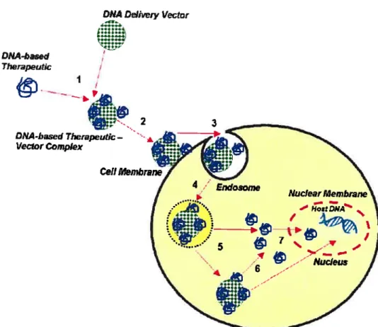
Figure 1.6 Schematic generalized representation of the delivery of a DNA based therapeutic using a non-viral vector (cationic polymers) (this figure is post on http://www. nano-lifescience. com/research/genedelivery. html): (1) Cationic polymers which promote particle (complexation) act as a scaffold for DNA condensation and coupling of bioactive ligands (binding with a high specificity and affinity to the recognition sites). (2) The complexation binding to the celi surface. (3) The complexation crosses the plasma membrane. (4) DNA escapes form the endosome into the cytoplasm. (5) Transport the DNA into the nucleus.
A!though the safety of the cationic polymers (in oncogenicity and immunogenicity) as non-viral vectors has been established in many studies,39’4° non-viral vectors have much lower transfection efficiency than viral vectors. The presence of positive charges at the surface of polyplexes can promote unwanted non-specific interactions with serum proteins and ce!! membranes.4’ Therefore, in vitro (without plasma issue), the efficiency of polyplexes is good, but in vivo, it is
poIyition with hgand bindnq to the receptor 2 endocytosîs 3 4 nucleus nuclear traffickin9 —z 11
Figure 3.9 ‘3C NMR spectrum ofmonomer 7 in CDC13 . 56
Figure 3.10 Mass spectrum ofmonomer 7 57
Figure 3.11 Scheme of synthesis for poly(NIPAM-co-CAMA- NH3C1 63 Figure 3.12 ‘H NMR spectra ofPCAMA-Boc $ and PCAMA-NH3CF 9 in
CD3OD 65
figure 3.13 FTIR spectra ofPCAMA-Boc 8, PCAMA-NH3C1 9 and PCAMA
NI-1210 66
Figure 3.14 Thermogravimetric analysis ofhomopolymers 68 Figure 3.15 Thermogravimetric analysis ofPCAMA-Boc 8 69 Figure 3.16 PCAMA-NH3C1 9 transmission changes as a function of pH at25
°C in methanol 70
Figure 3.17 ‘H NMR spectra ofNIPAM in CDCÏ3 and 3, 6, 10 and 20 moi% of monomer 7 in poly(NIPAM-co-CAMA-Boc) 12 in CD3OD 73 Figure 3.18 ‘H NMR spectra of 3, 6, 10 and 20 mol% ofmonomer 7 in
poly(N1PAM-co-CAMA-NHCl) 13in CD3OD 74
Figure 3.19 Thermogravimetric analysis of poly(NIPAM-co-CAMA-Boc)s 75 Figure 3.20 Thermogravimetric analysis of poly(NIPAM-co-CAMA-NH3Cl)s
76 Figure 3.21 The colTeÏation of poiy(N IPAM-co- 1 0%CAMA-NHCF)s
concentration and cioud point in aqueous solution 79 figure 3.22 Sait is generated with theaddition ofNaOH to solutions containing
poly(NIPAM-co-CAMA-NH3CF) 79
Figure 3.23 ColTelation of NaC1 concentration with cloud point of poly(NIPAM
co-CAMA-NH3CF) 80
figure 3.24 ColTelation ofpH value and cioud point ofpoly(NIPAM-co-CAMA
NH3CF) (1.5 wt/v% inwater) 82
Figure 4.1 Alternative synthetic strategy ofa cationic polymer containing cholic acid
LIST 0F TABLES
Table 2.1 Concentrations ofNaCl in the copolymers solutions. 41 Table 3.1 NMR chernical shifts of final monomers and intermediates 56 Table 3.2 The molecular weight ofPoly(NIPAM-co-CAMA-Boc)s 64 Table 3.3 FTIR adsorption of selected functional groups and classes of
compounds absorb 67
Table 3.4 The molar percent ofmonomer 7 in the copolymers 72 Table 3.5 Theoretical and experimental ratios ofthe monomer 7, and weight loss
ofBoc groups (WB0%) measured by TGA 78
Table 3.6 The concentration ofNaCl under different pH for all the poly(NIPAM
co-CAMA-NH3Cl) 81
LI$T 0F $YMBOLS AND
ABBREVIATION$
A Absorbance
A Collision frequency factor Frequency factor forpropagation
AIBN 2, 2’-Azobis(isobutylonitrile) arnu Atomic mass unit
atm Atmosphere
aq Aqueous
foc tert-butyloxycarbonyÏ Boc2O Di-tert-butyl dicarbonate
CA Cholic acid
CAME Methyl ester ofcholic acid
cm Centimeters
CP Cationic polymer
DMEDA N, N-D j methyl ethylenediamine
DCM Dichiorornethane
DMF N,N-Dimethylformamide
DNA Deoxyribonucleic acid E Arrhenius activation energy
E,, Activation energy for propagation
EDA Ethylenediamine
f
Initiator efficiencyg Gram
h Hour(s)
Hz Hertz
[J] Concentration of the initiator
Ic Rate constants of initiation
k,, Rate constants of propagation k, Rate constants of termination
LCST Lower critical solution temperature
M Molar per litre
[M] Concentration of the monomer
mg Milligram
iriL N4illiÏitre
mmol Millimole
mM Millimolar per litre
M, Number-average molecular weights
MS Mass spectrometry
Wight-average molecular weights
N IPAM N-Isopropylacrylaminde
nm Nanometer
NMR Nuclear magnetic resonance
DMSO Dimethyl sulfoxide
PAAM Polyamidoamine
PDEAM Poly(N,N-diethylacrylaminde)
PET Polyethyleneimine
PLL Poly-L-Iysine
PNIPAM Poly(N-isopropylacrylamide)
ppm Part per million
R Rate ofpolymerization
Retention factor
RNA Ribonucleic Acid
SEC Size exclusion chromatography
tBu tert—butyl
T Kelvin temperature
TEA Triethylamine
TLC Thin layer chromatography
T1 Melting temperature
TGA Thermogravimetric
THF Tetrahydrofuran
UV Ultraviolet
e Kinetic chain length
Number-average degree of polymerization
Percent transmittance Microlitre
CHAPTER 1
INTRODUCTION
1.1 Background
One of the most promising areas of therapeutic research is gene therapy. Therapeutic genes are ctsed to “reprogram” ceils into producing their own
therapeutic agents and tum off unwanted or detrimentai cell growth or activity. Many diseases are due to cellular processes which do flot function properiy at the molecular level. These cellular processes are regulated by proteins which are synthesized in the ccli according to the genetic information stored in DNA. By adding external DNA or RNA into the ceils to induce or suppress a specific function, gene therapy can be used to combat a wide range ofdiseases.
1.2 Vectors for gene therapy - Concept
Gene therapy faces numerous challenges, most notably getting the genes to, and then into, the targeted ceils in the body. Currentiy, there are two main approaches: viral and non-viral vectors.
Viral and non-viral vectors have been widely studied for the past decade. Viral vectors include retroviruses, adenoviruses, and adeno-associated viruses. Non-viral vectors include cationic lipids/iiposomes, and cationic poiymers. The fundamental principles by which these vectors access the target ccli are simiiar: the ceiis uptake the therapeutic gene carriers, transfect, and then replace the defective gene (Figure I.i). However, the propetties of such systems differ greatly.
DNAD&ivery Vector
DMA -based f
Therapeutc I
Figure 1.1 Generalized representation of the delivery of a DNA-based
therapeutic using a viral or non-viral DNA delivery vector: (1)
complexation and/or entrapment of DNA-based therapeutic with DNA
delivery vector; (2) interaction of DNA-based therapeutic-vector
complex with ceil membrane; (3) cellular intemalization via receptor- or
non-receptor-mediated endocytotic pathways; (4) endosomal breakdown;
(5) cytoplasmic release of DNA-based therapeutic-vector complex or
DNA-based therapeutic alone; (6) dissociation of DNA-based therapeutic
from vector; (7) nuclear translocation of viral vectors or DNA-based therapeutics.’
1.2.1 Viral Vectots
Viruses are very efficient for transfecting into ceils and producing new viral particles. A foreign gene can be placed into a virus, and these recombinant viral vectors are then used to enter the host ccli. The main idea of gene therapy using viral vectors is that vectors infect the ccli and change the cell’s defective genes with the new substituted ones. Viral vectors can be sub-divided into three categories: 2
Retroviruses are a large family of RNA viruses which inciude spumavirus (foamy viruses), lentivirus and moloney-murine-ientivirus-related viruses. The diameter oft-etroviral virions range from 80 to 130 nm, and their genomes consist of two identically positive-sense single-stranded RNA molecules. The genomes are encased inside a capsid (enveiop) aiong with the integrase and reverse transcriptase enzymes. Retroviral vectors are capable of transfecting high populations ofprimaly
human endothelial and smooth muscle cells, a class of cells that is generaliy extremely difficult to transfect.3 Retroviral vectors are the most widely used virai
vectors in clinical trials at present. The most important advantages of retroviral vectors are their ability to transform and stably integrate into the target cefl genome. However, they have been demonstrated to lose long-term expression capabiiity which limits their potential for clinical application. Furthermore, they are extremely difficuit to produce on a iarge-scaie because they are inherently unstable.
Adenoviruses are non-enveioped, icosahedral, doubie-stranded DNA viruses with a capsid diameter of 70—100 nm, and comprised of 252 capsomeres (240 hexons and 12 pentons). They are not incorporated into the genome of the target ccli (non-integrating), but remain as an extrachromosomal entity in the nucleus of the host ccli. Consequently there is no risk of insertional mutagenesis.3 Replication— defective recombinant adenoviruses are the second most commoniy used viral
vectors in clinical trials today. Adenovirai vectors are known to generate inflammatory responses in tissues with short-lived gene expression. There is concern over loss of transgenic expression upon tissue maturation when celis have
been transfected using viruses. 6 However, it has been demonstrated that
adenoviruses in formulations may lose their potency after storage in commonly used pharmaceutical vials.7
Adeno-associated viruses (AAV) are non-pathogenic human parvoviruses (parvoviruses are among the smallest, simplest eukaiyotic viruses) which depend on a helper virus, usually an adenovirus, to proliferate. Compared to the other vectors, adeno-associated vircises (AVV) medi ate stable transgenic expression in terminally difTerentiated celis without inducing significant inflammatory toxicity.8 Therefore,
9 10
AAV’s only slightly damage the target celis. ‘ However, there is evidence to suggest that AAVs are significantly less efficient than retroviral vectors at transducing primary—cell cultures.Il 12 In primary—cell transductions, most of the DNA does not integrate into the host genome but remains extrachromosomal, and this inefficiency might limit its use for in vivo application.
Viral vectors have shown excellent transfection efficiencies. However, the limitations restrict their clinic applications. Viral vectors can cause immune responses, creating a number of serious safety risks such as potential oncogenicity, toxicity and the development of high immunogenicity after repeated administration.13”4 Fctrthermore large scale production may be difficult to achieve. Because ofthese drawbacks, non-viral strategies are becoming more attractive.
1.2.2 Non-viral vectors
Although the number of clinical trials with viral carriers overshadows that of non—viral catTiers due to their inherently high degree of transfection and their increased persistence of gene expression, these carriers are prone to trigger host immunogenic responses and are limited in the transgene size.’5 Non-viral vectors can overcome many of the problems encountered with viral vectors. Non-viral methods of DNA transfer require only a small number of proteins; have a virtually infinite capacity; have little or no infectious or mutagenic capability and large scale production is possible using pharmaceutical techniques. 16, 17 DNA with cationic polymers are considered to be one of the most promising candidates for non-viral
gene delivery systems.’8”9’2° Cationic polymers, a non-viral system may, in the near future, overcome sorne of the problems inherent to currently employed viral gene delivery systems. Current studies of non-viral vectors are focused on cationic lipids/Iiposomes and cationic potymers.
(1) Cationic Iipids/liposomes
The preparation of liposomes requires molecules with three specific parts: a hydrophobic anchor, a Iinker, and a beaU group (figure 1.2).21 The hydrophobie anchor and positively charged beaU group offer the important characteristics of self assembly in aqueous solution. The cationic head group of the lipid compound associates with negatively charged phosphates on the nucleic acid, resulting in compaction of the nucleic acid in a liposome/nucleic acid complex.
o
CH +1
À
N—CH3b
CH3Hydrophobie Anchor Linker Head Group Figure 1.2 The general structure ofa cationic lipid.
As shown Figure 1.3, the hydrophobie end ofthe molecule avoids contact with water, while the positively charged head group seeks contact with water. In order to protect their hydrophobie end from water and expose the hydrophilic end, the molecules self-assemble into positively charged bilayers of phospholipids structures. When DNAs are introduced into the aqueous center of the liposomes, they are referred to as lipoplexes. Lipoplexes, a negatively charged DNA plasma in an aqueous compailment, is centered into a micelle-type structure. Lipoplexes mostly or totally protect the DNA from the outside environment and have a high affinity towards cell membranes.22
significantly lower, because of the non-specific binding in the blood plasma.42’43 To overcome these problems, the most attractive strategy is to replace non-specific electrostatic interactions between the transfection complexes and cefls with ccli specific interactions that trigger receptor-mediated endocytosis of the DNA complexes. Such an active targeting requires the use of ligands such as sugar residues, peptides, proteins and antibodies34 that bind with a high specificity and
afflnity to the recognition site (Figure 1 .6).
Cationic polymers are the frst suitable candidate of gene carriers, as stated before. CutTently, several cationic polymers are being studied and include PLL, PEI, chitosan and dendrimers (Figure 1 .7). Polymeric transfection systems are advantageous in that they are safer than viral systems and are non-oncogenic. However, each polymer used for this application bas its own specific advantages and disadvantages. NHH _ NH2 H NH2 Chitosan Poly(L-Iysine) HN L NH H2N NH N NH N Ç HN N N N N H H 2 Branched PoIy(ethyleneirnine) N 2 Poly(amidoamine) (Dendrimer) figut-e 1.7 Structures ofthe commonly used cationic polymers for gene therapy
(1) PLL
The biodegradable cationic poÏymer PLL bas been widely used as a non-viral vector because of its excellent DNA condensation propeiies.44 PLL protects DNA from the attack of nucleases efficiently and the polyplexes can be rapidly internalized to the ceIl. 46 However, it has the highest toxicity among the polycations, because the cations on its amino acid backbone interact with the cell’s surface, and reduces the recycling capacity of selected membrane components as well as membrane fluidity.47 Moreover, PLL-DNA complexes show a low level of transfection since the complexes Iack of normal communication with lysosomes and are therefore unable to be rapidly released from endosomes.46 Many copolymers have been studied to reduce the toxicity and to enhance the transfection.44 Many studies on copolymers with poly(ethylene glycol) (PEG) blocks bearing targeting ligands have shown that PEG shields the surface charges of PLL and can help in the steric stabilization due to its charge neutrality and water so1ubility.444S49 The presence of the ligands can prevent non-specific interactions of the polymer with cellular components.50’5’ PLL-PEG complexes with ligands-grafted micelles had high transfection efficiencies and low cytotoxicities compared to PLL alone.34
(2) PEÏ
Today, PET is the most commonly studied non-viral gene delivery polymer. Typically, the synthesis of PET is through acid-catalyzed, ring-opening cationic polymerization of aziridine, resulting in (NHCH2CH2) monomer units with branched or linear conformations. Protonation of these amines confers PEI with the highest cationic density of all synthetic polymers for the purpose of DNA condensation. Simultaneously, PET offers better protection against nuclease degradation and escape from the lysosomal compartment than other polycations, e.g. PLL, while also showing greater transfection.52’ However, a highly cationic PET gene carrier may also attract anionic components in the blood stream such as erythrocytes.54 Moreover, the unfavorable interactions may result in aggregation and removaÏ from the body though the reticuloendothelial system.5 Consequently,
there is an intricate balance for obtaining optimal cationic condensation of plasmid DNA witbout excessive cationic charge.6
PEI architecture has two forms: branched or linear. Both have advantages and disadvantages,7 but the majority ofPEI research reported in the litterature bas been conducted with brancbed PET, possibiy due to its increased number of primary amines that are widely availabie for conjugation to other modalities. In order to control toxicity whiie maintaining high transfection, research is culTently focused on iowering the moiecular weight of PET in combination with other moiecuies.58’ furthermore, PEI does not contain active groups for tissue or ccli targeting; therefore, researchers have attached a number of targeting modalities such as
cholesterol and antibodies.60’6’
(3) Chitosan
Chitosan, a neutral polysaccharide having structural characteristics similar to glycosaminoglycans, is non-toxic and biodegradable.62 It bas therefore been widely employed in pharmaceutical and biomedical fleids.63 In recent years, biocompatible chitosan microspheres and beads have been investigated as non-viral vectors because positively charged chitosan can be complexed with negatively charge DNA.64 Also, chitosan can effectiveiy bind DNA and protect it from nuciease degradation.6 In the manufacturing process of DNA-chitosan complexes, chitosan has many advantages during preparation and storage process. However, the levels of gene expression are very iow, and response time is very long.66’67 Furthermore, at high doses, chitosan causes hypercholesterolemia and therefore bas Timited applications.68
(4) Dendrimers
Dendrimers represent a promising ciass of motecules for use as vehicies for
drug deiivery.69’70’7’ The principie advantages that dendrimers offer inciude i) a well—defined composition, ii) multiple sites for manipulation and iii) a globular shape offering a protected hydrophobic interior that can be empioyed to solubiiize
guests. The most commonly used dendrimer for gene delivery is polyamidoamine (PAAM) (Figure 1.7). PAAM can be synthesized from the core, and outspread generation by generation with very low polydispersity. The 3-dimensional sphericai structure oF the resulting polymer can be used for gene therapy.7273 Because of its low polydispersity, it can lead to reproducible gene delivery and a clinically reliable formulation.74 The capability of dendrimers to transfect ceils is dependent upon the
size, structure and number of primary amino groups on the surface of the dendrimer. However an increased flexibiiity ofthe dendrimer with a better ability to complex DNA may be difficuit to achieve.7
In truth, cadi platform (linear or dendritic polymer) has its advantages and disadvantages. Efforts to understand the appropriate roles for each strategy wiil require investigations of ail systems at ail levels. The first generation of polymer bound therapeutics are in and emerging from clinical trials. These polymers convey three principal advantages to the drug: (1) they increase solubility; (2) they increase vascular circulation time; (3) they attenuate toxicity of the drug through selective targeting as a result of ligand/receptor interactions or enhanced permeability and retention that tumors show for large molecules. The success of these systems offers compelling motivation and proof of concept for tic examination of the next generation of polymer therapeutics.
1.3.2 pH sensitivity of cationic polymers in drug release
Cationic polymers were originally investigated for pH-sensitive controlled drug delivery in oral administration before the gene delivery. pH-sensitive polymers synthesized with either acidic or basic components demonstrate reversible swelling/deswelling in acidic or basic media. Because of the number of ionizable groups on its backbone, cationic polymers can respond to changes in pH (Figure 1.5). During tic passage of the drug delivery systems from the esophagus through the stomach to intestine, tic product is exposed to a variable pH ranging from 2-4 in the stomach and 6-8 in tic intestines. Utilizing this physiological property, many drugs have been developed for controlled release.
Hydrophobicity and degree of ionization play important roles in pH-controlled drcig systems. Traditionally, sustained delivery systems use the stomach as an area for drugs to dissociate from their host. In the intestines, the unique environmental conditions and the presence ofhigh concentrations ofbacteria can substantially alter the rate of drug release for the delivery system. Hydrogels made of cross-linked polyelectrolytes display large differences in swelling properties depending on the pH of the environment. For polycationic hydrogels, the swelling is minimal at neutral pi-I, thus minimizing drug release from the hydrogels. This property has been used to prevent release of foul-tasting drugs into the neutral pH environment of the mouth. Polycationic hydrogels would be ideal for localized deliveiy of antibiotics in the stomach.76
1.4 Thermo sensitive polymers V
Thermo sensitive polymers are hydrogels which are a group of stimuli-sensitive polymers that show a phase transition in response to a temperature change.77’ They have been extensively studied in the past decade because of their potential applications in many fields such as membranes, drug delivery systems,
80 81 $2 $3
ccli culture, the isolation of bio—molecules and enzyme activity control Many thermo sensitive polymers exhibit a lower critical solution temperature (LCST), which is defined as the temperature of the phase transition fiom a soluble to an insoluble state in an aqueous solution. It is presumed that the main mechanism of such a phase transition induced by a temperature change may be a drastic change in the hydrogen bonding between the polymer and surrounding water molecules. These polymers have attracted interest of many investigators because of their intelligent ability to deliver the drug they contain to the desired places under optimal conditions. Such polymers are physically characterized by their LCST or cloud point. The LCST of the polymers usually varies with the presence of additives such as electrolytes, organic solvents and surfactants.
Ihermo sensitive drug release polymers are designed to possess sensitivity
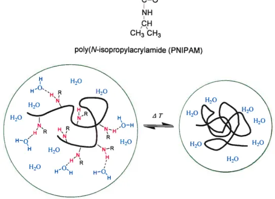
towards small changes of temperature (physiological temperatures), which leads either to an expansion or a contraction of their structure. Such systems exhibit dimensional changes leading to the release of their drug contents. Polymers such as poly(N-isopropylacrylamide (PNIPAM) (shown in figure 1.8) or their copolymers are ofien used to immobilize enzymes or as carriers of certain functional groups important for biochemical or bio-medical applications. Ihere are many
researchers who have worked on PNIPAM which is one of the most frequently
studied temperature-responsive hydrogels. PNIPAM exhibits a remarkable shrinkage with increasing temperature (LCST takes place around 32 °C).
Copolymerization ofNIPAM with other monomers can bring the LCST close to the
body temperature.86
tCH2ÇH+
CH3 CH3
ziT
poly(N-isopropylacrylamide (PNIPAM)
figure 1.8 The chemical structure of PNIPAM and the dehydration of
PNIPAM above its LCST
Figure 1 .8 also shows that PNIPAM chain changes its shape at the LCST from a random cou (left) to spherical globule (right). This phenomenon bas been reported widely.87’88 As tbe temperature increases, inter—molecular hydrogen—bonding witb water begins to break and the polymer changes conformation so as to favor intra molecular hydrogen-bonding among the amide groups along its backbone. Because the changes induced by temperature are strictly due to conformation, the response of PNIPAM to temperature stimuli is reversible and •fast.
1-lydrogels wbich are responsive to botb temperature and pH can be made by simply incorporating ionizable and hydrophobic (inverse thermo sensitive) ftinctional groups to the same hydrogels. When a small amount of cationic monomers bearing cationic groups (such as amine) is incorporated into a thermo sensitive polymei-, its LCST becomes dependent on tbe pH due to their ionization prope1ies. As the pH of the medium decreases below the pK1 of the amine groups, the LCST sbifts to bigher temperatures due to the increased bydrophilicity and charge repulsion. These copolymers could be customized to be doubly sensitive to external temperature and pH stimuli.
1.5 Backgtound on bile acids and their polymers
Bile acids are natural compounds which are in the family of steroids. Synthesized in the liver, they are generally conjugated witb glycine and taurine (e.g., glycocholic and taurocholic acids). Different groups on positions 7 and 12 define different types of bile acids (shown in Figure 1.9). The bile acids in the salt form are detergent-like substances secreted from the gallbladder and aid in the digestion and absorption of lipids. Like a detergent, bile acids contain hydrophobie and hydrophilic components. Because of their idiosyncratic structures, bile acids have facial amphiphilicity whicb causes them to form micelles and supramolecular structures, especially cholic acid (shown in figure 1.10). These surfactant properties are used to promote the absorption ofwater-insoluble compounds.
R1 R2
Cholic acid OH OH
Deoxycholic acid H OH
Chemodeoxycholic acid OH H
Lithocholic acide H H
Figure 1.9 The chemical structures of selected bile acids. Bile acids have a rigid planar structure with a hydrophobic face and a hydrophilic face, making them excel lent detergents.
Figure 1.10 The structure of cholic acid.89 Cholic acid is an amphiphilic molecule. It may be also important in disrupting membranes and activating
an inflammatory response by macrophages.
CH OH Hydrophobic 24 OH Hyd ro ph iii c 19
Cholic acid is one of the most abundant bile acids in humans, being present for the most part conjugated in amide linkage with the amino acids glycine or taurine, yielding glycocholate and taurocholate, respectively. Chemically, cholic acid is a cholan-24-oic (cholanic) acid (the terminal C24 of cholane becoming a —COOH group); biologically, cholic acid is derived from cholesterol ta cholestane derivative) and displays oxidation (OH groups) and orientation at positions 3, 7 and 12. Cholic acid is soluble in acetic acid and most organic solvents, and sparingly soluble in water. Quaternary ammonium bile salts were synthesized by some researchers, with very i nteresti ng pharmacological applications (such as antiviral and antifungal agents).9° furthermore, cationic bile salt conjugates with polyamines were shown to dramatically increase the cellular uptake ofDNA.9’
Cholesterol-modified short interfering RNA also known as “siRNA” (the siRNA interferes with the expression of a specific gene in the RNA interference pathway) showed increased stability and gene silencing activity in the investigation.92 As disease processes also depend on the activity of multiple genes, it is expected that in some situations tuming off the activity of a gene with a siRNA couÏd produce a therapeutic benefit. Because bile acids derivatives of cholesterol, it may also increase the tissue bioavailability in a variety of organs, just like cholesterol. The modified bile acids may also enhance the cellular penetration ofthe DNA or siRNA complex and overcome some of the problems of the transfection and toxicity in gene delivery systems.
Polymers made from the bile acids are extremely adaptable and present many properties which could be applied to both internal and extemal wrapping of pharmaceutical products. In certain cases they have been already used for such applications. Additional functionality can then be incorporated by using responsive polymers which can be triggered by a change in pH and temperature.‘
96, 97
Amino derivatives of bile acids obtained by the modification of carboxylic side chain or replacement of OH groups with amino groups were found useful for binding inorganic ions and DNA receptors.94’°8
Some of the polymers based on bile acids are hydrogels which can swell in water without dissolving. Hydrogels are usually made of hydrophulic polyrners which are cross-linked via chemical bonds, hydrogen bonding, ionic interactions, and bydrophobic interactions. The polymers of bile acids can preserve some ofthe properties of bile acids such as facial amphiphilicity, chirality and capacity to self assemble. Our group bas developed many bile acid-derived polymers. These polymers possess good biocompatibility and some of them are biodegradable, which present a potential for biomedical applications.94
1.6 Design of cationic polymers containing bile acid derivatives
Currently, non-viral vectors show rather lower transfection efficiency compared to viral vectors. In addition, many of the liposomes and polymers investigated thus far display considerable toxicity. Therefore, there is a great interest in improving the biocompatibility ofpolymeric gene vectors.
In the past, our group bas studied polymers containing derivatives of bile acids, whicb maintain some of the characteristics of bile acids and show interesting biocompatibility. Up to now, neutral and negatively charged polymers of bile acids have been studied in our group.94 To our knowledge, this work describes the first attempt to prepare cationic polymers containing derivatives ofcholic acid.
The hypothesis of this work is that the incorporation of derivatives of bile acids in cationic polymers will help alleviate certain issues related to their use. Since bile acids are endogenous compounds, polymers made with such materials may be more biocompatible compared to the currently used polymers for non-viral vectors. furthermore, the facial amphiphilicity of bile acids may promote complexation with DNA as well as transfection. The use of bile acids for preparing cationic polymers is considered justified given that polymers containing bile acids are adaptable and versatile.
In this research, the primary interest is to synthesize positively charged polymers containing derivatives of bile acids for use as a gene vector. Furthermore, since ail the charged polymers are sensitive to pH, this aspect will also be explored. Its copolymer with NIPAM should add the interesting feature of thermo sensitivity.
For this purpose, we plan to modify the carboxylic acid group on cholic acid to a quaternary amine. The alcohol on position 3 will be modified to a polymerizable group. Alternatively, the quaternary amine group on these monomers can be Ïeft as a free primaiy amine, thus confening pH sensitivity to the polymer. Finally, should solubility become an issue, these monomers could be copolymerized with other monomers such as NIPAM. The incorporation of NIPAM may also induce thermo
sensitivity in the resulting copolymers, thus allowing for another mechanism for
References
I Patil, S. D.; Rhodes, D. G.; Burgess, D. J. AAPS1 2005, 7, Article 9.
2 Bonadonna, G.; Hortobagyi, G. N.; Gianni, A. M. Textbook ofBreast Cancer, 2nd cd.; Martin Dunitz: London, 2001.
3 Garton, K. J.; FetTi, N.; Raines, E. W. Biotechniqttes 2002, 32, $30.
4 Bachrah, E.; Pelegrin, M.; Piechaczyk, M.; Pedersen, F. S.; Duch, M. Virology 2002, 293, 32$.
5 Wolff, J. A.; Malone, R. W.; Williams, P.; Chong, W.; Acsadi, G.; Jani, A.; Feigner, P. L. Science 1990, 247, 1465.
6 Cao, B.; Mytinger, J. R.; Huard, J. Microsc. Res. Tech. 2002, 58, 45.
7 Hoffiyian, C. N.; Cordova, E. A. Nat. Mcd. 1999, 5, 955.
$ Mizuno, M.; Yoshida, J.; Colosi, P.; Kurtzman, G. Jpn. J. Cancer. Res. 199$, 89, 76.
9 Muzyczka, N. J. Clin. Invest. 1994, 94, 1351.
10 Flotte, T. R.; Carter, B. J. Gene. Ther. 1995, 2, 357.
li Blomer, U.; Naldini, L.; Kafri, T.; Trono, D.; Verma, I. M. I Virot. 1997, 71, 6641.
12 Halbert, C. L.; Alexander, I. E.; Wolgamot, G. M.; Miller, A. D. J. Vira. 1995, 69, 1473.
13 Crystal, R. G.; Science 1995, 270, 404.
14 Tripathy, S. K.; Black, H. B.; Goldwasser, E.; Leiden, J. M. Nat. Mcd. 1996, 2, 545.
15 Machida, C. A. Viral VectorsJr Geite Therapy: inethods andprotocots, lst ed.; Humana Press: Totowa, NJ, 2002; p 2$7.
16 Jong, G. D.; Telenius, A.; Vanswebyl, S.; Meitz, A.; Drayer, J. Chromosome
Res. 2001, 9, 475.
17 Kreiss, P.; Cameron, B.; Rangara, R.; Mailbe, P.; Charriol, O. A.; Airiau, M.; Scherman, D.; Crouzet, J.; Pitard, B. NucÏeic Acid Res. 1999, 27, 3792.
1$ Shiba, M. H.; Yamauchi, K.; Harada, A.; Takamisawa, I.; Shimokado, K.; Kataoka, K. Gene ther. 2002, 9, 407.
19 Wagner, E.; Zenke, M.; Cotton, M.; Beug, H.; Birnstiel, M. L. Froc Natt. Acad. Sci. U. S. A. 1990, 87, 3410.
20 Pack, D. W.; Hoffiuian, A. S.; Pun, S.; Stayton, P. S. Nat. Rev. DrugDiscoveiy 2005,4, 581.
21 Kabanov, A. V.; Kabanov, V. A. Biocoittgate Chein. 1995, 6, 7.
22 I-long, S.; Leroueil, P. R.; Janus, E. K.; Peters, J. L.; KoBer, M. M.; Islam, M. T.; Orr, B. G.; Baker, J. R., Jr.; Banaszak, M. M. Bioconjugate Chein. 2006, 17, 728.
23 Cao, X.; Huang, L. Gene Ther. 1995, 2, 710.
24 Hwang, S. J.; Davis, M. E. Curr. Opin. Mot. Ther. 2001, 3, 183.
25 Wiethoff, C. M.; Middaugh, C. R.J. Pharmacot. Sel. 2003, 92, 203.
26 Segura, T.; VoIk, M. J.; Shea, L. D.I Controtted Retectse 2003, 93, 69
27 Treco, D. A.; Selden, R. F. Mot. lied. Today 1995, 1,314
28 BoussiI O.; Lezoualc’h, F.; Zanta, M. A.; Mergny, M. D.; Scherman, D.; Demeneix, B.; Behr, J. P. Froc. Natt. Acad. Sci. U. S. A. 1995, 92, 7297
29 Goddard, E. D.; Gruber, J. V. Principles ofPotyiner Science anct Technotogy in Cosinetics andPersonat Ccire; Marcel Dekker Inc.: New York, 1999; p 217. 30 Chiellini, E.; Sunamoto, J.; Migliaresi, C.; Ottenbrite, R. M.; Colin, D.
Biomedicat Potvnzers andPotyiner Therapeutics; Kiuwer Academic/Pl enuni Publishers: New York, 1999.
32 Hoffman, A. S. Macromo!. Symp. 1995. 98, 645.
33 Kim. L., Klibanov, A. M. TrenJs Biotechnot. 2005, 23, 343.
34 De-Smedt, S. C.: Demeester, J.; Hennink, W. E. Phono. Res. 2000, 17, 113.
35 Bruck, S. D. Contro!ÏedDrttgDefli’cn, CRC Press: Boca Raton, f1, 1983: Vol.1,p 149.
36 Schuber. F. Biochein. J. 1929, 1, 260.
37 Levine, R. R.; I Phannacot. Eap. Ther. 1961, 131, 328.
38 Li, S. D.; Huang, L. Gene Ther. 2006, 13, 1313.
39 Uchida, E.; Mizuguchi, I-1.; Watabe, A. I.; Hayakawa, T. Bio!. Phctnn. Biil!. 2002, 25, 891
40 Tan, Y.; Huang, L.J. Dnig Target. 2002, 10, 153.
41 Mumper, R. J.; Duguid. J. G.: Anwer, K.; Barron, M. K.: Nitta, H.; Rolland, A. P. Phctnn. Res. 1996, 13, 701.
42 Yang, Y.: Park, Y.: Man, S.; Liu, Y.; Rice, K. G.I Phono. Sci., 2001, 90, 2010.
43 Adami, R. C.; Rice. K. G.;]. Phono. Sci. 1999, 88, 739.
44 Zauner, W.; Ogris, M.; Wagner, E. Adv. DrugDetiven’ Rev. 1998, 30, 97.
45 Lacmmli, U. K. Proc. Ncït!. Acac!. Sel. U. S. A. 1975, 72, 4288.
46 Akinc, A.; Langer, R.Biotechno! Bloeng. 2002, 78, 503.
47 Fischer, D.; Li, Y.; Ahlemeyer, B.; Krieglstein, J.; Kissel, T. Bioniateria!s 2003, 24, 1121.
48 Choi, Y. H.; Liu, F.; Choi, J. S.; Kim, S. W.; Park, J. S. Hum. Genc Ther. 1999, 10, 2657.
49 Shiba, M. K.; Yamauchi, K.; Harada, A.; Takamisawa, I.; Shimokado, K.; Kataoka. K. Gene Ther. 2002, 9, 407.
50 Bikram, M.; Lee, M.; Chang, C. W.; Jana’t-Arnsbury, M. M.; Kem, S. E.; Kim, S. W. J. ContmltedRetease 2005, 103, 221.
51 Rihova, B. Adv. Drttg Delii’eiy Rev. 2002, 54, 653.
52 Boussif, O.; Lezoualc’h, F.; Zanta, M. A.; Mergny, M. D.; Scherman, D.; Demeneix, B.; Behr, J. P. Proc. NcttÏ. Acad. Sci.U. S. A. 1995, 92, 7297.
53 Fischer, D.; Bieber, T.; Li, Y.; Elsasser, H. P.; Kissel, T. Pliarin. Res. 1999, 16, 1273.
54 Ogris, M.; Brunner, S.; Schuller, S.; Kircheis, R.; Wagner, E. Gene Ther. 1999, 6, 595.
55 Florea, B. 1.; Meaney, C.; Junginger, H. E.; Borchard, G. AAPS PharmSci. 2002, 4, Article 1.
56 Guo, W. J.; Lee, R. J. AAPSPharinSci. 1999, 1, Article 19.
57 Lungwitz, U.; Breunig, M.; BÏunk, T.; Gopferich, A. Pur. J. Pharm. Biopharm. 2005, 60, 247.
5$ Godbey, W. T.; Mikos, A. G. I controtted Release 2001, 72, 115.
59 Petersen, H.; Fechner, P. M.; Fischer, D.; Kissel, T. Macromotecutes. 2002, 35, 6267.
60 Wang, D. A.; Narang, A. S.; Kotb, M.;Gaber, O.; Miller, D. D.; Kim, S. W.; Mahato, R. I. Biontacromotecttles 2002, 3, 1197.
61 Jeong, J. H.; Lee, M.; Kim, W. J.; Yockman, J. W.; Park, T. G.; Kim, Y. H.; Kim, S. W. J. Controt. Release 2005, 107, 562.
62 MuzzareÏli, R.; Baldassaarre, V.; Conti, F.; Ferrara, P.; Biagini, G. BiomateriaÏs, 198$, 9, 247.
63 Gebelein, C. G.; Dunn, R. L. Progress in Biomedical Polymers; Plenum Press: New York, 1990; p223.
65 Ccii, Z.; Mumper, R.].]. ControlÏed Release 2001, 75, 409.
66 Hoggard, M. K.; Tubulekas, I.; Guan, H.; Edwards, E.; Nilsson, M.; Varum, M. K.; Artursson, P. Gene. Ther. 8, 2001, 1108.
67 Erbacher, P.; Zou, S.; Bettinger, T.; Steffan, A. M.; Remy, J. S. Pharm. Res. 1998, 15, 1332.
68 LeI-Toux, J. G.; Grondin, F. Endocrinology 1993, 132, 1078.
69 Stririba, S. E.; Frey, H.; Haag, R. Angew. Chein. fnt. Ed. 2002, 41, 1329.
70 Aulenta, F.; Rayes, W.; Rannard, S. Eue. PoÏy. J. 2003, 39, 1741.
7 1 Patri, A. K.; Majoros, I. J.; Baker, J. R. Cure. Opin Chem Bio!. 2002, 6, 466. 72 Hughes, M. D.; Hussain, M.; Nawaz,
Q.;
Sayyed, P.; Akhtar, S. DrutgDiscov.Today 2001, 6, 303.
73 Arshady, R.; Gctyot, A. !vficrospheres liicrocapsules & Liposomes; Citus Books: London, 2002; Vol. 5, p31.
74 Kukowska-Latallo, J. F.; Bielinska, A. U.; Johnson, J.; Spindler, R.; Tomalia, D. D.; Baker, J. R., Jr. Froc. Natt. Acad. Sci. U. S. A. 1996, 93, 4897
75 Tang, M. X.; C. Redemann, T.; Szoka, F. C., Jr. Biocoiugate Chem. 1996, 7, 703.
76 Dc la Torre. P. M.; Torrado, G.; Torrado, S. J. Biomed. liciter. Res., Part B, 2005, 723, 191.
77 Chen, G.; Hoffman, A. S.; Nattire 1995, 373, 49
78 Jeong, B.; Bac, Y. R.; Lee, D. S.; Kim, S. W.; Nature 1997, 388, 860.
79 Shtanko, N. T.; Kabanov, V. Y.; Apel, P. Y.; Yoshida, M.; Vilenskii, A. I. I Membr. Sel. 2000, 179, 155.
80 Peppas, N. A.; Langer, R. Science 1994, 263, 1715.
81 von Recum, H. A.; Kim, S. W.; Kikuchi, A.; Okuhara, M.; Sakurai, Y.; Okana, T. J. Biomed. Mater. 3es. 1998, 40, 631.
82 Monji, N.; Hoffman, A. S. Appi. Biochem. Biotechno!. 1987, 14, 107.
83 Park, T. G.; Hoffrnan, A. S. J. Bioinater. Sci. Polym. Ed. 1993, 4, 493.
$4 Shiroya, T.; Yasui, M.; Fujimoto, K.; Kawaguchi, H. Cottoids Suif, 3 1995, 4, 275.
85 Alarcon, C. il.: Icimdiin. .:\ I andcr. C’. (lien,. Soe. RL\’. 2005. 31. 27(
86 Huang, G.; Gao, J.; Ru, Z.; St. John, J. V.; Ponder, 3. C.; Moro, D. J. Controtted ReÏecise 2004, 94, 303
87 Petriat, F.; Giasson, S. Langinuir 2005, 21, 7326.
88 Roux, E.; Lafieur, M.; Moreau, P.; Leroctx, J. C. Biomacromotecutes 2003, 4,
240.
$9 Davis, R. A.; Miyake, J. H.; Hui, T. Y.; Spann, N. J. J. Lipid. Res. 2002, 43, 533
90 Mukhopadhyay, S.; Maitra, U. Cttrr. Sel. 2004, 87, 1666
91 Howles, P. N.; Carter, C. P.; Hui, D. I Bio. Chem. 1996, 271, 7196
92 Soutschek, J.; Akinc, A.; Bramiage, B.; Charisse, K.; Constien, R.; Elbashir, S.; Geick, A.; Hadwiger, P.; Harborth, J.; John, M.; Kesavan, V.; Lavine, G.; Pandey, R. K.; Racie, T.; Rajeev, K. G.; RohI, 1.; Toudjarska, I.; Wang, G.; Wuschko, S.; Bumcrot, D.; KoteÏiansky, V.; Limmer, S.; Manoharan, M.; Vorniocher, H. P. Nature 2004, 432, 173
93 Virtanen, E.; Kolehmainen, E. Eur. I Org. Chem. 2004, 16, 3385
94 Zhu, X. X.; Nichifor, M. Ace. Chem. Res. 2002, 35, 539
95 Zhu, X. X.; Avoce, D.; Lin, H. Y.; Benrebouh, A. MacromoÏ. Svmp. 2004, 207, 187
96 Liu, H. Y.; Avoce, D.; Song, Z. J.; Zhu, X. X. !VfacromoÏ. Rapid. Coninutun. 2001, 22, 675
97 Avoce, D.; Liu, H.Y.; Zhu, X. X. PoÏymer2003, 44, 1081
B.; Brciker, K.; Axelrod, H. R.; Midha, S.; Babu, S.; Kahne, D. Proc. Natt. Acctd. Sci. U S. A. 1996, 93, 1580
99 Benrebouh, A.; Avoce, D.; Zhu, X. X. Polymer 2001, 42, 4031
CHAPTER 2
EXPERIMENTAL
2.1 Materials
Cholic acid (CA, 98%), NN-dimethyÏ ethylenediamine (DMEDA, 95%), N,N-ethylenediamine (EDA, 98%), di-tert-butyl dicarbonate (Boc2O, 97%), and N isopropylaciylamide (NIPAM, 97%) were purchased from Aldrich and were used as received without further purification unless specified in the text. 2,2-Azobis(isobutylonitrile) (AIBN) was purchased from Aldrich and recrystallized from methanol before use. Methaciyloyl chloride 80% was purchased from Aldrich and distilled immediately prior to use. Ail organic solvents were purchased from Aldrich. Methanol, chloroform, tetrahydrofuran (THF, dried with sodium), and triethylamine (TEA, dried with sodium), N,N-dimethylformamide (DMf, dried with potassium hydroxide) were dried using a column solvent purification system unless otherwise specified.
2.2 Synthesis of monomers
2.2.1 [(2 ‘-Dirnethylamino)ethylenej -3cJ-methacrytoyl-7cL,12cL-dihydroxy-5-cholanoamide 4
(1) Cholic acid methyl ester (2)’
In a 150 mL round-bottom flask equipped with magnetic stirrer and condenser, 70 mL ofmethanol and 14 g of 9$tY0 CA 1(32.8 mmol) were acidified with 0.5 mL HCI and heated to reflux for 1 h. The solution was allowed to cool to ambient temperature and the crude crystals of the methyl ester were separated and recrystallized in methanol. The purified crystals were collected and dried in a
vacuum oven at 40 oc overnight to yield 12.6 g (9 1%) of compound 2 (white powder; T11 = 120-121 oc).
‘1-1 NMR (CDC13) 6 shift: 0.69 (s, 3H, C1$-CH3), 0.89 (s, 3H, c19-C113), 0.9$ (U, 3H, C21-C113), 1.0-2.5 (various ring, aliphatic protons), 3.46 (m, 1H, c3-CH), 3.65 (s, 31-1, 0C113), 3.85 (s, 1H, c7-CH), 3.98 (s, 1H, c12-cH).
(2) [(2’ -Dimethylamino)ethylene]-3cx,7a,I 2a-trihydroxy-5 f3-cholanoamide (3)
In a 25 mL flask equipped with magnetic stiner and condenser, 15 mL of95% DMEDA and 6 g ofcholic acid methyl ester 2 (14.4 mmol) were heated to reflux for 14 h. After the reaction, 30 mL of a water and ice mixture were added to the solution. The resulting mixture was extracted three times with cH3cl (3 x 20 mL).
The combined organic extracts were washed with saturated brine, and then dried over anhydrous Na2SO4 The solvent was rernoved by rotary evaporation and the crude product was purified by column chromatography on silica gel with methanol as eluent (R= 0.12). 4.6 g (66%) ofa colorÏess compound 3 was obtained (T11 =
192-193 oc).
‘H NMR (cDcl3) 6 shift: 0.73 (s, 3H, C 18-Cil3), 0.94 (s, 3H, c19-d113), 1.05 (U, 3H, c21-dH3), 1.0-2.5 (various ring, aliphatic protons), 2.58 (s, 6H, N-(CH3)2), 2.84 (s, 2H, (cH3)2-N-dH2), 3.46(s, 2H, coNH-d112), 3.52(m, 1H, C3-CHOH
),
3.86 (s, 1H, c7-dHoH), 4.01 (s, 1H, c12-cHoH).(3) [(2’ -Dimethylamino)ethylene]-3 a-methacryloyl-7Œ, 1 2a-dihydroxy-5
f3-cholanoamide (4)Method 1
In a 150 mL three-necked flask equipped a magnetic stirrer, 0.06 mL of distilled methacryloyl chloride (0.57 mmol) was dissolved in 3 mL anhydrous THF was added dropwise under nitrogen to a solution made of 0.19 mL dried triethylamine (2.6 mmol), 250 mg compound 3 (0.52 mmol), and I5 mL anhydrous THF at O °C (ice bath). After the addition of the methacryloyl chloride, the
temperature was slowly raised to room temperature and the solution left to react for a further 24h. Under these conditions no reaction was evident by TLC.
Method 2
In a 150 mL three-neck flask equipped with a magnetic stirrer, 0.06 mL of distilled methacryloyl chloride (0.57 mmol) was dissolved in 3 mL anhydrous THF and added to a mixture of 0.16 mL pyridine (2.09 mmol), 250 mg of compound 3 (0.52 mmol), and 15 mL anhydrous THF at O oC (ice bath) under nitrogen purge.
After the addition of methacryloyl chloride, the temperature was slowly raised to room temperature and mixture was left to react for a further 24 h. Under these conditions no reaction was evident by TLC
Method 3
In a 50 mL three-neck fiask equipped with a magnetic stilTer, 0.06 mL of distilled methacryloyl chloride (0.57 mmol) was dissolved in 1 mL anhydrous DMF and added to a mixture of 0.19 mL triethylamine (2.6 mmol), 250 mg of compound 3 (0.52 mmol), and 7 mL anhydrous DMF at O °C (ice bath) under nitrogen purge. After the addition of methacryloyl chloride, the temperature was slowly raised to room temperature and the mixture was left to react for a further 24 h. Hydroquinone (inhibitor) (0.1 mg) was added, then DMF was removed by rotary evaporation at 50 °C. A drop ofa NaOH solution (—3.12 mmoi) and 8 mL methanol were added to the crude prodctct. The mixture was stirred for 1 h, the sait removed by filtration, and the solvent evaporated. The crude prodtict was purified by colunm chromatography on silica gel with methanol/THF (v/v = 1: Ï) as eluent (Rf = 0.3).
The yieid of4 was 25.6 mg (9%) (T01= 115 °C).
H NMR (CDCI3) shift: 0.73 (s, 3H, C18-CH3), 0.94 (s, 3H, C 19-Cil3), 1.02 (d, 3H, C21-d113), 1.0-2.5 (various ring, aliphatic protons), 1.94 (s, 3H CH2=C-Cil3), 2.37 (s, 6H, N-(CH3)2), 2.57 (ni, 2H, (CH3)2-N-CH2), 3.4 (iii, 2H, CONH
Cil2), 3.87 (s, 1H, C7-CHOH), 4.01 (s, 1H, C12-CHOH), 4.62 (ni, 1H, C3-Cil),
5.52 and 6.08 (d, 2H, CH2=C).
2.2.2 [(2 ‘-tert-butyloxycarboxamido)ethylenej -3c-methacryloy1-7,1 2i-dihydroxv-51-choIanoamide 7
(1) [(2’-Amino )ethylene]-3ci,7a,]2c-tri hydroxy-5 f3-cholanoam ide (5)2
In a 150 mL flask equipped with a magnetic stirrer and condenser, 50 mL of 98% EDA and 6 g cholic acid methyl ester 2 (14.4 mmol) were heated to reflux for 1 6 h. The mixture was cooled to room temperature and 40 mL of a water and ice mixture were added and stllTed for a further 5 h. The precipitate was filtered then
washed with water. The crude product was dried in a vacuum oven overnight at 40°C until a constant weight was obtained. 5.5 g (85%) of compound 5 was obtained (T 21 7 °C).
‘H NMR (MeOD) 3 shift: 0.73 (s, 3H, C 18-Cil3), 0.94 (s, 3H, C 19-Cil3). 1.06 (d, 3H, C21-C113), 1.0-2.5 (varions ring, aliphatic protons), 2.75 (t, 2H, NH-C112), 3.26 (U, 2H, CONH-CH2), 3.47 (iii, 1H, C3-Cil), 3.81 (s, 1H, C7-CHOH), 3.97 (s,
1H, C12-CHOH).
(2) [(2’ -tert-butyloxycarboxamido )ethylene]-3 cL,7a, I 2a-trihydroxy-5
f3
cholanoamide (6)In a 250 mL flask equipped with a magnetic stirrer and condenser, a solution of 97% Boc2O, 20.7 g (73.5 mmol) in 30 mL MeOH was added dropwise into 70 mL of MeOH containing 3 1.5 g (70 mmol) of compound 5. The reaction was left at room temperature for 5 h after the addition of Boc2O. The solvent was removed by evaporation under reduced pressure and then ethyl acetate (120 mL) was added to dissolve the residue. The organic phase vas washed with saturated brine twice, dried with NaSO4 and then concentrated under reduced pressure. The crude product was purified by column chromatography on silica gel with methanol and
ethyl acetate (v/v 1:1) as eluent (R1-= 0.21). Product was dried in a vacuum oven overnight to yield 34.7 g (90%) ofcompound 6 (T111 = 112-113 °C).
H NMR (CDCY3) 6 shift: 0.7 (s, 3H, C18-Cil3), 0.91 (s, 3H, C19-CH3), 1.02 (d, 3H, C21-CH3), 1.0-2.5 (various ring, aliphatic protons), 1.46 (s, 9H, COQ C(C113)3), 3.27 (s, 2H, CONH-CH2), 3.37 (s, 2H, C112-NH-Boc), 3.4$(m, 1H, C3-CII), 3.87 (s, 1H, C7-CHOH), 3.99 (s, 1H, C12-CHOH).
(3) [(2’ -tert-butyloxycarboxamido)ethylene]-3a-methacryloyl-7a, 1 2a-dihydroxy-5f3-cholanoamide (7)2
To a 150 mL three-neck fiask equipped with a magnetic stilTer, 0.67 mL of
distilled methacryloyl chloride (6 mmoÏ) dissolved in 10 mL dried THF was added dropwise to a solution containing 2.8 g of compound 4 (5.0 1 mmol), 7.3 mL of
TEA (10 mmol), and 45 ml of anhydrous THF at O °C under a nitrogen purge.
After the addition of methacryloyl chloride, the temperature was slowly raised to room temperature and the mixture was left to react for 24 h. The reaction mixture was filtered, and the solvent removed by rotary evaporation. Chloroform was added to dissolve the residue and was then washed twice with saturated brine. The mixture was dried with Na2SO4, and then the solvent was removed under vacuum. The crude product was purified by column chromatography on silica gel with methanol and ethyl acetate (v/v = 1:10) as eluent (Rr= 0.3). 1.8 g (57%) of 7 was
obtained (T11 = 105-106°C).
Elemental analysis: CaIc.: N: 4.53%, C: 67.96%, H: 9.39%; FoLLnd: N: 4.28%,
C: 67.18% H: 9.66%.
H NMR (CDC13) 6 shift: 0.73 (s, 3H, C18-CH3), 0.94 (s, 3H, C 19-Cil3), 1.02 (U, 3H, C21-CH3), 1.0-2.5 (various ring, aliphatic protons),1.46 (s, 9H, COQ C(CH3)3), 1.94 (s, 3H, CH2=C-CH3), 3.29 (s, 2H, CONH-CH2), 3.36 (s, 2H, Cil2-NH-Boc), 3.88 (s, 1H, C7-CilOH), 4.07 (s, 1H, C12-CHOH), 4.65 (ni, 1H, C3-CII), 5.53, 6.09 (d, 2H, C112=C).
3C NMR (CDC13) shift: 12.7 (1C, C18-CH3), 17.9$ (IC, C19-CH3), 18.76(ÏC, C21-CH3), 28.91 (3C, COO-C(CH3)3), 46.8$ (2C, CONH-CH2), 68.75 (1C, COO-C(CH3)3), 73.64 (1C, C7-CH), 75.20 tiC, C12-CH), 79.89 (1C, C3-CH), 125.4 tiC, CH7=C), 137.5 (1C,
cH7=c),
157.42 tic, NH-CO-O, Boc), 167.97 (ic,NH-CO-cH2), 175.39 tCH2=C-CO-O), 21.5-42.19 (various ring CI-12)2.3 Syntheses of polyrners
2.3.1 Homopolymers
Ail polymers were synthesized by free radical polymerization in anhydrous THF using AIBN as initiator. AIBN was purified by recrystallization in chioroform3 or ethanol4 and dried in a vacuum oven prior to use.
t
1) Poiy[t2 ‘-tert-butyloxycarboxamido)ethylene-3 a-methacryÏoyl-7a, 1 2a-dihydroxy-5 [3-cholanoamide] tPCAMA-Boc; 8)The polymerization was calTied out by the use of a previously reported procedure with some modiflcations.
In a 25 mL flask equipped with a magnetic stiner and condenser, 700 mg (1. 13 mmol) of monomer 7 and 4.5 1 mg tO.03 mmol) AIBN were introduced and the system purged with a stream of nitrogen for 30 minutes. 10 mL anhydrous and degassed THF was then added and the temperature was raised from room
temperature to 70 °c over a period of 2 h. The temperature was maintained at 70 °C for about 4$ h tuntil the polymerization to complete). After verification that the reaction was complete by H NMR spectroscopy, the solution was cooled to room temperature and THF was removed by rotary evaporation. The polymer was dissolved in a minimal amount of THF and precipitated in petroleum ether. The volume ratio of THF to petroleum ether was 1:3. The precipitate was collected by centrifugation of the reaction mixture. The white homopolymer was dried at so °C for 24 h in a vacuum oven. 0.61 g t87%) of compound 8 was obtained. The