HAL Id: tel-02861912
https://pastel.archives-ouvertes.fr/tel-02861912
Submitted on 9 Jun 2020HAL is a multi-disciplinary open access archive for the deposit and dissemination of sci-entific research documents, whether they are pub-lished or not. The documents may come from teaching and research institutions in France or abroad, or from public or private research centers.
L’archive ouverte pluridisciplinaire HAL, est destinée au dépôt et à la diffusion de documents scientifiques de niveau recherche, publiés ou non, émanant des établissements d’enseignement et de recherche français ou étrangers, des laboratoires publics ou privés.
Yangwei Deng
To cite this version:
Yangwei Deng. Polymersomes stimulables à base de polypeptoids. Polymères. Université Paris sciences et lettres, 2018. Français. �NNT : 2018PSLEC021�. �tel-02861912�
Préparée à Chimie ParisTech
Polymersomes stimulables à base de polypeptoids
Stimuli-responsive polymersomes based on polypeptoids
Soutenue par
Yangwei DENG
Le 23 novembre 2018
Ecole doctorale n° 406
Chimie Moléculaire de
Paris-Centre
Spécialité
Chimie Moléculaire
Composition du jury :
Patrick, KELLER Président
Directeur de recherche émérite, CNRS Institut Curie
Véronique, SCHMITT Rapporteur
Directeur de recherche, CNRS Centre de Recherche Paul Pascal
Sébastien, LECOMMANDOUX Rapporteur
Professeur des universités
Institut Polytechnique de Bordeaux
Yixian, WU Examinateur
Professeur des universités
Beijing University of Chemical Technology
Min-Hui, LI Directeur de thèse
Directeur de recherche, CNRS
Stimuli-Responsive Polymersomes
Based on Polypeptoids
I
Polymersomes Stimulables à Base de Polypeptoids
Comme un nanoconteneur robuste formé d’une bicouche de polymères amphiphiles, vésicule polymère, aussi appelée polymersome, a été considérée comme un vecteur de médicament potentiel dans le domaine biomédical, grâce à sa structure imitant les cellules, sa grande stabilité et sa capacité à être fonctionnalisée. Pour l’utilisation pratique des polymeromes dans des applications biomédicales, la biocompatibilité des copolymères qui composent les vésicules devient un sujet important. Le polypeptoid est une classe de dérivés polypeptides dont les atomes d’azote des amides sont substitués. En évitant la formation de liaisons hydrogène inter- et intramoléculaires, les polypeptoids ont des solubilités bien meilleures que les polypeptides dans des solvants organiques usuels, tandis que la biocompatibilité est bien maintenue. Des travaux sur l’auto-assemblage de copolymères à blocs contenant polypeptoids émergent dans la littérature. Cependant, les travaux sur les vésicules polypeptoids sont encore rares.
Dans cette thèse,notre objectif est de synthétiser de nouveaux copolymères à blocs amphiphiles contenant polypeptoids, de préparer les vésicules polypeptoids par auto-assemblage contrôlé des copolymères synthétisées, et de développer les polymersomes sensible à base de polypeptoids. Les copolymères à blocs amphiphiles contenant les polypeptoids ont été synthétisées par la polymérisation par ouverture de cycle (ROP) de N-thiocarboxyanhydrides d’acides aminés N-substitués (NNTA). Différentes techniques telles que la nanoprécipitation, l’hydratation de film mince et la double émulsion ont été utilisées pour réaliser les auto-assemblages dans le but d’obtenir des vésicules polypeptoids. Les auto-assemblages ont été étudiés en détail par DLS, cryo-EM, microscope confocale à balayage laser (CLSM) et 1H-RMN.
Synthèse des copolymères à blocs amphiphiles contenant polypeptoids
II
contenant polypeptoids. Dans la première famille de copolymères, la ROP de sarcosine NTA (Sar-NTA) a été initiée par le poly(ε-caprolactone) (PCL) à terminaison -oxyamine (PCL-ONH2 and H2NO-PCL-ONH2), pour obtenir le copolymère dibloc PCL-b-PSar,
et le copolymère tribloc PSar-b-PCL-b-PSar (Schème 1), avec différents poids moléculaires (MWs) et proportions hydrophiles. Parmi eux, PSar-b-PCL-b-PSar possède une structure ABA à peine rapportée avec PCL au milieu et les blocs PSar aux deux côtés. Les resultats de RMN et chromatographie sur gel perméable (GPC) révèlent la structure claire des copolymères à blocs (Figure 1 et Figure 2). Cependant, les degrés de polymèrisation (DPs) des blocs PSar ne peuvent pas être bien contrôlés avec la proportion du monomère sur l’initiateur, à cause de la faible activité d’initiation des oxyamines.
Schème 1. Préparation du copolymère dibloc PCL-b-PSar et le copolymère tribloc PSar-b-PCL-b-PSar.
III
Figure 1. Spectre 1H-RMN de PSar16-b-PCL40-b-PSar16 (CS10).
Figure 2. Traces GPC (0.05 M LiBr/DMF comme l’éluant) de PSar16-b-PCL40-b-PSar16
(CS10, B) et son initiateur H2NO-PCL40-ONH2 (PCL5, A).
La seconde famille de copolymères est basée sur un polypeptoid portant thioéthers, la poly(N-3-(méthylthio)propyl glycine) (PMeSPG). Le nouveau monomère, MeSPG-NTA, a été préparé par une procédure de « décarboxylation−N-carboxyméthylation » à
IV
partir de la méthionine, suivie d’une reaction de cyclisation (Schème 2). La ROP de MeSPG-NTA initié par polyéthylène glycol amine de DP = 45 (PEG45-NH2) donne les
copolymères à blocs amphiphiles PEG-b-PMeSPG (Schème 3B) avec différents MWs et proportions hydrophiles/hydrophobes, i. e., EM1 (PEG45-b-PMeSPG17), EM2
(PEG45-b-PMeSPG40) et EM3 (PEG45-b-PMeSPG71). Les copolypeptoids à blocs
amphiphiles PMeSPG-b-PSar est synthétisé par la ROP sequentielle de MeSPG-NTA et Sar-NTA initié par benzylamine (Schème 3C). Les réactions sont bien contrôlées et donnent les copolypeptoids, MS1 (PMeSPG45-b-PSar65), MS2 (PMeSPG60-b-PSar50) et
MS3 (PMeSPG75-b-PSar25). Les structures des copolymères sont confirmées par RMN
et GPC (Figure 2 et Figure 3).
V
Schème 3. Synthèses du homopolymère PMeSPG (A), le copolymère PEG-b-PMeSPG (B), et le copolypeptoid PMeSPG-b-PSar (C).
Figure 3. (A) Spectre 1H-RMN de EM3 (PEG45-b-PMeSPG71); (B) Les traces GPC
(0.01 M LiBr/DMF comme l’éluant) des copolymères EM1, EM2, EM3 et leur initiateur PEG45-NH2.
VI
Figure 4. Spectres 1H-RMN de (A) MS1, (B) MS2 et (C) MS3 révélant les structures
des premiers blocs (I) et des copolymères (II); Traces GPC de (D) MS1, (E) MS2 et (F) MS3 révélant les MWs des premiers blocs (rouge) et les copolymères (bleu).
VII
Auto-assemblage thermo-sensible de PSar-b-PCL-b-PSar
L’auto-assemblage de deux copolymères triblocs PCL-PSar, CS7 (PSar8-b-PCL28
-b-PSar8) et CS10 (PSar16-b-PCL40-b-PSar16) dans l’eau est étudié en détail à l’aide de
méthodes de nanoprécipitation et d’hydratation de film. Des feuilles et nanofibres unilamellaires (8−10 nm d’épaisseur et de diamètre) ont été obtenues par nanoprécipitation des copolymères à température ambiante (Figure 5). Ces lamelles et structures fibreuses peuvent être transformées en particules cylindriques et sphèriques (30−100 nm de diamètre) après chauffage à 65 °C (Figure 6A&B), au-dessus de la température de fusion (Tm) du PCL et de la température de transition vitreuse (Tg) du
PSar. Le chauffage à 90 °C conduit finalement aux polymeromes multilamellaires pour CS10 (100−500 nm de diamètre and 20−50 nm d’épaisseur), coexistant avec quelques sphères (Figure 6C). Alors que seules des sphères sont obtenues pour CS7 après le chauffage à 90 °C (Figure 6D). La mesure de la turbidité en fonction de la température révèle que la transformation morphologique commence autour du Tm des blocs PCL
(50−55 °C, en Figure 7). Il suggère un lien étroit entre l’auto-assemblage et la cristallisation des blocs PCL.
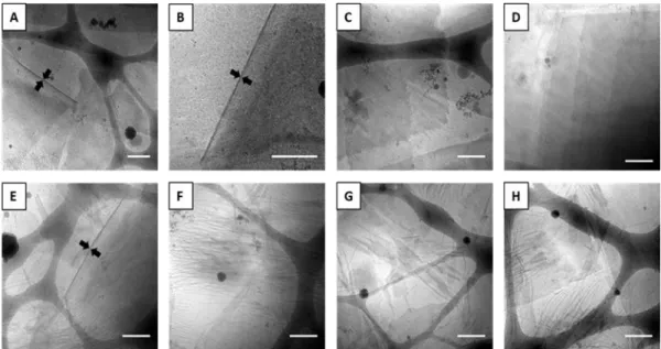
Figure 5. Images cryo-EM d’échantillons nanoprécipités de CS7 (A−D) et CS10 (E−H). Barre d’échelle = 300 nm.
VIII
Figure 6. Images cryo-EM d’échantillons nanoprécipités de: (1) CS7 (A) et CS10 (B) après le chauffage de 30 min à 65 °C; (2) CS7 (C) et CS10 (D) après le chauffage de 24 h à 90 ° C. Barre d’échelle = 100 nm en A et B. Barre d’échelle = 300 nm en C et D.
Figure 7. Evolution de la turbidité en fonction de la température pour les dispersions aqueuses d’auto-assemblages de CS7 (○) et de CS10 (■) préparées par nanoprécipitation, mesurées à une longueur d’onde de 600 nm avec un intervalle de température de 1 °C à la vitesse de 1 °C / min de 25 à 80 ° C. Les photographies à gauche révèlent directement le changement de turbidité des échantillons nanoprécipités de CS7 et CS10 après le chauffage.
IX
La transformation morphologique commence à partir des lamelles, cependant, la formation de vésicules CS10 semble différente de celle du Mécanisme I, où les lamelles se ferment en vésicules. Une analyse détaillée des polyméromes de CS10 révèle que les vésicules ne sont pas unilamellaires mais multilamellaires. Le scénario de formation des vésicules est proche du Mécanisme II, où les polymeromes multilamellaires sont formés par diffusion d’eau dans des sphères de polymère (c’est-à-dire des micelles plus grosses et complexes) lors du chauffage.
Une autre méthode d’auto-assemblage, l’hydratation du film, qui comprend également un traitement thermique au cours de l’étape d’hydratation, est réalisée sur CS7 et CS10, afin d’obtenir des vésicules de taille micrométrique. Comme indiqué par CLSM, des bâtonnets (Figures 8A&B) et quelques vésicules de taille micrométrique (Figures 8C&D) sont observées pour les deux copolymères.
Figure 8. Images CLSM d’auto-assemblages obtenues par l’hydratation du film de CS7 (A, C) et CS10 (B, D). Le rouge de Nil a été ajouté dans la préparation à un rapport pondéral de 1 : 100 (rouge de Nil au copolymère) pour observation. Des bâtonnets (A, B) et des vésicules géantes (C, D) sont observés. Barre d’échelle = 10 µm.
X
Pour évaluer la biocompatibilité, la solution aqueuse dialysée de CS10 provenant de la nanoprécipitation est utilisée dans le test de viabilité cellulaire avec le MTT comme indicateur. L’histogramme présenté à la Figure 9 indique que la viabilité relative des cellules testées dépasse 80% même avec les auto-assemblages de CS10 à une concentration de 0.69 mg/mL, ce qui confirme la biocompatibilité fiable des copolymères PCL-PSar et leurs échantillons d’auto-assemblage.
Figure 9. Viabilité cellulaire relative de CS10 pour les cellules L929 après 4 h d’incubation à 37 °C et à une concentration de 6.9, 34.5, 69.0, 345 et 690 μg/mL.
XI
Auto-assemblage de copolymères à blocs amphiphiles contenant PMeSPG
Les études d’auto-assemblage ont été menées sur les copolymères PEG-b-PMeSPG et PEG-b-PMeSPG-b-PSar, pour la formation de vésicules polypeptoids. Dans l’étude d’auto-assemblage par nanoprécipitation, trois copolymères PEG-b-PMeSPG, EM1 (PEG45-b-PMeSPG17, fPEG, wt = 45%), EM2 (PEG45-b-PMeSPG40, fPEG, wt = 26%)
et EM3 (PEG45-b-PMeSPG71, fPEG, wt = 16%), avec le MW croissant et le rapport
hydrophile décroissant, forment respectivement les nanosphères solides, les micelles sphériques et vermiculaire, ainsi que les lamelles et vésicules, comme les structures dominantes (Figure 10).
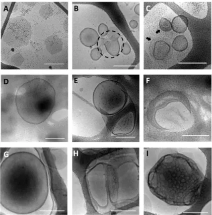
Figure 10. Images cryo-EM des auto-assemblages de EM1 (A−C), EM2 (D−F) et EM3 (G−I) par nanoprécipitation. Barre d’échelle = 100 nm pour A−F; barre d’échelle = 300 nm pour G−I.
XII
Tous les trois copolypeptoids PMeSPG-b-PSar, MS1 (PMeSPG45-b-PSar65, fPSar, wt
= 41%), MS2 (PMeSPG60-b-PSar50, fPSar, wt = 29%) et MS3 (PMeSPG75-b-PSar25, fPSar,
wt = 14%), peuvent former polyméromes par nanoprécipitation (Figure 11). La structure
des vésicules diffère dans les morphologies et les tailles en raison des différents rapports hydrophiles et MWs. Dans le même temps, les auto-assemblages des trois copolypeptoids sont évidemment non ergodique: les structures métastables, y compris micelles sphérique, lamelles, vésicules non fermés et vésicules de forme irrégulière coexistent dans le résultat du même copolypeptoid. Le comportement de PSar dans l’eau est analysé par la diffusion de neutrons à petit angle (SANS) et DLS, qui révèlent que PSar forme des grappes dans l’eau, plutôt que des bobines à chaîne unique comme le PEG. La différence de solubilité dans l’eau entre PSar et PEG est une des principales raisons de la différence d’auto-assemblage entre PEG-b-PMeSPG et PMeSPG-b-PSar, ainsi que la solubilité insuffisante de PSar dans l’eau est également probablement responsable de la nonergodicité de l’auto-assemblages de PMeSPG-b-PSar.
Figure 11. Images cryo-EM des auto-assemblages de MS1 (A−C), MS2 (D−F) et MS3 (G−I) par nanoprécipitation. Barre d’échelle = 300 nm.
XIII
En utilisant les propriétés thermodynamiques de PMeSPG, Vésicules sont obtenues de MS1 et MS2 par hydratation de film impliquant un chauffage (Figure 12). La transformation des vésicules MS2 de formes irrégulières à formes sphériques uniformes, parallèlement au temps du chauffage, confirme la possibilité de contrôler la morphologie d’auto-assemblage en fonction des conditions de préparation (Figure 13). Vésicules de taille micrométrique peuvent être obtenues de MS1−MS3 par la méthode de double émulsion, où les vésicules sont formées sous la direction de gouttelettes de double émulsion eau dans huile dans eau (W/O/W), pendant l’évaporation du chloroforme (Figure 14).
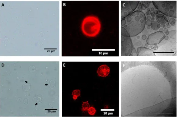
Figure 12. Images par microscopie optique à champ clair (A, D), CLSM (B, E) et cryo-EM (C, F) d’auto-assemblages de MS1 (A−C) et MS2 (D − F), par hydratation de film avec le chauffage de 48 h à 70 °C. Barre d’échelle = 300 nm pour C et F.
XIV
Figure 13. Images CLSM des vésicules MS2 par hydratation de film, après 48 h (A), 72 h (B), 144 h (C) et 168 h (D) du chauffage à 70 ºC.
Figure 14. Images microscopiques optiques à champ clair de l’émulsion double W/O/W de MS1 avant l’évaporation du chloroforme (A), après 12 h d’évaporation du chloroforme (B), et après 24 h d’évaporation du chloroforme (C).
MS3 forme les nanoparticules à structure bicontinue à l’intérieur, dans les auto-assemblage par nanoprécipitation et hydratation de film, en raison du rapport hydrophobe dominant (Figure 15). Les intermédiaires métastables sont capturés en tant qu’aspect de la nonergodicité des auto-assemblages de MS3, et l’analyse
XV
morphologique de ces intermédiaires peut prouver le mécanisme de formation de nanoparticules bicontinues par fusion interlamellaire (Schème 4).
Figure 15. Images cryo-EM des nanoparticules bicontinues et les structures intermédiaires de MS3 par nanoprécipitation (A−D) et double émulsion (E−H). Barre d’échelle = 300 nm.
Schème 4. La procédure de formation d’un attachement interlamellaire (ILA) entre deux lamelles approchées, dans le mécanisme de fusion interlamellaire.
XVI
Vésicules stimulables à base de PMeSPG-b-PSar
Le polypeptoid portant thioéthers est conçu pour ses caractéristiques stiumulables en réponse à l’oxydation. En effet, le desassemblage des vésicules polypeptoids de PMeSPG-b-PSar sensible à l’oxydation a été obtenu en raison de la transition « thioéther−sulfoxyde » (Figure 16) stimulée respectivement par le peroxyde d’hydrogène (H2O2) et par l’oxygène singulet (1O2) produit en presence de la lumière.
Les polyméromes et autres structures d’auto-assemblage de MS1 par nanoprécipitation sont désassemblés après le traitement avec 10 wt% (2.94 M) H2O2, ne laissant que peu
de grappes irrégulières lâches avec certaines cavités en forme de disques, révélés par cryo-EM (Figure 17).
Figure 16. (A) Schéme d’oxydation de PMeSPG avec thioéther à PMeSPGO avec
sulfoxyde. (B) PMeSPG et PMeSPGO suivis par spectres 1H-RMN (dans DMSO-d 6).
Les pics des protons adjacents à l’atome de soufre (a, b, c, tiret rouge) se déplacent vers le bas du champ (ao, bo, co, solide bleu) et confirment le passage du thioéther au
XVII
Figure 17. (A) et (B) Images cryo-EM des auto-assemblages MS1 obtenues par nanoprécipitation avant tout traitement (A) et après 90 minutes d’un traitement à 10 wt% H2O2 (B). (C) Évolution du diamètre hydrodynamique moyen (Dh) (sphères noires) et
des taux de comptage (triangles bleus) des auto-assemblages MS1 mesurés par DLS en fonction du temps du traitement de 10 wt% H2O2. (D) La distribution en taille des
auto-assemblages MS1 mesurée par DLS avant (rouge) et après (bleu) le traitement de 10 wt% H2O2 de 30 min.
La transformation détaillée en réponse à l’oxydation est enregistrée par CLSM des vésicules géantes MS2 par double émulsion, qui ont deux voies à distabliser sous la stimulation de 8.8 mM H2O2 (Figure 18). DLS et RMN sont utilisés pour confirmer
XVIII
Figure 18. (A) Illustration schématique de la destablization de la vésicule PMeSPG-b-PS en raison de la transition « thioéther−sulfoxyde » dans les blocs de PMeSPG sous la stimulation de H2O2. Le CLSM enregistre la rupture de la vésicule dans deux voies
différentes représentées par (B) et (C), sous la condition de 8.8 mM H2O2, avec le
rapport molaire de [H2O2]: [MeSPG] = 3.62 : 1. (B) voie IO: la vésicule passe d’abord
de vésicule ondulée et disquette à une vésicule bien arrondie dans les 89 premières secondes, puis la membrane commence à fluctuer de plus en plus intensément et de nombreux pores apparaissent autour de la surface de la vésicule (209 s) conduisant finalement à l’éclatement de la vésicule (225 s ). (C) Voie IIO: la vésicule change
d’abord en passant de vésicule ondulée et disquette à une vésicule bien arrondie au cours des 282 premières secondes; elle rapetisse en taille plus petite (exemple à 560 s) et a un bourgeonnement externe au dernier scène (754 s). Barre d’échelle = 25 µm.
XIX
Figure 19. (A) Le rapport des taux de comptage αt (αt = taux de comptage à l’instant t)
/ (taux de comptage à t = 0 avant l’addition de H2O2) en fonction du temps d’oxydation
t, à différentes concentrations de H2O2 (8.8 mM, 88 mM et 880 mM). (B) Spectres 1
H-RMN de polymersomes MS2 obtenus par émulsion double dans D2O en présence de
176 mM H2O2 avec le rapport molaire de [H2O2] : [MeSPG] = 3.62 : 1. Les
photographies d’un tube d’échantillon RMN à côté des spectres 1H-RMN montrent
clairement le changement de turbidité (de turpide à transparent) de la dispersion de polymersomes avant et après oxydation.
XX
Afin d’obtenir la vésicule sensible au stimulus distant et contrôlable, les polymersomes MS2 sont chargés avec le photosensibilisateur tétraphénylporphyrine (TPP), qui peut générer du 1O2 par l’activation de la lumière. Dans l’observation en
temps réel sous CLSM, la déstabilisation des polyméromes MS2 chargés en TPP est capturée, ce qui est initiée par le laser confocal localisé (HeNe 543 nm avec une puissance maximale de travail à 1.5 mW) du CLSM (Figure 20 et Figure 21). La déstabilisation photo-stimulable a des voies similaires à celles de la déstabilisation stimulée par H2O2, mais peut être obtenue avec une contrôlabilité et une efficacité bien
meilleures.
Figure 20. Instantanés de la séquence d’évolution de deux polymersomes MS2 marqués au rouge de Nil et chargés de TPP (rapport molaire de [TPP] : [MeSPG] = 1 : 30) sous l’éclairage du laser confocal (543 nm) de CLSM à la puissance de travail maximale de 1.5 mW. Deux voies sont observées pour la perturbation de polymersome. Barre d’échelle = 25 nm.
XXI
Figure 21. Les images CLSM d’une grande zone avant (I) et après (II) l’observation d’environ 15 minutes, avec deux points focalisés repérés par les cercles décrits en Figure 20. Les vésicules dans la mise au point du laser disparaissent, alors que celles des autres régions demeurent. Barre d’échelle = 25 nm.
1
Acknowledgements
Firstly, I would like to express my sincere gratitude to my supervisor Dr. Min-Hui Li for the continuous support of my Ph.D. study and related research, for her patience, motivation, and immense knowledge. Her guidance helped me in all the time of research and writing of this thesis. I could not have imagined having a better advisor and mentor for my Ph.D. study.
Besides my supervisor, I would like to thank the rest of my thesis committee: Prof. Sébastien Lecommandoux, Dr. Véronique Schmitt, Prof. Yixian Wu and Dr. Patrick Keller, for their insightful comments and encouragement, but also for the hard question which incented me to widen my research from various perspectives.
I thank my fellow labmates and friends: Yujiao Fan, Dapeng Zhang, Hui Chen, Bin Ni, Liang Zhao, Wei Qiang, Benoît Rhoné… for the stimulating discussions, for the sleepless nights we were working together before deadlines, and for all the fun we have had in the last three years. Also I thank my coworkers and friends in Zhejiang University. In particular, I am very grateful to Prof. Jun Ling and Dr. Xinfeng Tao for enlightening me the first glance of research.
I acknowledge the French National Research Agency (project ANR-16-CE29-0028) and the National Natural Science Foundation of China (project 21528402 and 21674091) for financial support. I also thank the China Scholarship Council for funding my Ph.D. scholarship in France.
Last but not the least, I would like to thank my parents for supporting me unswervingly throughout the accomplishment of the thesis. Their encouragement reinforced me to overcome every challenge coming from the work. My father was a diligent and respectable police officer, his hard work supported the whole family, but also damaged his health. He was diagnosed with cancer, and fought against it for five years. The news that I got an opportunity to persue the Ph.D. degree was his last comfort.
2
My mother is an incredible woman. She was not defeated by the sorrow and bitterness that life brought to her, but smiled to life, and kept a warm home in my back. Mother has an independent spirit, and persues a new life with considerable bravery and wisdom. She is a role model in my life. I will take my every effort to fulfill my dream, to be a responsible and respectable scientist. It would be the best present I can give back to my loving family and friends.
3
Outline
Acknowledgements ... 1 Outline... 3 Abstract ... 7 Résumé ... 9 Chapter 1. State of the Arts ... 11 1.1 Self-assembly of amphiphilic block copolymers ... 11 1.2 Polymersomes ... 13 1.3 Polymersomes in ROS-related applications... 30 1.4 Polymersomes based on polypeptides ... 38 1.5 Polymersomes based on polypeptoids ... 43 1.6 Conclusion ... 55 References ... 58 Chapter 2. Synthesis of Polypeptoid-Containing Amphiphilic Block Copolymers .... 73 2.1 Introduction ... 73 2.2 Experimental section ... 74 2.2.1 Materials ... 74 2.2.2 Synthesis of Sar-NTA ... 75 2.2.3 Synthesis of oxyamino-ended PCLs ... 75 2.2.4 Synthesis of PCL/PSar block copolymers ... 76 2.2.5 Synthesis of MeSPG-NTA ... 76 2.2.6 Polymerization of MeSPG-NTA ... 77 2.2.7 Synthesis of PEG-b-PMeSPG ... 77 2.2.8 Synthesis of PMeSPG-b-PSar ... 77 2.2.9 Characterizations ... 78 2.3 Results and discussion ... 78 2.3.1 PCL-b-PSar and PSar-b-PCL-b-PSar ... 78 2.3.2 PMeSPG and PEG-b-PMeSPG ... 83 2.3.3 Amphiphilic block copolypeptoid PMeSPG-b-PSar ... 88 2.4 Conclusion ... 91 References ... 92 Chapter 3. Thermo-Responsive Self-Assembly of PSar-b-PCL-b-PSar... 95 3.1 Introduction ... 954 3.2 Experimental section ... 96 3.2.1 Materials ... 96 3.2.2 Nanoprecipitation ... 96 3.2.3 Film hydration ... 96 3.2.4 Characterizations ... 97 3.3 Results and discussion ... 98 3.3.1 Thermodynamic behaviors of PSar-b-PCL-b-PSar ... 99 3.3.2 Self-assembly from nanoprecipitation ... 100 3.3.3 Morphological transformation upon heating treatment ... 102 3.3.4 Self-assembly from film hydration ... 109 3.3.5 Cell viability test ... 110 3.4 Conclusion ... 110 References ... 112 Chapter 4. Self-Assembly of PMeSPG-Containing Amphiphilic Block Copolymers ... 115 4.1 Introduction ... 115 4.2 Experimental sections ... 116 4.2.1 Materials ... 116 4.2.2 Nanoprecipitation ... 116 4.2.3 Film hydration ... 117 4.2.4 Double emulsion ... 117 4.2.5 Characterizations ... 117 4.3 Results and discussion ... 119 4.3.1 Self-assembly of PEG-b-PMeSPG by nanoprecipitation ... 119 4.3.2 Self-assembly of PMeSPG-b-PSar by nanoprecipitation ... 122 4.3.3 Behavior of PSar in water ... 126 4.3.4 Self-assembly of PMeSPG-b-PSar by film hydration. ... 131 4.3.5 Self-assembly of PMeSPG-b-PSar by double emulsion. ... 136 4.3.6 Bicontinuous structures and their formation mechanism ... 140 4.4 Conclusion ... 142 References ... 145 Chapter 5. Stimuli-Responsive Vesicles Based on PMeSPG-b-PSar ... 149 5.1 Introduction ... 149 5.2 Experimental section ... 150 5.2.1 Oxidation reaction of PMeSPG ... 150 5.2.2 Surface modification of coverslips ... 151 5.2.3 Characterizations ... 151
5 5.3 Results and discussion ... 152
5.3.1 H2O2-responsive PMeSPG-b-PSar nano-vesicles ... 152
5.3.2 H2O2-responsive PMeSPG-b-PSar giant vesicles ... 156
5.3.3 Photo-responsive TPP-loaded polymersomes ... 160 5.4 Conclusion ... 165 References ... 166 General Conclusion and Perspectives ... 169 List of Publications and Conference Abstracts during the PhD Thesis ... 172
7
Abstract
As a robust nanocontainer with polymeric bilayer structure, polymer vesicle, also called polymersome, has been considered as potential drug carrier in biomedical field, because of its cell-mimicking structure, high stability and capability to be functionalized. For practical use of polymersomes in biomedical applications, the biocompatibility of the copolymers that compose the vesicles become an important issue. Polypeptoid is a class of polypeptide derivatives of which the nitrogen atoms of amides are substituted. By avoiding the formation of inter- and intramolecular hydrogen bonds, polypeptoids have much better solubility than polypeptides in common organic solvent, while the biocompatibility is well maintained. Reports on self-assemblies of polypeptoid-containing block copolymers start to appear in the literature. However, works on polypeptoid vesicles are still scarce. In the present study, two families of polypeptoid-containing amphiphilic block copolymers are synthesized through the ring-opening polymerization (ROP) of N-substituted amino acid
N-thiocarboxyanhydrides (NNTAs). Various techniques like nanoprecipitation, film hydration and double emulsion are used to perform the self-assemblies with the objective to obtain polypeptoid vesicles. The self-assemblies are investigated in detail by DLS, cryo-EM, confocal laser scanning microscope (CLSM) and 1H-NMR.
In the first family of copolymers, ROP of sarcosine NTA (Sar-NTA) is initiated by oxyamino-ended poly(ε-caprolactone) (PCL), to obtain diblock copolymer PCL-b-PSar and triblock copolymer PCL-b-PSar-b-PCL-b-PCL-b-PSar. Unilamellar sheets and nanofibers are obtained by nanoprecipitation of PSar-b-PCL-b-PSar copolymers at room temperature. These lamellae and fibrous structures are transformed into worm-like cylinders and spheres after heating to 65 °C. Heating at 90 °C leads eventually to multilamellar polymersomes.
The second family of copolymers is based on a thioether-bearing polypeptoid, poly(N-3-(methylthio)propyl glycine) (PMeSPG). The new monomer, MeSPG-NTA is
8
obtained from a “decarboxylation−N-carboxymethylation” procedure from methionine, followed by cyclization. The amphiphilic block copolymers PEG-b-PMeSPG and PMeSPG-b-PSar are synthesized with different molecular weights and hydrophilic/hydrophobic ratios. Vesicles have been achieved with different methods, and the mechanism of vesicle formation through diverse nonergodic morphologies are discussed in detail.
The thioether-bearing polypeptoid is designed because of its oxidation-responsive features. Effectively, oxidation-oxidation-responsive disruption is achieved for the PMeSPG-b-PSar polypeptoid vesicles, based on the “thioether−sulfoxide” transition stimulated by hydrogen peroxide (H2O2) and light-induced singlet oxygen (1O2)
respectively. The oxidation- responsive and photo-responsive polypeptoid vesicles can be used as promising vehicles for smart drug release applications.
9
Résumé
Comme un nanoconteneur robuste formé d’une bicouche de polymères amphiphiles, vésicule polymère, aussi appelée polymersome, a été considérée comme un vecteur de médicament potentiel dans le domaine biomédical, grâce à sa structure imitant les cellules, sa grande stabilité et sa capacité à être fonctionnalisée. Pour l’utilisation pratique des polymeromes dans des applications biomédicales, la biocompatibilité des copolymères qui composent les vésicules devient un sujet important. Le polypeptoid est une classe de dérivés polypeptides dont les atomes d’azote des amides sont substitués. En évitant la formation de liaisons hydrogène inter- et intramoléculaires, les polypeptoids ont des solubilités bien meilleures que les polypeptides dans des solvants organiques usuels, tandis que la biocompatibilité est bien maintenue. Des travaux sur l’auto-assemblage de copolymères à blocs contenant polypeptoids émergent dans la littérature. Cependant, les travaux sur les vésicules polypeptoids sont encore rares. Dans cette thèse, deux familles de copolymères à blocs amphiphiles contenant les polypeptoids ont été synthétisées par la polymérisation par ouverture de cycle (ROP) de N-thiocarboxyanhydrides d'acides aminés N-substitués (NNTA). Différentes techniques telles que la nanoprécipitation, l'hydratation de film mince et la double émulsion ont été utilisées pour réaliser les auto-assemblages dans le but d'obtenir des vésicules polypeptoids. Les auto-assemblages ont été étudiés en détail par DLS, cryo-EM, microscope confocale à balayage laser (CLSM) et 1H-RMN.
Dans la première famille de copolymères, la ROP de la sarcosine NTA (Sar-NTA) a été initiée par le poly(ε-caprolactone) (PCL) à terminaison -oxyamine, pour obtenir le copolymère dibloc PCL-b-PSar, et le copolymère tribloc PSar-b-PCL-b-PSar. Des feuilles et nanofibres unilamellaires ont été obtenues par nanoprécipitation des copolymères PSar-b-PCL-b-PSar à température ambiante. Ces lamelles et structures fibreuses peuvent être transformées en particules cylindriques et sphèriques après
10
chauffage à 65 °C. Le chauffage à 90 °C conduit finalement à des polymeromes multilamellaires.
La seconde famille de copolymères est basée sur un polypeptoid portant thioéthers, la poly(N-3-(méthylthio)propyl glycine) (PMeSPG). Le nouveau monomère, MeSPG-NTA, a été préparé par une procédure de « décarboxylation−N-carboxyméthylation » à partir de la méthionine, suivie d'une cyclisation. Les copolymères à blocs amphiphiles PEG-b-PMeSPG et PMeSPG-b-PSar ont été synthétisés avec de différentes masses moléculaires et de différents rapports de blocs hydrophile/hydrophobe. Les études d’auto-assemblage ont été menées sur ces copolymères pour la formation de vésicules polypeptoids. Des vésicules ont été obtenues avec de différentes méthodes, et le mécanisme de la formation des vésicules à travers diverses morphologies non-ergodiques a été discuté en détail.
Le polypeptoid portant thioéthers est conçu pour ses caractéristiques stiumulables en réponse à l'oxydation. En effet, le desassemblage des vésicules polypeptoids de PMeSPG-b-PSar sensible à l'oxydation a été obtenu en raison de la transition « thioéther−sulfoxyde » stimulée respectivement par le peroxyde d'hydrogène (H2O2) et par l'oxygène singulet (1O2) produit en presence de la lumière.
Les vésicules polypeptoids oxydation-stimulable et photo-stimulable ainsi réalisés peuvent être des systèmes prometteurs dans des applications de relargages contrôlés des médicaments.
11
Chapter 1. State of the Arts
1.1 Self-assembly of amphiphilic block copolymers
Amphiphilic molecules can self-assemble in water, and form nano- or micro-structures of which the morphology is determined by the molecular properties and self-assembly conditions.1 As one kind of macromolecules with amphiphilic structure,
amphiphilic block copolymer observes the same principle and can self-assemble into various structures. As a dimensionless geometric parameter of the amphiphile, packing parameter, p, can be used to predict the self-assembly structure. Packing parameter is defined in equation: p = v/a0lc, where v is the volume of hydrophobic part, a0 is the
optimal area of the hydrophilic part at the interface, and lc is the length of hydrophobic
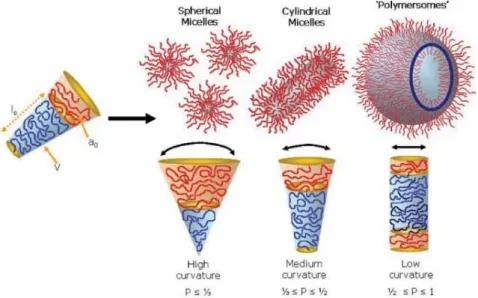
chain. Generally, amphiphilic block copolymers form spherical micelles when p ≤ 1/3, cylindrical micelles are favored from copolymers with 1/3 ≤ p ≤ 1/2, and for the copolymers with 1/2 ≤ p ≤ 1, copolymers tend to form vesicles, with enclosed membrane structure (Figure 1.1).1-3
Figure 1.1. Self-assembly structures of amphiphilic block copolymers: spherical micelles, cylindrical micelles and polymer vesicles, with corresponding packing parameters.3
12
Packing parameter reveals the inherent molecular curvature and favorable conformation of a block copolymer in water, which is determined by properties including chain structure, molecular weight, hydrophilic/hydrophobic ratio, chain rigidity and intermolercular interactions, etc. In the thermodynamic view, the self-assembly of amphiphilic block copolymers is driven by the minimization of free energy. Nevertheless, the relatively high molecular weight sets barriers for copolymers to adjust conformation and position towards global equilibrium state, which is much more difficult than small molecules. Consequently on the one hand, self-assembly structures of amphiphilic block copolymers have relatively high stability, on the other hand, it is often found that the self-assembled structures of copolymers stop at a metastable stage, which is called “kinetically frozen”.4 As the result, the self-assembly of block
copolymers shows nonergodic properties, with the coexistence of multiple morphologies.5 The nonergodicity endows polymeric self-assemblies the potential of
forming a large diversity of morphologies from the same block copolymer, which can be controlled by different preparing conditions. Various controlling or assistant methods come up in the research on block copolymer self-assemblies, including concentration variation,6 temperature variation,7 polarity of solvents,8 crystallization,9
addition of surfactant,10 emulsification,11 applying of electric field,12 etc. By combining
the inherent properties of block copolymers with the intervention of external process, structures with higher complexity can be generated, such as janus nanoparticles,13
cyclic micelles,14 alternating cylinders,15 helix structures,16 networks,17 lamellae,9
tubules,7 onion-like vesicles,18 faceted vesicles19 and ellipsoidal vesicles,20 etc.
For example, with the kinetic control, the team of Pochan and Wooley reported the preparation of cylinders and worm-like micelles with an alternating structure, by manipulating solvents in self-assembly.15 Water is slowly added to the solution of
poly(acrylic acid)-block-poly(methyl acrylate)-block-polystyrene (PAA-b-PMA-b-PS) and 2,2’-(ethylenedioxy)diethylamine (EDDA) in tetrahydrofuran (THF), forming separate spherical micelles (Figure 1.2A), where PMA and PS accumulate inside the
13 core, while the corona is composed of PAA, which is ionized by EDDA. THF is pipetted into the suspension quickly, turning the THF : H2O ratio from 1 : 4 to 2 : 1.
The system is forced back to a solvent composition where local lamellar structure is favored. As the result, spherical micelles turn into a disk-like shape, and tend to accumulate since the PAA-diamine pairs in the corona are immiscible in THF. With the anisotropic shape, disk-like micelles pack in a one-dimensional way, forming cylinders with an alternating structure (Figure 1.2B). The cylinders grow as the disks continue to pack, and develop into worm-like micelles (Figure 1.2C). In the late stage of growth, branches are formed when two disks pack to one end of the worm (Figure 1.2D).
Figure 1.2. (A) The spherical micelles formed by triblock copolymer PAA94
-b-PMA103-b-PS44 and EDDA (AA : EDDA = 1 : 1) in the mixture of THF : H2O = 1 : 4;
(B) cylinders resulted from the quick introfuction of THF; (C) the schematic illustration on the growth mechanism of ribbon-like structures; (D) worm-like micelles and separate disk-like micelles marked by black arrows; (E and F) branched structures formed in the late stage of growth. Scale bar = 200 nm.15
1.2 Polymersomes
Among the self-assembly structures of amphiphilic block copolymers, polymersome, that is polymer vesicle, has been a very attractive research object since the end of last century.21 Polymersome has a cell-mimicking membrane structure,
14
which is a curved bilayer composed of block copolymers, and an aqueous inner cavity. Based on the polymeric composition, polymersome has higher bilayer thickness (10−30 nm) than that of liposome (~5 nm), possessing relatively higher stability.21-22 Functional
polymersomes can be obtained by molecular design on block copolymers. These outstanding capabilities endow polymersomes with high potential in applications such as drug delivery vehicles and nanoreactors, harvesting lots of attentions in the field of new materials and biomedicine.
1.2.1 Self-assembly mechanism of polymersomes
Figure 1.3. Two mechanisms on the formation of polymersome: (a) in Mechanism I, polymersome is formed through the route of “spherical micelle−cylindrical micelle−bilayer−enclosed vesicle”; (b) in Mechanism II, the sperical micelle increases in size, and form a hydrophilic core by the rearrangement of copolymers, and polymersome is formed by water diffusing into the core.25
Polymersomes are favored to be formed from the self-assembly of the block copolymers with packing parameter 1/2 ≤ p ≤ 1. While the mechanism of polymersome formation which involves both thermodynamic and kinetic aspects, is still not fully understood. Two mechanisms were put forward to illustrate the process (Figure 1.3). In Mechanism I, spherical micelles are firstly formed instantly, then collide with each
15 other and slowly evolve into cylindrical micelles and in further open lamellae. Driven by minimization of edge energy, the lamellae close up and form vesicles.23 The
Mechanism II also starts from spherical micelle, however, instead of evolving into cylinder and lamella, the sphere grows bigger with the constituting copolymers reorganizing, and forming hydrophilic core inside. Water then diffuses into the core and forms a vesicle.24
Early in 1998, Eisenberg et al. reported the observation of lamellae with curved or folded structures, from the self-assembly of polystyrene-block-poly(ethylene glycol) (PS-b-PEG).26 The copolymers are firstly dissolved in N,N-dimethylformamide (DMF)
blended with some water (H2O wt% = 4.0−6.5%). Water is added to the system until
H2O wt% = 25%, and dialysis is conducted to remove DMF. According to the results,
if the copolymers are dissolved in DMF/water mixture for longer time, the obtained lamellae will have higher curvature, and more vesicles with complete enclosed structure will be found. It suggests that polymersomes are formed via Mechanism I, with the curved lamellae playing the role of intermediate.
In the self-assembly of PEG-block-polybutadiene (PEG-b-PBD), Discher and coworkers obtained nonergodic results from the blended system of PEG66-b-PBD170
and PEG150-b-PBD170, where multiple structures coexist.5 As revealed from the
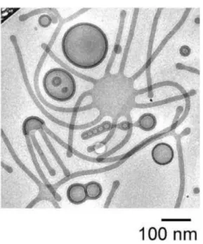
cryogenic electronic microscopic (cryo-EM) image (Figure 1.4), besides spherical micelles, worm-like micelles and vesicles, two octopus-like structures are observed. The right “octopus” stretches out 11 “arms” from the central disk, while the left one presents a semispherical shape. Both of them seem to be in the midway of bilayer enclosing to vesicle.
16
Figure 1.4. The nonergodic self-assembly result from the blended system of PEG66
-b-PBD170 (fPEG, wt = 24%) and PEG150-b-PBD170 (fPEG, wt = 42%).5
In the study of the polymerization-induced self-assembly (PISA), the mechanism of polymersome formation is further explored. Blanazs and coworkers reported the PISA using poly(glycerol methacrylate) as the macromolecular chain transfer agent (CTA), and initiating the reversible addition−fragmentation chain-transfer (RAFT) polymerization of (2-hydroxyl)propyl methacrylate (HPMA) in water.27 As HPMA is consumed, a series of self-assembly structures are observed
during the reaction, through the characterization under transmission electronic microscope (TEM). The mixture of spherical micelles and worm-like micelles are found from the system with monomer conversion at 55−68%, while for the system with conversion at 72−82%, intermediates with octopus- and jelly-fish-like structures emerge. Enclosed vesicles make up the majority of self-assemblies when the reaction completes with 100% conversion (Figure 1.5). In the procedure of PISA, monomers act as plasticizer, and render the formed copolymers mobile. At the late stage of reaction, most of monomers are consumed, leaving the copolymers with relatively longer chains and poorer mobility. Figure 1.5. shows all intermediates captured that help to describe the whole procedure of vesicle formation.
17 Figure 1.5. A series of self-assembly structures along with the monomer conversion of PISA, for the synthesis of PGMA47-b-PHMA200. Scale bar = 200 nm.27
Unlike the work above-discussed, the mechanism of polymersome formation of PEG-block-poly((2-N,N-diethyl)aminoethyl methacrylate) (PEG-b-PDEAMA) reported by Adams et al.25 is closer to Mechanism II. PEG45-b-PDEAMA81 is dissolved
in water with pH = 2, where the protonated PDEAMA make the copolymers totally dispersed individually. NaOH is added to deprotonate PDEAMA, driving copolymers to self-assemble. The results of dynamic light scattering (DLS) and cryo-EM reveal that neither cylindrical micelles nor lamella appear during the procedure. Instead spherical micelles, vesicles and spheres with loose cores are observed, which are supposed to be the intermediates in Mechanism II. The obtained vesicles cannot efficiently load hydrophilic fluorescent molecules, because in Mechanism II, the aqueous interior is formed by the diffusion of water molecules from outside to inside across rather thick barriers, and consequently water-soluble fluorescent molecules with rather high MW diffuse difficultly across these barriers.
1.2.2 Preparations of polymersomes
As the block copolymers have high molecular weight and their self-assembly shows nonergodic properties, the block copolymers with appropriate hydrophilic/hydrophobic ratios cannot self-assemble automatically into veiscles by just
18
adding them into water. Among various techniques to prepare polymersomes, nanoprecipitation, hydration and double emulsion are the three commonly used methods.
1.2.2.1 Nanoprecipitation
Nanoprecipitation is the most frequently employed method to perform the self-assembly of amphiphilic block copolymers. Generally, copolymers are first dissolved in an organic solvent, common for both polymer blocks and miscible with water. Water is then added to the system, driving the hydrophobic blocks to accumulate together. The self-assembling starts and continues upon the water addition. Finally the organic solvent is removed ether by dialysis againt water or by evaporation, leading to aqueous dispersions of polymer self-assemblies. In the procedure of nanoprecipitation, many factors such as the starting concentration, addition rate of water, water/organic solvent ratio, and polarity of the organic solvent, etc., can influence the morphology, the structure and the size of the obtained self-assemblies.
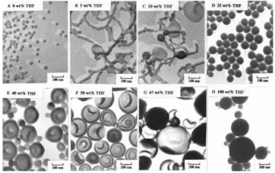
As a pioneer in the research of block copolymer self-assembly, the team of Eisenberg has reported early in 1997 the self-assembly of PS-b-PAA, through nanoprecipitation with organic solvents including THF, DMF, 1,4-dioxane (simply written as dioxane) and their mixtures as the starting solvents.28, 29 For example, in the
self-assembly of PS200-b-PAA18, spherical micelles are formed with DMF. When
DMF/THF mixture is used as solvent, and other conditions keep the same, the finally obtained self-assembly structures vary as a function of THF ratios (Figure 1.6): 5 wt% THF leads to the formation of worm-like micelles; 10 wt% THF leads to a mixture of worm-like micelles, lamellae and vesicles; 25−67 wt% THF allow the formation of polymersomes alone, and as the THF ratio increases further, the sizes of polymersomes get bigger. However, only large compound micelles are formed with pure THF as organic solvent. The difference in self-assembly may be explained by the difference of polarity of the organic solvent: THF has lower polarity than DMF, and comparatively
19 to DMF the polarity of THF is closer to that of PS. In the solvent with higher content of THF, on the one hand PS is more solvated, has higher degree of chain stretching, and keeps longer time of being solvated during the water addition procedure, which as the result gives the copolymers higher chance to adjust conformation towards the global equilibrium state. On the other hand, PAA blocks interact less with low-polar solvents, thus have less repulsion between corona chains, which is beneficial for the mutual fusion of self-assemblies for morphological evolution.
Figure 1.6. TEM images of self-assemblies of PS200-b-PAA18 from solutions in DMF,
THF and their mixtures: (A) pure DMF; DMF-THF mixtures of (B) 5 wt% THF; (C) 10 wt% THF; (D) 25 wt% THF; (E) 40 wt% THF; (F) 50 wt% THF; (G) 67 wt% THF; (H) pure THF.29
1.2.2.2 Hydration
In the hydration method, copolymer bulk is swollen by water to generate polymersomes. The copolymers are either dispersed directly in water, or processed first as thin film followed by hydration by water. The latter is referred as “film hydration” method, which origins from the preparation of liposomes, majorly for micrometer-sized giant vesicles.30 Copolymers are firstly dissolved in volatile organic solvent, and the
20
solution is deposited to a roughened surface (such as bottle bottom and Teflon plate). As the solvent evaporates, polymeric thin film is formed. The deposited surface is immersed in water. The film detaches from the surface and gets destabilized during the swelling of water, and external forces including heating, stirring, sonication and electric field can be introduced to accelerate the process.
Figure 1.7. A series of structures from the hydration process of PEG-block-poly(butylene oxide) (PEG-b-PBO).31
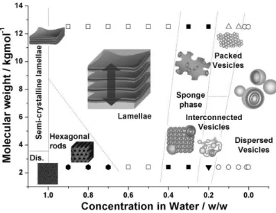
Battaglia and Ryan made detailed researches and reports on the mechanism of vesicle formation from hydration of copolymers.31, 32 Starting from the swollen lamellae
or hexagonal rods, series of structures are formed as the degree of hydration increases, including sponge phase, packed vesicles and interconnected vesicles. Dispersed vesicles are finally obtained when the system is diluted below a certain concentration (Figure 1.7).
The process of film hydration undergoes similar structural evolution. Through the observation under confocal laser scanning microscope (CLSM), Battaglia and Ryan captured a series of intermediate structures. Among them the multilamellar tubule is the most representative structure.33 Contacting with the surrounding water, the surface of
21 copolymer bulk and water, the wrinkling evolves into long tubular structures, which resemble the myelin connecting neuronal cells, and therefore are called “myelins”. As the further development of hydration, “myelins” destabilize into vesicles (Figure 1.8), where the concentration gradient works as a key factor. In the swelling process, more and more intermediates are formed because of the low mobility of copolymers, which lower the concentration gradient. The external driving force like electric field can be used to accelerate the transformation of intermediates, maintaining the concentration gradient, and consequently increase the efficiency of vesicle generation.12
Figure 1.8. (a) The formation of “myelin”, and the variation of size parameters during the transformation from wrinkling to “myelin”; (b) the mechanisms for “myelins” destablizing into vesicles.33
Lack of size control in the process, the vesicles obtained from film hydration method present often broad size distribution, and a coexistence of unilamellar and multilamellar vesicles. Further processes like sonication, extrusion and freeze-thaw cycles are needed to obtain mono-disperse and/or small vesicles.34-35 Based on the work
of Battaglia and Ryan, Howse and coworkers employed photolithography to modify a hydrophilic, fluorocarbon-decorated surface, with uniformly distributed hydrophilic domains.36 Through the spin-coating of copolymer solution, the surface is deposited
22
islands can generate unilamellar vesicles with uniform size in micrometer (Figure 1.9). The size of vesicle can be controlled by the size of hydrophilic domains.
Figure 1.9. (a) The formation of mono-distributed polymer islands through the patterned hydrophilic, fluorocarbon-decorated surface by photolithography, and spin-coating of copolymers; (b) observation of vesicle formation under CLSM.36
1.2.2.3 Double emulsion
Microemulsion are often used to form micrometer-sized spheres and capsules from crystallizable homopolymers, with the help of surfactants.37-39 In recent years the
emulsification-induced method, more precisely the method via water-in-oil-in-water (W/O/W) double emulsion, has become a versatile approach to study the self-assembly of amphiphilic block copolymers.11, 40-42 Copolymers are packaged in the W/O/W
emulsion composed of water and water-immiscible volatile organic solvent. With the amphiphilic property, copolymers accumulate in the W/O and O/W interfaces, stabilizing the emulsion as macromolecular surfactants. Upon the removal of organic solvent by evaporation, the monolayers of two interfaces meet with each other, forming the bilayer membrane of vesicles. In the work of Weitz et al., a capillary microfluidic device was employed to prepare double emulsions (Figure 1.10A).43-44 Three phases
23 are divided in the device, where the inner and outer phases are aqueous solutions with balanced osmolality, the middle phase is the organic solvent dissolving the copolymers. The inner aqueous droplet is formed in the dripping regime from a small injection tube, in the coaxial flow with the middle phase, while the middle oil droplet containing the inner droplet is formed from the breaking up of oil stream by the focused flow of the outer continuous phase. During the dewetting of emulsion droplets, acron-like structures are formed with oil droplet attached to the bilayer of copolymers (Figure 1.10B, C). Oil droplets shrink as the organic solvent evaporates, leading to vesicles with complete bilayer structure (Figure 1.10 D). With microfluidic control, the obtained polymersomes have a highly uniformed size. By adjusting the flow rates of inner and middle phases, and the number of inner tubes, the number of inner droplets inside an oil droplet can be controlled. In this way, multicompartment polymersomes from the stacking of plural vesicles can be obtained.45
Figure 1.10. (A) Schematic structure of the capillary microfluidic device generating W/O/W double emulsion droplets; (B) bright-filed microscope image of an acron-like structure formed from the partial dewetting of a double emulsion droplet, with the oil droplet on the left side, attched to the copolymer bilayer of an embryonic vesicle on the
24
right side, scale bar = 10 μm; (C) schematic illustration of the acron-like structue; (D) bright-field image of polymersomes after dewetting and solvent evaporation, with shrunk oil droplets, scale bar = 100 μm; (E) fluorescence microscope image of the same area as in (D), fluorescent HPTS solutes are encasulated inside polymersomes, by dissolution in the inner aqueous droplet in double emulsion preparation.44
1.2.3 Properties of polymersomes
In general, the properties of polymersomes depend on the properties of the copolymers that compose the vesiclular membranes. When compared with liposomes, polymersomes are distinguished by their larger membrane thickness, higher stability and lower permeability in most cases. These characteristics are the basis for the design of functional polymersomes, and determine their practical applications, especially as the encapsulating vehicles employed in biomedical fields.
1.2.3.1 Stability of polymersomes
Polymersomes have the high stability to keep their structure from the changes by external environment. The low mobility of block copolymers is a major factor. Polymer chains with higher MWs possess lower mobility, and higher chance for entanglement, which induces the barriers for rearrangement in conformation and position, keeping polymersomes from unwanted fluctuation and defect formation. Polymersomes can maintain its number, shape and encapsulated content basically unchanged over a month, while under the same condition, liposomes are found with the half-life only around 10−20 h.46 The low mobility also causes the formation and
long-term existence of metastable structures with irregular shapes.2 The shape of a vesicle
can be parameterized by a normalized volume-to-area ratio v, and a normalized area-difference ΔA.47 v = 6π1/2VA-3/2, where V and A respectively stand for the volume and
area of a vesicle, v actually represents the capsulated water content per membrane area;
ΔA = Aout – Ain, representing the difference of layer area between outside and inside
25 of the bilayer. For a uniformly spherical vesicle, v = 1 as the maximum, other non-spherical vesicle with the same membrane area entrap less water molecules, all with v < 1. ΔA is set as 1 for spherical vesicles, when ΔA > 1, the membrane tends to curve outward, causing the formation of vesicle budding and tubular structures; whereas ΔA
< 1 leads to an inwardly curved membrane, which leads to stomatocyte-like vesicles.48,
49 v can be adjusted by a “swelling-deswelling” process, and ΔA can be adjusted by the
flip-flop of molecules in the bilayer. Figure 1.11a shows the various morphological structures of vesicles formed with different v and ΔA in theory. In practice, v and ΔA could be adjusted respectively through water permeation and amphiphile exchange between layers and vesicles, leading to the uniformly spherical shape. However, due to the low mobility of copolymers, it is slow for polymersomes to reach the global equilibrium state, resulting in the kinetically trapped structures with metastable shapes. This phenomenon is also part of the nonergodic nature of polymer self-assemblies. Figure 1.11b shows the various forms of vesicles from the film hydration of PEG-b-PBO,2 which can basically correspond to the theoretical structures in Figure 1.11a.
Figure 1.11. Phase diagrams of (a) theoretical vesicle shapes, and (b) vesicles obtained from the film hydration of PEG-b-PBO. Ways to adjust the vesicle shapes are indicated in (a).2
As reported by the team of van Hest and Wilson, PS-b-PEG was found to form inwardly curved structure from nanoprecipitaion.50-52 The obtained vesicles have a
26
shape resembling stomatocyte, a kind of erythrocytes with slit-like central sink, thus are referred as “polymersome stomatocyte”. PS-b-PEG is dissolved in the mixture of dioxane and THF as the starting solvent, and water is then slowly added to preform self-assembling. After the formation of vesicles in the process of water addition, the exchange of water inside and outside the vesicle becomes difficult, due to the hydrophobicity of the polymer membrane. During the dialysis process, the osmotic pressure drives the organic solvent molecules to unidirectionally release outwards from the vesicle. Without appropriate amount of water entering the vesicle for supplement, the encapsulated content decreases, so does the v value, transforming the vesicle to a disk-like shape. The anisotropic arrangement in the membrane of disk-like vesicle is unstable, driving the vesicle to fold up into the stomatocyte-like shape. In this process, the organic solvent acts as plasticizer in the membrane, and gives the membrane certain mobility. As the solvent slowly dissipates, the PS region in the membrane enters glass state, which makes the like shape fixed as the final form. The stomatocyte-like morphology can be maintained over 4 months.50 There is a relationship between
the vesicle structure and the mixing ratio of the starting solvents: the opening of the polymersome stomatocyte decreases as the increase of THF ratio from 40% to 65% (Fig. 1.12). Because THF swells PS better than dioxane does. In the dialysis procedure, the amount of THF in the initial solvent mixture would influence the shape change, as higher THF ratio can increase the degree of swelling, and lower the rate of vitrification of the PS domain in the membrane. This would consequently results in a difference in the time allowed for the change of shape during dialysis. In the following studies, they adjusted the assembly conditions to precisely control the morphological structure of the obtained polymersomes.51-53 Platinum nanoparticles (PtNPs) can be loaded inside the
one-way opening inner cavity of the polymersome stomatocytes, to prepare nanomotors with active targeting function (Detailed discussion in 1.3.2 H2O2 gradient-sensitive
27 Figure 1.12. TEM images of polymersome stomatocytes obtained from dioxane-THF mixture as starting cosolvents, with THF ratio respectively at (A) 50%,(B) 55%,(C) 60% and (D) 65%. The Cryo-EM images of stomatocytes from cosolvents of THF ratio at (E) 50% and (F) 65%. (G) the plot of relationship between the size of stomatocyte opening and THF ratio of the starting cosolvent.
1.2.3.2 Permeability of polymersomes
As mentioned in above, polymersomes have relatively large membrane thickness (10−30 nm). Molecular dynamics simulation experiments show that the film thickness of the vesicles is positively correlated with the MW of the hydrophobic segment of the amphiphile. The bilayer film thickness d ~ MWphobb, for the amphiphilic
block copolymer, the coefficient b is about 0.55 (Figure 1.13a).56, 57 In the Fick’s
diffusion model driven by concentration gradient, the release rate of the encapsulated content from a vesicle is τ-1 = 3P/(2dR
V), where P is the permeability coefficient and
RV the vesicle radius.2 Therefore, Polymersomes composed of high-MW copolymers
28
Figure 1.13. (a) The schematic illustration on the relationship between the thickness of vesicle bilayer and the MW of hydrophobic segment of the composing amphiphiles; (b) curves of membrane properties including permeability, mobility and stability, in function of the MW of amphiphiles.57
Besides molecular weight, the permeability is also closely related to the state of membrane. The team of Eisenberg conducted in-depth studies on vesicles composed of PS-based block copolymers. By the addition of dioxane to the PS-b-PAA vesicle system, the release rate of doxorubicin (DOX) and the proton diffusion rate of vesicles are significantly increases along with the proportion of dioxane.58, 59 Dioxane acts as a
plasticizer which swells the PS region of the vesicle membrane and loosens its structure, thereby increasing the permeability. In the vesicle system from the self-assembly of PEG-b-PS-b-poly(2-(N,N-diethylamino)ethyl acrylate) (PEG-b-PS-b-PDEA) triblock copolymer, the hydrophobic region of the vesicle membrane consists of PDEA as the middle layer and PS on both sides.60 In basic environment, membrane structure is
compact and the permeability of the polymersome keeps low. As pH decreases, the PDEA layer gets protonated and swollen, increasing the membrane thickness and vesicle size. When the pH drops below 6, defects and breakages appear in the membrane (Figure 1.14). The change is clearly fed back on permeability, allowing the significantly increased penetration to water and protons.
29 Figure 1.14. (a) Cryo-EM images of PEG-b-PS-b-PDEA vesicle membrane at different pH; (b) schematic membrane structure corresponding to (a); (c) thickness of PS region, PDEA region and vesicle membrane at different pH.60
Another way to alter the permeability is to embed functional substances in the vesicle membrane. Based on polymersomes from the self-assembly of poly(2-methyloxazoline)-b-polydimethylsiloxane-b-poly(2-methyloxazoline) (PMOXA-b-PDMS-b-PMOXA), Meier and coworkers insert channel protein (porin) into the vesicle membrane, to regulate the exchange of certain molecules.61-63 Enzymes with specific
functions are encapsulated inside the vesicle. The enzyme catalyzes reactions on consuming substrates inside the vesicles, while porin channel let substrate and product circulate along the vesicle membrane. Then, the polymersome is turned into a nanoreactor with highly catalytic function. Encapsulation inside the vesicles allows to protect the enzyme from the surrounding environment and to improve its work efficiency. Meier et al. reported in 2013 another way to change the membrane permeability, by using 2-hydroxy-4’-(2-hydroxyethoxy)-2-methylpropiophenone (PP-OH) under UV light.64 UV-irradiation causes PP-OH to form two primary radicals
30
PP-OH. The attachment of the hydrophilic PP-OH increases the ability of vesicle membrane to allow hydrophilic molecules to penetrate. This reaction is applicable to a variety of vesicles, and does not change their size and structure. The enzyme-entrapped polymersome can also be beneficial, to achieve the nanoreactor function in a simpler way.
1.3 Polymersomes in ROS-related applications
The high stability and low permeability give polymersomes advantages in applications as drug carriers. With the robust membrane, polymersomes protect the encapsulated drug from the surrounding environment and minimize the loss of early release before the target. In the other view, the most direct and effective way to achieve stimuli-responsive release is to break the stability of membrane, causing the change in permeability or structural disintegration of vesicle under specific stimuli. pH variation and redox reactions have been reported to trigger the controlled release of polymersome.65, 66 The stimulation may cause the composing copolymers to change the
solubility,67, 68 to have degradation69 or special cleavage of bond linking hydrophilic
and hydrophobic blocks,70 leading to the increase of permeability or the bursting release
of polymersome. Some pathological bases such as the tissue of inflammation and tumor offer chemical environment differing from that of normal ones, and have specific accumulation of certain active substances. These active substances can be used as stimuli in the targeted sites.
Reactive oxygen species (ROS) refer to a class of byproducts from the process of oxygen-involving metabolism, including peroxide, superoxide, hydroxyl radical, singlet oxygen and alpha-oxygen, which have high oxidative activity.71 The high
oxidative property enables ROS to damage DNA and cell membrane structure, which can be used to destroy tumor cells in cancer therapy. On the one hand, for most of the radiotherapy and chemotherapy in cancer treatment, including the emerging photodynamic therapy (PDT), their mechanisms are based on or include the increase of
31 ROS concentration at target site.72-75 On the other hand, significantly higher ROS
concentrations found in some tumor and inflammatory tissues than in normal cells provide a means of targeted and ROS-responsive drug delivery.54, 71, 76, 77
1.3.1 Polymersomes as ROS generators or carriers
The commonly used anticancer drug, DOX, is considered in relation with the promotion of ROS production. 78 Lecommandox et al. reported that the intracellular
introduction of DOX-containing vesicles of poly(γ-benzyl L-glutamate)-block-hyaluronic acid (PGlu(OBn)-b-HYA) can results in the obvious increase of ROS concentration.79 Enzyme-catalyzed oxidation is one main source of ROS in vivo. Meier
and Bruns reported the encapsulation of a commonly used oxidase, laccase, inside the vesicle of poly(N-vinylpyrrolidone)-block-polydimethylsiloxane-block-poly(N-vinylpyrrolidone) (PNVP-b-PDMS-b-PNVP).80 PNVP-b-PDMS-b-PNVP vesicles
have high permeability for oxygen and ROS, but low permeability for laccase. Therefore the entrapped laccase is well protected by vesicle, and meanwhile can efficiently turn oxygen into ROS. The laccase-loading polymersomes work as a ROS-generating nanoreactor.
In the working mechanism of PDT, photosensitizer is activated by light stimulation, and turns oxygen-containing substrates into ROS, for example singlet oxygen (1O
2) from ground state oxygen.73 In the combination of PDT and polymersome,
photosensitizer is loaded on the membrane or inside the vesicle, for the generation and delivery of ROS. Chen and coworkers reported the use of polyion complex vesicle (PICsome) from PEG-block-poly(aspartic acid) (PEG-b-PAsp) and poly(5-aminopentyl asparagine) (P(Asp-AP)), as the carrier of photosensitizer Al(III) phthalocyanine chloride disulfonic acid (AlPcS2a).81 AlPcS2a-loaded PICsomes are
successfully delivered into the cells via endocytosis. Under the activation of light, AlPcS2a generates 1O2 and oxygen-containing free radicals, which in turn damage the