Université de Montréal
Augmentation sélective de l’expression protéique et de
1’ARNm de la synthase du monoxyde d’azote dans les
régions vulnérables du cerveau chez les rats déficients en
thiamine
par Milarca C. Kruse
Département de Sciences Biomédicales Faculté de Médecine
Mémoire présenté à la Faculté des études supérieures en vue de l’obtention du grade de Maîtrise
en Sciences Biomédicales
Août 2003
W
L)oLi
Direction des bibliothèques
AVIS
L’auteur a autorisé l’Université de Montréal à reproduire et diffuser, en totalité ou en partie, par quelque moyen que ce soit et sur quelque support que ce soit, et exclusivement à des fins non lucratives d’enseignement et de recherche, des copies de ce mémoire ou de cette thèse.
L’auteur et les coauteurs le cas échéant conservent la propriété du droit d’auteur et des droits moraux qui protègent ce document. Ni la thèse ou le mémoire, ni des extraits substantiels de ce document, ne doivent être imprimés ou autrement reproduits sans l’autorisation de l’auteur.
Afin de se conformer à la Loi canadienne sur la protection des renseignements personnels, quelques formulaires secondaires, coordonnées ou signatures intégrées au texte ont pu être enlevés de ce document. Bien que cela ait pu affecter la pagination, il n’y a aucun contenu manquant.
NOTICE
The author of this thesis or dissertation has granted a nonexclusive license allowing Université de Montréal to reproduce and publish the document, in part or in whole, and in any format, solely for noncommercial educational and research purposes.
The author and co-authors if applicable retain copyright ownership and moral rights in this document. Neither the whole thesis or dissertation, nor substantial extracts from it, may be printed or otherwise reproduced without the author’s permission.
In compliance with the Canadian Privacy Act some supporting forms, contact information or signatures may have been removed from the document. While this may affect the document page count, it does not represent any loss of content from the document.
Université de Montréal faculté des études supérieures
Ce mémoire intitulé
Augmentation sélective de l’expression protéique et de I’ARNm de la synthase du monoxyde d’azote dans les régions vulnérables du cerveau chez les rats déficients en
thiamine.
présenté par:
Milarca C. Kruse
a été évalué par un jury composé des personnes suivantes: Dr Gilles Pomier- Layrargues, président-rapporteur Dt Roger f. Butterworth, directeur de recherche
Résumé
La déficience en thiarnine entraîne la mort sélective de cellules neuronales dans les structures thalamiques.
Des études antérieures mettent en évidence le rôle du monoxyde d’azote dans la pathogenèse de la mort cellulaire causée par cette déficience vitaminique. Dans le but d’éclaircir l’origine de l’augmentation du monoxyde d’azote (NO), nous avons mesuré l’expression des isoformes de l’oxyde nitrique synthétase endothéliale (eNOS), inductible (iNOS) et neuronale (nNOS) dans le thalamus médian, le colliculus inférieur et le cortex frontal (une région cérébrale non-atteinte) chez les rats où la déficience en thiamine fut induite par un régime déficient en thiamine et aussi par l’administration quotidienne de pyrithiamine, un antagoniste central de la thiamine.
Nous avons observé une augmentation significative des niveaux de l’ARN messager et de l’expression protéique de l’isoforme endothéliale (eNOS) proportionnelle à la sévérité de la dégénérescence neurologique et de la perte neuronale dans le thalamus médian et le colliculus inférieur.
Aucun changement dans l’expression des autres isoformes du NOS n’a été observé. Ces résultats suggèrent que l’endothélium vasculaire est un site important de production de NO dans les dans le cas de le cerveau des rats déficients en thiamine et que celui-ci pourrait être la cause des dommages aux structures vulnérables dans cette pathologie.
Mots-clés: Déficience en thiamine, Oxyde nitrique (NO), Endothélium vasculaire, Oxyde nitrique synthétase (NOS), Oxide nitrique synthétase endothéliale (eNOS), Oxide nitrique synthétase inductible (iNO$), Oxide nitrique synthétase neuronale (nNOS), Thalamus médian, Colliculus inférieur, Léon oxydative, Encéphalopatie de Wernicke (EW), Système nerveux central (SNC).
iv
Abstract
Thiarnine deficiency is a cause of selective neuronal ceil death in thalamic structures. Previous studies provide evidence for a role for nitric oxide (NO) in the pathogenesis of ceil death due to thiamine deficiency. In order to ascertain the origin of the increased NO in the thiarnine deficient brain, expression of the nitric oxide synthase isoforms endothelial (eNOS), inducible (iNOS) and neuronal (nNO$) was measured in the media! thalamus and the inferior colliculus, and compared to the expression in the frontal cortex (a spared region in this particular pathology) of rats in which thiamine deficiency was induced by feeding of a thiarnine-depleted diet and the daily administration of pyrithiarnine, a central thiamine antagonist.
Endothelial NOS rnRNA and protein expression were significantly increased as a function of the severity of neurological impainnent and degree of neuronal ce!! loss in the medial thalamus and inferior colliculus. No significant alterations in the expression of other NOS isoforms were observed. These findings suggest that the vascular endothelium is a major site of NO production in the brain in thiamine deficiency and that eNOS—derived NO may account for the selective injury to histologically targeted regions in this disorder. Keywords : Thiamine Deficiency, Nitric Oxide (NO), Vascular endothelium, Nitric oxide synthase (NOS), Endothelial nitric oxide synthase (eNOS), Inducible nitric oxide synthase (iNOS), Neuronal nitric oxide synthase (nNOS), Medial Thalamus, Inferior Colliculus, Oxidative damage, Wernicke Encephalopathy (WE), Central nervous system (CNS).
Table des matières
Introduction. 10
Conclusions. 82
Liste des tableaux
1. Characteristics of NOS isoforrns.Liste des figures
1.1- Chemical structures ofthiamine and pyrithiamine
1.2- Schematic diagram ofthe glycolytic and TCA pathways. (Adapted from Hazeil et
al., 1998).
viii
À
Guy P.Kruse Brooke, Estrella Liergo, Guy P.Kruse JIL Karen E.Kruse, Sarah L. Whatïey, Donald et Peggy Murray et Brenagh Fitzpatrick.Remerciements
To D’ Pan! Desjardins and Mi Darren Navarrofor titeir invattiabte assistance, kindness,
patience anct geiterosity with theirtbne andknowteclge ait clwithont whonz titis work
Introduction
1-Sommaire
L’Encéphalopathie de Wernicke (EW) est une atteinte neurologique sérieuse causée par la déficience en thiamine (vitamine Bi) et est sotivent associée à un état général de malnutrition. Dans le monde occidental, on retrouve principalement cette pathologie chez les patients souffrant d’alcoolisme chronique, de vomissements associés à la grossesse sévères, d’autant que par les anorexiques et les sidéens. D’autre part, la consommation abusive d’alcool entraîne également des changements rendant ainsi les patients plus susceptibles à cette pathologie.
L’EW a été initialement décrite en 1881 chez trois patients souffrant d’ataxie, de nystagmus vertical et de changement cognitifs. Depuis lors, plusieurs chercheurs ont tenté
de déterminer l’étiologie de cette pathologie. Il est maintenant établi qu’une insuffisance en
thiamine est la cause principale de l’EW. Cependant, les mécanismes physiopathologiques menant à l’apparition de lésions histologiques dans certaines régions spécifiques du cerveau demeurent jusqu’à maintenant inconnus.
La déficience en thiamine induite par la pyrithiamine (DTP) chez le rat est un modèle qui émule I’EW en entraînant la dégénérescence sélective de certaines structures cérébrales. Chez le rat, la DTP évolue progressivement et récapitule les symptômes neurologiques et la neuropathologie de l’EW, soit l’apparition de lésions focales symétriquement distribuées (Troncoso et al., i98i).
La pyrithiamine est un analogue central de la thiamine qui franchit la barrière hémato-encéphalique et entraîne chez l’animal des symptômes neurologiques sévères (ataxie, opisthonus, nystagmus, perte du réflexe de redressement, convulsions et finalement
le coma). La pyrithiarnine s’accumule dans les structures cérébrales (Rindi et Perri, 1961;
Rindi et al., 1963) et entraîne une diminution rapide des taux de thiamine cérébrale en inhibant la thiamine pyrophosphokinase (Gubler, 1968; Johnson et Gubler, 1968), l’enzyme
responsable de la conversion de la thiarnine en ester diphosphate. De plus, la pyrithiarnine inhibe la capture de la thiarnine dans les tranches de cerveau (Nose et al., 1976) et peut déplacer la thiamine dans des préparations nerveuses (Cooper, 1968).
Le radical libre monoxyde d’azote (NO) possède des effets toxiques sur le système nerveux central (SNC) (ladecola, 1997). Cette substance cause des dommages oxydatifs en réagissant avec les anions de superoxyde (O2-) pour former le péroxynitrite (ONOO-), un puissant anion oxydant qui se est décompose rapidement en radical hydroxyle OH, un oxydant hautement réactif qui réagit de façon non spécifique (Merriil et Murphy, 1996). Une insulte oxydative peut mener à la peroxidation des lipides, la décomposition de l’ADN et à l’inactivation des enzymes piégeuses de radicaux libres. Les producteurs de radicaux libres dans le SNC sont les mitochondries, la xanthine oxydase, le métabolisme de l’acide arachidonique et l’oxydation spontanée de molécules telles que les catécholarnines et l’hémoglobine.
Les anions de superoxyde formés par diverses réactions sont normalement transformés en peroxyde d’hydrogène (f1202) de façon spontanée ou aussi par les actions de
la superoxyde dismutase (SOD). Une étude par Langlais et al. en 1997, a démontré la présence de substances oxygénées réactives dans le thalamus des animaux DTP. Jusqu’à maintenant la source exacte de la production excessive de substances oxygénées réactives dans la DTP n’était pas claire.
Les cellules endothéliales et les astrocytes sont des producteurs importants de NO (Murphy et al., 1990; Moncada et al., 1991). Il a été démontré que le NO peut endommager la BHE (Au et al., 1985). On sait aussi que les radicaux libres peuvent augmenter la perméabilité des cellules endothéliales (Chan et al., 1991). Il est donc possible que le NO puisse modifier l’intégrité fonctionnelle de ce type de cellule et ainsi jouer un rôle important dans la rupture de la BHE chez les animaux DTP (Calingasan et al., 1 995a; Harata et Iwasaki, 1995).
12
Même lorsque l’activité totale de NOS (l’enzyme responsable de la production de NO), dans le cerveau est réduite dans les animaux traités à la pyrithiamine (Rao et al., 1996), l’expression de l’isoforme endothéliale est augmentée dans les régions vulnérablesdu cerveau ainsi que dans les vaisseaux sanguins.
De plus, le traitement de rats au deprenyl, un piégeur de radicaux libres, accroît la survie neuronale chez les rats DTP (Todd et Butterworth, 1 99$b), ce qui indique que les substances réactives oxygénées, dont le NO, jouent un rôle critique dans la pathogenèse des atteintes cérébrales induites par la déficience en thiamine.
Dans une importante étude par Calingasan et al. (2000) le rôle de l’induction de eNOS par la voie dépendante de 1’ ICAM-i est un facteur critique dans la mort neuronale causée par les dommages oxydatifs dans les neurones dont le métabolisme est compromis.
General Introduction
1-1 CHEMICAL STRUCTURE 0F THIAMINE, SOURCES AND NEEDS
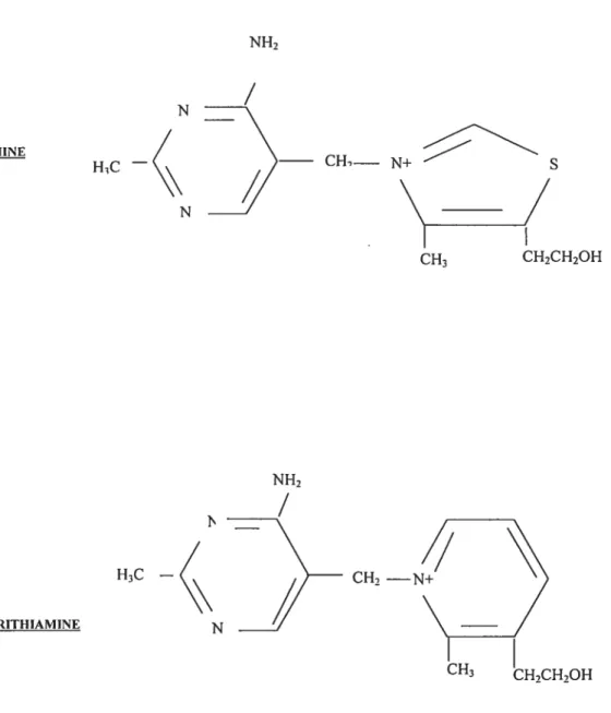
Thiamine, which is also known as vitamin Bi is a water-soluble vitamin whose structure contains a pyrimidine ring and a thiazol cycle bonded to a quatemary ammonium by a methyiene bridge (Figure 1.1)
CH2 — CH3 CH2CH2OH CH1— N+ CH3 CH2CH2OH N TIIIAMINE
/
HC N H3C — PYRITHIAMINE N NH214
Thiamine is a vitarnin essential to most mamrnals, since they are incapable of autonomous synthesis. The main sources of this nutrient are cereal products, vegetables, meat, fish, nuts and dairy (Robinson, 1966; Thiessen, 1978). The brain itself contains 1 pglg of fresh tissue (Davis and Icke, 1983). Thiamine plays an extremely important role in glucose rnetaboÏism that takes place in order to fulfihi cellular energetic processes; therefore the necessary intake to satisfy those needs must be adjusted according to need.
Theoretically, an average daily intake of approximately 1.0 and 1.5 mg per diem would be sufficient (Dietary Reference Intake or RDI, 2003).
In the brain, thiamine is most ofien encountered as thiamine diphosphate (TDP) (Spector, 1982; Davis and Icke, 1983) in contrast with the haematological pathway where the prefelTed form is that of thiarnine monophosphate (TMP). This difference is indicative of the probability that thiamine transport in the central nervous system (CNS) is dependent on a phosphorylation step (Barchi and Braun, 1972; Nose and al, 1974; Spector, 1976).
The existence of a specific membrane phosphatase that is favorable to transport would explain the reason why certain regions of the brain would be less vuinerable to the depletion ofthiamine stores than others.
Thiarnine plays its most significant role in thc CNS as an enzyme cofactor. There are three thiarnine-dependant enzymes in the CN$: Pyruvate Dehydrogenase Complex (PDHC), Œ-Ketoglutarate Dehydrogenase (aKGDH) and Transketolase (TK).These three enzymes are critical factors in glucose metabolism.
1.3 CLINICAL IMPLICATIONS 0F THIAMINE DEFICIENCY.
1.3.1 Wernicke —Korsakoff Syndrome
Wernicke encephalopathy part of the Wernicke-Korsakoff syndrome, is a neurological disorder whose aetiology is generalized thiamine deficiency, usually the product of poor nutritional status. In economically deveÏoped countries, the highest incidence is found amongst the chronic alcoholic patient population. Aïcohol abuse in itself leads to dysfunction of thiamine absorption making these sarne individuals even more vulnerable to this vitamin deficiency.
While experimenting by feeding pigeons with a thiamine-free diet, Sir Rudolph Peters (Kinnersley and Peters, 1930) successftilly dernonstrated the accumulation of pyruvate and lactate at the brainstem that preceeded the appearance of neurological symptoms. This discovery was the first to suggest the existence of a « biochemical lesion».
Later on, Bowman et al. (1939) described the therapeutic effect of thiamine administration in the treatment of a patient suffering from cognitive changes (which are one ofthe symptoms encountered in this pathology). Thiamine was used successfully to reverse the ophthalmological as well as cognitive symptoms by Joliffe et al. in 1941.
On the other hand, the residual psychosis, also known as Korsakoff psychosis, seems to be refractory to this treatment modality. Thus, treatment of Wernicke encephalopathy with thiamine has been standard for several decades but the pathophysiology ofthis disorder has flot been entirely explored.
16
1.3.1.1 Clinical presentation
This syndrome can be described as having an acute stage, that of Wernicke encephalopathy which is sensitive to treatment with thiamine and the more chronic stage, that of Korsakoff psychosis which seems refractory to this treatment rnodality. Both stages are respectively the neurological and psychiatrie facets of a same syndrome which can be found amongst affected patients.
Three types of symptoms such as cognitive deficits, ataxia and also the ophthalmological type of symptoms such as ophthaimoplegia or vertical nystagmus characterize Wernicke encephalopathy (EW). Ail of these symptoms are seldom found in conjunction, but combinations of some of them and most frequently, ataxia and ophthalmological manifestations are the most common clinical profile presented in practice.
The characteristic cognitive alterations seen in this syndrome is inclusive of a significant level of apathy, difficulty concentrating on a particular task or thought as well as amnesia and perceptional alterations. In a few cases, the reversai of these behavioural and cognitive symptoms is achieved upon therapeutic thiamine administration. In rnost patients, improvement of the symptoms is progressive and sometimes is replaced by learning disabilities and (short-term) memory abnormalities. Memory integrity is rarely recovered.
Ataxia which is an important symptom for an accurate diagnosis is harder to confirm clinically, since it can vary from a subtie motor coordination problem to a severe handicap and it is only after performing a specific neuroiogical examination, that the diagnosis can be rendered. Thiamine is oniy marginally successflul as a treatment for this symptom.
Ophthalmologic manifestations are important landmarks and can therefore assist in rendering differential diagnosis. These include nystagmus (85%), paresthesis of lateral
abductors (54%) and paresthesis of coordinated vision (44%). Ophthalmological symptoms
are rapidly alleviated afier therapeutic thiamine administration.
Korsakoff psychosis is a very unique and characteristic set of symptoms in this disease process and it presents with short-term memory problems and learning deficiencies. Approximately 20% of patients recover completely from these symptoms and an additional 20% of them is non-responsive to treatment, with most of the patients falling in between these extremes.
1.3.1.2 Neuropathology
In an important series of studies, Victor et al. (1989) showed afier the post-mortem evaluation of the brains of WE patients, macroscopic histopathological changes that are inclusive of an increase in size of the third and lateral ventricles and the thalamic subventricular regions, which are affected by lesions. On the other hand, cerebral atrophy is found in 20% ofthe patients and most often seen in the frontal lobes (Harper, 1983).
Rapid and accurate diagnosis of the disease is effective not only towards promptly improving the neuropathological abnormalities. The selectively vuinerable regions do not have a common embryological source (Hamilton et al., 1952) and do not share vascularisation.
The earliest histological changes in thiamine deficiency are morphological alterations in glia (Collins, 1967; Robertson et al., 1968). Macroscopic atrophy of the mamillary bodies can be seen when the dissection of a fixed brain is performed. This lesion is symmetrical with pinpoint hemorrhages in some cases.
Lesions found in the gray matter are similar and can be observed around the aqueduct, inferior and superior colliculus and the inferior portion of the IVth ventricle. The cerebellar vermis is mainly targeted at the folia, anterosuperior to the prirnary fissure. In cases of both Korsakoff psychosis and WE the lesion distribution is similar, with the
18
exception that in the latter the additional implication of lesions in the medial thalamus and hypothalamus has been found. Other targeted regions are the thalamus; hypothalamus, rnidbrain, inferior olive, cerebellar vermis and the vestibular lateral nuclei (Torvik, 1985; Mair et al.1979; Victor, 1976) as well as the basal nucleus ofMeynert (Arendt et al., 1983).
An interesting particularity of this pathology is that although the periventricular regions are the most affected by lesions, most of its neighboring structures are spared. This is a good example of the regional specificity of the lesions occuning in this nutritional disorder. At histological observation it is important to discern that the damage seen in the rnammillary bodies and sub-ependymal area is principally caused by chronic degeneration of neuropil, which develops spongiosis, and loss of myelinated fibers. These types of lesions are due to severe endothelial inflammation but in this case, neurons are not especially targeted (Torvik, 1985; Victor, 1976).
Contrary to this, in the thalamus and inferior olive, the exact opposite type of lesions are found, so that neurons are particularly damaged, and relatively sparing the neuropil (Torvik, 1985; Victor, 1976). Torvik’s 1985 study suggested the presence of two different types of lesions and indicates the possibility that cellular brain injury rnight be the product of several mechanisms.
Lesions to the cerebellar hemisphere and to the anterior vermis are characterized by the decrease in Purkinje celis and the augmentation in Bergman astrocytes that in more severe cases are accompanied by an important neuronal loss in the granular layer.
1.3.1.3 Thiamine status and alcoholism
In economically developed countries, the most frequent cause of thiamine deficiency is chronic alcohol abuse. This is caused by the generally poor nutritional status seen in these individuals and the insufficient thiamine consumed in their diets (Leevy et al.,
1965; Neville et al., 1968). To add to this insuit, excessive alcohol consumption alters normal thiamine phosphorylation.
It has been shown in both humans, (Tomasulo et al., 1968; Thomson et aI., 1970) as well as in a rat model (Hoyumpa et al.. 1978), that ethanol reduces intestinal absorption of this substance. It was shown that following oral administration of 2Omg of thiamine, maximal absorption is of 4.77±0.36mg in normal patients but on the other hand, the absorption is decreased to 1 .50±0.3mg in alcoholic patients.
The mechanism proposed to explain this phenomenon was the inhibition of active transport (Hoyumpa et al., 1975) through suppression of NaKATPase in the enterocytic basolateral membranes (Hoyumpa et al., 1977).
Intestinal concentration seems to be a determining factor for inefficient absorption more so than the length of time of the exposure of the enteric tissues (Hoyumpa et al., 1978). Another aggravating factor is the common co-deficiency in alcoholics of vitarnins 36 and 312. This leads to a reduction in thiamine absorption (Thomson et al., 1972; Howard
et al., 1974; Nishino and Itokawa, 1977). Adding folates to the diet improves this effect
(Davis and Srnith, 1974).
Alcohol abuse also reduces the stores and capture of thiamine in the liver and also increases the depletion ofthis substance (Heaton, 1977; Howard et al., 1974; Chi-Po Chen, 1978). The consequence is that in the alcoholic patient the depletion ofthe hepatic thiamine stock is more rapid and its replacement mechanism is less efficient.
Alcohol abuse slows down TPKase activity which catalyses the synthesis of TDP, the active form of thiamine when it is utilized as a cofactor (Davis et Icke, 1983; Leevy and Baker, 1968). This downregulation of the conversion rate could explain why WK patients are less responsive to thiamine treatrnent.
20
1.4 EXPERIMENTALWERNICKE ENCEPHALOPATHY
1.4.1 Treatment with Pyrithiamine (PTD)
Pyrithiamine-induced thiamine deficiency or PTD, is a recognized experimental model that is used to reproduce and study the pathophysiological changes of the human version of this disease, which is Wernicke encephalopathy, because the biochemical changes and selective histological lesions seen in the human disease are closely mirrored in this animal model (Troncoso et al., 1981).
Experimental thiamine deficiency in rodents is an excellent means to study the different mechanisms through which a general and chronic dysfunction of oxidative metabolisrn can lead to selective neuronal loss. The targeted brain regions usually start showing alterations after 10 consecutive days of treatment, narnely symmetricai lesions in the thalamus, mammillary bodies, the pons and the gray matter in the periaqueductal area. The cerebellum, cortex, and striatum are mostly spared. Lesions are characterized by severe inflammation and hemorrhagic necrosis that affects astrocytes, myelinated layer and dendrites. At the symptomatic stage (afier the loss ofthe righting reflex), these lesions become more extensive and reach the thalamus, hippocampus, the inferior colliculus, the lateral vestibulary nuclei, the inferior olive, and the globus pallidus (Troncoso et al., 1981; Irle and Markowitsch,
1983).
Neuropathologically, the first change in the vulnerable regions is the disruption ofthe blood-brain barrier (BBB) (Calingasan et al., 1998). Ccli death and increased immunoreactivity accumulation to B amyloid precursor protein (APP) in perikaya and abnormal neurites around the lesions follow this (Calingasan et al., 1995). Mechanisms responsible for the region specific BBB disruption, the cellular loss, and the altered expression of APP in the thiamine deficient rat are so far not ftilly understood. A favored hypothesis is the one that proposes that certain
substances released by endothelial celis and microglia such as nitric oxide (NO) or other producers of free radicals are the likely sources of damage. Relatively recently, an increase in the production of free radical species was shown in thiarnine deficiency (Langlais et al., 1997).
NOS (the subject of this study) plays an important role in CNS physiology but in some cases, it is also a powerftil mediator for neurotoxicity in several of the pathologies that affect the central nervous system. Several prior studies in other neuropathologies, have proposed that the excessive production of NO can be related or even be the cause of the increased permeability of the BBB (Mulligan et al., 1991; Corbet et al., 1992; Boje, 1996). Microgliaare involved in the pathogenesis of neuronal ccli loss in certain neuropathologies sucli as Alzheimer’s (AD) or Parkinson’s (PD) Diseases (Barron, 1995). It has been demonstrated in in vitro
models, that activated microglia damage neuronal ceils through NO (Chao et al., 1992).
On the other hand, oxidative stress derived from mitochondrial dysfunction
is also critical in the development of several pathologies including the aforementioned ones (AD and PD) as welÏ as ischemia, Huntington’s disease (HD) and amyotrophic lateral sclerosis (ALS) (reviewed by Fiskum et al., 1999). The majority of these diseases have shown several common mechanisms that contribute to the neurodegenerative process interestingly and among others, inhibition of a ketoglutarate dehydrogenase (ŒKGDH) which is a thiamine-dependent enzyme in the TCA cycle that is affected by several factors (revised by Gibson et al. 2000).
Thiamine deficiency leads to oxidative mitochondrial dysfunction and thus to neuronal death (Héroux and Butterworth, 1992). Mitochondrial dysftinction can be reversible afier thiamine cofactor administration (Sparacia et al., 1999).
22
This inhibition of ŒKGDH is the eariiest metabolic pathological event seen (Butterworth et al., 1986, 1989). It leads to alteration in mitochondrial function which is conducive to a cascade of abnormalities that end in oxidative damage.
In the PTD rats, this damage is inclusive of the factors mentioned earlier such as heightened BBB permeability, augmentation in free radical production and increase in the expression of superoxide dismutase (SOD) (Todd and Butterworth, 1997) which is accompanied by increased microglial activity (Todd and Butterworth, 1999). Other important effects are increased production of NO (Calingasan et al., 1998) and glutamate excitotoxicity caused by the increase in extracellular glutamate concentrations and the impaired function of the glutamate transporters (Hazell et al., 1993, 2001).
1.4.1.2 The PTD Rat Protocol
The animais used in this particular study, were male $prague-Dawley rats weighting approximately between 200 and 225 g. They were weighed daily and were given a subcutaneous (sc.) injection of pyrithiamine (0.5mg/kg). Rats in the control group were pair fed to the ones in the PTD (“symptomatic”) group and fed the same amount of thiamine deficient food. The control animais also received a supplemental sc. injection ofthiamine (0.1 mg/kg).
Afier 12 days of continuai treatment, an additional group was designated amongst the PTD-treated rats and selected for a “pre-symptomatic” group afier careful observation and evaluation for characteristic symptoms and signs such as development of rotational movement and tendency towards backwards movement without the presence of seizures or loss ofthe righting reflex (Hazell et al., 1998a). The rats in this group were sacrificed at this time.
Treatment was continued in the rest of the groups, as previously described, until they displayed ioss of righting reflex, that is, when they were unable to turn over when placed on their backs. This was considered as the acute symptomatic stage.
1.4.1.3 Thiamine Levels
Thiamine deficiency is generaiized and systemic. The brain is the last organ to be affected, which is evidence ofhow this structure is particularly protected.
Neurological damage is only apparent when thiamine serum levels reach 20% of their normal values (Aikawa et ai., 1984). Neurological symptoms are flot manifested until TDP decreases in the brain (Pincus and Wells, 1972). In order for the neurological repercussions to appear, thiamine levels must reach approximateiy 20 to 50% of their control value in the brain, (McCandiess and Schenker, 1968; Pincus and Weiis, 1972; Aikawa et ai., 1984; McCandiess, 1985). Afier examination of the different regions, the pons and midbrain showed a significant decline in the TDP levels with regard to the cerebellum and cortex which are relatively spared (Pincus and Weiis, 1972; Pincus and Grove, 1970). It has aiso been demonstrated that in asymptomatic animals the TDP level was around 35% of its control value but in those animais that were symptomatic this value was only of 25%.
In 1973, Murdock and Gubier showed that the use of pyrithiamine was significantly more effective in producing thiamine deficiency than strictly dietary deficiency. It has been aiso shown that if neurologicai manifestations become noticeabie when brain thiamine level attains 20%, these symptoms are easiiy reversed afier a minimal increase of this nutrient (26%) (McCandless and Shenker,
24
1.4.1.3 Cerebral energy metabolism
Previous studies have shown that cerebral vulnerability is related to the decrease in adenosine triphosphate (ATP) levels in the regions that are targeted by histological lesions (Aikawa et al., 1984). Since ŒKGDH is a key rate limiting enzyme of the ATP-yielding TCA cycle, specific reductions in its activity (Gibson et al., 1984; Butterworth et al., 1986; Butterworth and Héroux, 1989) are probably responsible for the decline in the total energy production and by this means of the decrease in ATP in the sensitive regions of the brain during PTD. This likely is a significant contributor in determining the survival of celis in particular brain regions during the course ofthe protocol.
PTD leads to a decline in generalized glucose utilization. A localized rise, then a decline follows this initial decrease, and this phenomenon is limited to the histologically targeted regions (Hakim and Pappius, 1983). The alterations in glucose utilization precede the appearance of the neurological symptoms. As demonstrated by Aikawa et al. in 1984, the fall in ATP and phosphocreatine (PCr) levels is probably linked to the final decrease in glucose utilization. Administration of thiamine afler these developments, is only partiaÏly effective in restoring normal glucose utilization and it has been shown that this effect is improved if administration of thiamine takes place prior to the temporary increase stage (Hakim et al., 1983). These types of studies suggest that the histologically targeted regions in thiamine deficiency probably develop functional impairments before the acute symptomatic stage occurs.
1.5 The « Biochemical >Lesion.
As it was stated in the general introduction, three major enzymes depend on thiamine in the form of TDP for their function. These are PDHC, aKGDH and TK.
In early studies by Peters (1936 and 1969), thiamine deficit brought about by a diet based on polished rice, ied to the occurrence of opistonus and accumulation of lactate in brainstem structures in a pigeon model that occurred prior to the development of the structural lesions. These metabolic abnormalities which were reversed in vitro gave risc to the development of the concept of a “biochemical lesion” caused by abnormal pyruvate oxidation.
In more recent studies, it has been shown that this lesion is the product of a decline in the activity of brain aKGDH that occurs during PTD treatment in rats (Gibson et al., 1924; Butterworth et al., 1986). Also, the fail in the activity of aKGDH in the symptomatic PTD rat brain has been associated with an increase in alanine (Gibson et al., 1984; Butterworth and Héroux, 1989) and lactate (McCandless, 1922; Hakim, 1984; Munujos et al., 1993), supporting the hypothesis that suggests that there is a decrease in pyruvate entering the tricarboxylic cycle flux (TCA). ŒKGDH activity is severely restricted in most regions that eventually develop histological lesions in PTD animais (Gibson et al., 1984; Butterworth et al., 1986; Butterworth and Héroux, 1989) particularly, in the medial thalamus.
The reduction in total aKGDH activity is thought to be the cause for the reversible reduction in the concentrations ofneuroactive aminoacids such as: glutamate, aspartate, and GABA, which demonstrates the importance that the activity of these amino acids may have on the formation of this biochemical lesion. Despite this, normalization of ŒKGDH, GABA and aspartate activities afier thiamine replenishment is only partial which might be a sign of the permanent structural damage that takes place in this neuroanatomical region.
26
GLUCOSE
4,
______ ______
GLUCOSE -6-P _ RIBOSE-5-P XYLLOSE-54’
4,
JTKÏ GLYŒRALDEHYOE 3-P SEDOHEPTULOSE -7-P LAATE LIPIOS ACh ACETYL CoA ASPARTATEk
OXALOAŒTATE . OETRATEI
OSACONITATE MALATE lSDcITRATt FUMARATE OXALOSUCONATE SUCCINATE KETOGLUTARATET ‘. SUCC1NVL CoA KGOHC
GLUTAMATE
SUCCINYL
SEMALDEHYOE GABA
1.3 Schematic diagram ofthe glycolytic and TCA pathways. Thiamine dependent enzymes are indicated with an asterisk. (Adapted from Hazeli et al., 1 99$a).
Although the changes in pyruvate and lactate levels would suggest that there could possibly exist a reduction in PDHC levels, these remain unperturbed during PTD (Gibson et al., 1984; Butterworth et al., 1985; Elnageh and Gaitonde, 198$).
Btit dramatic and ofien penrianent decreases have been observed in transketolase (TK) levels in both vuinerable and refractory regions to the characteristic histological lesions in the brain (McCandless and Shenker, 196$; McCandless et al., 1976; Gibson et al., 1984; Giguère and Butterworth, 1987) (figure 1.3). Moreover, progression towards the manifestation of neurological symptoms has been correlated to reduced levels in TK activity in the brain (Dreyfus, 1962,1967). TK plays a role in the pentose phosphate shunt, which is a pathway that produces both ribose sugar residues needed for nucleic acid synthesis, and fatty acids necessary for the adequate sustenance of the membrane phospholipid integrity.
It was shown in 1996, that destabilization of TK protein or decrease in mRNA translation of this enzyme produced by thiamine deficiency. is the principal cause for the decrease in activity (Sheu et al., 1996). On the other hand, despite the reduction in TK activity, the total activity of the pentose phosphate shunt seems undisturbed in PTD-treated animals (McCandless et al., 1976). This leads to the notion that this pathway may well be an important metabolic store. With the evidence accumulated so far on the subject, the conclusion can be drawn that modifications in PDHC and TK activity levels are probably not significant factors in the development of the reversible neurological symptoms of PTD. In contrast to this, a prolonged period of reduced TK activity is likely to lead to insufficient production of nucleic acids, lack of integrity of the cellular membranes and the ensuing demyelination that is a characteristic of WE.
In studies made in 1940 by Mann and Quastel on the development of the neurological symptoms that accompany thiamine deficiency, it was shown that the rate of
28
synthesis of acetylcholine was significantly eievated in brain suces of thiamine deficient pigeons after in vitro addition of thiamine. More recently, it was reported that thiamine
deficiency is a cause for the decline in levels of the precursor for acetylcholine, Acetyl coenzyme A (Heinrich et al., 1973) in the brain, and logically those of acetylcholine in the sarne organ (Lissak et al., 1943; Cheney et al., 1969; Heinrich et al., 1973). These findings were contested since other research teams were flot able to reproduce these resuits and found no alterations in brain acetylcholine metabolism in the same model (Reynolds and Blass, 1975,Vorhees et al., 1977). In support of this point of view, the activities of a cholinergic marker, the enzyme choline acetyltransferase remained stable throughout the duration of the treatment (Thompson and McGeer, 1985; Amstrong —James et al., 1988) which means that there is no significant loss of neurons in this model. Furthermore, administration of eserine, which is an acetylcholinesterase inhibitor, before the onset of seizures, increases PTD rat survival time (Cheney et al., 1969). Moreover, afier observation of the effects of cholinergic agents on the ability of thiarnine deficient animais to perform a string test test in which they cross the length of a string, and also on open fteld staring behaviour, it has been established that a central muscarinic cholinergic lesion is develops early in the pathology (Gibson et al., 1982).
Afier gathering the existing evidence, it can be concluded that there are dysfunctions in the cholinergic system in thiamine deficiency yet these are more likely to be due to diminished acetylcholine synthesis than to a structural lesion (Gibson et al., 1984).
As stated in the previous section, pyrithiamine induced thiamine deficiency has distinct and specific effects on thiamine-dependent enzymes that play a major role in ce!! energy metabolism.
Their obvious decrease when there is a general depletion of the cofactor, leads to decreased efficiency of the TCA cycle and consequently of lipid, acetylcholine, ATP and associated neurotransmitter synthesis and logically to CNS pathology (Dreyftis and Hauser,
1965; McCandless and $henker, 1968; Hollowach et al., 1968; Butterworth, 19$5b).
1.5.1.1 PDHC (Pyruvate dehydrogenase complex)
PTD treatment produces an increase in the levels of pyruvate and lactate. This is an effect that is particularly evident in the thalamus and vestibular lateral nuclei and sparing the cerebellum and cerebral cortex (Ho!Iowach et aI., 1968; Collins and Converse, 1970). In PTD, there is a decline in pyruvate oxidation (Bennett et al., 1966; Gubler, 1961). A few studies have found changes in the activity ofthis enzyme in the brain.
In 196$, Hollowach et al. observed a 25% decrease in this enzyme and proposed that this change could be the cause of the neurological alterations that develop in this pathology. Gibson et al. (1985) only found a significant reduction (32%) in the vuinerable regions (mammillary bodies) but it remained unchanged in the cortex. In that same year, it was shown that PDHC activities are selectively downregulated from 15 to 30% in the rnidbrain and pons (vestibular lateral nuclei). Thiamine administration reversed the characteristic neurological symptoms and simultaneously diminished the abnormalities in brain PDHC, but this was only true in the case of purely dietary thiamine deficiency. In the case of PTD, although the pathology is significantly accelerated, selective regional modifications in PDHC activity are not present (Butterworth et al., 1985).
30
PTD treatrnentin the rat has much more serious consequences and more extensive neurological repercussions than those caused by purely dietary thiamine deficiency. In PTD, ŒKGDH activities are substantiaily reduced in ail areas ofthe brain with the most dramatic alterations occurring in the sites most vuinerable to pyrithiamine treatment (lateral vestibular nuclei, midbrain, hypothalamus, meduila and pons).
In some cases changes in ŒKGDH precede the onset of neuropathoiogicai symptoms. These reductions in ŒKGDH may explain prior observations of iocalized seiective changes in energy metabolism and decreases in synthesis of glucose-derived neurotransrnitters (acetyichoiine, GABA, glutamate) in PTD animais.
Afier thiamine administration to PTD animais, characteristic symptoms are eiiminated and the activity of aKGDH is restored in the brain. These observations indicate that the reversible symptoms of WE in humans are likely to be caused by deficient ŒKGDH function in the vuinerable regions ofthe CNS (Butterworth et al., 1986).
1.5.1.3 Transketolase (TK)
In PTD, total transketoiase (TK) activities are aiways reduced in ail of the structures observed (McCandless et ai., 1976; Gibson et aI., 1984). The activity of this enzyme is significantiy reduced before the onset of neurologicai symptoms and the appearance of the symptoms is mirrored by an even stronger decline of TK.
In this model, transketolase activities are decreased equally in both vuinerable and refractory regions and these modifications remain unchanged after reversai of the neurologicai symptoms with therapeutic thiamine treatment (Giguère et al., 1987).
1.5.1.4 ATP Level
Some studies undertaken in the 1960’s implicate the decrease in ATP levels in the developrnent of necrotic oedematous lesions that are seen early in the pathogenesis and suggest that the fali in PDHC activity might be cause of this event (Robertson et al., 1 96$). On the other hand, a consistent level of ATP has been observed despite the decrease in PDHC, concluding that PDHC affects the diseases process by other means than those related to ATP production (McCandless et aI.1968, 1976).
More recently, studies done in the PTD model (Aikawa et al., 1984) have shown that there are decreases in ATP and phosphocreatine (PCr), an energetically rich phosphate
in the brainstem and the diencephalon and as expected that the cortex and cerebellum are
characteristically spared. These declines become more severe after the onset of neuroÏogicaÏ symptoms. A few years later, these observations were confirrned but the same types of lesions were found also in the cerebral cortex (Takahashi et al., 1988).
These abnorrnalities in mitochondrial rnetabolism in thiamine deficiency are a product of the reduction in the synthesis of the thiamine-dependent enzymes which leads to
a lowered ATP production. This is one possible source of the irreversible neurological
changes in this model and in WE, which was also observed in a study by Parker et al. (1984).
1.5.1.5 Lactic Acidosis
Lactate accumulation is a weÏl-known feature of thiarnine deficiency (Kinnersley and Peters, 1930; Holowach et al., 1968, McCandless and Schenker, 1968). The
n
-D
augmentation in lactate levels and the consequent acidosis this provokes, have been shown to be lirnited to the regions that later develop characteristic histological damage (McCandless, 1982; Hakim, 1984; Munujos et al., 1993).
In the rat model this causes changes in pH (acidification) in both in isolated ceils and tissues (Mutch and Hansen, 1984). This is also an important factor in associated syndromes such as hypoxic and ischemic cerebral lesions (Myers, 1979; Pulsinelli et al.,
1982; Plum, 1983; Yoshida et al., 1985).
In related pathologies, oedema has been shown, particularly in guai ceils (Myers, 1979; Kalirno et al., 1981; Jenkins et al., 1984; Siesjô, 1985) and oedema can be produced by acidosis inside and well as in the exterior cellular environment (Collins, 1967; Robertson et al., 1968; Watanabe and Kanabe, 1978; Watanabe et al., 1981). Lactate accumulation and the change in pH that take place are probably factors contributing to neuronal ceH death in both this model and WE (Hakim, 1984).
1.6 MECHANISMS 0F NEURONAL CELL DEATU IN THIAMINE
DEFICIENCY.
1.6.1 NMDA-receptor mediated excitotoxicity
Aherations in glutamate homeostasis are often suggested as being the sources of the pathology that develops in the CNS in the PTD animal. Glucose levels are decreased in the totality of the brain (Butterworth, 1982), and also in the characteristically vuinerable regions such as the thalamus and the pons (Butterworth and Héroux, 1989). These observations support studies that describe a decrease in the conversion of glucose radioactively labeled with [‘4C] into glutamate in PTD rats (Gaitonde et al., 1975). Ca2
dependent glutamate release was also shown to be reduced in hippocampal suces of symtomatic animals (Lê et al., 1991).
During observations carried out in 1988 by Amstrong-James et al. it was recorded that progression and general aspect of damage in lesions to the medial thalamus which present during thiamine deficiency, have a similar appearance to those lesions present when excitatory arninoacids are administered The ultrastructural aspect of the affected thalamus
is also similar to that observed in necrosis mediated by excitotoxicity (Olney, 1978). To
support this finding, it was shown that it is possible to limit the damage caused by ceil death in the vuinerable regions in this pathology using MK-80 1 which is a non-competitive antagonist of the NMDA receptor (Langlais and Mair, 1990; Todd and Butterworth, 1 99$a). f urthermore, another supporting factor that points to glutamate excitotoxic mechanisms is that extracellular glutamate concentrations were found to be augmented in the posterior thalamus during PTD (Hazeil et al., 1993; Langlais and Zhang, 1993). Significant neuronal loss bas been observed and it becomes apparent before the onset of neurological symptoms (Leong et al., 1994; Todd and Butterworth, 1998a; Hazeil et aL,
1 998a).
Increases in the extraceilular glutamate concentration in the vuinerable regions such
as the thalannis in PTD animais can iead to unrestricted entry of Ca2 into the celi. The loss
of calcium homeostasis bas been shown to be an important cause of cdl death through excitotoxic processes in several of the neurodegenerative pathologies (Olney et al.,
1971; Schanne et al., 1979; farber et al., 1981; Raichie, 1983; Siesji and Bengtsson, 1989).
Depolarization caused by glutamate can lead to the activation of both types of calcium channels: those that are NMDA-receptor mediated and also voltage sensitive calcium channels (VSCCs). This in turn can result in an excessive influx of Ca2 into neurons. In a study published in 1998, in vivo binding of [3H] nimopidine was analyzed in
34
PTD animais (Hazeli et ai., 19985). This ligand is an antagonist ofVSCCs that specificaliy binds to brain regions touched by ischemic insuit which are iikely to deveiop infarction (Hakim and Hogan, 1991). An increase in specific binding of [3H] nimopidine was seen in the thalamus while binding in other regions is mostly non-specific. These modifications are seen before the deveiopment of significant ceil necrosis and subsequent to the loss of the righting reflex, and this is probably evidence for depolarization and activation of the L-type of VSCCs in the thalamus. This may indicate the likelihood that an excitotoxic mechanism is invoived in lesions developed in this neuroanatomical region.
Some observations suggest that the eievation in extraceiluiar glutamate in the thalamus of acute symptomatic animais can resuit from a Ca2 independant process (Hazeil and Hakim, 1994). These findings support the hypothesis that the increase in extracellular glutamate ieveis seen in the thalamic region of symptornatic PTD animais, is due to a decrease in transmitter glutamate release and a rise in the non-vesicuiar component efflux from cytopiasmic reservoirs.
1.6.1.2 Selective reduction in astrocytic glutamate transporters
Increases in extracellular glutamate concentrations have been observed, particuiarly in the thalamus, one of the targeted regions in the PTD model. Despite this, in a recent study, a decrease in the protein expression ievels of the astrocytic glutamate transporters GLT-1 and GLAST was shown in vivo (Hazeil et ai., 2001).
It was described a few years ago, that in the mediai thalamus, apoptotic processes occur prior to the onset of symptoms in this region (Matsushima et al., 1997) or to the increase in extracellular glutamate leveis (Todd and Butterworth, 1998). Later, the thalamus develops significant histologicaÏ lesions that are characterized by pannecrosis, which might 5e indicative of an excitotoxic process. The proposed mechanisms for the
increase of this neurotransmitter include an elevation of excitation of glutamatergic neurons possessing NMDA receptors (Langlais and Mair, 1990), a decline in glutamate reuptake caused by depolarization (Hazeli et al., 199$b), an increase in calcium independent glutamate release (Hazeli et al.,2001) and dysfunction ofthe glutamate transporters.
In PTD rats at the acute symptomatic stage, a selective increase in extracellular concentration levels has been shown in the thalamus (Hazell et al., 1993; Langlais and Zhang, 1993). The fall in GLT-1 and GLAST is flot apparent in pre-symptomatic animais at the 12th day of treatment, which is concordant with prior observations that confirmed normal glutamate levels were maintained until the onset of the symptomatic stage (Hazell et al., 1993) and this supports the hypotheses that states that this decrease is a late event in this model. The results of these studies indicate that PTD leads to the loss of a critical astrocytic function, namely the maintenance of homeostasis of extracellular glutamate levels. This is caused by a malftinction of the transport of this neurotransmitter derived from the decline of GLT- 1 and GLA$T.
Glutamate transporter activity is also modulated by protein kinase C (PKC) and cyclic AMP (cAMP). Therefore alterations in phophorylation of the transporter and in cAMP levels could also lead to the downregulation of GLT-1 and GLAST protein expression. Another factor that might affect this expression, is the increase in reactive oxygen species (ROS) and induction of nitric oxyde synthase (NOS), which is an important souce of free radical production in the thalamus of PTD rats (Langlais et al., 1997; Calingasan et al., 199$). The increase in production of free radicals can also be a causal agent ofthe downregulation in glutamate transporters (Gegelashvili and Schousboe, 1997).
36
1.7 The Blood Brain Barrier (BBB)
It was suggested several decades ago, that loss ofblood-brain barrier integrity rnight be a source ofthe brain lesions encountered in WE. Since then, a few studies have focused on the study of the integrity of this neuroptotective mechanism, particularly in the purely dietary experimental model and have reported changes in BBB permeability (Warnock and Burkhalter, 1968; Robertson and Manz, 1971; Manz and Robertson, 1972; Phillips and Cragg, 1984). Studies done in a more direct manner in the PTD mode!, utilizing immunohistochemical techniques, have shown disruption of the BBB that allows for passage of large molecules such as IgG or albumin before and during the onset of the symptomatic stage (Harata and Iwasaki, 1995; Calingasan et al., 1995a). Furthermore, the haernorraghic lesions classically seen in the vuinerable regions (Witt, 1985; Vortmeyer and Colmant, 198$; Calingasan et al., 1995a), rather indicate that loss of integrity of the capillary vessel walls takes place in PTD. Analogous findings have been reported in relationto the brains ofWE patients (Schroth et al., 1991).
Pathophysiological processes may produce alterations in BBB integrity in PTD. The optimal function of the BBB is principally sustained by the cerebral endothelium. The low permeability characteristic of the BBB, is possible because of the presence in endothelial ceils of tight junctions and limited vesicular profiles (Reese and Karnovsky, 1967; Brightman, 1977).
Mechanical opening of endothelial tight junctions are an unusual finding in the brains of PTD anirnals. When this structure has been observed, tight junction integrity has been described (Calingasan et al., 1995a; Watanabe, 197$). The augmentation in vesicular transport across endothelial ceils seen in thiamine deficiency (Manz and Robertson, 1972; Calingasan et al., 1 995a) has also been observed in other neuropathologies (Hirano et al., 1994).
On the other hand, prior studies demonstrating increased transport of high molecular weight molecules through the BBB have linked this phenornenon to augrnented 333 permeability and also to cerebral oedema (Joô, 1971; Westergaard and Brightman, 1973). These particular studies suggest that the enhanced immunostaining seen in the cases of IgG and albumin in the PTD, are evidence of the likely access of detrimental products imported via the bloodstream into the brain parenchyma. An increase in 333 permeability has been shown as early as the iO day of pyrithiamine treatment and this trend continues as the pathology progresses towards the symptomatic stage (Hazeli and Butterworth, 1997).
Aithougli it is possible that the focal lesions seen in this model might be a product of loss of function of the BBB, this remains sornewhat controversial. Sorne of the structures that are characteristically targeted in this inodel, such as the inferior colliculus, present a haemorraghic type lesion, in contrast to the ischemic type lesion seen in the thalamus (Vortmeyer and Colmant, 1978). Studies examining the brains of WE patients and those of PTD animais confirm that the thalamus is affected in a different manner than other targeted neuroanatornical structures in this particular pathology (Torvik, 1985; Matsushima et ai.,
1997; Hazeil et al., 199$a).
Another possibility that has been explored in regards to 333 vulnerability is the one that suggests that this effect might be caused by a biochernically-mediated process. Recent studies have shown the presence of amyloid precursor protein (APP) in the targeted regions (thalamus and inferior colliculus) (Calingasan et al., 1995a, 1996). Studies recently undertaken, have demonstrated that the 3 arnyloid peptide (Af3), which has consistentiy been shown to form accumulations in numerous other neurodegenerative pathologies such as Alzheimer’s (AD), Parkinson’s (PD), Creutzfeld-Jakob (CJD), Huntington’s (HD) and Down’s syndrome (DS), and that is a fragmentation of APP, significantly increases the
j
fluidity of the neuronal membrane as well as contributes to lipid peroxidation (Avdulov et al., 1997)
These modifications in the membrane’s lipid environment could be influential towards causing the BBB deterioration seen in PTD. furthermore, Af3 has been shown to enhance endothelial ceil permeability to albumin (Blanc et al., 1997). This might be a reason for the easier access of albumin across the brain parenchyma observed in this model (Harata and Iwasaki, 1995). This effect has been shown to be reversible when antioxidants were administrated (Blanc et al., 1997). Other studies have also reported that Af3 might be a possible cause of excitotoxic damage (Koh et al., 1990; Yanker et al., 1990; Mattson et al., 1992) and can stimulate apoptotic processes (Forloni et al., 1993; Loo et al., 1993). These resuits indicate that this substance contributes to the increased BBB permeability and the cell death encountered in PTD.
1.8 Oxidative Stress
The free radical nitric oxide (N0) is known for having neurotoxic effects on the CNS (ladecola, 1997). N0 causes oxidative damage when it reacts with superoxide anions Ç02) which leads to the formation of peroxinitrite (0N00) which is a potent oxidative anion that reacts non-specifically (Merrill et Murphy, 1996). Oxidative damage can cause lipid peroxidation, DNA fragmentation and enzyme inactivation, including ftee radical scavengers. There are several sources of superoxide in the CNS and amongst these are mitochondria, xanthine oxidase, and metabolism of arachidonic acid and also oxidation of substances such as catecholamines and hemoglobin.
Under normal conditions, superoxide anions derived from various sources are transformed into hydrogen peroxide (F1202) spontaneously, or catalysed by superoxide dismutase (SOD). Langlais et al. in 1997 described the presence of reactive oxygen species
in the thalamus of PTD-treated animals. The possible cause for the increase in free radical production in the case ofthiamine deficiency is until now flot fully understood but amongst the probable culprits that have been suggested are increased extracellular glutamate concentration levels and activation of NMDA receptors, alterations of the BBB, and activation of microglia and astrocytes. Activated microglia, as was shown in a recent study (Todd and Butterworth, 1998c), may be responsible for the enhanced production of free radicals. Reactive microglia are also characteristic of WE, presenting the possibility that free radical production might be a component of the human pathophysiology of this disease.
Augmented endothelial transport has been suggested as being a contributing factor to the dysftmction ofthe BBB seen in PTD (Calingasan et al., 1995a; Harata and Iwasaki. 1995). This indicates that the function of this type of cells might be comprornised in this case. NO can penetrate the BBB (Au et al., 1985). Endothelial celis and astrocytes are known to be sources of NO production (Murphy et al., 1990; Moncada et al., 1991). Also free radicals can enhance permeability of endothelial celis in the brain (Chan et al., 1984). Therefore this free radical is likely to contribute to BBB disruption in PTD.
Despite a finding that describes a decline in total NOS activity in PTD animals (Rao et al., 1996) it has been shown in several studies including the present one that there is a significant increase in the endothelial (eNOS, NOS III) isoform of this enzyme in the classically targeted regions ofthe brain both by immunohistochemical methods (Calingasan et al., 1998) or as will be discussed later, by the increases observed in both protein and mRNA expression.
40
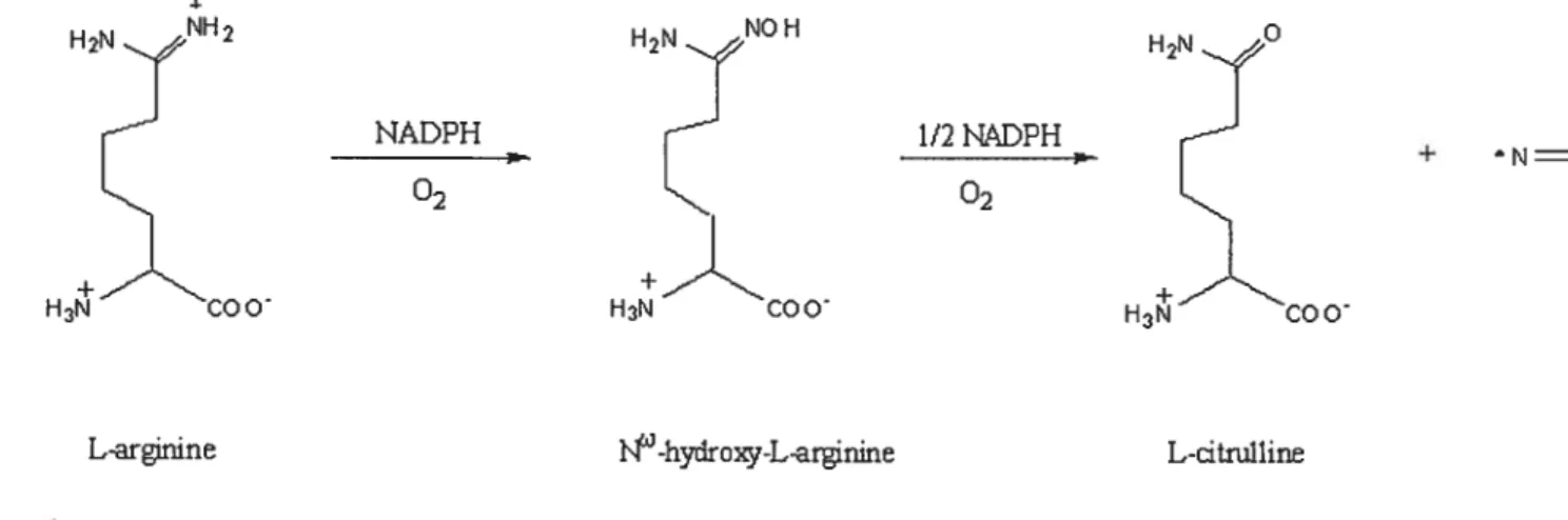
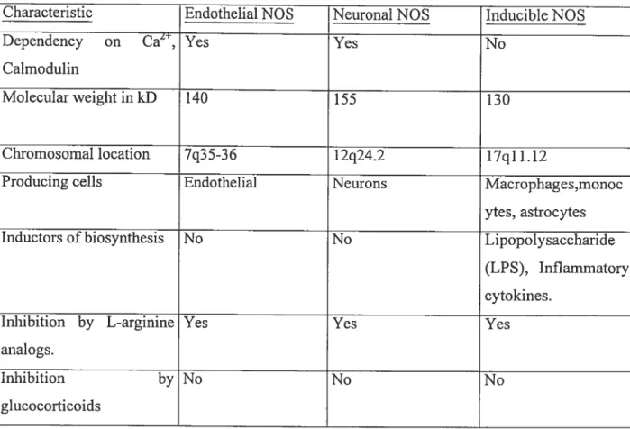
Nitric Oxide (NO) is a small, reactive molecule that is an important bioregulator. NO is used for a number of cellular signaling functions including blood vessel dilation, neuronal signal transmission, cytotoxicity against pathogens and turnours, coordination of heart rhythm and the regulation of celi respiration activity (Murad, 1999). NO is produced by the heme-containing metalloenzyme, nitric oxide synthase (NOS) (f ig. 1.4). There are three NOS isoforms, of which two are thought to be constitutive (nNOS and eNOS) in nature and one which is inducible (iNO$) (Marletta, 1993). (Table 1.0)
Endothelial NOS (eNOS) is mostly membrane bound and formed only in endothelial celis (furchgott, 1999). Neuronal NOS (nNOS), was identified in the cytosol of central and peripheral fleurons. NO derived from its constitutive isoforms acts as a physiological regulator by relaxing vascular smooth muscle or by functioning as a neurotransmitter. These isoforms produce small amounts of NO for short periods of time in a Ca2/Calmodulin dependent manner upon stimulation. Endothelial NOS with the endothelial cell acting as a transducer, releases NO continously in varying arnounts to regulate blood-vessel tone and by the same means the blood flow and pressure. Large amounts of NO produced for a prolonged time can cause vasodilation and hypotension. On the other hand, insufficient NO prodution can be a cause of hypertension.
H2NJlH2 H2N° NADPH 1/2KDPH
H
+ N0 HÀCOO H3’COO 02 H3COOL-arfrmne -hydroxy-L-arginine L-dtnilline
42
Characteristic Endothelial NOS Neuronal NOS Inducible NOS
Dependency on Ca2, Yes Yes No
Calmodulin
Molecular weight in kD 140 155 130
Chromosornal location 7q35-36 12q24.2 17q1 1.12
Producing celis Endothelial Neurons Macrophages,monoc
ytes, astrocytes
Inductors of biosynthesis No No Lipopolysaccharide
(LPS), Inflammatory cytokines.
Inhibition by L-arginine Yes Yes Yes
analogs.
Inhibition by No No No
glucocorticoids
Table 1. Characteristics of human NOS isoforms
This suggests that NO is involved in the regulation of the vascular system (Ignarro, L., 1999). Within the CNS, NO is released in response to increases in intracellular Ca2 that follow stimulation of glutamate receptors. The other function of NO, on which we focus this study relates to its neurotoxic effects since its excessive release is known to lead to ceil death processes.
The third isoform of NOS is flot present in resting celis but instead, the ceils must be induced to express the enzyme, ergo the nomenclature inducible NOS (iNOS). Stimuli typically includes cytokines and lipopolysaccharide (LPS), and once expressed, the enzyme
generates copious arnounts of NO. A number of cytokines are involved in the production of iNOS. Among them are IfN-y, IL-1, IL-6, THF-Œ, GM-CSF (granulocyte-macrophage colony stimulatory factor) and PAF (platelet activating factor) exert the stimulatory effect whereas the suppression has been observed in the cases of IL-4, IL-8, IL-10, TGF-t3 (transforming growth factor), PDGF (platelet-derived growth factor) and MDF (macrophage deactivating factor).
NO may react with superoxide to form the highly toxic peroxynitrite anion:
NO + O2 ONOOE
Which in turn may be transformed in an acidic environment to peroxynitrite and subsequently to the hydroxyl radical:
ONOO+H ONOOH
ONOOH . OH+NO2 NO+ H
Independent pathways are involved in the synthesis of reactive oxygen intermediates (ROI) and reactive nitrogen intermediates (RNI).
The basis of the functional activity of NO is its dual action on some enzymes of target cells. The small amount of NO released by the constitutive isoforms is sufficient for the activation of the NO-sensitive enzymes (guanylate cyclase and ADP-ribosyl transferase) that participate in NO signaling pathways. The larger amonts of NO generated by iNOS can also activate the NO-sensitive enzymes, but in several celi types the increased production of NO also exceeds the necessary concentration treshold to inhibit the action of
44
certain fe+-containing enzymes, namely aconitase, NADPH-ubiquinone oxidoreductase, succinate-ubiquinone oxidoreductase, ribonucleotide reductase, NADPH oxidase and glyceraldehyde-3 -phosphate dehydrogenase.
Activation of soluble guanylate cyclase by NO leads to the synthesis of cGMP, which in tum, causes the relaxation of vascular smooth muscle ceils, inhibition of platelet adherence and aggregation, inhibition of neutrophil chemotaxis, and signal transduction in the central and peripheral nervous systems. NO causes autoribosylation of glyceraldehyde 3-phosphate dehydrogenase, which inactivates this glycolytic enzyme. NO also inhibits three mitochondrial enzymes: aconitase of the TCA cycle and NADPH ubiquinone oxidoreductase and succinate —ubiquinone oxidoreductase ofthe electron transport chain.
1.8.1.2 Nitric oxide effects in celi metabolism
NO is known to inhibit cytochrome C oxidase by binding to its heme group and leading to an upregulated generation of superoxide in the mitochondrial respiratory chain (Ghafourifar and Richter, 1997). In turn superoxide reacts with NO to yield peroxynitrite (Ghafourifar et al., 1999). NO or peroxynitrite block respiratory chain complexes I, III, and TV as well as the activity of cis-aconitase, an enzyme in the TCA cycle, by binding to the iron-sulfur centers. This inhibition leads to the truncation of the metabolism of acetyl coenzyme A to carbon dioxide, which is a crucial step in production of nicotinamide adenine dinucleotide (NADH), which is necessary to fuel oxidative phosphorylation. These effects severely impair the cell’s ability to conserve and manufacture an adequate reservoir ofATP.
On the other hand peroxynitrite decomposes into the hydoxyl radical (0H) which is the most reactive species among those known to cause extensive ceT! and tissue damage in inflammation. It is widely known to cause lipid peroxydation, DNA mutations and protein modifications, and has apoptotic and cytotoxic effects on various ceils. The other product
of the decomposition of peroxynitrite is the NO2 radical that causes nitration of the tyrosine residues ofproteins.
NO is capable of inducing apoptosis depending on its concentation, flux and the ceil type on which it is enacting. It activates the transduction pathways that lead to apoptosis. Pro-apoptotic effects are ofien observed when NO reacts with superoxide to yield the highly toxic peroxynitrite. Contrary to this, it also can protect ceils against spontaneous or induced apoptosis. NO inactivates caspases through oxidation and S-nitrosylation of the active cystein, stimulation of cGMP-dependent protein kinase, modulation of Bcl-2/Bax farnily, induction of heat shock protein Hsp 70 and interaction with the ceramicle pathway. The reduction-oxidation state of the ceils appears to be determinant of the ultimate effects of NO on celi proliferation and survival.
1.8.1.3 Vascular f actors in $elective Neuronal Loss in PTD
A recent study by Calingasan et al. (2000) examines the importance and influence of oxidative stress and vascular changes that are seen in PTD. They approached this by testing the role of ICAM-1 which is an inducible ecu-surface glycoprotein expressed in endothelial celis, leukocytes, macrophages and dendritic celis and that of eNOS which is the subject of the present study and is known to be influenced by inflammatory processes and therefore may lead to BBB degradation.
It has commonly been suggested that iNOS might be the cuiprit of ceil death and damage in this particular neuropathology but it has been demonstrated in a prior study (Calingasan et al., 1998) that it is not necessary to induce the pathology cascade in PTD since deletion of the iNOS gene did flot mitigate the subsequent and characteristic neurodegeneration or the induction of heme oxygenase 1 (HO-1), an indicator of oxidative
46
stress. Induction of HO- 1, has been correlated to neuronal loss in the thalarnic region in PTD (Calingasan et al., 1998).
In this study, transgenic mice missing the genes encoding ICAM-1 aiid eNOS were used, to demonstrate that both are critical factors in the selective neuronal loss ofPTD.
The resuits showed that although eNOS is an effector of basal vasodilation, excessive endothelium-derived NO is Ïikely to damage neurons.
The resuits obtained in the studies perforrned in the transgenic eNOS-null mice, suggest that the beneficial effects of vasodilation are less significant than the toxic effects it
lias on PTD. Indeed, the targeted disruption of the eNO$ gene, blunted the deleterious
effects on neurons typical of PTD. This is confirmatory evidence for the hypothesis that states that NO produced by endothelial cells contributes to TD—induced neurodegeneration.
4$
2.1 OBJECTIVES 0F THE STUDY
There is a growing body of evidence that implicates excessive or inappropriate production of nitric oxide (NO) in disturbances of the the electron transport chain and also
in the disruption ofthe BBB as was mentioned in the introduction.
This smdy seeked to explore temporal gene and protein alterations of the different NOS isoforms in the PTD model ofneurodegeneration.
mRNA and protein expression of eNOS was determined by RT-PCR and Western Blot respectively in the medial thalamus, inferior colliculus and frontal cortex of PTD rats
at the presymptomatic and acute symptomatic stages with respect to pair-fed controls treated following the protocol outlined in the introduction. Appropriately designed oligonucleotides were used in the RT-PCR experiments and monoclonal antibodies specific for each NOS isoform were used for the Western Blot protein expression studies. A confirmatory immunohistochernical study was also carried out, using paraffin embedded sections of PTD rat brains at the progressing stages of the pathology, using the same monoclonal antibodies.
Earlier studies in TD have reported increased immunorectivity to NOS in different ceil types (Calingasan et al., 199$ and 1999), but this is the first study to report consistent alterations in gene expression in one ofthe NOS isoforms in TD.
The resuits obtained demonstrated a rise in eNOS rnRNA and an increase in eNOS protein expression in the PTD model. This augmentation of eNOS is concordant with the resuits reported in prior immunohistochemical studies (Calingasan et ai, 1998) that were confirrned by immunohistochemistry also in the present study and moreover, provide further information with respect to the changes in eNO$ expression in the PTD mode!.
2.2 Incrcased brain endothelial nitric oxide synthase gene expression in thiamine deficiency: relationship to selective vulnerability.
50
Increased brain endothelial nitric oxide synthase gene expression
in thiamine deficiency: relationship to selective vulnerability.
Neurochemistry International
Milarca Kruse ,Danen Navarro, Paul Desjardins and Roger f. Bufferworth*
Neuroscience Research Unit, CHUM (Campus Saint-Lue), University ofMontreal, Montreal, Quebec, Canada.
Running Titie: eNOS in thiamine deficiency
*Address for reprints and correspondence: Roger. F. Butterworth, Ph.D, D.Sc. Neuroscience Research Unit CHUM (Campus Saint-Luc) University of Montreal 105$, Saint-Denis Street Montreal, Quebec, H2X 3J4 Canada. Phone: (514) 890-$310 ext. 35759 FAX: (514) 412-7314 roger.butterworthumontrea1.ca
Keywords: Thiamine deficiency Nitric oxide
Endothelial nitric oxide synthase Medial Thalamus
Oxidative stress
52
Abstract
Thiamine deficiency resuits in selective neuronal cdl death in thalamic structures. Previous studies provide evidence for a role implicating nitric oxide (NO) in the pathogenesis of ccli death due to thiamine deficiency. In order to ascertain the origin of increased NO in the thiamine deficient brain, expression of endothelial nitric oxide synthase isoform, (eNOS), was meastired in the medial thalamus and in the inferior colliculus and compared to the frontal cortex (a spared region) of rats in which thiamine deficiency was induced through a feeding protocol of thiamine-deficient diet concomitant with daily administration of pyrithiamine, a central thiarnine antagonist. eNOS mRNA and protein expression were significantly increased as a function of the severity of neurological impairment and the degree of neuronal cdl loss in the medial thalamus and in the inferior colliculus. These findings suggest that the vascular endothelium is a major site of NO production in the brain in thiamine deficiency and that eNOS-derived NO could account for the selective damage to the thalamic structures as welI as for those seen in the inferior colliculus that are observed in this particular disorder.