Copyright ©1990, American Society for Microbiology
Characterization
of
an
Enterococcus hirae
Penicillin-Binding
Protein
3
with Low Penicillin Affinity
GRAZIELLA
PIRAS,'
ABOUBAKER ELKHARROUBI,l
JOZEF VAN BEEUMEN,2 ETIENNECOEME,'
JACQUES COYETTE,1*
AND JEAN-MARIEGHUYSEN'
Service deMicrobiologie, Universite deLiege, Institut de Chimie, B6, B4000 Sart Tilman (Liege
1),'
and Laboratoriumvoor
Mikrobiologie
en MicrobieleGenetica,
Rijksuniversiteit-Gent, Ledeganckstraat,
35,
B-9000Gent,2
Belgium
Received 23 May 1990/Accepted27 September 1990
Enterococcus hirae S185, a clinical isolate from swine intestine, exhibits a relatively high resistance to penicillin and contains two 77-kDa penicillin-bindingproteins 3 of high (PBP3s)and low (PBP 3r)affinity to
penicillin, respectively. A laboratorymutant S185' hasbeenobtained which overproducesPBP3randhas a
highly increased resistance to penicillin. Peptidefragments specifically produced by trypsin and SV8 protease digestions of PBP 3r were isolated, and the amino acid sequences of their amino terminal regions were
determined. On the basis of these sequences, oligonucleotides were synthesized andusedasprimerstogenerate, by polymerization chain reaction, a233-bp DNAfragmentthe sequence of whichtranslatedintoa 73-amino-acid peptide segment of PBP 3'. These structural data led tothe conclusion that theE.hirae PBP3' and the methicillin-resistant staphylococcal PBP 2' are members of the same class of
high-M,
PBPs. As shown byimmunological tests, PBP3' isnotrelated to PBP38 but,in contrast, is relatedtothe71-kDa PBP5oflow
penicillin
affinity
which isresponsible for penicillin resistance inE.hirae ATCC 9790 andR40.The emergence among important bacterial pathogens of
high-M,
penicillin-binding
proteins (PBPs) having low af-finity for the drug is a serious threat for the future ofchemotherapy (19, 24). Resistance may arise bythe
remod-eling of some targetted PBPs into altered forms which exhibit low intrinsic sensitivity, by de novo synthesis ofa
PBP of lowaffinity, orby overproduction ofa preexisting highly resistantPBP(9, 22, 23).
The relatively low
sensitivity
of Enterococcus hiraeATCC 9790 topenicillin has been attributed to the occur-rence,insmall amounts,ofa
high-Mr
PBPoflowaffinity,the71-kDa PBP 5. Laboratory mutants such as E. hirae R40
have been obtained which overproduce PBP S and, as a
corollary, arehighly penicillin resistant (9).
In contrast to E. hirae R40, E. hirae S185, another
penicillin-resistant
strain isolated from swineintestine,
con-tained a small amount ofPBP 5 but alarge amount ofthe
77-kDaPBP 3. Given that PBP 3 in strains ATCC 9790and R40is very sensitive to
penicillin,
experiments wereunder-takentounravel theunderlying mechanism ofthisnew type
of penicillin resistance.
(Thiswork wasconducted by G. Pirasinpartial fulfillment oftherequirements forthe Ph.D.degree fromtheUniversity of Liege, Liege, Belgium, 1990.)
MATERIALS ANDMETHODS
Bacterial strains and MIC determination. E. hirae S185 was agift from L. Devriese, University of Ghent, Ghent,
Belgium. E. hirae R40 (9), NT1/20 (2), and
Revl4
(10) weregifts from R. Fontana and P. Canepari, University of
Ve-rona, VeVe-rona, Italy. MAX Efficiency Escherichia coli
DH5aF'IQ
competent strain was from Bethesda ResearchLaboratories,
Inc. (Gaithersburg, Md.). E. coli HB101 wasalso used.
MIC values were determined in liquid medium as
de-scribed previously(4).
*Correspondingauthor.
Membranes. E. hirae cells grown unshaken at37° C in 500 ml of SB medium
(5)
and collected atthe lateexponential
phase(A550
=6.0)weresuspended in 100 ml of 5 mM sodiumphosphate (pH 7.0) containing1 mMMgCl2andlysed witha
mixture of
lysozyme (10
mg), DNase(200 ,ug), RNase(100
,ug),
and muramidase(1
mg) as described previously(8).
Membraneswerepurified by several washings and
centrifu-gations. They
were stored in the frozenstate(10mg of totalproteins
ml-')
in 40 mMsodium phosphate (pH 7.0)contain-ing 5% (vol/vol) glycerol. The proteins were measured
by
usingtheLowry methodas modified by Coyetteetal.(3).
Labelingwith
benzyl['4C]penicillin,
SDS-PAGE, andfluo-rography. Samples were labeled with
benzyl[14C]penicillin
(54
Cimol-';
Amersham International,Buckinghamshire,
United Kingdom) and subjected to sodium dodecyl
sulfate-polyacrylamide gelelectrophoresis (SDS-PAGE), and
fluo-rographyofthegelswasperformed asdescribed
previously
(8).
The PBPswere estimatedby densitometry of thefluo-rogramsby using a model 620 densitometer (Bio-Rad) and
Streptomyces R61 PBPas the standard (11). The values of the second-order rate constant of protein acylation and the antibiotic concentrations and incubation times necessary to
achieve a certain extent of saturation of the PBPs were
calculated asdescribed previously (12, 15).
Amino acid sequencing. Depending on the molecular mass, the peptides were subjected to SDS-PAGE (8.5 or 15%
acrylamide)and electroblottedonpoly(vinylidenedifluoride)
Immobilon membrane filters (Millipore Corp.) by using a
Bio-Rad Mini Trans-Blot cell (17). Automated microse-quenceswereperformedon a477-A pulsed liquid sequenator with on-line analysis of the amino acidphenylthiohydantoin
derivatives by usinga 120-A analyser (Applied
Biosystems,
Foster City, Calif.).
Amplification by PCR, cloning, andnucleotide sequencing. The DNA recombinant techniques used were described previously (20). The E. hirae S185' DNA was prepared as
described previously (16), which includes treatments with
lysozyme in the presence of sucrose, SDS, and protease K. The two nucleotide primers (see Results and Fig. 8) were 6856
synthesized by Eurogentec, Liege, Belgium. Polymerase
chain reaction (PCR) amplificationwasperformed on 100-1.l
samples containing the E. hirae S185' DNA (2
[ug),
theprimers (1 pLM each), deoxynucleoside triphosphates (200
piM),
the TaqDNA polymerase (2.5 U; Perkin Elmer-Cetus, Norwalk, Conn.), and0.2% (wt/vol)gelatin. The buffer was 10 mM Tris hydrochloride (pH 8.4) containing 50mM KCland 2 mM MgCl2. Samples were coveredwith paraffin and
submitted to 30 amplification cycles in a programmable heater as follows: 1 min ofdenaturation at
94TC,
1.5 min ofannealing at 550C, and 1.5 min ofpolymerization at 720C. The 233-bp DNA product was treated as follows: (i)
sub-jected to PAGE (7% acrylamide) in TBE buffer (1 mM
EDTA-40mMTris-boratebuffer [pH 8]) by using a Bio-Rad
Mini-Protean apparatus; (ii) eluted from the gel by shaking the relevant strip for 15 h at 37° C in 1 ml of 100 mM Tris
hydrochloride (pH 8.0)containing 500mMNaCl and 5 mM
EDTA; (iii) filtered on a
0.22-t>m-pore-size
membrane filter(Millipore);
(iv) precipitated with ethanol; (v) digested with BamHIand EcoRI restriction enzymes (BethesdaResearchLaboratories); and (vi) cloned in M13tg130 and M13tgl31
(Amersham
International, Buckinghamshire, UnitedKing-dom). MAX Efficiency E. coli DH5aF'IQ competent cells servedfor transformation experiments. Nucleotide
sequenc-ing
wasperformedwith theSequenase kit (U.S. BiochemicalCorp.,
Cleveland, Ohio) by using the dideoxynucleotide chain termination method (21).Antibodies. Two antisera were used. One of them was
raised against the largest water-soluble tryptic fragments
derived from PBP5 of strain R40 (7). The other was raised
against a large tryptic fragment prepared from PBP 3' (the
64-kDa t-PBP 3r; see Results). Adult rabbits were injected
three times at 15-day intervals with 100 ,ug of the peptide
emulsified in complete Freund adjuvant. Anti-PBP 3r
anti-bodieswerepurified by immunoadsorptionon E.coliHB101
and E. hirae Revl4 cell lysates (13). Immunodetection was
performed
with theantiserumat afinal5,000-fold dilution onproteins
or peptides transferred from polyacrylamide slabgels
onto0.45-,um-pore-size
HA type nitrocellulose sheets(Millipore).
Transfer was made by using a Bio-RadTrans-BlotSD cell and 20
mM
Tris-192mMglycine buffer (pH 8.3)containing
20%(vol/vol)
methanol. The antibody-antigencomplexes
weredetectedby usingan alkalinephosphatase-coupled
anti-rabbit goat antiserum (Bio-Rad instructionmanual, Immun-blot assay kit, catalog no. 170-6509 and
170-6511).
RESULTS
PBPprofilesinE.hirae ATCC9790, R40, S185, andS185r: occurrence in strains S185 and S185' of two 77-kDa PBP3S and PBP3rofhighand lowpenicillin sensitivity, respectively. E. hirae S185rwas derived from strain S185 by four serial
cultureson agar
plates
containing 32, 64, 128, and256jig
ofbenzylpenicillin
ml-'.
E. hiraeATCC9790, R40, S185, andS185r contain the six
species-specific
membrane-bound PBPs, namely,PBP 1(119 kDa), PBP2 (84kDa), PBP 3 (77kDa), PBP 4(75 kDa), PBP 5(71 kDa), and PBP 6 (43 kDa)
(Fig.
1, lanes1 through 4). The 69-kDaPBP4* is a sponta-neousbreakdown productof PBP 4 formed duringprepara-tion and storage ofthe membranes (4).
Although
theyhave similar PBPprofiles,
thefour entero-coccalstrains differfromeach otherbythe amounts of PBPs 3 and 5 thatthey contain. In comparisonwith strain ATCC9790
(Fig. 1, lane 1),
membranes of strain R40 containedrelatively
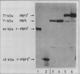
less PBP 3 and more PBP5 (lane 2). Incontrast,FIG. 1. SDS-PAGE and fluorography of
benzyl["4C]penicillin-labeled membranes ofE. hirae strains and water-soluble tryptic peptide fragments 64-kDa t-PBP 3r, 42-kDa t-PBP 3r, and 58-kDa
t-PBP 3S. Lanes: 1 through 4, membranes (200 pug of protein) ofE. hiraeATCC 9790, R40, S185, andS185r,respectively;5,membrane (200 pLg) of E. hirae S185r in which PBP3r wasselectivelylabeled
with100 puMbenzyl[14C]penicillinfor 60min;6and 7, supernatant fractions of the S185r membranes (300 jug) after treatment with trypsinatpH 7.0 (lane 6)oratpH 7.8 and in thepresenceof10mM
MgCl2 (lane 7); 8, membranes (200,ug)ofE.hirae R40in whichPBP
5was selectivelylabeled with 100,M benzyl[14C]penicillin for 60
min;9 and 10, membranes (150,ug)ofE.hiraeNT1/20assuch(lane
9)orinwhich PBP 3swasselectively protected against labelingwith
100 ,uM benzyl[14C]penicillin for 60 min (lane 10); 11 and 12, supernatant fractions ofthe membranes shown in lanes9and 10, respectively, aftertreatmentwith trypsin. Theacrylamide
concen-tration used for the SDS-PAGEwas7.2%(wt/vol).
membranes of strain S185 contained more PBP 3 and less
PBP 5 (lane 3). In membranes of strain S185 these
differ-ences were morepronounced.PBP 3andPBP5represented
23 and 12%, respectively, ofthe total PBPsinmembranesof strain ATCC 9790, 10 and 47% in strain R40,41 and 6% in strain S185, and 76 and 2% in strain S185r.
The four enterococcal strains also have widely different benzylpenicillin MIC values: 1 ,ug ml-' for strain ATCC 9790, 16 ,ug ml-' for strain S185, 80 jig
ml-'
forstrain R40,and 175 ,ug ml-' for strainS185r. Figure2showstheeffectof increasing concentrations of benzylpenicillin on bacterial
growth in SBa broth (5).
Penicillin resistance of strain R40 was proposed to be attributabletothe presence ofahigh level of PBP 5 which
hasalowaffinity for penicillin (9). Given that PBP 3 in strain
ATCC 9790 (and R40) isverysusceptibletoderivatization by benzylpenicillin (4, 9), the high level of penicillin resistance
of strains S185 and S185r (which contain large amounts of
PBP 3andsmallamountsofPBP5)wasunexpected. Further
study revealed that saturation of PBP 3 by benzylpenicillin
wasmonophasic in strains ATCC 9790 andR40,biphasic in
strains S185 and S185 (Fig. 3), and strongly suggestedthe
concomitantpresenceofahighlypenicillin-sensitive77-kDa
PBP 3S and a much-less-penicillin-sensitive 77-kDa PBP 3.
Asderived from benzylpenicillin concentrations requiredto achieve50%saturation after 5minofincubationat37° C,the
values of the second-orderrateconstantofprotein acylation
were 5,000 M-1 s-1forPBP 3s (as observedwith PBP 3 of
PBFS kOa 1 120- ** 2
84
-. 3?77
4 75 =_ 4* 69- 64kOa
6 43 49 1 2 3 4 5 6 7 8 9 1011 12c Co vn Dl w 0 90 180 270 0 90 180 270(min)
FIG. 2. Effects of increasing concentrations of benzylpenicillin
ongrowth in SBa broth of E. hirae ATCC 9790, R40, S185, and
S185r.Absorbancesarecorrectedtoagreewith Beer Lambert's law.
Benzylpenicillin,attheindicatedfinal concentrations(1 ,uM=0.356
,ugml-';5 p.M=1.78 ,ugml-';25 pM=8.9 pugml-')wasaddedat
timezero.
strainATCC9790) and 20 M1 s-' for PBP3r(asobserved with PBP 5 of strain R40).
To discriminate PBP 3' from PBP3S, membranes of strain
S185' (and strain R40 used as control) were firstincubated with 15 ,uM nonradioactive benzylpenicillin for 10 min at
37° C. This antibiotic concentration was 10-foldhigher than
thatnecessary to saturatePBP 3S (as wellas PBPs 1 and 2)
and20-fold lower than thatnecessarytosaturate PBP3ror
PBP5. Ina secondstep, the PBPs left in afreeformin the
membranes were labeled by reaction with 100 p.M benzyl['4C]penicillin for 60 min at370C, thus causing
com-plete derivatization of both PBP 3Y and PBP 5. Asaresult of
this competition experiment involving nonradioactive and radioactive penicillin, the only labeled PBP seen in vast amounts was the PBP 3r in the membranes of strain S185r (Fig. 1, lane 5) and the PBP 5 in the membranes of strainR40
(Fig. 1, lane 8). The PBP contents of E. hirae ATCC 9790,
R40, S185, and S185r wereestimated on the basis of these
andother data (Table 1). E. hirae ATCC 9790 and R40 lack PBP3r. PBP 3S and PBP 3Yoccur in approximately
equiva-lentamounts(0.3% of total membrane protein) in strain S185 and in a ratio of approximately 1 to 4 (0.4% and 1.6%,
respectively) in strain S185r.
Trypsin digestion of the 77-kfla PBP3r:the64-kDa t-PBP 3'
1000
[Benzylpenicitlinl jiM
FIG. 3. Saturationbybenzyl['4C]penicillin of77-kDaPBPs 3 of membranes ofE. hiraeATCC 9790 (A), S185 (B), and S185r (C). Membranes (100pgin10p.l)wereincubated for5minat370C in the presenceofincreasing concentrations ofbenzyl['4C]penicillin. Mi-crodensitymeasurementsof the 77-kDa PBPs 3 weremadeonthe fluorograms.
and 42-kDa t-PBP 3'. Isolation of the membrane-bound 77-kDaPBP 3F was not attempted. Instead, membranes of strain S185' in which PBP 3r was selectively radioactively labeled as described above (Fig.1,lane5)weredigestedwith trypsin(type XI; Sigma Chemical Co.) under the following conditions: 300 pLg of total membrane proteinplus 12 ,ug of
trypsinin 30 p.1of 25 mM sodiumphosphate (pH 7.0);30min at370C. Thistrypsintreatmentresulted in the releasetothe
100,000 x g supernatant ofthree water-soluble radioactive peptides of 64, 42, and 29 kDa(Fig. 1, lane6). The relative amountsof each of thesepeptidefragmentscould be modi-fied to some extentby adjustingtheconditions of proteolytic treatment.Inparticular,theaddition of 10 mMMgCl2and an elevated pH of 7.8 favored the production of the 42- and
29-kDa fragments (Fig. 1, lane 7). (Note that the 29-kDa fragment is not shown in Fig. 1.)
Membranes of strainR40 inwhich PBP 5 wasspecifically radioactivelylabeled(Fig. 1, lane8)werealso submittedto trypsin digestion. As shown in detail elsewhere (7), the
tryptic digest profileof PBP 5frommembranesof strain R40 differed from that of PBP3'frommembranesof strain S185.
Trypsindigestion of the77-kDa PBP 3s: the 58-kDa t-PBP 3S
peptide fragment. Identification of the
trypsin
degradation product(s) of the 77-kDa PBP 3s rested upon the use of E. hirae NT1/20. Strain NT1/20 has a PBP profile similar to thatTABLE 1. PBPs as percentage of totalproteins of the membranes ofE.hirae strains
% of totalproteinsin strain: PBP ATCC9790 R40 S185 S185r 1 0.34 0.18 0.25 0.11 2 0.22 0.17 0.14 0.09 3s 0.4 0.2 0.32 0.4 3 0.0 0.0 0.32 1.6 4 0.31 0.26 0.19 0.25 5 0.22 0.94 0.09 0.05 6 0.26 0.24 0.26 0.14 0.1
-L
.Sojjm.
P.64-kDa t-PBP3r--_ 58 -kOat-P8P3S- _
42-kOat-PBP3r.
__qw.
2 3 M
FIG. 4. SDS-PAGEand Coomassie blue staining of t-PBP3Yand t-PBP 3S peptide fragments. Lanes: 1, 64-kDa t-PBP 3' (3 pug of protein);2, 58-kDa t-PBP3S(-8.5
jig);
3, 42-kDa t-PBP 3'(7.5jig);
M, protein markers (66.3-kDa bovine serum albumin; 42.7-kDa ovalbumin; 38-kDaStreptomyces R61 PBP; 29-kDa carbonic anhy-drase).of strainATCC 9790 and thus lacks PBP 3', but it also lacks PBP 2(Fig. 1, lane 9) (2). Asacorollary, the PBP which has
the highest affinity for cefotaxime is PBP 3s (second-order rateconstant of protein acylation, 4,400 M- so')(6).
Membranes (300 ,ug of total proteins in 15 AL of 40 mM
sodiumphosphate [pH 7.0] containing 1 mM MgCl2 and 5% [vol/vol] glycerol) as suchorpretreated with 5
RxM
cefotax-ime for 10 min at 370C were labeled with 100 jiM benzyl[14C]penicillin for 60 min at 370C and then digested
with 3 jigof trypsin for10 minat370C. Notethat the5 jiM
cefotaximeconcentrationwasfivefold higher than that
nec-essaryto saturatePBP3S almost completely (Fig. 1, lane 10).
The membranes that were not pretreated with cefotaxime yielded three major radioactively labeled peptides of69,58,
and 44kDa, respectively (Fig. 1, lane 11). Thepretreatment
with cefotaxime selectively prevented the 58-kDa peptide
from reacting with radioactive penicillin (Fig. 1, lane 12). Thus, thispeptide wasconsideredtooriginate from PBP 3s
and therefore was called 58-kDa t-PBP 3S. The 69- and
44-kDapeptideswereconsideredtobethe t-PBP4*andone
of the t-PBP 5fragmentsstudiedpreviously (7), respectively. Properties and purification ofthe64-kDa t-PBP 3r, 42-kDa
t-PBP 3r, and 58-kDa t-PBP 3S. As derived from saturation curves,the 64-kDa t-PBP3rand the 42-kDa t-PBP3r peptide fragments reacted withbenzylpenicillin witha low
second-order rate constant (10 M-1 so1) very similar to that ob-served with the membrane-bound PBP 3r (-20 M-1
s'1),
andthe 58-kDa t-PBP3s peptide fragmentreacted withahigh
second-order rate constant (5,000 M-1 so1) very similarto that observed with the membrane-bound PBP 3S.
Accord-ingly, ateach stepofthe purification procedure, thetryptic fragments were identified on the basis of their molecular massand affinity for benzyl[14C]penicillin.
Themembranes fromstrain S185 or S185rand the
condi-tions oftrypsin digestion varieddependingontheparticular
tryptic fragment tobe isolated in sufficient quantity.
What-ever the case, the peptide fragment(s) of interest could be isolated by using the two-step procedure described below
withatrypsin digest of membranes ofstrainS185rcontaining
1.5gof totalproteins (60mgoftrypsinand 200mlof25mM
sodiumphosphate [pH 7]; 30minat370C).
In step1,the supernatantwasfiltered througha
0.22-jim-pore-sizemembrane filter(Millipore), supplementedwith 25 ml of 1 M Tris hydrochloride (pH 7), and loaded onto a
FIG. 5. Reaction of anti-64-kDa t-PBP 3rantibodies with 64-kDa t-PBP3' (lane 1), 42-kDa t-PBP3'(lane 2), PBP 5 of membranes of E. hirae ATCC 9790 and R40 (lanes 3 and 4), and PBP 3' of membranes of E. hirae S185 and S185' (lanes 5 and 6). Theamounts ofproteins used were 2 jig (lanes 1 and 2) and 100 jig (lanes 3
through 6). The same pattern was obtained with the anti-t-PBP 5
antiserum.
Pharmacia Q-Sepharose fast-flow column (2.6 by40cm) (gel volume, 58 ml; total capacity, 1 g). Agradient of NaCl(Oto 1M)in 25 mM Trishydrochloride-25 mM sodiumphosphate (pH 7) eluted the 64-, 42-, and 58-kDapeptide fragments at 0.17to0.22 MNaCl.
FIG. 6. Degradation of 64-kDa t-PBP 3' into subfragments 3' Sal, 3' Sa2, 3' Sa3, and3' Sa4by treatment with S. aureus SV8 protease. ThefigurewasobtainedbySDS-PAGE(15% acrylamide-2 Murea)and Coomassie bluestainingof the electroblotmadeonan
Immobilon membrane filter(Millipore).The 64-kDat-PBP3rreacted withbenzyl[14C]penicillin. Subfragments 3' Sal,3rSa2,and3rSa4, butnot3rSa3,wereradioactive(not shown).Proteinmarkersareas follows: 66.3-kDa bovineserumalbumin, 42.7-kDaovalbumin, 38-kDaStreptomyces R61PBP,29-kDacarbonicanhydrase, 21.5-kDa trypsin soybean inhibitor,and 17.2-, 14.6-,and 8.2-kDamyoglobin CNBrcleavage products. 77-kOa
PBp3r
_71-k~a
PBP5- UiIM464-kDa t-PBP3r
_42-kOa
t-PBP3 4 1 2j3
4 5 6 .IS.aureus PBP2' : 1) 58-kDa t-PBP3 : 2) 3rSa3 140 150 160 170 180 ....QKDQSIHIENLKSERGKILDRNNVELANTGTHMRLGIVPKNVSK. * A A *A** AA TGQLYKGSEVVKAKRGTIYDRNGVALAEDATSYVDKA... *A A**** AA AA AAAA ATRGNILDRNGEPLATTGKLKQLGVWPSKLG... 320 330 340 350 360 370 380 390 400 S.aureus PBP2': TLIEKKKKDGKDIQLTIDAKVQKSIYNNMKNDYGSGTAIHPQTGELLALVSTPSYDVYPFMYGMSNEEYNKLTEDKKEPLLNKFQIT... r AA* AA*A A*A*****
A*A 3) 42-kDa t-PBP3 : VLIECEVQNGKDIKLTIDAKAQKTAFDSLGGKAGSTVAT... 4) 3rSa4 5) PBP3r-PCR A A (XXXXXXX)NPEQPFIARFATG...
AAAA AAAAA* AA ** A A AA AA** A ***A A A A A A At
QNGKDIKLTIDAKAQKTAFDSLGGKAGSTVATrPKTGDLLALASSPSYDPNKMTNGISQEDYKAYEENPEQPF
FIG. 7. Amino acidalignmentof thepeptidefragmentsof E.hirae PBP 3S(sequence 1)and PBP3Y(sequences2through 5)with the peptide segmentsQ137-K180andT314-T400of the methicillin-resistantPBP 2'ofS.aureus. Forunderlined sequences, seeFig.8.
In step 2, the 0.17 to 0.22 M NaCi fractions were pooled andconcentratedto 5mlby filtrationon a YM10 membrane
filter (Amicon Corp.). Theresulting solutionwasbrought to 1.7 M(NH4)2SO4 in50 mM sodiumphosphate (pH 7), and
samples containing at most 10 mg of total protein were
filtered on a 1-mlphenyl-Superose HR5/5 column (Pharma-cia). Upon treatment with a decreasing gradient of
(NH4)2SO4 concentration in the same buffer, the 58-kDa t-PBP 35 and the 42-kDa t-PBP3' fragmentseluted at about 0.75 M (NH4)2SO4 (overall yield,
'20%)
and the 64-kDa t-PBP 3r eluted at 0.22 M (NH4)2SO4 (overall yield, 60%).The64-kDa t-PBP 3r thusobtainedwas95 to100%pure(Fig.
4,lane1). Inturn,it wasestimated (Fig.4, lane 2) that about 50% of the total proteins wereaccounted for by the 58-kDa t-PBP 3S andthatthe 42-kDa t-PBP 3r was aminor compo-nent of this fraction. Improved yield (about 40%) in the 42-kDa t-PBP 3r required trypsin treatment of the
mem-branes atpH 7.8 in the presence of10 mM
MgCl2
(Fig. 4,lane3).
Specificity profile of the anti-64-kDa t-PBP
3F
and anti-t-PBP5antibodies. Theantibodies raised againstthepurified 64-kDa t-PBP 3r and those raised against purified t-PBP Sfragments ofE. hirae R40 reacted with the 64- and42-kDa
t-PBPs 3'(Fig.5,lanes1and 2), the PBP 3r ofmembranesof strains S185 (lane 5) and S185r (lane 6), the PBP 5 of membranes of strains ATCC 9790 (lane 3) and R40 (lane 4),
andthepurifiedt-PBP S fragments (not shown in Fig. 5). A very small amount of membrane-bound PBP 5 was found in strains S185 and S185' (lanes5 and 6). Treatmentwith the
antisera failed to abolish penicillin binding. Theantibodies
didnot reactwith the 77-kDa PBP3sin membranes of strains ATCC 9790 and R40 (lanes 3 and 4) nor with the 58-kDa t-PBP 3s (lane 2).
SV8 protease hydrolysis of the 64-kDa t-PBP3' tothe
3'
Sal, 3' Sa2, 3r Sa3, and 3' Sa4 peptide fragments. Edman
degradation ofthe purified 64-kDat-PBP 3r failed.
Conse-quently, the peptide, previously labeled by reaction with
benzyl[14C]penicillin,
was carboxymethylated and then di-gested with theStaphylococcusaureusSV8 protease [60pug of peptide, 1.2pLg of protease (Miles Scientific, Naperville,Ill.), and 250 ixl of 100 mM (NH4)2CO3 containing 1 mM
CaCl2; 8 h at37'C]. SDS-PAGE (15% acrylamide) followed by Coomassie blue stainingandfluorographyrevealedthree
radioactively labeled peptides of 19.7 kDa (3rSal), 17.5 kDa (3r Sa2), and 8.5 kDa (3r Sa4), and one nonradioactively
labeledpeptide of 9.6 kDa(3r Sa3) (Fig. 6).
Amino acid sequences. Samples containing the purified
42-kDa t-PBP 3r and 58-kDa t-PBP 3s and samplescontaining
the fourpeptide fragments (3r Sal, 3r Sa2,3rSa3,and 3r
Sa4)
were subjected to SDS-PAGE. After electroblotting, each
purified peptide (about 500 pmol) was subjected to
auto-Otigonucleotide n9 1 G
S G T A G A 3
GIGGATC
CCA -AA -6 G-AA -GAT-ATT-AABamHI A C TC A C 0. - N - G - K - 0 - I -(K) Oligonucleotide n2 2 G G 3 G A T A TT -GG -CTT-GT -GG -AAA-TA-CTTAAGG A T C T C C EcoRI N - P - E - - P - F -(I)
FIG. 8. Synthetic nucleotides used as primers for PCR
amplifi-cation ofa233-bp DNA fragment encoding the peptidePBP 3r-PCR showninFig. 7.Oligonucleotide 1encodes thesequenceQ8-K14of
the 42-kDa t-PBP 3'. Oligonucleotide 2 is complementary of the nucleotide sequence encoding thesequence N8-114 of3rSa4. The amino acidsequencesare underlined in Fig.7.
FIG. 9. PAGE analysis of the PCR products generated by using thetwooligonucleotides of Fig. 8asprimers and the DNA from E.
hirae R40 (lane 1) and E. hirae S185' (lane 2) astemplate. Lane 3
showsDNAsize markers (298, 220, and 200 bp of the 1-kb ladder) (BRL). 1 2 3 5' --
298
_. 220200
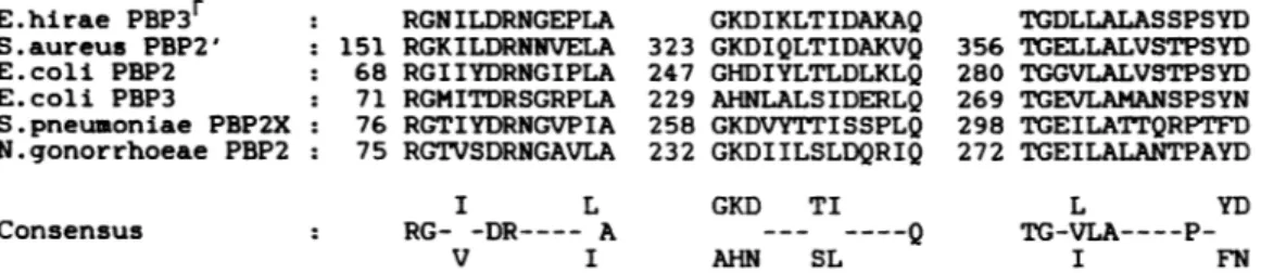
E.hirae PBP3r S.aureus PBP2' E.coli PBP2 E.coli PBP3 S.pneumoniae PBP2X : N.gonorrhoeae PBP2 : Consensus 151 68 71 76 75 RGNILDRNGEPLA RGKILDRNNVELA RGIIYDRNGIPLA RGMITDRSGRPLA RGTIYDRNGVPIA RGTVSDRNGAVLA 323 247 229 258 232 I L RG- -DR---- A V I GKDIKLTIDAKAQ GKDIQLTIDAKVQ GHDIYLTLDLKLQ AHNLALSIDERLQ GKDVYTTISSPLQ GKDIILSLDQRIQ GKD TI
-__
----Q AHN SL TGDLLALASSPSYD 356 TGELLALVSTPSYD 280 TGGVLALVSTPSYD 269 TGEVLAMANSPSYN 298 TGEILATrQRPTFD 272 TGEILALANTPAYD L YD TG-VLA----P-I FNFIG. 10. Homologous boxesoccurringin theamino-terminal regions of severalhigh-MrPBPs, i.e., S. aureus PBP 2' (22), E.coliPBP 2 (1), E. coliPBP3 (18), Streptococcus pneumoniae PBP 2X (14), and Neisseria gonorrhoeae (23).
mated microsequence analysis. Comparison of the data thus
obtained with PBP 2', of known primary structure, of methicillin-resistant S. aureus (22) led to the following observations (Fig.
7).--(i) The 37-amino-acid amino-terminal region of the 58-kDa t-PBP3S aligned with the peptide stretch Q137-4173 ofPBP 2', yielding 11 identities from residue 145 to residue 167.
(ii) The 31-amino-acid amino-terminal region of the 9.6-kDa 3r Sa3 peptide (originating from the 64-9.6-kDa t-PBP 3r)
alignedwith the peptide stretch S149-S179 of PBP 2',
yield-ing 15 identities. Note that the 58-kDa t-PBP 35 and the
9.6-kDa 3' Sa3 aligned within the same region of PBP 2'.
(iii) The 39-amino-acid amino-terminal region of the 42-kDa t-PBP 3raligned with the peptide stretch T314-1352 of PBP2', yielding 18 identities.
(iv) The amino-terminal region of the 8.5-kDa 3r Sa4
[(ct0)7NPEQPFIARFATG]
lacked similarity with anypep-tide segment of PBP 2', but it was necessarily located
downstream ofthat region of the 42-kDa t-PBP 3r which had
been sequenced. Accordingly, the two oligonucleotides shown in Fig. 8 were synthesized. Oligonucleotide 1 had a BamHI site at the 5' OH end and coded for the sequence
Q8-K14of the 42-kDa t-PBP 3r, andoligonucleotide2had an EcoRIsiteatthe 5' OH end and was complementary of the
nucleotide sequence coding for the sequence N8-114 of 3r Sa4. Amplification by the PCR technique with these two
oligonucleotides asprimers and the E. hirae S185r DNA as template generated a 233-bp DNA fragment (Fig. 9, lane 2), the sequenceofwhich translated into a 73-amino-acid
pep-tide. This peptide, called PBP 3rPCR, aligned with the
K321-L393 segmentof PBP 2', yielding 33 identities (Fig. 7). Notethat the233-bp DNAfragmentwasnotproduced when theE. hirae R40 DNAwas used as thecontrol(Fig. 9, lane
1).
(v) The19.7-kDa3rSal and 17.5-kDa 3r Sa2 (not shown in
Fig. 7)aroseby cleavageofthe E-Vbond, atpositions6 and 7of the 42-kDa t-PBP 3 .
DISCUSSION
From thework presentedhere, onecandraw thefollowing conclusions.
(i) Development of resistance to penicillin among
entero-coccicanbe the result of the emergence ofa novel 77-kDa PBP3rwhich ismuch less susceptibletopenicillin thanthe normal 77-kDa PBP 3S.
(ii) The 77-kDa PBP 3 , like the E. hirae PBP 5 of low
penicillin affinity, is another member of that class of
physi-ologicallyimportanthigh-MrPBPstowhich themethicillin-resistant staphylococcal PBP 2' belongs. Indeed, several
peptide fragments of PBP 3r align well with two
peptide
segments which in the staphylococcal PBP 2' extend from S149 to S179 (31 residues) and from T314 to T400 (87
residues), respectively.Alignmentof the 118 amino acids of PBP 3r and PBP 2' generates 52 strict identities (40%) and highlights acommon signature consisting ofthreeboxes of
veryhigh homology. The fact that these boxes areconserved
inthe amino-terminal domains ofother high-Mr PBPs (Fig.
10)strongly suggests that they are markers of structural and
functional significance.
(iii) The E. hirae 77-kDa PBP 3r and 71-kDa PBP 5 are
immunologically related and are acylated bybenzylpenicillin
with the same low second-order rate constant (-10 to 20
M`
s-1). PBP3r and PBP 5 are probably similarproteins,yetthey have different tryptic digest profiles. In addition,the oligonucleotide primers used in this work discriminate the PBP 3r and PBP5-encodinggenes. Consequently,the PBP
3F
gene is only present in E. hirae S185r, while the PBP 5 gene is present inbothE. hirae S185r and R40.(iv)The E. hirae PBP 3r (or the PBP5) isnot
immunolog-ically related to PBP 3S, suggesting that PBP 3r is not a protein mutantthatwould have emerged by limited
remod-eling of the penicillin-binding domain of PBP 3S. PBP 3S,
however, possesses at least the same marker (Fig. 10,
R151-A163)asthat foundin PBP
3F
and in thestaphylococcalPBP
2'.
ACKNOWLEDGMENTS
This work was supported in part by the Fonds National de la RechercheScientifique (toJ.C.),theFondsde laRecherche Scien-tifique Mddicale(contract3.4537.88),theBelgian Government (con-vention 86/91-90), the Fonds deRecherche de laFacultd de Med-ecine ULg, andatripartiteagreementbetween the WalloonRegion,
SmithKline Beecham, United Kingdom, and the University of Liege. G.P.was aFellow of theInstitutpourl'Encouragement dela Recherche Scientifique dans l'Industrie et l'Agriculture, Brussels. Theproteinsequencework inGentwassupportedby the National lncentive Program on Fundamental Research in Life Sciences initiated bythe Belgian Science Policy Programming Department andbytheFund forJoint Basic Research(contract2.0042.85).
LITERATURE CITED
1. Asoh,S.,H.Matsuzawa, F.Ishino, andJ.L.Strominger. 1986. NucleotidesequenceofthepbpAgene andcharacteristics ofthe deducedaminoacidsequenceofpenicillin-binding protein2of EscherichiacoliK12. Eur. J. Biochem. 160:231-238.
2. Canepari, P.,M.Del MarLleo,R.Fontana,and G. Satta. 1987. Streptococcusfaecium mutantsthat are temperaturesensitive for cellgrowth and show alterations inpenicillin-binding pro-teins. J. Bacteriol. 169:2432-2439.
3. Coyette,J., J.M.Ghuysen,andR.Fontana. 1978.Solubilization andisolationof the membrane-bound DD-carboxypeptidase of Streptococcusfaecalis ATCC 9790. Eur. J. Biochem. 88:297-305.
4. Coyette, J.,J.M.Ghuysen,andR.Fontana. 1980. The penicil-lin-bindingproteinsinStreptococcusfaecalisATCC9790. Eur. J. Biochem. 110:445-456.
J. M.Ghuysen. 1974. Membrane-bound DD-carboxypeptidase andLD-transpeptidase of Streptococcusfaecalis ATCC 9790. Eur. J.Biochem. 44:459-468.
6. Coyette, J., A. Somze, J. J. Briquet, J. M. Ghuysen, and R. Fontana. 1983. Function of penicillin-binding protein 3 in Strep-tococcusfaecium, p. 523-530. In R. Hakenbeck, J. V. Holtje, and H. Labischinski (ed.), The target of penicillin. W. de Gruyter and Co., Berlin.
7. ElKharroubi, A., P. Jacques, G. Piras, J. Coyette, and J. M. Ghuysen.1988. Characterizationof the trypsin-solubilized pen-icillin-binding proteins of Enterococcus hirae (Streptococcus faecium), p. 367-376. In P. Actor, L. Daneo-Moore, M. L. Higgins,M. R.J. Salton, and G.D.Shockman (ed.), Antibiotic inhibition of bacterial cell surface assembly and function. Amer-ican Society for Microbiology, Washington, D.C.
8. El Kharroubi, A., G. Piras, P. Jacques, I. Szabo, J. Van Beeumen, J. Coyette,andJ.M.Ghuysen. 1989. Active-site and membranetopology of theDD-peptidase/penicillin-binding pro-teinn06 of Enterococcus hirae (Streptococcus faecium) ATCC 9790. Biochem. J. 262:457-462.
9. Fontana, R.,R. Cerini,P. Longoni,A.Grossato, and P. Cane-pari. 1983. Identification ofa streptococcal penicillin-binding protein that reacts very slowly with penicillin. J. Bacteriol. 155:1343-1350.
10. Fontana, R., A. Grossato, L. Rossi, Y. R. Cheng, and G. Satta. 1985.Transition from resistance to hypersusceptibilityto
P-lac-tamantibiotics associated with loss of a low-affinity penicillin-binding protein in a Streptococcusfaecium mutant highly resis-tanttopenicillin. Antimicrob. Agents Chemother. 28:678-683. 11. Frere, J. M., J. M. Ghuysen, H. R. Perkins, and M. Nieto. 1973.
Kinetics of concomitant transfer and hydrolysis reactions cata-lysed by the exocellular DD-carboxypeptidase-transpeptidase of Streptomyces R61. Biochem.J. 135:159-165.
12. Ghuysen, J. M., J. M. Frere, M. Leyh-Bouille, M. Nguyen-Disteche, and J. Coyette. 1986. Active-site serine D-alanyl-D-alanine-cleaving peptidase-catalysed acyl-transfer reactions. Procedures for studying the penicillin-binding proteins of bac-terialplasma membranes. Biochem. J.235:159-165.
13. Helfman,D. M., and S. H. Hughes. 1987. Use of antibodies to
screencDNAexpression libraries prepared in plasmidvectors. Methods Enzymol. 152:451-457.
14. Laible, G., R.Hakenbeck, M. A. Sicard, B. Joris, andJ. M. Ghuysen.1989. Nucleotide sequences of the pbpX genes encod-ing thepenicillin-binding proteins 2X from Streptococcus pneu-moniae R6 and a cefotaxime resistant mutant, C506. Mol. Microbiol. 3:1337-1348.
15. Leyh-Bouille, M.,M.Nguyen-Disteche,S.Pirlot,A. Veithen, C. Bourguignon, and J. M. Ghuysen. 1986. Streptomyces K15 DD-peptidase-catalysed reaction with suicideP-lactamcarbonyl donors. Biochem. J. 235:177-182.
16. Loureiro DosSantos,A. L., and A. L. Chopin. 1987. Shotgun cloning in Streptococcus lactis. FEMS Microbiol. Lett. 42:209-212.
17. Matsudaira, P. 1987. Sequence from picomole quantities of proteins electroblotted onto polyvinylidene difluoride mem-branes. J. Biol. Chem.262:10035-10038.
18. Nakamura, M., I. N. Maruyama, M. Soma, J. I. Kato, H. Suzuki,and Y. Hirota.1983. On the process of cellular division inEscherichiacoli:nucleotide sequence of the gene for penicil-lin-binding protein3. Mol.Gen. Genet. 191:1-9.
19. Reynolds,P. E.1984.Resistance of theantibiotic target site.Br. Med. Bull. 40:3-10.
20. Sambrook, J.,E. F.Fritsch,and T. Maniatis. 1989. Molecular cloning:alaboratory manual. Cold Spring Harbor Laboratory, ColdSpring Harbor,N.Y.
21. Sanger, F.,S.Nicklen,and A. R.Coulson. 1977. DNA sequenc-ing with chain-terminating inhibitors. Proc. Natl. Acad. Sci. USA 74:5463-5467.
22. Song,M.D.,M.Wachi,M.Doi,F.Ishino,and M. Matsuhashi. 1987. Evolution of an inducible penicillin-target protein in methicillin-resistant Staphylococcus aureus by gene fusion. FEBSLett. 221:167-171.
23. Spratt, B. G. 1988. Hybridpenicillin-binding proteins in peni-cillin-resistant strains of Neisseria gonorrhoeae. Nature (Lon-don)332:173-176.
24. Spratt, B.G., and K. D. Cromie. 1988. Penicillin-binding pro-teins of Gram-negative bacteria. Rev. Infect. Dis. 10:699-711.
![FIG. 1. SDS-PAGE and fluorography of benzyl["4C]penicillin- benzyl["4C]penicillin-labeled membranes of E](https://thumb-eu.123doks.com/thumbv2/123doknet/6142264.156974/2.918.487.832.103.401/page-fluorography-benzyl-penicillin-benzyl-penicillin-labeled-membranes.webp)
![FIG. 3. Saturation by benzyl['4C]penicillin of 77-kDa PBPs 3 of membranes of E. hirae ATCC 9790 (A), S185 (B), and S185r (C).](https://thumb-eu.123doks.com/thumbv2/123doknet/6142264.156974/3.918.98.439.97.620/fig-saturation-benzyl-penicillin-pbps-membranes-hirae-atcc.webp)