HAL Id: inserm-02482159
https://www.hal.inserm.fr/inserm-02482159
Submitted on 17 Feb 2020
HAL is a multi-disciplinary open access
archive for the deposit and dissemination of
sci-entific research documents, whether they are
pub-lished or not. The documents may come from
teaching and research institutions in France or
abroad, or from public or private research centers.
L’archive ouverte pluridisciplinaire HAL, est
destinée au dépôt et à la diffusion de documents
scientifiques de niveau recherche, publiés ou non,
émanant des établissements d’enseignement et de
recherche français ou étrangers, des laboratoires
publics ou privés.
High-scale expansion of melanoma-reactive TIL by a
polyclonal stimulus: predictability and relation with
disease advancement
Marie-Christine Pandolfino, Nathalie Labarrière, Marie-Helene Tessier, Alain
Cassidanius, Sylvain Bercegeay, Philippe Lemarre, Frederic Dehaut, Brigitte
Dréno, Francine Jotereau
To cite this version:
Marie-Christine Pandolfino, Nathalie Labarrière, Marie-Helene Tessier, Alain Cassidanius, Sylvain
Bercegeay, et al.. High-scale expansion of melanoma-reactive TIL by a polyclonal stimulus:
pre-dictability and relation with disease advancement. Cancer Immunology, Immunotherapy, Springer
Verlag, 2001, 50 (3), pp.134-140. �10.1007/PL00006683�. �inserm-02482159�
ORIGINAL ARTICLE
Marie-Christine Pandol®no á Nathalie LabarrieÁre
Marie-HeÂleÁne Tessier á Alain Cassidanius
Sylvain Bercegeay á Philippe Lemarre
FreÂdeÂric Dehaut á Brigitte DreÂno á Francine Jotereau
High-scale expansion of melanoma-reactive TIL by a polyclonal
stimulus: predictability and relation with disease advancement
Received: 16 November 2000 / Accepted: 18 January 2001
Abstract The rationale of treating melanoma patients by
infusion with tumor-in®ltrating leukocytes (TIL) is to
perform an adoptive therapy through injection of
tu-mor-speci®c T cells. Nonetheless, methods currently
used for ex vivo TIL expansion have not been evaluated
for their ecacy to expand TAA-speci®c T cells. We
have addressed this question here, using a culture
method in which high TIL growth was induced by a
polyclonal T cell stimulus. Intracellular cytokine assays
were performed to measure the proportion of T cells
responding to autologous tumor cells among the
lym-phocytes from lymph node biopsies (TIL) of 26 patients
with stage III melanoma. The data show that TIL from
18 of these patients contained detectable amounts of
tumor-speci®c T cells before expansion. Although they
decreased somewhat in percent abundance during
expansion, they were still present afterwards, ranging
from 0.3 to 13.8%. Since a median number of
1.7 ´ 10
10TIL was obtained from these patients
(start-ing from 3.6 ´ 10
6TIL), a total amount of
tumor-reactive cytokine-secreting TIL of between 2.8 ´ 10
6and 1.12 ´ 10
9was obtained in each case from 18
patients. The TIL populations from 8 patients did not
contain tumor-reactive T cells: neither before expansion,
nor after expansion. Lack of tumor-reactive TIL only
occurs for patients bearing several tumor-invaded lymph
nodes (40%), but not for those having a single invaded
lymph node. Therefore, high numbers of tumor-reactive
T cells can be produced, through a polyclonal TIL
stimulation, from most early stage III melanoma
pa-tients but from only about half of the papa-tients with a
more disseminated disease. For this last group, the
possibility of getting tumor-reactive TIL can be
pre-dicted by checking the presence of these cells before
expansion.
Key words Immunotherapy á Lymphocytes á Cytokines
Introduction
The adoptive transfer of TAA-speci®c CD8+ T cells
was shown to induce tumor rejection in dierent animal
tumor models [14, 15]. Although the mechanisms of this
eect are far from being elucidated, data indicate that
both lysis and cytokine secretion functions are critical
for the anti-tumor eect of adoptively transferred T cells
[1, 16]. Because human melanoma tumors frequently
contain cytotoxic and cytokine-producing TAA-speci®c
CD8+ T cells [8, 13, 17, 25], an approach based on
high-scale expansion and infusion of tumor-in®ltrating
lym-phocytes (TIL) has been proposed to treat melanoma
patients [20, 25]. Interestingly, it was retrospectively
shown that objective tumor regressions observed were
frequently associated with TAA-speci®c or
TAA-epi-tope-speci®c responses of the infused TIL [11, 21, 22].
Several culture methods have been designed to grow
large numbers of human TIL for use in immunotherapy
[2, 9, 24]. However, although the presence of
tumor-reactive T cells among expanded TIL was reported in
some cases, the ecacy of these methods to grow
TAA-speci®c T cells or tumor-reactive T cells was not
ana-lyzed systematically, essentially because appropriate
methods to measure these cells were not available. Such
methods have now been developed. The most recent
consists of using soluble HLA-peptide tetramers to label
T cells speci®cally for a given epitope [18, 19]. However,
M. C. Pandol®no and N. LabarrieÁre contributed equally to this work.
M.-C. Pandol®no á A. Cassidanius á S. Bercegeay á P. Lemarre F. Dehaut á B. DreÂno
Unite de TheÂrapie Cellulaire et GeÂnique, CHRU de Nantes, 9 Quai Moncousu, 44093 Nantes cedex 1, France
M.-C. Pandol®no á N. LabarrieÁre á B. DreÂno á F. Jotereau(&) Unite INSERM U463, Institut de Biologie,
9 Quai Moncousu, 44093 Nantes cedex 1, France e-mail : jotereau@nantes.inserm.fr
Tel.: +33-2-40084720; Fax: +33-2-40356697 M.-H. Tessier á B. DreÂno
DeÂpartement de Dermatologie duCHRU de Nantes, 1 rue Gaston Veil, 44093 Nantes cedex 1, France
this method is neither suitable to quantify T cells speci®c
for unknown tumor epitopes, nor to screen polyclonal
populations of highly diverse speci®city. In contrast,
both are possible through the detection of cytokine
re-sponses at the single cell level, using either the ELISPOT
assay [7] or the intracellular cytokine labeling technique
[10]. Here we used the latter technique to make a relative
quanti®cation of T cells reactive to autologous
mela-noma cells among melamela-noma-invaded lymph node
lymphocytes (TIL) of stage III melanoma patients. This
quanti®cation was done among shortly-cultured TIL
and among the same TIL after high-scale expansions for
therapy, which were undertaken according to a culture
method that we have described previously [9]. This
method allows high, rapid and reproducible expansion
of TIL populations, and most of these populations still
exhibit speci®c lysis of autologous tumor cells after a
3-week expansion [9, 23]. The present study shows that
tumor-reactive, cytokine-producing T cells can also be
eciently expanded by the same culture method.
Material and methods
Cell lines
Autologous melanoma cell lines were obtained by culturing small fragments of tumor-invaded lymph node biopsies, as described [5]. This was successful for 26 out of 44 patients. The LAZ 388 cell line, an Epstein Barr virus-transformed B-cell line, was a gift from Thierry Hercend. All cell lines were cultured in RPMI 1640 (Life Technol-ogies, Cergy-Pontoise, France) containing 10% FCS (Eurobio, Les Ulis, France), 100 U/ml penicillin, 100 lg/ml streptomycin (Life Technologies) and 1 nM glutamine (Life Technologies). TIL culture
TIL lines were produced in `Good Manufacturing Practice' con-ditions in the Unit of Cellular and Genetic Therapy (CHRU, Nantes, France) according to a procedure described previously [9, 23]. Brie¯y, TIL were isolated by culturing cryopreserved fragments of stage III tumor-invaded LN in two 12-well tissue culture plates with X-Vivo 15 serum-free medium (Bio*Whittaker, Walkersville, Md., USA) containing 150 U/ml rIL2 (Eurocetus, Rueil-Malmai-son, France) and 1nM glutamine (Bio*Whittaker) for 10±14 days. To perform high- fold expansion, 1.8 ´ 106of these
short-term-cultured TIL were plated at 300 viable lymphocytes/well with irradiated feeder cells into U-bottom microplates in 200 ll of rIL-2 medium. PHA-P (Difco, Detroit, Mi., USA) was added on day 0 (15 lg/ml). After 48 h, most PHA was removed by replacing the culture medium. Ten days later, lymphocytes were recovered from the culture plates, adjusted to 1 ´ 106cells/ml in r-IL2 medium
and transferred into culture trays for an additional 10 days. The ®nal TIL harvest was performed by centrifuging, washing and suspending the TIL in 4% human serum albumin (LFB, Les Ulis, France). A second TIL expansion was performed within 1 month of the ®rst one, starting from 1.8 ´ 106cryopreserved
short-term-cultured TIL. Aliquots of TIL suspensions injected to the patients were cryopreserved for the present study, which could be done retrospectively once the autologous tumor cell line had been established in culture.
Cytokine production
Samples of 1 ´ 105TIL were stimulated by 3 ´ 105stimulator cells
(melanoma cells) in 200 ll of RPMI 1640 containing10% FCS
and 10 lg/ml brefeldin A (Sigma, St. Louis, Mo., USA) in round-bottom 96-well plates. The cultures were incubated for 6 h at 37 °C in a 5% CO2humidi®ed atmosphere. For intracytoplasmic
cyto-kine staining, cells were then ®xed 10 min at room temperature in a solution of PBS containing 4% paraformaldehyde (Sigma), washed and stored at 4 °C until labeling.
T-cell responses were considered signi®cant when the mean ¯uorescence labeling of TIL stimulated by the autologous tumor cell line exceeded, by at least half a log, the mean ¯uorescence of the background responses of non-stimulated TIL and/or of TIL stimulated by an HLA-mismatched melanoma line. The value of 0.3% was considered as the signi®cant threshold.
Flow cytometry analysis of intracellular cytokines
Stimulated TIL were ®xed and stained for cytokines using the method described by Jung et al. [10]. Brie¯y, ®xed cells were stained for 30 min at room temperature with the dierent mAbs at the concentration of 5 lg/ml, which was shown to give optimal stain-ing. Anti-human cytokine mAbs (IFN-c, TNF-a, IL-2 and GM-CSF) were purchased from Pharmingen. After two washes, cells were incubated with Fab'2 fragments of goat anti-mouse IgG (Bio-Atlantic, Nantes, France). Reagent dilutions and washes were performed with PBS containing 0.1% BSA and 0.1% saponin (Sigma). After staining, cells were resuspended in PBS and 5 ´ 103
events were analyzed with a FACScan ¯ow cytometer, using Cell Quest software (Becton Dickinson, Grenoble, France).
Results
Quanti®cation of autologous tumor-reactive
T cells among short-term-cultured TIL
To evaluate the ecacy of a TIL expansion method to
also expand TAA-speci®c T cells, we started measuring
the fraction of TAA-speci®c TIL before expansion.
Twenty-four populations of short-term-cultured TIL
could be analyzed and as shown in Table 1 and Fig. 1A,
16 of these were found to contain a signi®cant
propor-tion of T cells secreting IFN-c in response to autologous
tumor cells. This fraction ranged from 0.3 to 17.9%
(mean 6%). Table 1 also shows that most
IFN-c-re-sponding TIL populations also contained a similar
percentage of TNF-a-responding tumor-speci®c T cells,
ranging from 1.1 to 15.8%. GM-CSF-responding T cells
were also present among the same TIL populations,
although in lower proportions than IFN-c-, and in most
cases also than TNF-a-responding T cells. Sixteen TIL
populations were analyzed for their capacity to secrete
IL-2 in response to autologous tumor cells. Six of these
populations contained detectable fractions of
IL-2-secreting TIL, ranging from 0.3% to 4.8%.
Quanti®cation of autologous tumor-reactive
T cells among highly expanded TIL
For each patient, two independent high-fold expansions
of short-term-cultured TIL (R1 and R2) were
per-formed at a 1-month interval, using PHA and feeder
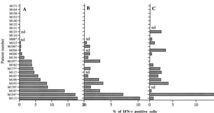
cell stimulation [9]. As shown in Table 2 and Fig. 1B,
C, 18 TIL populations, among the 26 analyzed,
tained a detectable fraction of T cells, secreting IFN-c
and TNF-a in response to autologous tumor cells, after
the ®rst (R1), second (R2), or both ex-vivo expansions.
Figure 2 shows some examples of IFN-c intracellular
labeling. Proportions of IFN-c-secreting T cells ranged
from 0.35 to 13.8%. The median level of these
TAA-speci®c T cells among positive TIL was 2.7% (n 26
for R1 + R2). Respectively, 12 out of 24 and 7 out of
19 expanded TIL populations contained T cells capable
of secreting GM-CSF and IL-2, in response to
auto-logous tumor cells.
Estimation of the amount of tumor-speci®c TIL
obtained from each patient
The amounts of TIL obtained from each patient by the
®rst (R1) and second (R2) TIL expansions are shown in
Table 3. The average combined number of TIL obtained
from each of these two expansion cultures was
1.7 ´ 10
10. Table 3 also shows the amounts of R1 and
R2 TIL that exhibited a speci®c IFN-c response to
tu-mor cells. From these results we were able to calculate
the total amount of tumor-reactive TIL obtained per
patient. As shown in Table 3, high-fold expansions of
TIL from eight patients failed to induce any detectable
amount of tumor-reactive T cells. However, the data in
Table 1 show that TIL from these patients did not
contain detectable fractions of tumor-reactive TIL
be-fore expansion. For the other 18 patients, variable
fractions of tumor-reactive T cells had been detected
among TIL before expansion (see Table 1) and the two
high-fold expansion cultures, R1 and R2, yielded a total
amount of tumor-reactive TIL ranging from 2.8 ´ 10
6to
1.12 ´ 10
9.
Table 1 Cytokine responses of short-term-cultured TIL toautologous melanoma cells. TIL were stimulated for 6 h by autologous melanoma cells in the presence of brefeldin A, then ®xed, permeabilized, stained for cytokine production and analyzed by FACScan. (ND, not done)
Patient % IFN-c % TNF-a % IL-2 % GM-CSF
M88a ND ND ND ND M154a 17.2 15.8 2.9 7.7 M170a 8.7 2.5 ND 4.7 M177a 4.2 1.6 0.5 2 M196a 0.4 ND 2.6 ND M197a 3.7 ND ND ND M110b ± ± ND ± M113b 17.9 8.1 ND 14.4 M117b 4.5 ± ND ± M125b ND ND ND ND M131b ± ± ND ND M132b ± ± ± ± M134b 1.4 1.1 0.3 ± M140b ± ± ND ± M153b ± ± ± ± M158b ± ± ± ± M164b ± ± ND ± M167b 5.7 3.5 ± 1.4 M171b ± ± ± ± M182b 3.8 1.8 ± 2.4 M187b 13.9 6.7 4.8 7 M193b 8.4 5.5 ± 1.4 M199b 6.4 5.2 2.1 4.8 M200b 0.7 2.8 ± 2.3 M204b 0.5 ± ± ± M212b 0.3 ± ± ±
aMelanoma patients bearing only one invaded lymph node bMelanoma patients bearing more than one invaded lymph nodes
Fig. 1A±C Percentage of IFN-c-positive cells detected in TIL populations by intracellular labeling in response to autologous melanoma cells. A Before expansion; B after the ®rst expansion; C after the second expansion. (*Patients bearing only one invaded lymph node)
Melanoma-speci®c responses are mainly elicited
by CD8+ lymphocytes
We performed double staining of CD8 and CD4 IFN-c
on seven TIL populations in response to class
II-posi-tive autologous melanoma cell lines. As shown in
Table 4, CD8-speci®c responses were found in higher
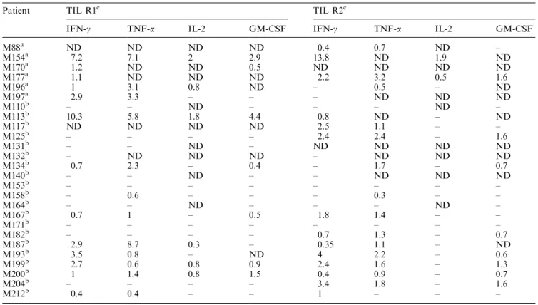
Table 2 Percentages of highly expanded TIL reactive against autologous melanoma cells. TIL were stimulated for 6 h by autologous melanoma cells in the presence of brefeldin A, then ®xed, permeabilized, stained for cytokine production and analyzed by FACScan. (ND, not done)
Patient TIL R1c TIL R2c
IFN-c TNF-a IL-2 GM-CSF IFN-c TNF-a IL-2 GM-CSF
M88a ND ND ND ND 0.4 0.7 ND ± M154a 7.2 7.1 2 2.9 13.8 ND 1.9 ND M170a 1.2 ND ND 0.5 ND ND ND ND M177a 1.1 ND ND ND 2.2 3.2 0.5 1.6 M196a 1 3.1 0.8 ND ± 0.5 ± ND M197a 2.9 3.3 ± ± ± ND ND ND M110b ± ± ND ± ± ± ND ± M113b 10.3 5.8 1.8 4.4 0.8 ND ± ND M117b ND ND ND ND 2.5 1.1 ± ± M125b ± ± ± ± 2.4 2.4 ± 1.6 M131b ± ± ND ± ND ND ND ND M132b ± ND ND ND ± ND ND ND M134b 0.7 2.3 ± 0.4 ± 1.7 ± 0.7 M140b ± ± ND ± ± ND ND ND M153b ± ± ± ± ± ± ± ± M158b ± 0.6 ± ± ± 0.3 ± ± M164b ± ± ND ± ± ± ND ± M167b 0.7 1 ± 0.5 1.8 1.4 ± ± M171b ± ± ± ± ± ± ± ± M182b ± ± ± ± 0.7 1.3 ± 0.7 M187b 2.9 8.7 0.3 ± 0.35 1.1 ± ND M193b 3.5 0.8 ± ND 4 2.2 ± 0.6 M199b 2.7 0.6 0.8 0.9 2.4 1.6 ± 1.3 M200b 1 1.4 0.8 1.5 0.4 0.9 ± 0.7 M204b ± ± ± ± 3.4 1.8 ± 1.6 M212b 0.4 0.4 ± ± 1 ± ± ±
aMelanoma patients bearing only one invaded lymph node bMelanoma patients bearing more than one invaded lymph node
cR1 and R2 are the TIL populations obtained from the ®rst and the second high- scale expansion, respectively
Fig. 2 Labeling of IFN-c-secreting T cells among polyclonal TIL in response to autologous tumor cells. TIL were stimulated for 6 h by autologous tumor cells, at a ratio of 1: 3 respectively (lower panel), or by HLA-mismatched allogeneic tumor cells as a negative control (upper panel), in the presence of brefeldin A (10 lg/ml). After ®xation, TIL were stained with anti-IFN-c mAb (5 lg/ml) in the presence of saponin and analyzed by FACScan (5 ´ 105events)
proportions than CD4 responses for each tested
pop-ulation.
Discussion
We have undertaken, for the ®rst time, a systematic
quanti®cation among melanoma patients of
tumor-reactive T cells that can be expanded for cellular
thera-py. This was possible retrospectively for 26 TIL-treated
patients, after the autologous melanoma cell line had
been derived from these patients. We used intracellular
cytokine assays to measure TIL fractions that
speci®-cally responded to the autologous melanoma cells.
Eighteen of the TIL populations analyzed contained
tumor-reactive T cells in proportions ranging from 0.3 to
17.9% before expansion. We showed that using a
poly-clonal TIL expansion method, based on the use of PHA
and feeder cell stimulation, a fraction of these
tumor-reactive T cells was systematically expanded. For the
TIL populations that contained over 4% tumor-reactive
T cells before expansion, increases in the numbers of
these cells were observed in the two high-scale
expan-sions performed. However, for some of the TIL
popu-lations, in which the fraction of tumor-reactive T cells
was below 4% before expansion, a detectable growth of
these cells was successful in only one out of the two TIL
cultures. During expansion the initial fraction of
tumor-reactive TIL usually decreased, but the ®nal fraction was
correlated with the fraction present before expansion.
Therefore, checking TIL speci®city before high- scale
expansion allowed prediction of whether the recovery of
tumor-reactive T cells would be feasible for an
immu-notherapy.
In the present study, the TIL of 8 out of 26 stage III
melanoma patients apparently lacked fractions of
tu-mor-reactive T cells detectable by the cytokine assays
used. These populations derived from patients bearing
more than one invaded lymph node. This suggests that
either the presence or the reactivity of TAA-speci®c T
cells inside melanoma-invaded lymph nodes could be
in¯uenced by the advancement of the disease. Indeed, we
could obtain melanoma-speci®c TIL from the six
pa-tients bearing only one invaded lymph node and from
only 12 out of 20 patients of the other group. However,
this dierence is not yet statistically signi®cant due to the
low representation by patients of the ®rst group. If it
were con®rmed on a larger group of patients, this
observation would directly support the existence of T
cell inactivation mechanisms inside tumor-invaded
lymph nodes.
Another new result from the present study was to
allow a ®rst estimation of the total amount of
tumor-reactive T cells that can be obtained to treat melanoma
patients via a given TIL expansion method. TIL
pro-duction was performed here twice, at a 1-month interval,
by two independent TIL cultures. Each expansion
cul-ture was initiated from 1.8 ´ 10
6TIL, either fresh or
thawed, which is a minimal TIL amount that can be
easily obtained from melanoma biopsies. The median
number of TIL obtained after two 3-week periods of
culture was 1.7 ´ 10
10. The median ampli®cation
induced was therefore an approximately 5 ´ 10
3-fold
increase. The total amounts of tumor-reactive T cells
obtained from these cultures were calculated from the
percent of IFN-c-responding T cells. These were quite
variable, ranging from 2.8 ´ 10
6to 1.12 ´ 10
9. This is
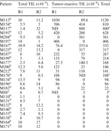
Table 3 Amounts of TIL and tumor-reactive TIL obtainedthrough high-scale expansions
Patient Total TIL (´10)9) Tumor-reactive TIL (´10)6) Total
R1 R2 R1 R2 M113b 10 11.2 1030 89.6 1120 M154a 5.5 3 396 414 810 M117b 1.4 32 ND 800 800c M193b 12 5.2 420 208 628 M204b 9.5 16.5 0 561 561 M197a 14 1 406 0 406 M167b 10.9 14.2 76.4 255.6 332 M125b 12 13.2 0 317 317 M187b 8 10 232 35 267 M199b 5 3.3 135 79 214 M177a 2.5 6.4 27.5 140.5 168 M200b 12.3 5.8 123 23 146 M212b 7.8 11 31 110 141 M170a 9 0.5 108 ND 108c M134b 13.5 9 94 0 94 M196a 8.6 14 86 0 86 M182b 0.6 3.1 0 22 22 M88a 6 0.7 ND 3 3c M110b 1.1 1 0 0 0 M131b 8.5 7 0 0 0 M132b 8 12.2 0 0 0 M140b 2.7 1.1 0 0 0 M153b 7.4 9.3 0 0 0 M158b 8 10.7 0 0 0 M164b 10 17 0 0 0 M171b 10 12 0 0 0
aMelanoma patients bearing only one invaded lymph node bMelanoma patients bearing more than one invaded lymph nodes cAmount of speci®c TIL infused through only one TIL injection
Table 4 The proportions of CD4- and CD8-speci®c lymphocytes in TIL populations in response to autologous melanoma cell lines expressing HLA class II antigen. Shortly-cultured TIL were stimulated for 6 h by autologous melanoma cells in presence of brefeldin A, after which the cells were labeled with CD4-PE or CD8-PE antibodies and then ®xed, permeabilized and stained for cytokine production
Patient Proportion of CD4-speci®c T cells among tumor-speci®c lymphocytes
Proportion of CD8-speci®c T cells among tumor-speci®c lymphocytes M125 4% (R2) 96% (R2) M167 28% (R1) 72% (R1) M170 0% (R1) 100% (R1) M187 18% (R1) 82% (R1) 16% (R2) 84% (R2) M193 0% (R1) 100% (R1) M197 0% (R1) 100% (R1) 0% (R2) 100% (R2) M204 40% (R2) 60% (R2)
probably a minimal estimation, however, since
intra-cellular cytokine labeling fails to detect low-avidity
T cells, and even some fractions of high-avidity T cells
that are transitorily refractory to cytokine production
in vitro [3, 8].
As previously shown with melanoma-speci®c CTL
clones, much lower fractions of TIL responded to
autologous tumor cells by GM-CSF and IL-2 secretion
than by IFN-c and TNF-a secretion. This results in part
from dierent antigen thresholds being necessary for the
secretion by T cells of these cytokines [3, 4, 12]. These
data therefore suggest that an important proportion of
melanoma invaded lymph node lymphocytes speci®c for
autologous tumor cells are of relatively low avidity for
tumor cells. Avidity is considered to be an important
parameter for the ecacy of T cell-dependent responses.
Nonetheless, the potential role of the dierent cytokines
secreted by CTL in the success of anti-tumor responses
remains unclear.
The contribution of TAA-speci®c CTL in limiting
tumor development has been established in animal
models, in great part through adoptive transfers of
speci®c T-cell populations [1, 14, 15, 16]. Since high
numbers of TAA-speci®c T cells of de®ned speci®city [6,
26] and high numbers of tumor-reactive TIL (the present
results) can now be readily produced from melanoma
patients, it will be feasible to ascertain their potential
role in driving an ecient response against tumors by
developing randomized adoptive transfer treatments.
Acknowledgements This work was supported by grant FK-ERC INSERM program 97/11, by grant number 6494 from the ``Asso-ciation pour la recherche contre le cancer'', by funds from the ``Ligue Nationale contre le cancer, axe immunologie du cancer'', the ``Ligue departementale de Loire Atlantique'' and by grant PHRC 93 from the CHR de Nantes.
References
1. Aruga A, Shu S, Chang AE (1995) Tumor-speci®c granulocyte/ macrophage colony-stimulating factor and interferon gamma secretion is associated with in vivo therapeutic ecacy of ac-tivated tumor-draining lymph node cells. Cancer Immunol Immunother 41: 317±324
2. Chin Y, Janssens J, Smeyers E, Bleus J, Zhang J, Raus J (1992) Large scale expansion of human tumor in®ltrating lymphocytes with surface-modi®ed stimulator cells for adoptive immuno-therapy Anticancer Res 12: 733±736
3. FonteneauJF, Le Drean E, Le Guiner S, Gervois N, Diez E, JotereauF (1997) Heterogeneity of biologic responses of mel-anoma-speci®c CTL. J Immunol 159: 2831±2839
4. Gervois N, Guilloux Y, Diez E, Jotereau F (1996) Suboptimal activation of melanoma in®ltrating lymphocytes (TIL) due to low avidity of TCR/MHC-tumor peptide interactions. J Exp Med 183: 2403±2407
5. Gervois N, Heuze F, Diez E, Jotereau F (1990) Selective ex-pansion of a speci®c anti-tumor CD8+ cytotoxic T lympho-cyte clone in the bulk culture of tumor-in®ltrating lympholympho-cytes from a melanoma patient: cytotoxic activity and T cell receptor gene rearrangements. Eur J Immunol 20: 825±831
6. Gervois N, Labarriere N, Le Guiner S, Pandol®no MC, Fon-teneau JF, Guilloux Y, Diez E, Dreno B, Jotereau F (2000) High avidity melanoma-reactive cytotoxic T lymphocytes are
eciently induced from peripheral blood lymphocytes on stimulation by peptide-pulsed melanoma cells. Clin Cancer Res 6: 1459±1467
7. Herr W, Schneider J, Lohse AW, Meyer zum Buschenfelde KH, Wolfel T (1996) Detection and quanti®cation of blood-derived CD8+ T lymphocytes secreting tumor necrosis factor alpha in response to HLA-A2.1-binding melanoma and viral peptide antigens. J Immunol Methods 191: 131±142
8. Itoh Y, Germain RN (1997) Single cell analysis reveals regu-lated hierarchical T cell antigen receptor signaling thresholds and intraclonal heterogeneity for individual cytokine responses of CD4+ T cells. J Exp Med 186: 757±766
9. JotereauF, Pandol®no MC, Boudart D, Diez E, Dreno B, Douillard JY, Muller JY, LeMevel B (1991) High-fold ex-pansion of human cytotoxic T-lymphocytes speci®c for autol-ogous melanoma cells for use in immunotherapy. J Immunother 10: 405±411
10. Jung T, Schauer U, Heusser C, Neumann C, Rieger C (1993) Detection of intracellular cytokines by ¯ow cytometry. J Im-munol Methods 159: 197±207
11. Kawakami Y, Dang N, Wang X, Tupesis J, Robbins PF, Wang RF, Wunderlich JR, Yannelli JR, Rosenberg SA (2000) Rec-ognition of shared melanoma antigens in association with major HLA-A alleles by tumor in®ltrating T lymphocytes from 123 patients with melanoma. J Immunother 23: 17±27 12. Labarriere N, Diez E, Pandol®no MC, Viret C, Guilloux Y, Le
Guiner S, Fonteneau JF, Dreno B, Jotereau F (1997) Optimal T cell activation by melanoma cells depends on a minimal level of antigen transcription. J Immunol 158: 1238±1245
13. Labarriere N, Pandol®no MC, Raingeard D, Le Guiner S, Diez E, Le Drean E, Dreno B, JotereauF (1998) Frequency and relative fraction of tumor antigen-speci®c T cells among lymphocytes from melanoma-invaded lymph nodes. Int J Cancer 78: 209±215
14. Lynch DH, Miller RE (1991) Immunotherapeutic elimination of syngeneic tumors in vivo by cytotoxic T lymphocytes gen-erated in vitro from lymphocytes from the draining lymph nodes of tumor-bearing mice. Eur J Immunol 21: 1403±1410 15. Melief CJ, Kast WM (1995) T-cell immunotherapy of tumors
by adoptive transfer of cytotoxic T lymphocytes and by vac-cination with minimal essential epitopes. Immunol Rev 145: 167±177
16. Nagoshi M, Goedegebuure PS, Burger UL, Sadanaga N, Chang MP, Eberlein TJ (1998) Successful adoptive cellular immunotherapy is dependent on induction of a host immune response triggered by cytokine (IFN-gamma and granulocyte/ macrophage colony-stimulating factor) producing donor tu-mor-in®ltrating lymphocytes. J Immunol 160: 334±344 17. Pandol®no MC, Viret C, Gervois N, Guilloux Y, Davodeau F,
Diez E, JotereauF (1992) Speci®city, T cell receptor diversity and activation requirements of CD4+ and CD8+ clones de-rived from human melanoma-in®ltrating lymphocytes. Eur J Immunol 22: 1795±1802
18. Pittet MJ, Valmori D, Dunbar PR, Speiser DE, Lienard D, Lejeune F, Fleischhauer K, Cerundolo V, Cerottini JC, Romero P (1999) High frequencies of naive Melan-A/MART-1-speci®c CD8(+) T cells in a large proportion of human histocompatibility leukocyte antigen (HLA)-A2 individuals. J Exp Med 190: 705±715
19. Romero P, Dunbar PR, Valmori D, Pittet M, Ogg GS, Ri-moldi D, Chen JL, Lienard D, Cerottini JC, Cerundolo V (1998) Ex vivo staining of metastatic lymph nodes by class I major histocompatibility complex tetramers reveals high numbers of antigen-experienced tumor-speci®c cytolytic T lymphocytes. J Exp Med 188: 1641±1650
20. Rosenberg SA, Packard BS, Aebersold PM, Solomon D, To-palian SL, Toy ST, Simon P, Lotze MT, Yang JC, Seipp CA (1988) Use of tumor-in®ltrating lymphocytes and interleukin-2 in the immunotherapy of patients with metastatic melanoma. N Engl J Med 319: 1676±1680
21. Rosenberg SA, Yannelli JR, Yang JC, Topalian SL, Sch-wartzentruber DJ, Weber JS, Parkinson DR, Seipp CA, Ein-139
horn JH, White DE (1994) Treatment of patients with meta-static melanoma with autologous tumor-in®ltrating lympho-cytes and interleukin 2. J Natl Cancer Inst 86: 1159±1166 22. Schwartzentruber DJ, Hom SS, Dadmarz R, White DE,
Yannelli JR, Steinberg SM, Rosenberg SA, Topalian SL (1994) In vitro predictors of therapeutic response in melanoma pa-tients receiving tumor-in®ltrating lymphocytes and interleukin-2. J Clin Oncol 12: 1475±1483
23. Tessier MH, Pandol®no MC, Jotereau F, Boudart D, Litoux P, Dreno B (1996) Home therapy with autologous tumour-in®l-trating lymphocytes and subcutaneous interleukin-2 in meta-static melanoma. Eur J Cancer 32A: 735±736
24. Topalian SL, Muul LM, Solomon D, Rosenberg SA (1987) Expansion of human tumor in®ltrating lymphocytes for use in immunotherapy trials. J Immunol Methods 102: 127±141 25. Topalian SL, Solomon D, Rosenberg SA (1989)
Tumor-spe-ci®c cytolysis by lymphocytes in®ltrating human melanomas. J Immunol 142: 3714±3725
26. Yee C, Savage PA, Lee PP, Davis MM, Greenberg PD (1999) Isolation of high avidity melanoma-reactive CTL from heter-ogeneous populations using peptide-MHC tetramers. J Im-munol 162: 2227±2234