Acute Tubular Dysfunction With Fanconi Syndrome: A New
Manifestation of Mitochondrial Cytopathies
François-Guillaume Debray, MD,
1Aicha Merouani, MD,
2Marie Lambert, MD,
1Pierre Brochu, MD,
3Chantal Bernard, MD,
4Brian H. Robinson, PhD,
5and Grant A. Mitchell, MD
1INDEX WORDS: Mitochondria; tubulopathy; Fanconi syndrome; metabolism, inborn error; acidosis; episodic.
M
itochondrial diseases are a heterogeneous
group of energy metabolism disorders
that can affect almost any organ.
1Kidney disease
is well-known in patients with mitochondrial
disorders, most commonly manifesting as
proxi-mal tubulopathy with chronic tubular acidosis,
typically a minor feature in pediatric patients
with severe involvement of brain, liver, heart, or
other organs.
2-5Other renal presentations
in-clude chronic tubulointerstitial nephropathy,
6,7progressive glomerular diseases,
8,9and nephrotic
syndrome.
10Acute, severe, and fluctuant tubular
dysfunction has not been previously described.
CASE REPORTS
Clinical Histories
Patient 1
A girl born to first-cousin Turkish parents was admitted at age 2 weeks for failure to thrive, tachypnea, and fever. Blood chemistry results were as follows: pH, 7.07; PCO2, 57.6 mm Hg; bicarbonate, 16 mEq/L (mmol/L); anion gap, 16 mEq/L (mmol/L); lactate, 74 mg/dL (8.27 mmol/L; normal,⬍20 mg/dL); and lactate-pyruvate molar ratio, 41.9 (normal, ⬍17). She received antibiotics for suspected sepsis. Acid-base status thereafter normalized. Plasma phosphorus level was 6.9 mg/dL (2.3 mmol/L), and tubular reabsorption of phosphate was 91%. The patient was readmitted at 4 months for fever. Blood chemistry showed pH of 7.19; PCO2of 43.2 mm Hg; bicarbonate of 15.3 mEq/L (mmol/L); anion gap of 10.1 mEq/L (mmol/L); and lactate level of 17.7 mg/dL (1.97 mmol/L). Urinary pH was 6.0 and glycosuria was absent. Renal tubular acidosis was suspected and bicarbonate was administered. Psychomotor delay and neurosensory deaf-ness were documented, and she required nasogastric tube feeding.
Because of multisystemic involvement, a mitochondrial disease was suspected, and at 7 months, the patient was electively admitted for investigations. Blood analyses showed the following values: pH, 7.33; PCO2, 39 mm Hg; bicarbon-ate, 22.0 mEq/L (mmol/L); and anion gap, 12.0 mEq/L (mmol/L). Urinary pH was 8.0, and there was generalized aminoaciduria without glycosuria (Fig 1A). Plasma phospho-rus level was 3.3 mg/dL (1.10 mmol/L; normal, 1.45 to 2.1 mmol/L), and tubular reabsorption of phosphate was 72%. Muscle biopsy results were normal, but liver showed marked mitochondrial proliferation and ultrastructural anomalies.
At 1 year, the patient was readmitted for fever. Blood analysis results were as follows: pH, 7.35; PCO2, 26.2 mm Hg; bicarbonate, 14.4 mEq/L (mmol/L); anion gap, 15.6 mEq/L (mmol/L); and lactate, 40 mg/dL (4.44 mmol/L). Urinary pH was 6.0, and glycosuria showed glucose of 252 mg/dL (14 mmol/L). Bicarbonate administration was tran-siently doubled. Two weeks later, electrolyte imbalances resolved, and she returned to her previous level of bicarbon-ate supplementation. At this age, renal investigations repeat-edly showed generalized aminoaciduria, normoglycemic gly-cosuria (glucose, 180 mg/dL [10 mmol/L]), proteinuria (protein, 0.12 g/dL [1.19 g/L]), hypophosphatemia (phos-phate, 1.5 mg/dL [0.5 mmol/L]), low tubular reabsorption of phosphate (52%), hypercalciuria (urinary calcium-creatinine ratio, 5.96), and mild nephrocalcinosis. Blood pH was 7.31, plasma bicarbonate level was 19 mEq/L (mmol/L), and urinary pH was 7.0. Plasma creatinine level was 0.2 mg/dL (18 mol/L), and glomerular filtration rate measured by using creatinine clearance was 110 mL/min/1.73 m2(1.83 mL/s/1.73 m2). Phosphorus was administered.
At 15 months, another major metabolic crisis occurred. Initial laboratory values were pH, 7.11; PCO2, 31 mm Hg; bicarbonate, 11 mEq/L (mmol/L); anion gap, 14 mEq/L (mmol/L); lactate, 62 mg/dL (6.88 mmol/L); and urinary pH, 7.0. Bicarbonate requirements increased 10-fold (to 22 mEq [mmol]/kg/d intravenously), and phosphorus, to 95 mg/kg/d (Fig 2A). Glycosuria exceeded 1 g/L (⬎55 mmol/L). Unex-pectedly, tubular losses improved and all electrolyte supple-ments were stopped within 5 weeks. For 5 additional weeks, mean plasma bicarbonate and phosphorus concentrations
From the Divisions of1
Medical Genetics and2
Nephrol-ogy, Department of Pediatrics; and3
Department of Pathol-ogy, CHU Sainte-Justine;4
Department of Pathology, Mon-treal Children Hospital, McGill University, MonMon-treal, Québec; and 5
Metabolism Research Program, Research Institute, Department of Pediatrics and Biochemistry, The Hospital for Sick Children, University of Toronto, Toronto, Ontario, Canada.
Received June 8, 2007. Accepted in revised form Novem-ber 13, 2007.
Address correspondence to Grant A. Mitchell, MD, CHU Sainte-Justine, 3175 Côte Sainte-Catherine, Montréal, Qué-bec, H3T 1C5, Canada. E-mail: grant.mitchell@recherche-ste-justine.qc.ca
© 2008 by the National Kidney Foundation, Inc. 0272-6386/08/5104-0020$34.00/0
doi:10.1053/j.ajkd.2007.11.024
were 24.8⫾ 1.9 (SD) mEq/L (mmol/L) and 5.1 ⫾ 0.3 mg/dL (1.7⫾ 0.1 mmol/L), respectively. Glycosuria disappeared. Hypophosphatemia and acidosis recurred thereafter, with intermittent glycosuria (glucose, 0 to 100 mg/dL [0 to 5 mmol/L]), and supplementations were reintroduced (Fig 1A).
The patient had severe encephalopathy and developed hyp-sarrhythmia and sideroblastic anemia requiring multiple transfusions.
At 21 months, the patient was admitted for vomiting and fever and rapidly developed profound metabolic acidosis uncontrolled by bicarbonate infusion. Lactic acidemia in-creased to 315 mg/dL (35 mmol/L), and she died of multiple organ failure. Autopsy included muscle and kidney biopsies performed 1 hour postmortem. Muscle was normal, but proximal tubular cells showed massive mitochondrial prolif-eration (Fig 3A).
Patient 2
This girl born to unrelated French-Canadian parents at 35 weeks of gestation was admitted at age 2 months for irritabil-ity and poor weight gain. Blood analysis showed the follow-ing values: pH, 7.42; PCO2, 26 mm Hg; bicarbonate, 17.0
mEq/L (mmol/L); anion gap, 11 mEq/L (mmol/L); lactate, 70 mg/dL (7.73 mmol/L), and lactate-pyruvate ratio, 40.6. Glycosuria and aminoaciduria were absent. Nasogastric tube feeding was introduced and bicarbonate was administered (Fig 1B). At age 9 months, serum creatinine level was 0.73 mg/dL (65mol/L), and isotopic glomerular filtration rate was 42 mL/min/1.73 m2 (0.70 mL/s/1.73 m2). Urinalysis showed normoglycemic glycosuria (glucose, 100 mg/dL [5.5 mmol/L]), proteinuria (protein, 0.63 g/L), pH of 7, and generalized aminoaciduria. Plasma phosphorus concentra-tion was normal (4.5 mg/dL [1.50 mmol/L]), and plasma bicarbonate level was 20 mEq/L (mmol/L). Renal biopsy at 9 months, normal by means of light microscopy, showed mitochondrial proliferation and abnormalities by electron microscopy in proximal tubular cells (Fig 3B). During the following months, bicarbonate supplementation was in-creased to 7 mEq (mmol)/kg/d, but growth remained im-paired. She walked unaided at 16 months and language development was normal.
At 30 months, the patient presented with a severe acidotic crisis requiring continuous intravenous bicarbonate infusion (up to 33 mEq [mmol]/kg/24 h) for nearly 1 month (Fig 2B). At admission, she was polypneic and mildly dehydrated (weight loss⬍ 3%). Laboratory values were as follows: pH, 7.07; PCO2, 26.9 mm Hg, bicarbonate, 3.3 mEq/L (mmol/L);
lactate 195 mg/dL (21.7 mmol/L); anion gap, 23.7 mEq/L (mmol/L); and glucose, 150 mg/dL (8.3 mmol/L). Urinalysis showed pH 5.0, glycosuria with glucose of 252 mg/dL (14 mmol/L), and proteinuria with protein of 0.10 g/dL (1 g/L). There were no neurological signs, and after an initial period of hydration and bicarbonate infusion, she appeared
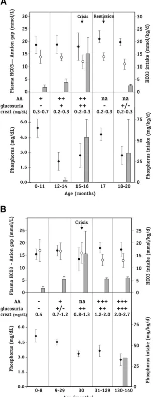
clini-4™™™™™™™™™™™™™™™™™™™™™™™™™™™™™™
Figure 1. Lifelong course of patients (A) 1 and (B) 2. Boxes represent mean for bicarbonate (upper right-side y-axis) and elemental phosphorus (lower right-side y-axis) administrations, mean plasma bicarbonate (closed circle; upper left-side y-axis ), mean anion gap (open circle; upper left-side y-axis), and mean plasma phosphorus (closed diamond; lower left-side y-axis). Error bars are SD. Abbre-viations: AA, aminoaciduria; creat, plasma creatinine. To convert creatinine in mg/dL tomol/L, multiply by 88.4; phosphorus in mg/dL to mmol/L, multiply by 3.01.cally well, energetic, and in no distress. Bicarbonate admin-istration was progressively decreased over 4 weeks to 6 mEq (mmol)/kg/d orally. She had a milder acidotic episode at age 4 years, but none thereafter. Chronic renal failure has progres-sively developed (Fig 1B). In the last 6 months, plasma phosphorus level decreased, with tubular reabsorption of phosphate of 50% and radiological evidence of early rickets. At 11 years, renal evaluation shows proteinuria with protein
4™™™™™™™™™™™™™™™™™™™™™™™™™™™™™™
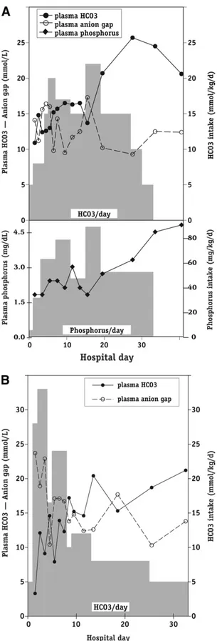
Figure 2. Chronology of biological parameters and electrolyte administration during acidotic episodes of pa-tients (A) 1 and (B) 2. Grey boxes show supplementation levels for bicarbonate (upper right-side y-axis) and phos-phorus (lower right-side y-axis). Shown are plasma bicar-bonate (closed circle; upper left-side y-axis), anion gap (open circle; upper left-side y-axis), and plasma phospho-rus (closed diamond; lower left-side y-axis). To convert phosphorus in mg/dL to mmol/L, multiply by 3.01.Figure 3. Electron micrographs show widespread pro-liferation of mitochondria with bizarre shape and hypertro-phic cristae occupying most of the volume of proximal tubular cells. (A) Patient 1 (original magnification⫻3,000); (B) patient 2 (original magnification⫻2,800).
of 0.15 g/dL (1.48 g/L), massive aminoaciduria and glycos-uria (glucose ⬎ 1 g/L), plasma creatinine level of 2.34 mg/dL (207mol/L), and creatinine clearance of 29.2 mL/ min/1.73 m2(0.03 mL/s/1.73 m2). She receives phosphorus and 1,25-hydoxyvitamin D3. Of note, development and neurological examination findings are normal.
Additional Investigations
Respiratory chain enzyme activities were measured in fibroblasts and liver as reported.11,12Blue native gel studies
were performed in fibroblast and liver, as described.13
Spec-trophotometric assays showed complex IV deficiency in the liver of patient 1 (0.2moles of reduced cytochrome c per gram of wet weight tissue per minute; controls, 0.6 to 2.4). Blue native gel studies confirmed decreased levels of com-plex IV in the liver of patient 1 and showed deficiency of complex I in fibroblasts of patient 2 (not shown).
Clinical Follow-up
Metabolic parameters and tubular function were assessed from the patients’ medical charts. The clinical course was divided into periods during which parameters of tubular function remained constant, shown inFig 1. Major changes in treatment, such as initiation or cessation of electrolyte supplementation, were used to define new periods. Acute acidotic episodes are presented chronologically (Fig 2).
DISCUSSION
Both patients fulfilled the diagnostic criteria of
Fanconi syndrome: aminoaciduria,
normoglyce-mic glycosuria, chronic tubular acidosis, and
impaired renal phosphate reabsorption. In stable
periods, when the patients were acidotic, urinary
pH was appropriate for the level of acidosis,
indicating preserved distal tubular acidifying
ca-pacity. Conversely, in other instances, alkaline
urine was observed with normal plasma
bicarbon-ate levels, suggesting a proximal origin of
tubu-lar acidosis. In addition to chronic tubulopathy,
both experienced major acidotic crises requiring
massive bicarbonate administration (up to 22 and
33 mEq/kg/d). Without consideration of renal
function, such episodes might falsely be
attrib-uted solely to hyperlactatemia. Although lactic
acid levels increased during episodes and
contrib-uted to the metabolic acidosis, anion gaps
re-mained normal or only modestly increased,
indi-cating that in the absence of digestive losses,
acidosis most probably was related to renal
tubu-lar dysfunction. Taken together with other signs
of proximal tubulopathy, the huge amount of
bicarbonate required to maintain acid-base
equilibrium and observation of alkaline urine
at admission of crisis in patient 1 suggest that
acidotic crises were caused by renal bicarbonate
losses. In patients with mitochondrial diseases,
tubulopathy is often considered a biological
marker of little clinical significance, but this
report shows that acute tubular failure can be life
threatening.
In patients with Fanconi syndrome, tubular
losses generally are considered fairly constant.
14In addition to acidotic crises, patient 1 had
tran-sient phosphaturia and glycosuria that resolved
in parallel with the tubular acidosis. Patient 2
presented with very late-onset hypophosphatemic
rickets, unusual in patients with Fanconi
syn-drome and particularly surprising regarding her
chronic renal failure. Mitochondrial diseases
should be considered in patients with atypical
Fanconi syndromes and unexplained multiple
tubular dysfunctions.
The reported spectrum of renal disease in
patients with mitochondrial diseases is listed in
Table 1
, with usual age at presentation,
occur-rence of extrarenal symptoms, and biochemical
or molecular findings. End-stage renal failure is
rare in patients with mitochondrial diseases. In
39 patients with mitochondrial disease and
tubu-lopathy, only 2 had moderate renal failure.
2In
another prospective study, none of 35 patients
had serum creatinine levels greater than 0.8
mg/dL (
⬎71
mol/L).
5Chronic renal failure
was described in a patient with mitochondrial
diseases and tubulointerstitial nephritis without
tubulopathy.
7Mitochondrial DNA mutations can
cause segmental glomerulosclerosis,
8leading to
end-stage renal failure.
9In these cases, renal
insufficiency manifested later than in our patient
2, and without tubulopathy. The kidney biopsy of
patient 2 was performed at age 9 months, and her
current glomerular histological state is unknown.
Patient 2 shows that mitochondrial diseases can
present as a primary renal disease without
neuro-logical, hepatic, or cardiac involvement. She also
shows that chronic renal failure, rare in patients
with mitochondrial diseases, can become the
major problem of some patients.
The pathophysiological characteristics of acute
tubular dysfunction are unclear, but several
obser-vations may be pertinent. Proximal tubules have
a high metabolic rate, generating more than 95%
of their adenosine triphosphate by means of
oxidative metabolism.
19Recent in vitro
experi-ments showed that nephrotoxicity of some drugs
is related to inhibition of mitochondrial
respira-tion, producing adenosine triphosphate depletion
and secondary tubular damages.
20Functional
coupling between ion transport and aerobic
respi-ration was demonstrated in vitro.
21Moreover,
even in normal proximal tubule cells,
physiolog-ical energy demand is close to the capacity for
adenosine triphosphate generation, shown by the
rapid decrease in adenosine triphosphate level
after stimulation of epithelial sodium transport.
22This constant dependence of proximal tubular
cells on high levels of energy supply resembles
that of the central nervous system. Acute
neuro-logical crises, termed “metabolic stroke,” are
well-known in patients with mitochondrial
dis-eases and may be related to imbalance between
energy requirement and production, leading to
cell death. Of note, metabolic strokes in patients
with mitochondrial diseases occur after such
stresses as minor infections, during which
meta-bolic demands are presumably increased,
compro-mising energy homeostasis in tissues with
im-paired energy production capacity. Perhaps the
episodes of acute tubular dysfunction in our
patients fall within a similar framework.
ACKNOWLEDGEMENTS
The authors thank Dr Eric Shoubridge (McGill Univer-sity, Montreal, Canada) for blue native gel studies.
Support: This work was supported in part by the Brandon Teresi Foundation.
Financial Disclosure: None.
REFERENCES
1. Munnich A, Rötig A, Chretien D, Saudubray JM, Cormier V, Rustin P: Clinical presentations and laboratory investigations in respiratory chain deficiency. Eur J Pediatr 155:262-274, 1996
2. Niaudet P, Rötig A: The kidney in mitochondrial cytopathies. Kidney Int 51:1000-1007, 1997
3. Rötig A: Renal diseases and mitochondrial genetics. J Nephrol 16:286-292, 2003
4. Martin-Hernandez E, Garcia-Silva MT, Vara J, et al: Renal pathology in children with mitochondrial diseases. Pediatr Nephrol 20:1299-1305, 2005
5. Neiberger RE, George JC, Perkins LA, Theriaque DW, Hutson AD, Stacpoole PW: Renal manifestations of congeni-tal lactic acidosis. Am J Kidney Dis 39:12-23, 2002
Table 1. Renal Manifestations of Mitochondrial Disorders
Kidney Involvement Age Extrarenal Symptoms Biochemical/Molecular Diagnosis Reference
Tubulopathy
Renal tubular acidosis or Fanconi syndrome
I, C Neurological disease, multivisceral involvement
Numerous isolated respiratory chain deficiencies
(complexes I, II, III, IV)
1-5
I Neurological and liver disease Complex III deficiency, BCS1L mutation
15
I, C Neurological disease Complex IV deficiency, COX10 mutation
16
I Sideroblastic anemia, exocrine pancreas dysfunction
Pearson marrow syndrome (mtDNA large deletion)
17
Adol Neurological disease, pigmentary retinopathy, cardiopathy (heart block)
Kearns Sayre syndrome (mtDNA large deletion)
2
Tubulointerstitial nephropathy
C, Adol None or late-onset neurological disease
Mt DNA large deletion 6,7
Glomerulopathy Segmental
glomerulosclerosis
Adol, adult None or deafness, diabetes, MELAS syndrome
MtDNA point mutation (AC_000021.2:3243G⬎A)
8,9
Glomerulosclerosis, nephrotic syndrome
(I), C, Adol Neurological and multivisceral disease
MtDNA deletion, complex III and CoQ10 deficiency, multiple complex deficiencies 4,18,23 Congenital nephrotic syndrome NN, I Progressive neurological disease, cardiomyopathy
Single or multiple respiratory chain deficiencies
10
Abbreviations: Adol, adolescence; C, childhood; I, infancy; MELAS, mitochondrial encephalomyopathy, lactic acidosis, stroke like episodes; mtDNA, mitochondrial DNA mutation; NN, neonatal period.
6. Szabolcs MJ, Seigle R, Shanske S, Bonilla E, DiMauro S, D’Agati V: Mitochondrial DNA deletion: A cause of chronic tubulointerstitial nephropathy. Kidney Int 58:1388-1396, 1994
7. Rötig A, Goutières F, Niaudet P, et al: Deletion of mitochondrial DNA in patient with chronic tubulointerstitial nephritis. J Pediatr 126:597-601, 1995
8. Jansen JJ, Maassen JA, Van der Woude FJ, et al: Mutation in mitochondrial tRNAleu(UUR) gene associated with progressive kidney disease. J Am Soc Nephrol 8:1118-1124, 1997
9. Hotta O, Inoue CN, Miyabayashi S, Furuta T, Takeuchi A, Taguma Y: Clinical and pathologic features of focal segmen-tal glomerulosclerosis with mitochondrial tRNALeu(UUR) gene mutation. Kidney Int 59:1236-1243, 2001
10. Goldenberg A, Huynh Ngoc L, Thouret MC, et al: Respiratory chain deficiency presenting as congenital ne-phrotic syndrome. Pediatr Nephrol 20:465-469, 2005
11. Carter SL, Rennie CD, Hamilton SJ: Changes in skeletal muscle in males and females following endurance training. Can J Physiol Pharmacol 79:386-389, 2001
12. Cameron JM, Levandovskiy V, MacKay N, Robinson BH: Respiratory chain analysis of skin fibroblasts in mito-chondrial disease. Mitochondrion 4:387-394, 2004
13. Nijtmans LGJ, Henderson NS, Holt IJ: Blue native electrophoresis to study mitochondrial and other protein complexes. Methods 26:327-334, 2002
14. Brodehl J: The Fanconi syndrome, in Edelman (ed): Pediatric Kidney Disease. Boston, MA, Little, Brown, 1996, pp 1841-1871
15. de Lonlay P, Valnot I, Barrientos A, et al: A mutant mitochondrial respiratory chain assembly protein causes
complex III deficiency in patients with tubulopathy, encephalopathy and liver failure. Nat Genet 29:57-60, 2001 16. Valnot I, von Kleist-Retzow JC, Barrientos A, et al: A mutation in the human heme A:farnesyltransferase gene (COX10) causes cytochrome c oxidase deficiency. Hum Mol Genet 9:1245-1249, 2000
17. Rötig A, Cormier V, Blanche S, et al: Pearson’s marrow-pancreas syndrome. A multisystem mitochondrial disorder in infancy. J Clin Invest 86:1601-1608, 1990
18. Rötig A, Appelkvist EL, Geromel V, et al: Quinone-responsive multiple respiratory chain dysfunction due to widespread coenzyme Q10 deficiency. Lancet 356:391-395, 2000
19. Epstein FH: Oxygen and renal metabolism. Kidney Int 51:381-385, 1997
20. Engbersen R, Masereeuw R, van Gestel MA, van der Logt EMJ, Smits P, Russel FGM: Glibenclamide depletes ATP in renal proximal tubular cells by interfering with mitochondrial metabolism. Br J Pharmacol 145:1069-1075, 2005
21. Balaban RS, Mandel LJ, Soltoff SP, Storey JM: Coupling of active ion transport and aerobic respiratory rate in isolated renal tubules. Proc Natl Acad Sci U S A 77:447-451, 1980
22. Beck JS, Breton S, Mairbaurl H, Laprade R, Giebisch G: Relationship between sodium transport and intracellular ATP in isolated perfused rabbit proximal convoluted tubule. Am J Physiol 261:F634-F639, 1991
23. Diomedi-Camassei F, Di Giandomenico S, Santorelli FM, et al: COQ2 nephropathy: A newly described inherited mitochondriopathy with primary renal involvement. J Am Soc Nephrol 18:2773-2780, 2007