Varicella-Zoster Virus IE63, a Virion Component Expressed during Latency and
Acute Infection, Elicits Humoral and Cellular Immunity
C. Sadzot-Delvaux, A. M. Arvin, and B. Rentier Division of Infectious Diseases, Department of Pediatrics, Stanford University School of Medicine, Stanford, California; Laboratoire de Virologie fondamentale, Universite´ of Lie`ge, Lie`ge Belgium Varicella-zoster virus (VZV) latency in human dorsal root ganglia is characterized by the
tran-scription of large regions of its genome and by the expression of large amounts of some polypeptides, which are also expressed during lytic cycles. The immediate early 63 protein (IE63) is a virion component expressed very early in cutaneous lesions and the first viral protein detected during latency. Immune response against IE63 has been evaluated among naturally immune adults with a history of chickenpox: Specific antibodies were detected in serum, and most subjects who had a T cell proliferation with unfractionated VZV antigens had T cell recognition of purified IE63. The cytotoxic T cell (CTL) response to IE63 was equivalent to CTL recognition of IE62, the major tegument component of VZV, whose immunogenicity has been previously described. T cell recogni-tion of IE63 and other VZV proteins is one of the likely mechanisms involved in controlling VZV reactivation from latency.
As a member of the Alphaherpesviridae, varicella-zoster vi- then confirmed in human dorsal root ganglia [10]. This protein is one of the virion components [11]; it is expressed very early rus (VZV) shares many characteristics (e.g., neurotropism or
the ability to become latent after a primary infection and to in culture [9] and in skin lesions, in which it is detectable much earlier than are late proteins, and sometimes it is expressed in reactivate many years later) with the other members of this
virus subfamily (i.e., herpes simplex type 1 or 2 [HSV-1 or the absence of a lesion [12]. During acute infections, IE63 shows mostly a nuclear localization, while it seems to accumu--2]). However, the mechanisms involved in latency of VZV
and HSV-1 and -2 seem to be completely different: During late in cytoplasm of latently infected neurons [10]. However, it is still unknown whether the protein has the same characteris-HSV latency, the viral genome is found in neurons only, and
transcription is limited to one region of the genome that pro- tics during productive and latent infections or whether differ-ences (e.g., phosphorylation) can be observed in either case. duces latency-associated transcripts (i.e., antisense of the
ICP-0 transcripts) [1]. These transcripts do not seem to be translated, More recently, other proteins corresponding to the transcripts and no viral protein has been detected during HSV latency. previously detected during the dormant phase have been
de-On the contrary, the nature of cells in which VZV remains tected in human material [13].
quiescent is still debated because in patient samples, the ge- These observations indicate that the mechanisms involved nome has been detected in neurons only [2, 3], in satellite cells in VZV latency are completely different from those acting surrounding neurons only [4], and in both cell types [5]. The in other alphaherpesvirus-infected cells, and the role of viral most striking difference between HSV and VZV latency is that proteins alone or in interaction with cellular proteins has to be several immediate early (IE) and early genes of VZV, such as hypothesized.
genes encoding for IE4, IE62, IE63, and MDBP (major DNA One of the parameters that could be critical to the control binding protein), are transcribed in latently infected rat dorsal of VZV infection is host immunity, as suggested by Hope-root ganglia [6] and in human ganglia [7, 8]. Moreover, VZV Simpson in 1965 [14]: VZV primary infection is limited by latency is characterized by the expression of large amounts of the host immune response, which remains high for many years, IE63, as demonstrated in the rat experimental model [9] and with a slow decrease in time. According to his hypothesis, frequent virus reactivations or contact with infected persons could counterbalance this progressive decrease; however, host resistance may still reach a critical level that is too low to
Informed consent was obtained from all subjects according to the US Depart- control virus reactivation. Many clinical observations show that ment of Health and Human Services and Stanford University guidelines for
the frequency and the severity of virus reactivations increase
research involving human subjects.
Financial support: NIH (AI-20459, AI36884, and CA-49605 to A.M.A.). in patients whose cellular immunity is impaired due to age, While performing this work, C.S.-D. was a VZV Research Foundation and a
pathologic disorders, or immunosuppressive treatments prior
Belgian American Educational Foundation fellow and was supported by a
to transplantation. These observations confirm Hope-Simpson’s
NATO and a Francqui fellowship.
Reprints or correspondence: Dr. C. Sadzot-Delvaux, Laboratoire de Virologie hypothesis on the role of host immune response, even if it now fondamentale, Pathologie B23, B-4000 Lie`ge, Belgium ([email protected]). appears obvious that it is mostly the cell-mediated immunity The Journal of Infectious Diseases 1998; 178(Suppl 1):S43 – 7 that limits virus reactivation; this is suggested by the fact that
q 1998 by the Infectious Diseases Society of America. All rights reserved.
Assays for cellular immunity to VZV. Peripheral blood mono-since the herpes zoster episode is not correlated with
hypogam-nuclear cells (PBMC), which had been separated from 50 mL of maglobulinemia.
heparinized blood by ficoll-hypaque gradient, were used for T Previous studies have shown that protein IE62 (the major
lymphocyte proliferation, cytokine, or cytotoxicity assays. tegument protein) and gC, gE, gG, and gI (the major
glycopro-Specific T lymphocyte proliferation was assayed as previously teins) are highly immunogenic and elicit a long-term cellular
described [17]. In brief, PBMC were recovered from 50 mL of immune response after natural VZV infection or immunization
heparinized blood by ficoll-hypaque gradient. Cells were cultured with a live attenuated vaccine strain (see [15] for review). in 96-well plates at 31 105
cells/well and stimulated in triplicate The immunogenicity of IE63 has not been evaluated, and its with VZV or control antigens or with GST or GST-IE63 fusion expression during latency and acute infection suggests that this proteins (3 – 25 mg/mL) whose concentration had been optimized protein probably plays a crucial role and could be an important previously. Preliminary experiments done with a broad range of fusion protein concentration (3 – 100 mg/mL) indicated that with target for the immune system.
ú25 mg/mL, the response to GST alone was too high. Phytohemag-As previously described [16], we have evaluated the immune
glutinin (100 mg/mL) and RPMI 1640 were used as positive and response against IE63 among 15 healthy adults with a history
negative controls, respectively. After 5 days, T cell proliferation of chickenpox as verified by detection of VZV IgG
anti-was measured by [3
H]thymidine uptake and expressed as a stimula-bodies. As controls, we used 2 healthy adults who had no
tion index (i.e., the ratio of mean counts per minute [cpm] in VZV history of varicella and no measurable anti-VZV antibodies.
or GST-IE63 – stimulated wells to that of control or GST wells). All experiments were done with the IE63 protein, which was
A stimulation index§2.0 was conventionally defined as a positive expressed in bacteria and fused to glutathione-S-transferase response.
(GST), as a stimulating antigen [9]. The IE63-specific cytotoxic response was detected by use of
limiting-dilution assays and compared with the cytotoxic T lym-phocyte response to IE62 as previously described [16]. Effector Materials and Methods
cells were either CD4/and CD8/cell populations or
unfraction-Study populations. Blood was obtained from 15 healthy adults ated T cells recovered from PBMC by separation with Lympho-with a history of varicella as confirmed by detection of IgG anti- kwik-T (One Lamba, Los Angeles). CD4/and CD8/cells were
bodies to VZV by ELISA. Two healthy adults who had no history purified by positive selection with magnetic cell sorting (MACS; of varicella and no measurable VZV IgG antibodies served as Miltenyi Biotec, Auburn, CA) [16], and their purity was verified
controls. by fluorescence-activated cell sorter analysis: Positive selections
GST and GST-IE63 production and purification. The GST and yielded§90% CD4/or CD8/cells. The contaminating population
GST-IE63 proteins were produced from the plasmids pGex-3X was£5%, and CD16/cells constituted£10% of the effector cells
and pGex-3X/ORF63, which have been described [9]. In brief, in all experiments.
plasmid-transformed bacteria were induced by 1 mM isopropyl- VZV-specific effector T cells were generated by incubation (2 b-D-thiogalactopyranoside for 3 h at 377C, pelleted, and sonicated weeks in 24-well plates) of 21 106
cells/well with whole VZV in PBS Triton X-100 (1% wt/vol). The GST and GST-IE63 pro- antigen in the presence of 106autologous irradiated PBMC (2700
teins that were released into the supernatant were purified by affin- rads) as feeders. Cells were refed every third day with 10% fetal ity chromatography on glutathione-Sepharose-4B (Pharmacia- calf serum, VZV antigens, and 10 U/mL recombinant human in-LKB, Uppsala, Sweden) and eluted with reduced glutathione (10 terleukin (IL)-2 (R&D Systems, Minneapolis). Target cells were mM). The proteins were quantified by use of a Bradford assay autologous EpsteBarr virus – transformed B cells that were in-(Bio-Rad, Hercules, CA) and loaded onto SDS – 10% polyacryl- fected with vaccinia recombinants expressing IE62 or IE63 [15].
amide gel to verify their purity. Mock-infected targets or cells infected with vaccinia virus devoid
Whole VZV antigen preparation. Melanoma cells cultured in of any VZV gene were used as controls. After 14 h of incubation Dulbecco’s modified Eagle medium supplemented with 10% fetal at 377C, targets were centrifuged, resuspended in culture medium, calf serum were infected with a laboratory strain (Chase) of VZV, and incubated with 300 mCi of51Cr (Amersham) for 4 h. Labeled
dispersed with glass beads when 90% cytopathic effect was ob- cells were washed, resuspended at 31 104
cells/mL, and added served, washed with PBS, and sonicated for 60 s. After centrifuga- to 96-well V-bottom plates at 31 103cells/well in 0.1 mL.
tion at 47C for 10 min, supernatants were frozen and thawed three To measure cytotoxicity, effector cells were added to lymphoid times, and aliquots were stored at0707C. An uninfected cell con- cell line targets at effector-to-target ratios of 30:1, 15:1, 3:1, 1:1,
trol was prepared in parallel. and 1:3 in 8-well replicates. After 4 h at 377C,51
Cr release was
Western blot assay. Western blot analysis was used to deter- measured by counting 100 mL of supernatant in a gamma counter. mine if anti-IE63 antibodies were in the sera. Purified GST and The spontaneous release of51
Cr and the maximum release after GST-IE63 were electrophoresed and transferred onto a nylon mem- lysis with 1% Nonidet P40 detergent were determined for each brane. To avoid nonspecific labeling, the membrane was saturated target cell preparation. The percentage of specific lysis was calcu-with nonfat milk (5 g/L). The membrane was incubated for 1 h at lated for each target as the [(mean experimental release)0 (mean room temperature with the donor’s serum (1/250), washed, and spontaneous release)]/[(mean maximal release)0 (mean spontane-incubated with a peroxidase-conjugated anti-human IgG (1/2000, ous release)]1 100.
Dakopatts, Glostrup, Denmark). Antibodies were detected using a Lysis of VZV-specific targets was considered specific when it Western blotting detection reagent (ECL; Amersham Life Science, was at least 10% higher than lysis of control targets. The responder cell frequency (RCF) for each assay was determined by applying Amersham, UK).
the maximum likelihood method to standard limiting-dilution plots, using a computerized analysis [18]. Replicates were scored as positive if the cpm was higher than the mean cpm for the corresponding spontaneous release plus 3 SD.
Assays for cytokine production. Cytokine production was eval-uated by stimulating 31 105
PBMC with GST-IE63 antigen or GST (1.25 mg/well) in 96-well plates. Supernatants were harvested from replicate wells on days 2, 4, and 6 and tested for IL-2, g-interferon (g-IFN), and IL-4. Cytokine release was quantitated by use of commercial ELISAs with sensitivities of detection defined by reference standards in each assay. The cytokine response was calculated as the amount of cytokine produced with GST-IE63 minus the amount elicited by GST. Commercial assays were used to measure IL-2 (Genzyme, Cambridge, MA) and g-IFN (Endo-gen, Cambridge, MA). The presence of IL-4 was assessed using an ultrasensitive assay (Cytoscreen; BioSource International, Ca-marillo, CA).
Results
The presence of anti-IE63 – specific antibodies was deter-mined by use of Western blot analysis with GST-IE63: Serum from all VZV-immune subjects bound to a band corresponding to IE63, indicating the presence of specific antibodies. This anti-IE63 humoral immune response was not quantified, but the faint reactivity in Western blotting indicated that concentra-tions of IE63-specific antibodies were low in naturally immune
Figure 1. Proliferation induced by stimulation of T cells with un-subjects. No reactivity to IE63 was observed with control sera.
fractionated VZV antigens or with immediate early (IE) 63 protein. Since cell-mediated immunity seems to be the most im- Stimulation index (SI) for each subject was calculated as ratio between portant criteria in latency, as previously discussed, we searched mean counts per minute for GST-EI63 – purified protein or VZV anti-for a memory immune response to IE63 by T cell lymphoproli- gens and GST-purified protein or control antigen. Mean SI{ SE is
indicated by black square with error bars. feration and cytotoxicity assays.
After samples were stimulated with VZV antigens, the stimu-lation index (SI) in those from the VZV-immune subjects
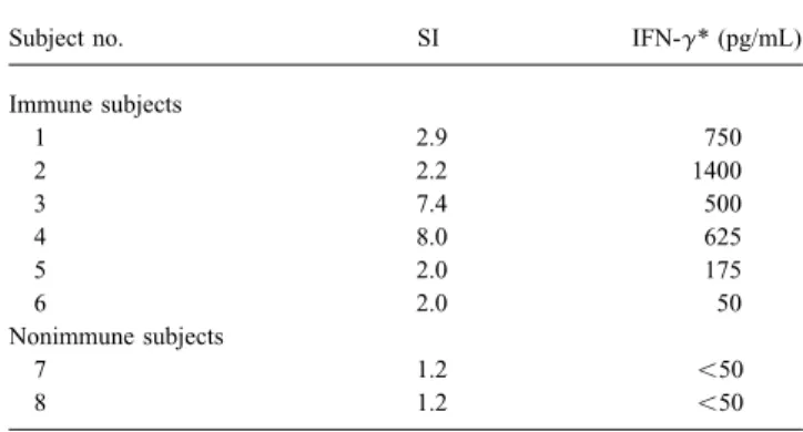
ranged from 2.1 to 52 (mean, 14.3{ 10.2 [SE]) (figure 1), in all samples from VZV-immune subjects. This g-IFN produc-tion was lower in the PBMC cultures of the 2 VZV-immune while the SIs in samples from the nonimmune subjects were
2.1 and 1.6. The SI after stimulation with GST-IE63 was 1.2 subjects, who had a low SI in response to IE63 stimulation. These results have to be compared with the absence of g-IFN and 1.4 for the nonimmune subjects and ranged from 1.8 to
8.0 (mean, 3.4{ 1.5) for the VZV-immune subjects. Among in the samples from nonimmune subjects and are in accordance with the pattern observed in T cells stimulated in standard the 15 immune subjects, 9 (60%) had a clear T cell proliferation
in response to GST-IE63; the remaining 6 (40%) had an SI of PBMC culture with unfractionated VZV antigens [19]. However, the absence of measurable IL-2 and IL-4 could 2.0 despite SIs to VZV antigen ranging from 5.4 to 11.0.
How-ever, the latter showed a strong stimulation with GST alone, be explained by the fact that these cytokines could be produced very early and could be bound directly to their receptors, thus making difficult any interpretation of the specific stimulation
by IE63. No correlation was found between the magnitude of being undetectable by the techniques used in this study. In this case, it would be useful to search for the cytokines earlier the SI measured in response to GST-IE63 and total VZV
anti-gens (data not shown). after stimulation or to search for their transcripts by reverse
transcript – polymerase chain reaction. It is also likely that the For 6 immune and 2 nonimmune subjects, we measured the
cytokine release by PBMC stimulated with IE63 for 2, 4, or 6 production of some cytokines is regulated by other ones. Even if the detection of other cytokines is needed to confirm these days. IL-2, IL-4, and g-IFN released in the culture supernatant
were quantified by ELISA. The results were expressed as the preliminary observations, g-IFN production indicates that T cell stimulation by IE63 leads mostly to a Th1 proliferation and difference between the cytokine concentration detected after
stimulation by GST-IE63 and by GST alone (table 1). IL-2 that the cytolytic component of cellular immunity is enhanced. Using unfractionated T cells from 4 patients, we observed and IL-4 were not detected; however, significant amounts (50 –
Table 1. Interferon-g (IFN-g) production by IE63-stimulated pe- the immune response to other major VZV immunogenic pro-ripheral blood mononuclear cells from 6 VZV-immune and 2 nonim- teins, such as IE62 and gE.
mune subjects with no history of varicella infection. The implication of IE63 recognition by the immune system in the control of latency has to be documented using animal
Subject no. SI IFN-g* (pg/mL)
models or by studies involving more subjects, especially
sub-Immune subjects jects with a high probability to reactivate the virus. In fact, one
1 2.9 750 of the main characteristics of IE63 is the fact that it is expressed
2 2.2 1400
during productive infection and during latency. This raises the
3 7.4 500
question of the relationship between the immune system and
4 8.0 625
the nervous system. Indeed, the nervous system is protected
5 2.0 175
6 2.0 50 from immune recognition by physical barriers first and then
Nonimmune subjects by the absence of classical MHC molecules at the surfaces of
7 1.2 õ50
neurons. On the contrary, satellite cells surrounding neurons
8 1.2 õ50
express MHC and could play an important role in antigen
NOTE. IFN-g was measured on days 2, 4, and 6 of stimulation, and levels presentation. It is therefore critical to clearly define in which
correspond to maximum amount detected. SIÅ stimulation index. cells the virus remains quiescent: Viral genomes have been * Expressed as difference between IFN-g concentration detected after
stimu-detected in neurons only, in satellite cells only, and in both
lation by glutathione-S-transferase (GST)-IE63 and by GST alone.
cell types, whereas IE63 protein has been observed only in neuron cytoplasm during latency. If neurons are the only cells expressing viral antigens, the mechanisms leading to the recog-was equivalent to that observed with targets expressing IE62
nition of viral peptides has to be clarified. It is possible that (25% – 48%) and was significantly higher than that observed
nonclassical MHC proteins are expressed at the cell surface in for mock-infected or vaccinia-infected targets used as controls.
response to viral infection as has been suggested for HSV The mean effector frequency for T cells that recognized IE63
infection [20]. However, in HSV-infected cells, peptide presen-was 1:31,000{ 16,000 SE and was not significantly different
tation by MHC molecules seems to be impaired because of a (PÅ .8) than the RCF for cells expressing IE62 (1:44,500 {
viral protein that inhibits peptide processing [21 – 22]. Such a 22,500 SE).
mechanism has not been demonstrated for VZV and must be Both CD4/and CD8/T lymphocyte populations lysed IE63
explored. and IE62 targets, as demonstrated by assays using purified
It will be of interest to characterize the immune response to CD4/and CD8/populations as effectors. The RCF of CD8/
IE63 in elderly and immunocompromised subjects, who have cells recognizing IE63 (1:30,500{ 13,700 SE) was not
statisti-a high incidence of herpes zoster, to exstatisti-amine possible correlstatisti-a- correla-cally different (PÅ .9) from the RCF of CD8/cells recognizing
tions between the risk of virus reactivation and diminished IE62 (1:28,500{ 10,100 SE), indicating that both viral proteins
responses to IE63. Enhancing the immune response to IE63 were recognized with the same efficiency by CD8/ cells. In
protein may be an important strategy to prevent VZV reactiva-the case of IE63 recognition, reactiva-the frequency of T cells lysing
tion from latency. If it is, IE63 protein could be a suitable IE63 targets was equivalent (PÅ .97) for CD4/(1:31,450{
candidate for incorporation into a therapeutic VZV vaccine for 7500 SE) and CD8/populations of effector cells.
administration to a population at high risk for herpes zoster.
Discussion Acknowledgments
We thank Paul Kinchington (University of Pittsburgh, Pitts-Our experiments demonstrate that most VZV-immune
indi-burgh) and Serge Debrus (University of Lie`ge) for providing vac-viduals have memory immunity against IE63, as shown by
cinia constructs and GST-IE63 fusion protein, respectively. specific antibody detection, and by proliferation of T cells when
stimulated by a GST-IE63 fusion protein. The cytokines
pro-duced by these proliferating T cells suggest that the stimulation References by IE63 leads mostly to a proliferation of Th1 cells and that
1. Stevens JG, Wagner EK, Devi-Rao GB, Cook ML, Feldman LT. RNA
the cytolytic component of the cellular immunity is enhanced, complementary to a herpesvirus alpha gene mRNA is prominent in which is confirmed by the lysis of targets expressing IE63. As latently infected neurons. Science1987; 1056 – 9.
2. Hyman RW, Ecker JR, Tenser RB. Varicella-zoster virus RNA in human
with IE62 and VZV glycoproteins, IE63 protein has an amino
trigeminal ganglia. Lancet1983; 2:814 – 6.
acid sequence that can be presented by the class I and II major
3. Gilden DH, Rozenman Y, Murray R, Devlin M, Vafai A. Detection of
histocompatibility complex (MHC) pathways, as indicated by
varicella-zoster virus nucleic acid in neurons of normal human thoracic
the participation of CD4/and CD8/in the cytotoxic response.
ganglia. Ann Neurol1987; 22:377 – 80.
IE63 is thus highly immunogenic and leads to a long-term 4. Croen KD, Ostrove JM, Dragovic LJ, Straus SE. Patterns of gene expres-sion and sites of latency in human nerve ganglia are differnet for
cella-zoster and herpes simplex viruses. Proc Natl Acad Sci USA1988; tory proteins during latency. Proc Natl Acad Sci USA1998;95:7080 –
85:9773 – 7. 5.
5. Lungu O, Annunziato PW, Gershon A, et al. Reactivated and latent vari- 14. Hope-Simpson RE. The nature of herpes zoster: a long-term study and a cella-zoster virus in human dorsal root ganglia. Proc Natl Acad Sci new hypothesis. Proc R Soc Med1965; 58:9 – 20.
USA1995; 92:10980 – 4. 15. Arvin AM. The T-lymphocyte response to varicella-zoster virus and its 6. Sadzot-Delvaux C, Debrus S, Nikkels A, Piette J, Rentier B. Varicella- relevance to vaccine development. Rev Med Virol1994; 4:161 – 75.
zoster virus latency in the adult rat is a useful model for human latent 16. Sadzot-Delvaux C, Kinchington PR, Debrus S, Rentier B, Arvin AM. infection. Neurology1995; 12:S18 – 20. Recognition of the latency-associated immediate early protein IE63 of 7. Vafai A, Murray RS, Wellish M, Devlin M, Gilden D. Expression of varicella-zoster virus by human memory T lymphocytes. J Immunol
varicella-zoster virus and herpes simplex virus in normal human trigemi- 1997; 159:2802 – 6.
nal ganglia. Proc Natl Acad Sci USA1988; 85:2362 – 6. 17. Arvin AM, Kinney-Thomas E, Shriver K, et al. Immunity to varicella-8. Meier JL, Holman RP, Croen KD, Smialek JE, Straus S. Varicella-zoster zoster viral glycoproteins, gpI (90/58) and gpIII (gp118), and to a non
virus transcription in human trigeminal ganglia. Virology 1993; 193: glycosylated protein, p170. J Immunol1986; 137:1346 – 51.
193 – 200. 18. Fazekas de St. Groth S. The evaluation of limiting dilution assays. J 9. Debrus S, Sadzot-Delvaux C, Nikkels AJ, Piette J, Rentier B. Varicella- Immunol Methods1982; 49:R11 – 23.
zoster virus gene 63 encodes an immediate early protein abundantly
19. Zhang Y, Cosyns M, Levin MJ, Hayward AR. Cytokine production in expressed during latency. J Virol1995; 69:3240 – 5.
varicella zoster virus-stimulated limiting dilution lymphocyte cultures. 10. Mahalingam R, Wellish M, Cohrs R, et al. Expression of protein encoded
Clin Exp Immunol1994; 98:128 – 33. by varicella-zoster virus open reading frame 63 in latently infected
20. Pereira RA, Tscharke DC, Simmons A. Upregulation of class I major human ganglionic neurons. Proc Natl Acad Sci USA1996; 93:2122 – 4.
histocompatibility complex gene expression in primary sensory neurons, 11. Kinchington PR, Bookey D, Turse SE. The transcriptional regulatory
pro-satellite cells and Schwann cells of mice in response to acute but not teins encoded by varicella-zoster virus open reading frames (ORFs) 4
latent herpes simplex virus infection in vivo. J Exp Med1994; 180: and 63, but not ORF61, are associated with purified virus particles. J
841 – 50. Virol1995; 69:4274 – 82.
21. Tomazin R, Hill AB, Jugovic P, et al. Stable binding of the herpes simplex 12. Nikkels AF, Debrus S, Sadzot-Delvaux C, Piette J, Rentier B, Pie´rard
virus ICP47 protein to the peptide binding site of TAP. EMBO J1996; GE. Immunochemical identification of varicella-zoster virus gene
63-15:3256 – 66. encoded protein (IE63) and late (gE) protein on smears and cutaneous
22. Ahn K, Meyer TH, Uebel S, et al. Molecular mechanism and species biopsies: implications for diagnostic use. J Med Virol1995; 47:342 – 5.
specificity of TAP inhibition by herpes simplex virus protein ICP47. 13. Lungu O, Panagiotidis CA, Annunziato PW, Gershon AA, Silverstein