Université de Montréal
Diencephalic and mesencephalic substrate for brain
stimulation reward
Par Marc Fakhoury Département de Neurosciences Faculté de Médecine Thèse présentéeen vue de l’obtention du grade de doctorat en neurosciences
Avril 2018
Université de Montréal Faculté des études supérieures
Cette thèse intitulée:
Diencephalic and mesencephalic substrate for brain stimulation reward
présentée par : Marc Fakhoury
a été évaluée par un jury composé des personnes suivantes :
John Kalaska, président-rapporteur Pierre-Paul Rompré, directeur de recherche
Stéphanie Fulton, membre du jury Andreas Arvanitogiannis, examinateur externe
iv
Résumé
La stimulation électrique de certains sites cérébraux chez les animaux de laboratoire induit un effet de récompense suffisamment fort pour obtenir une réponse opérante; par exemple, les rats apprendront à appuyer sur un levier pour recevoir une salve de pulsions électriques dans ces régions. Ce comportement, appelé autostimulation intracérébrale (ASI), a été largement étudié pour caractériser les substrats neuronaux de la récompense et des comportements dirigés. Plusieurs études au cours des dernières années suggèrent que le système dorsal diencéphalique (SDC) ainsi que la queue de l'aire tegmentale ventrale (qATV) sont impliqués dans le phénomène de récompense induit par l’ASI. Cependant, malgré des progrès significatifs dans la recherche, les mécanismes par lesquels le SDC et qATV participent à la transmission du signal de récompense induit par l’ASI restent largement inconnus.
Le principal objectif de cette thèse est d'étudier le rôle du SDC et du qATV dans la récompense induite par stimulation intracérébrale. Trois articles de recherche sont présentés dans cette thèse. Le premier article évalue l'effet de lésions électrolytiques au niveau du SDC et du faisceau médian prosencéphalique (FMP) dans le phénomène de récompense induit par la stimulation électrique de l'hypothalamus latéral (HL) et du raphé dorsal (RD) chez le rat. Les résultats montrent que des lésions effectuées au niveau du SDC et du FMP produisent une plus grande atténuation du phénomène de récompense induit par l’ASI que des lésions effectuées sur une seule de ces voies seulement, et ont un effet plus important sur le signal de récompense induit par la stimulation électrique du HL. Dans le deuxième article de cette thèse, la technique de marquage immuno-histochimique pour la protéine Fos a été utilisée en combinaison avec l’ASI et des lésions électrolytiques du SDC pour déterminer si les mêmes noyaux qui sont actifs chez des rats n’ayant pas reçu de lésion continuent à être actif après une lésion du SDC. Les résultats montrent que des
v
lésions du SDC réduisent l’expression de la protéine Fos induite par la stimulation électrique du HL dans certains sites du cerveau antérieur, du mésencéphale et du tronc cérébral. Enfin, le troisième article de cette thèse examine le rôle du qATV dans le phénomène de récompense induit par l’ASI et dans l'activité locomotrice. Une attention particulière a été accordée aux fonctions comportementales des récepteurs glutamatergique AMPA et NMDA, ainsi que les récepteurs opioïdes de type mu exprimés au niveau du qATV. Les résultats montrent que le blocage pharmacologique des récepteurs AMPA et NMDA, ainsi que l'activation des récepteurs opioïdes de type mu, dans certains sites du qATV entrainent une augmentation de l’activité locomotrice et de la récompense induite par l’ASI. Les résultats montrent aussi que la diminution de l’expression des récepteurs NMDA du qATV avec les petits ARN interférents ne modifie pas la récompense induite par l’ASI mais provoque une diminution marquée de la réponse opérante maximale. Les résultats obtenus dans cette thèse apportent une meilleure compréhension du substrat neuronal de la récompense induite par l’ASI, et suggèrent que (i) le SDC constitue une voie fiable pour la transmission de la récompense induite par l’ASI, (ii) le SDC est connecté avec certaines régions du cerveau antérieur, du mésencéphale et du tronc cérébral, et que (iii) la transmission glutaminergique et opioïde au niveau du qATV régule l'activité locomotrice et le phénomène de récompense induit par l’ASI. Ce travail pourrait avoir d'importantes répercussions sur la compréhension des comportements appétitifs, tels que l'alimentation et la consommation d'alcool, ainsi que sur les troubles psychiatriques, tels que la dépression, la schizophrénie, et les troubles liés à l'utilisation de substances.
Mots-clés: activité locomotrice; autostimulation intracérébrale; queue de l'aire tegmentale
vi
Abstract
Electrical stimulation of certain brain regions in laboratory animals induces a rewarding effect that is strong enough to support operant responding; for instance, rats will learn to press a lever to receive a short train of electrical pulses in these regions. This behavior, termed intracranial-self-stimulation (ICSS), has been extensively employed to characterize the neural substrate underlying reward and goal-directed behaviors. Evidence over the past few years suggests that the dorsal diencephalic conduction system (DDC) and the tail of the ventral tegmental area (tVTA) are involved in the rewarding effect of ICSS (or brain stimulation reward). However, despite significant progress in research, the underlying mechanisms remain largely unknown.
The overarching goal of this thesis is to investigate the role of the DDC and the tVTA in brain stimulation reward. Three research articles are presented in this thesis. The first article evaluates the effect of serial electrolytic lesions at the DDC and the medial forebrain bundle (MFB)— another pathway involved in brain stimulation reward—on the reward signal triggered by ICSS of the lateral hypothalamus (LH) and the dorsal raphe (DR) in rats. Results show that lesions at both the DDC and MFB produce larger and longer-lasting attenuations in brain stimulation reward than individual lesions at either pathway alone, and are more effective in attenuating the reward signal induced by LH self-stimulation. In the second article of the thesis, stimulation-induced Fos-like immunoreactivity (FLIR), a marker of cellular activity, was combined with electrolytic lesions at the DDC to determine whether the same nuclei that are active in lesion-naïve rats continue to be active following the lesions. Results show that electrolytic lesions at the DDC cause a marked reduction in stimulation-induced FLIR in certain forebrain, midbrain and brainstem regions that are activated by ICSS. Finally, the third article of the present thesis examines the role of the tVTA in brain stimulation reward and locomotor activity. Special attention was given to the behavioral
vii
functions of the glutamate receptors AMPA and NMDA, as well as the mu opioid receptors (MORs) expressed in the tVTA. Results show that pharmacological blockade of AMPA and NMDA receptors as well as activation of MORs in certain sites of the tVTA produce rewarding and locomotor stimulant effects. Results also show that downregulation of NMDA receptors in the tVTA using the small interfering RNA (siRNA) technique fails to alter brain stimulation reward, but causes a marked decrease in the maximal rate of operant responding for ICSS.
The findings obtained from this thesis shed new light on the neural substrate underlying brain stimulation reward with respect to brain regions, connectivity, and neurotransmitter systems. They suggest that (i) the DDC constitutes a viable route for the transmission of brain stimulation reward and merges with the MFB on a common reward integrator, (ii) the DDC is functionally connected to forebrain, midbrain and brainstem regions that are activated by ICSS, and that (iii) glutamate and opioid transmission in the tVTA are major regulators of brain stimulation reward and locomotor activity. This work could have important implications for understanding appetitive behaviors, such as eating and drinking, and psychiatric conditions, such as substance use disorder, depression and schizophrenia.
Keywords: brain stimulation reward; dorsal diencephalic conduction system; intracranial
viii
TABLE OF CONTENTS
Résumé………...iv Abstract………..vi List of tables………xiii List of figures………..xivList of initials and abbreviations………...xvi
Acknowledgments………...xxii
CHAPTER 1: GENERAL INTRODUCTION………1
1.1 Intracranial self-stimulation: a putative model to study the brain reward system….1 1.1.1 Context and discovery………..1
1.1.2 Reinforcing properties of ICSS………...……….3
1.1.3 Response-frequency function and the curve-shift paradigm………...6
1.1.4 Methodological and technical considerations………10
1.2 Anatomy, neurochemistry, and pharmacology of the brain reward system………..10
1.2.1 Brain regions and neural pathways………10
1.2.2 Central role of dopamine in reward………...14
1.2.3 Glutamate transmission: overview and implication in reward processing………18
1.2.4 The opioid system: overview and implication in reward processing……….24
1.3 The dorsal diencephalic conduction system in the brain reward circuitry: spotlight on the anatomy and function of the habenular complex (Adapted from the review paper Fakhoury 2018. Behavioral Brain Research, Accepted in Press)………...28
1.3.1 The habenula: morphological, cellular and electrophysiological profile………...29
ix
1.3.3 The habenula: a major regulator of monoaminergic systems………36
1.3.3.1 The habenula and the DA system……….……….37
1.3.3.2 The habenula and the 5-HT system………...40
1.3.4 The DDC in reward and aversion………..43
1.3.4.1 Electrophysiological and neuroimaging studies………43
1.3.4.2 Electrical stimulation studies………...46
1.3.4.3 Optogenetic stimulation studies………...49
1.4 The tail of the ventral tegmental area in behavioral processes and in the effect of psychostimulants and drugs of abuse (Adapted from the review paper Fakhoury 2018. Prog Neuropsychopharmacol Biol Psychiatry, 84:30-38)……….…….54
1.4.1 Context and overview………54
1.4.2 Cellular, synaptic and electrophysiological profile of the tVTA………...56
1.4.3 Afferents and efferents of the tVTA………..57
1.4.4 The tVTA: a source of negative reward signals……….61
1.4.5 The tVTA: from aversion expectation value to avoidance behavior……….64
1.4.6 The tVTA in responses to psychostimulants and drugs of abuse………..66
1.4.7 Summary and perspectives………....74
1.5 Thesis proposal: overview, hypotheses and objectives……….76
1.5.1 Article 1: Serial lesions at the DDC and MFB………..76
1.5.2 Article 2: Anatomical disconnection following a single lesion at the DDC………..77
1.5.3 Article 3: Manipulation of glutamate and opioid transmission in the tVTA……….79
x
2.1 ARTICLE 1: Role of the dorsal diencephalic conduction system in the brain reward
circuitry (Published in Behavioral Brain Research, 2016)………..…...81
Author contributions………..81
Abstract………..82
Introduction………83
Material and Methods………86
Results………90 Discussion………..96 Conclusion………...101 Acknowledgments………101 Figure legends………..103 Figures………..105 References……….………...113
2.2 ARTICLE 2: Effect of electrolytic lesions of the dorsal diencephalic conduction system on the distribution of Fos-like immunoreactivity induced by rewarding electrical stimulation (Published in Neuroscience, 2016)………...118
Author contributions………118
Abstract………119
Introduction………..120
Material and Methods………..122
Results………..129
Discussion………131
xi
Acknowledgments..………..138
Figure and table legends………..139
Tables………...………141
Figures………..142
References….………...149
2.3 ARTICLE 3: Modulation of brain stimulation reward and locomotor activity by ionotropic glutamate and mu opioid receptors in the tail of the ventral tegmental area (in preparation)……157
Author contributions………157
Abstract………158
Introduction………...159
Material and Methods………..161
Results………..166 Discussion………170 Acknowledgment……….175 Supplementary methods………...176 Figure legends………..179 Figures………..183 Supplementary figures……….188 References………....193
CHAPTER 3: GENERAL DISCUSSION AND CONCLUSION………..202
3.1 The DDC constitutes a route for the transmission of the reward signal triggered by ICSS………...202
xii
3.2 The DDC and MFB are functionally interconnected and merge on common
reward-relevant neural elements………205
3.3 The DDC is functionally connected to brain regions activated by ICSS…………..207
3.4 AMPA and NMDA receptors of the tVTA are major inhibitors of brain stimulation
reward and locomotor activity………..210
3.5 MORs of the tVTA are major enhancers of brain stimulation reward and locomotor
activity………...……..214 3.6 NMDA receptors of the tVTA are involved in incentive salience and brain stimulation reward…...………..217
3.7 Conclusion: implications for psychiatric diseases………...219
xiii
List of tables
General introduction
Table 1. The DDC in reward processing……….51 Table 2. The tVTA and the effect of drugs………70
Results of original contributions Article 2
xiv
List of figures
General introduction
Figure 1. ICSS apparatus and properties………...5
Figure 2. Schematic illustration of representative parameters used during an ICSS paradigm…….7
Figure 3. Schematic illustration of the theoretical functions that relate response rates to stimulation frequency under different current intensities and effort requirements…...8
Figure 4. Non-exhaustive illustration of regions involved in brain stimulation reward…………..12
Figure 5. Ionotropic and metabotropic glutamate receptors………20
Figure 6. Glutamate transmission in the ventral midbrain exerts opposite roles………23
Figure 7. Model of opposing tonic effects of opioid receptors on mesolimbic dopaminergic cells.26 Figure 8. Habenula afferents and efferents connections……….35
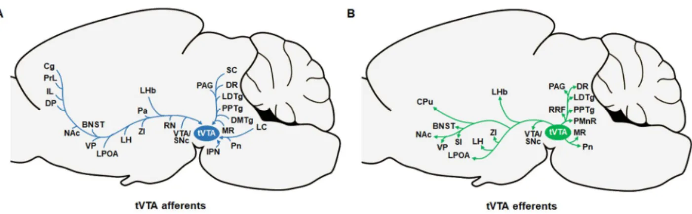
Figure 9. Main afferents and efferents of the tVTA………59
Results of original contributions Article 1 Figure 1………..…………..105 Figure 2…..………..106 Figure 3………...……….107 Figure 4………..…………..108 Figure 5………..…………..109 Figure 6………..…………..110 Figure 7………..…………..111 Figure 8………..…………..112
xv Article 2 Figure 1………..…………..142 Figure 2………..…………..143 Figure 3………..………..…144 Figure 4………..…………..145 Figure 5………..…………..146 Figure 6………..…………..147 Figure 7………..…………..148 Article 3 Figure 1………..…………..183 Figure 2………..…………..184 Figure 3………..…………..185 Figure 4………..………..186 Figure 5………..…………..187 Supplementary Figure 1………...188 Supplementary Figure 2………...189 Supplementary Figure 3………...190 Supplementary Figure 4………...191 Supplementary Figure 5………...192
General discussion and conclusion Figure 1. Schematic overview of article 1, 2, and 3 of the present thesis………220
xvi
List of initials and abbreviations
6-OHDA 6-hydroxydopamine
β-EP β-endorphin
A cyclase Adenylyl cyclase
ADHD Attention deficit hyperactivity disorder
Amy Amygdala
AMPA Amino-3-hydroxy-5-methyl-4-isoxazolepropionate
AMPAR AMPA receptor
ATP Adenosine triphosphate
B.C. Before Christ
BLA Basolateral amygdala
BNST Bed nucleus of the stria terminalis cAMP Cyclic adenosine monophosphate
Cg Cingulate cortex
CNS Central nervous system
CPu Caudate putamen
DA Dopamine
DBS Deep brain stimulation
DDC Dorsal diencephalic conduction system DMTg Dorsomedial tegmental area
DNQX 6,7-dinitroquinoxaline-2,3-dione
DR Dorsal raphe
xvii DOR Delta opioid receptor
DP Dynorphin
DPC Dorsal peduncular cortex
EAAT Excitatory amino acid transporter
EM-1 Endomorphin-1
EM-2 Endomorphin-2
EPSC Excitatory postsynaptic current
GABA Gamma-aminobutyric acid
GHB γ-hydroxybutyric acid sodium salt
Gi Inhibitory G protein
Glu Glutamate
Gly Glycine
GTP Guanosine triphosphate
I Intensity
ICSS Intracranial self-stimulation
IL Infralimbic cortex
IPN Interpeduncular nucleus
IPSC Inhibitory post-synaptic current
Hb Habenula
HVA Homovanillic acid
Hz Hertz
k Current density
xviii
KAR Kainate receptor
LC Locus coeruleus
LH Lateral hypothalamus
LHb Lateral habenula
log Logarithm
LPO Lateral preoptic area
LTDg Laterodorsal tegmental nucleus
M50 Stimulation frequency that maintains half-maximal responding MFB Medial forebrain bundle
mGLuR Metabotropic glutamate receptor mGlut Metabotropic glutamate
MOR mu-opioid receptor
MR Median raphe
ms Millisecond
mPFC Medial prefrontal cortex
Nac Nucleus accumbens
nAChR Nicotinic acetylcholine receptor antagonist
NBQX 2,3,-Dioxo-6-nitro-1,2,3,4-tetrahydrobenzo(f)quinoxaline-7-sulfonamide
NMDA N-methyl-D-aspartate
NMDAR NMDA receptor
OFC Orbitofrontal cortex
Pa Parafascicular thalamic nucleus
xix
PCP Phencyclidine
PLC Phospholipase C
PM Posterior mesencephalon
PMnR Paramedian raphe nucleus
Pn Pontine reticular nucleus
PN Pontine nuclei
PPPA (2R,4S)-4-(3-Phosphonopropyl)-2-piperidinecarboxylic acid PPTg Pedunculopontine tegmental nucleus
PrL Prelimbic cortex
R/F Response/Frequency
r Radius of stimulation
RMTg Rostromedial tegmental nucleus
RN Red nucleus
RRF Retrorubral field
RT-PCR Reverse transcription-polymerase chain reaction
s Second
SC Superior colliculus
SI Substantia innominate
siRNA Small interferon RNA
SNc Substantia nigra pars compacta SNr Substantia nigra pars reticulata
T0 Theoretical point at which the stimulation becomes rewarding tVTA Tail of the ventral tegmental area
xx VGLUT Vesicular glutamate transporter VGLUT1 Vesicular glutamate transporter 1 VGLUT2 Vesicular glutamate transporter 2
VM Ventral midbrain
VP Ventral pallidum
VTA Ventral tegmental area
xxi
To my father
For his unconditional love and support
xxii
Acknowledgments
Well, here it is. This thesis represents one of the major early milestones of my career as a scientist. It is the product of tireless efforts and an act of dedication, but also one of pride. This thesis would not have been completed without the support and encouragement of many special individuals.
First and foremost, I would like to thank Dr. Sandra Boye, with whom I initially started my doctoral research. The unparalleled excellence of the creative scientist and humble person that she was gave me the inspiration to strive for perfection. I am truly grateful to her for her full support in applying for a doctoral scholarship, which eventually helped me receive an award from the Centre de Recherche en Neuropsychologie et Cognition (CERNEC) and the prestigious Alexander Graham Bell Canada Graduate Scholarship award from the Natural Sciences and Engineering Research Council of Canada (NSERC). Thank you Sandra! Wherever you are, I hope you will find this thesis as a true reflection of the inspiring vision and dream that we shared.
I am also immensely indebted to my current supervisor, Dr. Pierre-Paul Rompré. I don’t have words enough to express my gratitude to him for allowing me to continue my research under his supervision. He has instilled in me a strong sense of passion and discipline, and has helped me grow as an independent scientist. His invaluable advice and scientific insight have been very instrumental in guiding my research towards its successful completion. I feel truly privileged for being able to work with him!
I wish to thank the staff and veterinarians of the animal research facility, and all of my colleagues, in particular Giovanni Hernandez, Claude Bouchard, Sergio Dominguez Lopez and David Voyer for their help in performing the intricate experimental procedures. Many thanks also go to my PhD
xxiii
committee members, Dr. John Kalaska and Dr. Anne-noël Samaha, for their continuous support and invaluable feedbacks throughout the course of my research.
This thesis would not have seen the light without the financial support from generous contributors. All the work presented in this thesis was supported by NSERC. I also gratefully acknowledge the doctoral scholarships from CERNEC, NSERC and the Faculté des études supérieures et postdoctorales (FESP). These scholarships not only encouraged me to work harder, but also enabled me to focus entirely on my research without having to worry about my finances. Over the course of my doctoral studies, I was able to contribute to 31 research articles/reviews in peer-reviewed journals with 25 as first author, 15 abstracts/presentations, and 1 book chapter. I hope anyone who is reading this can take this personal accomplishment as an example of how anything is possible with the right mindset.
Last but not least, I would like to thank my family. My father’s unconditional support is the main reason I was able to pursue my education in Canada. He has stimulated and inspired me immensely, and continues to do so every day. I know he is up there right now smiling at me with pride. I would also like to thank my mother and two brothers who gave me continued encouragement and love throughout the years. None of this would have been possible without you.
1
CHAPTER 1: GENERAL INTRODUCTION
1.1 Intracranial self-stimulation: a putative model to study the brain reward system 1.1.1 Context and discovery
Throughout their lives, humans learn different patterns of behavior to adapt to their environment and meet their vital needs. One of the most fundamental processes of learning is the ability to effectively associate different stimuli for acquiring or strengthening a behavior. In the early 20th century, Ivan Pavlov, a Russian physiologist, introduced the notion of classical conditioning, in which subjects learn to associate a previously neutral stimulus to an unconditioned stimulus that reliably elicits a response (Pavlov, 1927). In his experiments, he observed that dogs would normally salivate upon presentation of food (the unconditioned stimulus). However, dogs would also drool whenever they saw the lab coats that were worn by the technicians who normally feed them, even when there was no food in sight. Pavlov therefore conducted an experiment in which a bell was rung every time food was presented to the animals. As predicted, the sound of the bell (the neutral stimulus) was able to trigger the salivation response in dogs. Pavlov concluded that any event or object that the animals learn to associate with a reward (in this case food) would trigger the same behavioral response obtained upon presentation of the reward itself. Pavlov's research on classical conditioning have set the ground for much of the subsequent research on conditioning, and stills serves as a historical backdrop for current learning theories. Influenced by Pavlov's experiments on classical conditioning, John B. Watson, an American psychologist, established the first basic principles of behaviorism, which are described in the article “Psychology as the behaviorist views it” (Watson, 1913). In this article, Watson argued that psychology should be viewed as an objective experimental branch of science whose theoretical goal is the understanding of the behavior of humans or animals, rather than their consciousness or internal
2
state. Watson's theory of behaviorism played a significant role in paving the way for the changes in psychological research that ensued. In 1938, Burrhus F. Skinner introduced the notion of operant conditioning based on the observation that animals would quickly learn to press a lever (the operant response) in order to receive food (Skinner, 1938). He coined the terms positive reinforcers for stimuli that increase the likelihood of the operant response to occur, and punishers for stimuli that decrease the likelihood of the operant response to occur. Most of Skinner’s ideas, however, are built upon Edward Thorndike’s law of effect, which states that the behavioral responses that are followed by pleasant consequences are more likely to occur again in the same situation, while the behavioral responses that are followed by unpleasant consequences are less likely to be repeated (Thorndike, 1911). Nonetheless, Skinner’s theories on conditioning behavior differ from those of Thorndike insofar as they established a distinction between positive and negative reinforcers, the latter being defined by stimuli whose removal increases the likelihood of the operant response to occur. An example of negative reinforcement is when animals need to press a lever to avoid receiving aversive stimuli such as electrical foot shocks. Skinner also found that the way in which reinforcers are scheduled can significantly affect the rate of lever press (or operant response rate) and the rate at which the operant response is extinguished (or extinction rate) (Fesrter and Skinner, 1957).For instance, a continuous reinforcement, where food is delivered after every lever press, would produce a slow rate of responding and a relatively fast extinction rate, whereas a variable ratio reinforcement, where food is delivered after an unpredictable number of lever presses, will tend to sustain high rates of responding with relatively slow extinction rates (Fesrter and Skinner, 1957).
Although research on classical and operant conditioning in the first half of the 20th century has shaped our understanding of motivated behaviors, it failed to characterize its underlying neural
3
basis. One of the first studies that provided insights into the neural substrate underlying reward and motivated behaviors was by James Olds and Peter Milner in the early 1950s. In their experiments, the electrode that was initially intended to be implanted into the reticular formation missed its target and ended up in the septal area. They discovered that the subject would readily press a lever to receive trains of electrical stimulation within this region, implying that the stimulation was intrinsically rewarding (Olds and Milner, 1954). In additional experiments, Olds and Milner demonstrated that rats would also press the lever for stimulation of other brain areas, including the tegmentum, subthalamus, and cingulate gyrus of the cortex (Olds and Milner, 1954). This behavioral paradigm was termed intracranial-self stimulation (ICSS) on the basis that rats had to perform a specific response to receive a train of electrical pulses. The discovery that ICSS of certain brain areas could serve as a reinforcer spurred intense interest among psychologists and neuroscientists, and led to a wide array of subsequent experiments on the neural substrate underlying brain stimulation reward. An urgent requirement at that time was to discover all the brain areas that could support ICSS, and to determine whether this behavior could be reproduced in other species (Milner, 1991). Following Olds and Milner’s observations, the reinforcing properties of ICSS have been described in numerous brain regions (Milner, 1991) and in several species, including monkeys (Bursten and Delgado, 1958), dogs (Sadowski, 1972), cats (Wilkinson and Peele, 1963), chicks (Andrew, 1967), and humans (Bishop et al., 1963).
1.1.2 Reinforcing properties of ICSS
The usefulness of the ICSS paradigm stems from the fact that the rewarding effectiveness of the electrical stimulation can by-pass sensory systems and natural physiological processes such as satiation, thus providing a powerful tool to directly study the brain reward system (Wise, 2002).
4
Electrical stimulation of certain brain regions triggers a remarkably strong operant behavior in rodents, so strong that they will continuously press a lever over a long period of time to receive the rewarding stimulation (Olds, 1958a). When given the choice between palatable food and electrical stimulation, rats will vigorously work for the rewarding stimulation to the point of self-starvation (Routtenberg and Lindy, 1965). Rats will also exert extra effort, such as crossing a foot-shock-delivering floor grid (Olds, 1958b) or galloping uphill along a runway (Edmonds and Gallistel, 1974) to access the lever that elicits the delivery of the rewarding stimulation.
In a typical ICSS paradigm, rodents are individually placed in stimulation chambers and trained to self-administer a rewarding electrical stimulation through electrodes implanted in certain regions of their brain (Figure 1A). Operants such as nose poke or lever press can be used to study the behavior of the animal in response to the rewarding electrical stimulation. Only neural elements that are located directly around the uninsulated electrode tip and within a certain radius will be stimulated. A common first step in the ICSS procedure is the adjustment of the current intensity for each subject. A reliable estimation of the suprathreshold radius of stimulation for a given current intensity can be obtained by the equation r² = I/k, where I is the current intensity of the stimulation in μAmp, k the current density in μAmp/mm2, and r = the radius of stimulation around the electrode tip in mm2 (Fouriezos and Wise, 1984; Yeomans et al., 1986). This estimation is, however, only valid for reward-relevant axons and for selected values of k, and provided that each pulse generates a single action potential, and that the impedance around the electrode as well as the density of activated neurons remain homogeneous within a given radius (Fouriezos and Wise, 1984; Yeomans et al., 1986). An electrical stimulation of high current intensity will activate a high proportion of reward-relevant neurons, but may also activate neural elements that are not involved in reward (Figure 1B), thus resulting in unwanted motoric effects. Conversely, a stimulation of
5
Figure 1: ICSS apparatus and properties. (A) Operant self-stimulation chamber. Each lever press
triggers the delivery of a short train of electrical pulses to a specific brain region through an implanted electrode. The stimulation electrode is electrically insulated except for the dome-shaped tip. Reproduced from Schultz, 2015. (B) Influence of the current intensity and frequency of the stimulation on the activation of neural elements. The current intensity determines the population of neurons that are activated, while the frequency determines the number of times a given neuron is activated.
low current intensity will activate a small proportion of reward-relevant neurons (Figure 1B). Therefore, caution should be taken while choosing the current intensity of the stimulation during an ICSS test. Ideal parameters of stimulation should be able to sustain a reliable rate of responding with minimal motoric effects.
The reinforcing properties of ICSS also depend on the stimulation frequency (Gallistel and Leon, 1991; Mark and Gallistel, 1993). Increasing the frequency of the stimulation will trigger more action potentials in the directly stimulated neurons, which will then increase the rewarding value of the stimulation. One fundamental property of the rewarding electrical stimulation is the ability to add the reinforcing effect of several pulses arriving over a short period of time; a property referred to as temporal summation (Gallistel, 1974; Milner, 1991). When the duration of the
6
stimulation is constant, the total number of firings in the population of the directly stimulated neurons is determined by the product of the pulse frequency and the current intensity (Gallistel, 1974). Thus, an x-fold increase in current intensity (spatial integration) will have the same effect on the reward intensity as an x-fold increase in pulse frequency (temporal integration) assuming an even density of reward-relevant neurons within the radius of the stimulation. This model of spatial-temporal integration has been dubbed the “counter model” (Gallistel, 1978; Gallistel et al., 1981). When the current intensity of the stimulation remains constant, the rewarding value of the stimulation becomes a function of the pulse frequency; rats will respond more vigorously for stimulation of higher frequencies, and will stop responding at very low stimulation frequencies. The function that describes the relationship between the resulting operant response and the frequency of the stimulation follows a curve of quasi-sigmoidal shape known as the response/frequency (R/F) curve.
1.1.3 Response-frequency function and the curve-shift paradigm
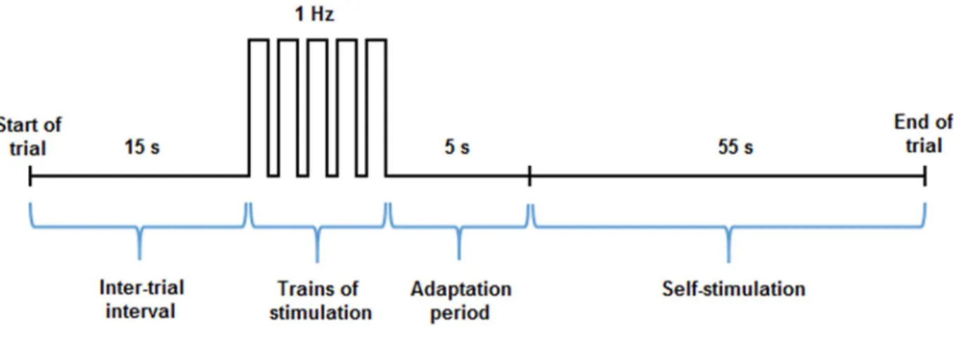
In the curve-shift paradigm of ICSS, animals are tested during several trials of varying stimulation frequencies and a constant current intensity so as to maintain the population of neurons that is stimulated unchanged. Although the parameters used for ICSS can greatly vary from one study to another, each trial typically begins with a 15 s inter-trial interval during which no electrical stimulation is provided, followed by the delivery of 5 trains of non-contingent priming stimulation, and a 5 s adaptation period (Figure 2). The priming trains of stimulation are typically delivered at a rate of 1 Hz, and are used to signal the arrival of discrete 55 s trials during which the animals will be allowed to self-stimulate at constant stimulation parameters (Figure 2). At the end of each trial, the frequency (i.e. the number of pulses per train of stimulation) is lowered by approximately
7
Figure 2: Schematic illustration of representative parameters used during an ICSS paradigm. The
nose poke response or number of lever presses of the animal is recorded during discrete 55 s trials. Trials are separated by a 15 s inter-trial interval (or time-out period), followed by the delivery of 5 trains of priming stimulation (delivered over 5 s) and a 5 s adaptation period. The stimulation frequency is reduced by approximately 0.1-log unit after each trial, and at the end of the session, a plot of the response rate as a function of the pulse frequency is generated.
0.1-log units, and at the end of the entire session, a R/F curve illustrating the rate of responding (number of lever presses or nose poke response) versus the stimulation frequency is obtained. During the discrete 55 s trials of ICSS, each operant response triggers the delivery of a single 400 ms train of rectangular cathodal pulses of very short duration (0.1 ms) so as to induce only one action potential per stimulated fiber. Cathodal currents are used instead of anodal currents because they are more effective in triggering action potentials (Ranck, 1975). Moreover, the electrode is connected to ground between deliveries of each pulse so that there is no build up of charge at the tip; building up of charge can be detrimental particularly with anodal stimulation. The delivery of each train of cathodal pulses is followed by a 600 ms period during which the pulse generator could not be triggered. The introduction of a fixed-interval delay after the delivery of each rewarding stimulation prevents the summation between two consecutive trains (Fouriezos, 1995)
8
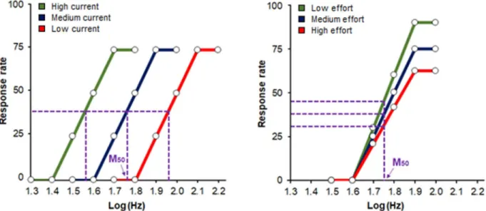
Figure 3: Schematic illustration of the theoretical functions that relate response rates to stimulation
frequency under different current intensities (A) and effort requirements (B). The stimulation frequency that maintains half-maximal responding (M50) is indicated by the pink arrow. Changes in the current intensity of the stimulation cause a lateral displacement of the R/F curve (A) while changes in task difficulty cause a vertical displacement of the R/F curve (B).
and enables a clear control over reward density, thus ensuring that the amount of reward received by the animal does not depend on its speed of responding, but rather on the rewarding effectiveness of the stimulation (Boye and Rompre, 1996).
The plot of the rate of responding of the animal for different pulse frequencies yields an R/F curve of quasi-sigmoidal shape characterized by a bottom portion, a rising portion, and an upper limit (also called plateau). Theoretical examples of R/F curves generated under different experimental conditions are illustrated in Figure 3A and 3B. When the current intensities and pulse duration of the stimulation are constant, the magnitude of the reinforcing effect of ICSS is function of the pulse frequency (Gallistel et al., 1981); animals will respond at negligible rates at low frequencies, intermediate rates for moderate frequencies, and maximal or asymptotic rates at high frequencies (Figure 3A and 3B). The most intuitive measure of the reinforcing efficacy of
9
the stimulation is the brain reward threshold, or M50, which corresponds to the pulse frequency sustaining a half-maximal rate of responding. Manipulations that increase or decrease the rewarding efficacy of ICSS shift the M50 towards lower or higher values, respectively, thereby causing a lateral displacement of the R/F curve (Edmonds and Gallistel, 1974). A decrease in the current intensity of the stimulation increases the M50, while the opposite effect is observed when the current intensity of the stimulation is increased (Figure 3A).
An advantage of using the curve-shift paradigm is that reward threshold that is derived from this method is remarkably stable over several months, thus allowing for multiple testing of each experimental animal over a long period of time (Stoker and Markou, 2011). The curve-shift paradigm also enables experimenters to evaluate the capacity of the subject to self-stimulate. Manipulations that induce a change in the capacity of the animal to self-stimulate (such as increasing or decreasing the effort needed to perform the operant response) lead to upward or downward shifts in the maximum response rate (Miliaressis et al., 1986). Animals will typically have a higher rate of responding when the effort required to self-stimulate is low, and a lower rate of responding when the effort required to self-stimulate is high (Figure 3B).When the reduction in maximum response rate is proportional at each of the pulse frequency tested, the M50 remains the same (Figure 3B). However, the curve shift paradigm does not necessarily enable a clear dissociation between reward threshold and maximum reponse rate since increases in task difficulty can displace the R/F curve laterally (Frank and Williams, 1985; Miliaressis et al., 1986; Fouriezos et al., 1990). The curve-shift paradigm is also insufficient inasmuch as it does not dissociate between the change in the subjective intensity and the cost of reward; this could be better addressed by the "reinforcement mountain" model (Arvanitogiannis and Shizgal, 2008).
10
1.1.4 Methodological and technical considerations
Since its discovery and implementation, the ICSS paradigm has become a powerful tool to study the neural substrates underlying reward and goal-directed behaviors. A major strength of this paradigm is that the electrical stimulation directly activates the brain reward circuitry, bypassing sensory systems and natural physiological processes like satiation. The ICSS procedure also provides quantitative measurements of brain stimulation reward that are robust and stable over long periods of time, thus allowing the experimenter to perform multiple tests or longitudinal studies on the same subjects (Stoker and Markou, 2011). Another advantage of ICSS is that the stimulation can be delivered to desired regions of the brain with extremely high temporal accuracy (in the order of ms). This can be achieved by controlling external parameters such as the pulse frequency and duration of the stimulation. However, studies employing ICSS with the use of an electrical stimulation have been plagued with a lack of anatomical specificity. The main issue is that the electrical stimulation of the brain does not allow the distinction between reward-relevant neural elements and those that do not play a role in reward, thus making the identification of the neural substrate for brain stimulation reward extremely challenging (Murray and Shizgal, 1996b). However, despite its technical limitations, electrical stimulation is still widely used in rodents for the identification of the neuroanatomical substrates of reward, and has proven valuable in the study of the reinforcing effects of drugs of abuse.
1.2 Anatomy, neurochemistry, and pharmacology of the brain reward system 1.2.1 Brain regions and neural pathways
The discovery by Olds and Milner that rats would readily press a lever to obtain pulses of electrical stimulation in certain brain regions (Olds and Milner, 1954) ushered in a series of
11
investigations aiming at determining the neuroanatomical substrate underlying brain stimulation reward. Shortly after this discovery, studies showed that electrical stimulation of forebrain and hypothalamic structures (Olds, 1956b, a) could also generate high response rates in an ICSS paradigm, which unequivocally pointed to the medial forebrain bundle (MFB) as a key neural pathway in the brain reward system. The MFB is a large tract of ascending and descending axons that span the entire length of the brain, passing through the basal forebrain and the lateral hypothalamus (LH) in a rostral-caudal direction. MFB fibers also course through midbrain regions including the ventral tegmental area (VTA), to terminate into several nuclei of the brainstem including the dorsal raphe (DR) nucleus (Nieuwenhuys et al., 1982), thus relaying information from one pole of the brain to the other. In the following paragraphs, evidence implicating the MFB in brain stimulation reward are discussed, with an emphasis on mapping, anatomical, and lesion studies. The dorsal diencephalic conduction system (DCC), which is another neural pathway of the brain reward system, will be discussed in details in the next section.
Following the identification of the MFB as a major substrate for ICSS, numerous brain mapping studies, where the anatomical site of the stimulation electrode is manipulated, were conducted to characterize the brain regions that could support ICSS (Figure 4). The emerging picture resulting from these studies suggests that operant responding for ICSS could be obtained along brain regions known to send or receive MFB fibers, including the orbitofrontal cortex (OFC) (Mora et al., 1980), the medial prefrontal cortex (mPFC) (Bielajew and Trzcinska, 1998), the caudate-putamen (Bielajew and Trzcinska, 1998), the lateral preoptic area (Bushnik et al., 2000), the ventral
12
Figure 4: Non-exhaustive illustration of regions involved in brain stimulation reward. This
simplified diagram illustrates key brain regions in the MFB (in red) and the DDC (in purple) that support operant responding for ICSS (citation in blue) and/or that result in an attenuation of the rewarding effects of MFB self-stimulation following a lesion (citation in green). Abbreviations: amygdala (Amy); caudate-putamen (CPu); dorsal raphe (DR); habenula (Hb); lateral hypothalamus (LH); lateral preoptic area (LPO); medial prefrontal cortex (mPFC); nucleus accumbens (NAc); orbitofrontal cortex (OFC); posterior mesencephalon (PM); ventral pallidum (VP); ventral tegmental area (VTA).
pallidum (Panagis et al., 1995), the amygdala (Kane et al., 1991), the pontine tegmentum (Rompre and Boye, 1989), the median raphe (MR) (Rompre and Miliaressis, 1985), and the DR (Corbett and Wise, 1979; Rompre and Boye, 1989). ICSS has also been reported in the habenula (Sutherland and Nakajima, 1981; Nakajima, 1984), the olfactory bulbs (Phillips, 1970), the hippocampus (Ursin et al., 1966; Phillips et al., 1977), the nucleus accumbens (NAc) (Mogenson et al., 1979), the VTA (Fibiger et al., 1987), and the cerebellum (Ball et al., 1974; Corbett et al., 1982). However, one of the most extensively studied substrates to date for brain stimulation reward is the LH owing
13
to its critical role in energy homeostasis and motivated behaviors (Berthoud and Munzberg, 2011; Stuber and Wise, 2016).
The LH is a heterogeneous area of the MFB located posterior to the preoptic area and anterior to the VTA. It resides dorsoventrally between the zona incerta and the base of the brain, and mediolaterally between the optic tract and the fornix. Studies employing electrical or optogenetic stimulation of the LH have implicated this structure in feeding (Delgado and Anand, 1953) and reward-seeking (Olds and Milner, 1954; Kempadoo et al., 2013) behaviors. Anatomically, the LH comprises several distinct nuclear subgroups that receive a wide array of internal and external information, making it well-suited to mediate functions across major output axes (Berthoud and Munzberg, 2011).Afferents to the LH have been classically studied using injections of a retrograde tracer (i.e., transport of the dye occurs in the reverse direction, from the axon terminal back to the cell body), the results of which have demonstrated the existence of projections originating from the bed nucleus of the stria terminalis (BNST), the diagonal tract of Broca, the caudate-putamen, the NAc, the lateral septal nuclei, the lateral preoptic area, the amygdala, and the zona incerta (Barone et al., 1981; Kita and Oomura, 1982). On the other hand, studies employing anterograde tracing techniques (i.e., transport of the dye occurs in the forward direction, from the cell body out to the axon terminal) showed that LH neurons project to distinct areas of the brain including the hypothalamic paraventricular nucleus, the lateral habenula (LHb), the VTA, the mesencephalic and pontine central gray, the lateral parabrachial nucleus, and the raphe nucleus (Kita and Oomura, 1982; Larsen et al., 1994).
Numerous studies have also employed lesions along certain brain structures or pathways in order to evaluate their impact on the reinforcing properties of ICSS (Figure 4). The reasoning behind this technique is based on the assumption that destruction of axons or cellular elements involved
14
in brain stimulation reward should result in an attenuation of the reinforcing effects obtained from ICSS. Therefore, assessing the strength of the reinforcing effect of ICSS before and after a lesion could help characterize the underlying neural circuit of brain stimulation reward. A common finding is that electrolytic lesions or knife cuts along the MFB cause a rightward shift in R/F curves obtained from LH (Janas and Stellar, 1987; Gallistel et al., 1996)or VTA (Simmons et al., 1998) self-stimulation, indicating a sustained attenuation of the rewarding effectiveness of ICSS. Lesions encompassing the habenula (Morissette and Boye, 2008), the cortical and adjacent amygdaloid subnuclei (Bielajew et al., 2002),the lateral preoptic area (Waraczynski, 1988), and the posterior mesencephalon (PM) (Boye, 2005) also cause a rightward shift in R/F curves for MFB self-stimulation. However, several other studies employing electrolytic lesions failed to observe decreases in the rewarding effectiveness of MFB stimulation. In particular, lesions at the amygdala (Waraczynski et al., 1990), the dorsomedial hypothalamus (Waraczynski et al., 1992), the parabrachial nucleus (Waraczynski and Shizgal, 1995), the rostral LH (Gallistel et al., 1996), and the lateral PM (Boye, 2005) have not resulted in consistent and noticeable changes in the rewarding effectiveness of MFB stimulation. A hypothesis to account for these negative findings is that the neural network subserving ICSS is anatomically diffuse, collateralized, and highly heterogeneous (Lorens, 1966; Simmons et al., 1998), and may be comprised of several pathways that are functionally interconnected. As such, the loss of reward-relevant neurons within one pathway would be compensated by the other, and vice versa, thereby enabling the integration and transmission of reward signals in the brain.
15
Dopamine (DA) is a neurotransmitter of the catecholamine family and plays a crucial role in a variety of processes within the central nervous system (CNS). In the brain, DA is synthesized from dopaminergic neurons of the VTA, substantia nigra pars compacta (SNc), retrorubral field (RRF), and hypothalamic arcuate and periventricular nuclei, and is transmitted via distinct dopaminergic pathways that play unique functions (Bjorklund and Dunnett, 2007; Luo and Huang, 2016). Dopaminergic neurons of the SNc project into the caudate-putamen via the nigrostriatal pathway, which play a key role in the regulation of voluntary movements (Luo and Huang, 2016). On the other hand, VTA DA neurons project to the NAc and other structures of the limbic system via the mesolimbic pathway, and to the prefrontal cortex via the mesocortical pathway (Luo and Huang, 2016). The mesolimbic and mesocortical pathways are primarily involved in emotion-related behavior and are often collectively referred to as the mesocorticolimbic pathway because of their significant overlap and association (Arias-Carrion and Poppel, 2007). Given the wide range of functions mediated by dopaminergic pathways, it is not surprising that abnormalities in DA transmission have been associated with numerous neuropathological conditions, including schizophrenia, major depressive disorders, substance use disorder, and Parkinson’s disease (Birtwistle and Baldwin, 1998; Dunlop and Nemeroff, 2007; Volkow et al., 2009).
Midbrain DA neurons have been extensively studied in the context of reward-related processes owing to their ability to encode information about motivational salience (Bromberg-Martin et al., 2010; Schultz, 2015). In monkeys, these neurons show increased firing rate following the delivery of unpredicted food or liquid rewards, and following the presentation of reward-predicting stimuli (Ljungberg et al., 1992; Schultz et al., 1997; Schultz, 2010). DA neurons not only serve as a global reward signal, but also function as a reward prediction error signal, that is, the degree of discrepancy between the actual reward and its prediction (Schultz, 2016). In the context of ICSS,
16
early evidence implicating DA in brain stimulation reward comes from studies showing that electrical stimulation of DA-containing cell bodies in the midbrain (Crow, 1972a, b; Wise, 1981) and diencephalic areas that are traversed by DA fiber bundles (Corbett and Wise, 1980) produces positive reinforcement. In the study by Corbett & Wise (1980), reward thresholds obtained from stimulating ascending midbrain dopaminergic pathway were negatively correlated with the density of DA neurons surrounding the tip of the stimulation electrode, suggesting that DA transmission is strongly involved in mediating the rewarding effectiveness of ICSS. Neurochemical studies employing fast-scan cyclic voltammetry and/or in-vivo microdialysis to measure the extracellular release of DA in the brain have also provided robust evidence for the involvement of DA in the rewarding effect of ICSS. These studies reported long-lasting increases in the extracellular concentration of DA in various brain regions, including the NAc (Nakahara et al., 1989; You et al., 2001) and mPFC (Bean and Roth, 1991; Nakahara et al., 1992) following electrical stimulation of the MFB. Increased release of DA metabolites, including 3,4-dihydroxyphenylacetic acid (DOPAC) and homovanillic acid (HVA), were also observed in the striatum, NAc, and olfactory tubercle of rats following VTA self-stimulation (Fibiger et al., 1987). Consistently, DA release in the NAc was significantly increased following VTA self-stimulation (Phillips et al., 1992; Hernandez and Shizgal, 2009), and was found to be inversely correlated with reward thresholds for MFB self-stimulation (Yavich and Tanila, 2007). Last but not least are electrophysiological findings showing that the majority of midbrain DA neurons ( >70%) are activated by rewarding electrical stimulation of the PM (Moisan and Rompre, 1998), thus concurring with the view that DA participates in the reinforcing effects of ICSS.
In addition to electrophysiological and neurochemical findings, data from pharmacological studies also provide robust evidence for the role of DA transmission in brain stimulation reward.
17
DA binds to and activates five different receptor subtypes that are divided into two major subclasses: D1-like receptors, which include the D1 and D5 subtypes, and D2-like receptors, which include D2, D3 and D4 subtypes (Jaber et al., 1996; Beaulieu and Gainetdinov, 2011). Administration of pimozide and chlorpromazine, two selective antagonists for DA D2 receptors, produce a rightward shift in ICSS reward thresholds, indicating that the rewarding efficacy of the stimulation is decreased (Gallistel and Karras, 1984; Miliaressis et al., 1986; Gallistel and Freyd, 1987). Reduced brain stimulation reward was also obtained following blockade of DA D2 receptors (Schaefer and Michael, 1980; Benaliouad et al., 2007) and DA D1 receptors (Nakajima and McKenzie, 1986), suggesting that DA transmission is critical in the objective reinforcement associated with ICSS. Accordingly, pharmacological manipulations that enhance DA transmission, such as agonist-mediated activation of DA receptors (Gilliss et al., 2002), blockade of DA transporter (Rompre and Bauco, 1990; Maldonado-Irizarry et al., 1994), and administration of psychostimulants such as amphetamine and cocaine (Colle and Wise, 1988; Straub et al., 2010), produce a leftward shift in brain reward thresholds. However, other studies failed to observe changes in the rewarding efficacy of ICSS following administration of DA receptors agonists (Malanga et al., 2008) or antagonists (Fibiger et al., 1976), suggesting that the mechanisms underlying the reinforcing effect of ICSS might not entirely depend on DA transmission. Consistent with this view, rats that received 6-hydroxydopamine (6-OHDA)—a neurotoxin that selectively destroys DA neurons—by intracerebroventricular injection (Sidhu et al., 1993) or bilaterally into the substantia nigra (Ornstein and Huston, 1975) showed normal rates of responding for ICSS. Altogether, the aforementioned studies suggest that although DA transmission plays a crucial role in the reinforcing effects of ICSS, there are most likely other systems involved in brain stimulation reward. Among those, the glutamate and opioid systems
18
have garnered considerable attention as important mediators of reward-related processes owing to their critical role in controlling DA neuronal activity (Le Merrer et al., 2009; D'Souza, 2015). In the following sections, an overview of evidence implicating these modulatory systems in reward processing is presented.
1.2.3 Glutamate transmission: overview and implication in reward processing
Glutamate is the most abundant excitatory neurotransmitter in the brain and accounts for the majority of synaptic transmission (Niciu et al., 2012). Present in high concentrations in the CNS, glutamate is a non-essential amino acid that can be synthesized from glucose and a variety of other sources (Dingledine and McBain, 1999). Glutamate also serves as a metabolic precursor to gamma-aminobutyric acid (GABA), the main inhibitory neurotransmitter in the brain, the latter being synthesized from the former by the enzyme glutamic acid decarboxylase (Fenalti et al., 2007). As an amino acid and neurotransmitter, glutamate is involved in a wide range of physiological processes, and perturbations in glutamate transmission can result in deleterious effects. For instance, glutamate-mediated excitotoxicity, which occurs as a result of glutamate receptors overactivation and excessive entry of calcium inside the cells, can cause neuronal damage and/or death (Sattler and Tymianski, 2001), and has often been associated with numerous neurodegenerative conditions such as Alzheimer’s disease (Hynd et al., 2004), Parkinson’s disease (Beal, 1998), and amyotrophic lateral sclerosis (Shaw and Ince, 1997). Because disruptions in glutamate transmission have also been linked to imbalances in the DA system (Kretschmer, 1999) and changes in reward sensitivity (Bechtholt-Gompf et al., 2010), a growing number of studies are focusing on the glutamatergic system as a therapeutic target for psychiatric conditions such as schizophrenia, depression, and substance use disorder (Javitt, 2004; Kerner, 2009).
19
In order to prevent neuronal damage that might occur as a result of glutamate-mediated excitotoxicity, the extracellular fluid concentration of glutamate is tightly regulated by excitatory amino acid transporters (EAATs) (Zhou and Danbolt, 2014). The EAATs are located on glutamatergic terminals and presynaptic glial cells, and are responsible for the removal of glutamate from the synaptic cleft (D'Souza, 2015). The vesicular glutamate transporters (VGLUTs), which constitute another class of glutamate transporters, are responsible for the uptake and sequestration of glutamate into presynaptic vesicles, and are dependent on a proton gradient that is generated upon the hydrolysis of adenosine triphosphate (ATP) (Liguz-Lecznar and Skangiel-Kramska, 2007). Once stored in vesicles, glutamate gets released into the synaptic cleft by exocytosis, and binds to either metabotropic or ionotropic glutamate receptors (Figure 5). Metabotropic glutamate receptors (mGluR) are slow-acting G-protein coupled receptors located in presynaptic and postsynaptic terminals, which are subclassified into three groups based on anatomical and functional homology: group I includes mGluRs 1 and 5, Group II includes mGluRs 2 and 3, and Group III includes mGluRs 4, 6, 7 and 8 (Niswender and Conn, 2010). Conversely, ionotropic glutamate receptors are fast-acting ligand- gated ion channels which include amino-3-hydroxy-5-methyl-4-isoxazolepropionate (AMPA), N-methyl-D-aspartate (NMDA), and kainate receptors.
AMPA receptors are tetramers composed of four subunits designated as GluR1, GluR2, GluR3
and GluR4 (Ward et al., 2010). Each of the GluR1-4 subunits exists in two different forms (“flip” and “flop”) created by alternative splicing, thus conferring different signalling properties to AMPA receptors (Ozawa et al., 1998). Kainate receptors are also tetramers that form ligand-gated ion channels and that can be assembled from a combination of five different subtypes of subunit: GluK1-3 and GluK4-5 (Fisher and Fisher, 2014). Both AMPA and kainate receptors interact with
20
Figure 5: Ionotropic and metabotropic glutamate receptors. NMDA receptors interact with
glutamate, glycine, Mg2+, Zn2+ and polyamines, and contain a channel that allows the passage of Ca2+, Na+ and K+ ions. AMPA and kainate receptors interact only with glutamate and their specific agonists, and contain a channel that is permeable to Na+ and K+ ions. On the other hand, mGluRs are members of the G-protein-coupled receptor superfamily. They regulate ion channels and downstream signalling by activating a guanosine triphosphate (GTP)-binding protein (Gq for Group I, and Gi/G0 for Group II and Group III mGluRs), which in turn modulates the function of various effector molecules including the enzymes adenylyl cyclase and phospholipase C. Abbreviations: adenylyl cyclase (A cyclase); AMPA receptor (AMPAR); glutamate (Glu); glycine (Gly); kainate receptor (KAR); metabotropic glutamate (mGlut); NMDA receptor (NMDAR); phencyclidine (PCP); phospholipase C (PLC). Adapted from Kritis et al., 2015.
glutamate and their specific agonists, and their associated channels are permeable to various cations including Na+ and K+ (Kritis et al., 2015). On the other hand, NMDA receptors are tetramers composed of two obligatory GluN1 subunits (for which there are several splice variants), and two of the following subunits: GluN2A, GluN2B, GluN2C, GluN2D, GluN3A and GluN3B (Paoletti et al., 2013). The two non-GluN1 subunits can be identical or different, thus giving rise to di-heteromeric or tri-heteromeric receptors (Paoletti et al., 2013). Unlike AMPA and kainate receptors, NMDA receptors need to be co-activated by two ligands: glutamate and either D-serine
21
or glycine (Kleckner and Dingledine, 1988). In addition, the NMDA receptor pore is blocked by Mg2+ ions in a voltage-dependent manner, however, this block can be dislodged following sufficient membrane depolarization such as opening of AMPA receptor channels (Gass and Olive, 2008). Other regulatory sites on NMDA receptors are recognized by the dissociative anesthetics phencyclidine (PCP), which selectively antagonizes the response to NMDA in a non-competitive manner, and by Zn2+ ions, which produce a voltage-independent block.
Converging evidence implicates glutamate transmission in the regulation of DA neuronal activity. Under resting conditions, a small proportion of DA neurons are non-responsive to excitatory inputs, while the majority of DA cells fire spontaneously in two distinctive patterns of activity: (i) a tonic or single spike mode, and (ii) a fast phasic burst firing (Grace and Bunney, 1984a, b). Activation of glutamatergic afferents in the ventral midbrain mediates the switch from pacemaker tonic to phasic burst firing of DA neural activity (Johnson et al., 1992; Chergui et al., 1993; Lodge and Grace, 2006), a mode that is associated with increased DA release (Gonon, 1988; Overton and Clark, 1997). Besides its role in switching the firing mode of DA cells, glutamate afferents also establish synaptic contacts with local GABAergic interneurons of the midbrain, thereby enhancing the inhibitory drive onto DA neurons (Dobi et al., 2010; Omelchenko and Sesack, 2010). In light of these observations, studies examining the capacity of glutamate receptor agonists to alter DA release have reported conflicting results. For instance, both increases and decreases in DA release were observed in the striatum and NAc following activation of metabotropic (Ohno and Watanabe, 1995; Feenstra et al., 1998; Verma and Moghaddam, 1998) and ionotropic (Karreman et al., 1996; Wu et al., 2000) glutamate receptors. Moreover, activation (Kretschmer, 1999) and blockade (Narayanan et al., 1996; Cornish et al., 2001) of ionotropic glutamate receptors in the midbrain enhances locomotor activity, a DA-dependent behavioral
22
measure. The discrepancies in the results observed concur with the view that glutamate exerts opposite regulatory functions on DA neuron activity, and raise the possibility that such opposite modulation may be mediated by different glutamate receptor subtypes.
Because of its differential role in the regulation of DA neuron activity, glutamate has been
proposed to mediate opposite effects on reward by acting on different glutamatergic receptor subtypes. To further clarify the role of midbrain glutamatergic transmission in reward, a series of elegant studies has evaluated the effect of ionotropic glutamatergic receptors blockade on the reinforcing efficacy of ICSS. In rats, intra-VTA injection of the AMPA receptor antagonist, 2,3,-Dioxo-6-nitro-1,2,3,4-tetrahydrobenzo(f)quinoxaline-7-sulfonamide (NBQX), dose-dependently attenuates the rewarding efficacy of ICSS, most likely as a result of reduced glutamatergic excitatory inputs to midbrain DA neurons (Ducrot et al., 2013). The view that AMPA receptor blockade causes a marked reduction in brain stimulation reward is in line with studies showing that injection of AMPA antagonists into the VTA blocks the development of a conditioned place preference to cocaine (Harris and Aston-Jones, 2003) and morphine (Harris et al., 2004). However, it is in contrast with a previous report showing that mice can learn to self-administer the AMPA receptor antagonist, 6,7-dinitroquinoxaline-2,3-dione (DNQX), into the VTA (David et al., 1998). Such discrepancies in the results could be attributable to differences in experimental approaches (i.e., drug and dose used) and injection sites (rostral versus caudal regions of the VTA). A study by Ducrot et al. (2013) also investigated the effect of (2R,4S)-4-(3-Phosphonopropyl)-2-piperidinecarboxylic acid (PPPA)—an NMDA receptor antagonist with a preferred action on GluN2A subunits—on the rewarding efficacy of ICSS, showing that PPPA injection into the VTA produces a time-dependent increase in brain stimulation reward. The reward-enhancing properties of PPPA are very similar to those reported by other studies using the same dose and injection site
23
Figure 6: Glutamate transmission in the ventral midbrain exerts opposite roles. On the one hand,
glutamate can switch the firing pattern of DA neurons from tonic to phasic burst firing. On the other hand, glutamate afferent terminals can make synaptic contact with local GABAergic interneurons, thereby increasing the inhibitory drive onto DA neurons. The inhibitory effect of glutamate on DA neuronal activity is most likely mediated by GluN2A-containing NMDA receptors located on afferent terminals. Abbreviations: ventral midbrain (VM). Adapted from Hernandez et al., 2015.
(Bergeron and Rompre, 2013; Hernandez et al., 2016), and suggest that GluN2A-containing NMDA receptors are mainly expressed on GABAergic neurons of the VTA. Using the small interferon RNA (siRNA) technique, Hernandez et al. (2015) also showed that downregulation of NMDA receptors in the ventral midbrain attenuates brain stimulation reward, most likely as a result of decreased glutamate-mediated excitability of DA neurons. However, the siRNA-mediated downregulation of NMDA receptors failed to alter the reward-enhancing effect of PPPA,
24
indicating that GluN2A-containing NMDA receptors are most likely located on glutamate afferent terminals of the ventral midbrain (Figure 6) (Hernandez et al., 2015).
Taken together, the aforementioned findings suggest that glutamate signalling is highly implicated in reward and motivational processes as a result of its strong influence over DA neuron activity. This could have important implications for psychiatric conditions such as major depressive disorders, where abnormalities in reward processing are frequently observed (Admon and Pizzagalli, 2015; Whitton et al., 2015). Ketamine, a pharmacological agent that targets glutamate NMDA receptors, has already proven successful in producing rapid and long-lasting attenuations of depressive symptoms in patients (Berman et al., 2000; Messer and Haller, 2017), however, evidence supporting its use as an antidepressant is still very limited and requires further investigation.
1.2.4 The opioid system: overview and implication in reward processing
Early evidence for the existence of an opioid system stems from the use of opium by the Sumerians in 3400 B.C., who called it "gil"—the word for joy (Brownstein, 1993). Opium is obtained from the seed capsules of the opium poppy, Papaver somniferum, and is the source of a family of drugs referred to as opiates or opioids. The opioids have been used for both recreational and medicinal purposes for thousands of years due to their principal effect in euphoria and pain reduction. In the beginning of the 19th century, Sertürner isolated the active ingredient of opium, and named it morphine after the Greek god of dreams, Morpheus (Brownstein, 1993). In addition to morphine, opium contains other closely related opioids including codeine and thebaine. However, despite being effective in pain relief, these opium-derived compounds were found to be highly addictive and not very safe to use. In an effort to develop a safer opiate for pain relief,