Prevalence of naturally occurring cartilage defects in the ovine knee
J.-M. Vandeweerd
y
*, F. Hontoir
y
, N. Kirschvink
y
, P. Clegg
z
, J.-F. Nisolle
x
, N. Antoine
k
,
P. Gustin
{
y Integrated Veterinary Research Unit (IVRU), Namur Research Institute for Life Sciences (NARILIS), Department of Veterinary Medicine, University of Namur, Belgium
z Department of Musculoskeletal Biology, Faculty of Health and Life Sciences, University of Liverpool, Neston, UK x Department of Diagnostic Imaging, Université Catholique de Louvain, Mont Godinne, Belgium
k Department of Morphology and Pathology, Faculty of Veterinary Medicine, University of Liège, 4000, Sart-Tilman, Belgium
{ Department of Functional Sciences, Unit of Pharmacology, Faculty of Veterinary Medicine, University of Liège, 4000, Sart-Tilman, Belgium
a r t i c l e
i n f o
Article history: Received 17 December 2012 Accepted 13 May 2013 Keywords: Sheep Cartilage defects Prevalence Stifle Knees u m m a r y
Objective: To determine the prevalence, anatomical location and severity of cartilage defects in the stifle (knee) within a population of adult ewes (N ¼ 65).
Materials and methods: Articular cartilage (AC) of the distal femur, proximal tibia and patella was assessed using Osteoarthritis Research Society International (OARSI) recommendations for macroscopic and microscopic scoring of ovine cartilage. Synovial fluid analysis and histology of the synovial membrane were performed. All limbs were examined by computed tomography.
Results: Twenty-eight sheep (n ¼ 28; 43%) presented at least one score 2 or score 3 lesion. Twenty-two (n ¼ 22; 34%) sheep were macroscopically normal. Most frequent localizations of lesions were: axial aspect of the central third of the medial tibial condyle (32.7% of the lesions), middle third of the medial femoral condyle (29.4%), middle third of the articular surface of the patella (9.8%), and axial aspect of the central third of the lateral tibial condyle (9.8%). Grade of macroscopic lesions was significantly (H (3) ¼ 29.31, P 0.000) affected by age. Macroscopic score correlated well with histological changes that can be found in osteoarthritis (OA) (r 0.83; P 0.000). Neither clinical signs of OA, nor cytological and histo-logical signs of inflammation were identified, while imaging abnormalities were very rare.
Conclusions: Our data seem to indicate that naturally occurring OA exists in ageing sheep, at least sub-clinically. It might be useful to take into account prevalent cartilage defects at baseline in studies using ovine models.
!2013 Osteoarthritis Research Society International. Published by Elsevier Ltd. All rights reserved.
Introduction
Articular cartilage (AC) defects of the knee are common in humans and are encountered in approximately 60% of knee ar-throscopies1. The extent of pathology ranges from small focal defects to widespread damage to the articular surface. In animals, there is limited information about the prevalence and impact of AC defects. Palmar osteochondral disease (POD)2and AC defects in the meta-carpo-phalangeal joint3are prevalent in Thoroughbred racehorses. In sheep, microscopic lesions of osteochondrosis were reported to be frequent in fast-growing lambs although few defects progressed to gross lesions4. Spontaneous non-infectious osteoarthritis (OA)
has not been reported in this species. Proteoglycan loss, cartilage matrix atrophy and erosion of the cartilage consistent with OA change occur spontaneously in the unprotected region of the medial tibial plateau in goats as young as 2 years of age5. As ovine models of
knee OA are an essential modality for exploring the pathophysiology and therapy of the disease6e15, information about the prevalence of AC abnormalities in this joint would be useful.
The objective of this study was to determine the prevalence, anatomical location and severity of naturally occurring cartilage defects in the knee within a population of adult ewes enrolled for research.
Material and methods Animals
Hind limbs (n ¼ 130) from 65 crossed Texel ewes (N ¼ 65), euthanatized for reasons other than hind limb lameness, were * Address correspondence and reprint requests to: J.-M. Vandeweerd, Integrated
Veterinary Research Unit (IVRU), Namur Research Institute for Life Sciences (NAR-ILIS), Department of Veterinary Medicine, University of Namur, Rue de Bruxelles 61, 5000 Namur, Belgium. Tel.: 32-81-72-43-79.
E-mail address:[email protected](J.-M. Vandeweerd).
1063-4584/$ e see front matter ! 2013 Osteoarthritis Research Society International. Published by Elsevier Ltd. All rights reserved. http://dx.doi.org/10.1016/j.joca.2013.05.006
disarticulated at the level of the coxo-femoral joint and collected within 6 h of euthanasia. Sheep ranged from 6 months to 11 years old (mean 5.9; standard deviation [SD] 3.1) and weighed between 35 and 72 kg (mean 57.5; SD 9.0). The animals came from the Ovine Research Centre of the University of Namur. The experimental protocol (KI 10/148) was approved by the local ethical committee for animal welfare. Sheep included in this study were euthanized from May 2011 to February 2013. Each limb was identified by a number.
Outcome measures Computed tomography
Immediately after euthanasia, all limbs (n ¼ 130) were exam-ined with an Emotion 6 (Siemens). Acquisition protocol was: 130 KV, 80 mAs, pitch 0.4, collimation 0.6 mm and rotation time of the tube 0.6 s. Images of 0.6 mm were reconstructed with an increment of 0.3 mm. From this isotropic volume of images, coro-nal, sagittal and transversal reformations were obtained (1 mm thickness and 1 mm space between slices). Field of view extended from the base of the patella to 6 cm below the tibial plateau. Images were reconstructed on a 512e512 matrix, and in-plane resolution was 0.2 mm. Images were then transferred on a medical digital imaging system (PACS, TELEMIS). All reconstructed images were prospectively stored on CD roms. Later, the knees were examined by a specialist in medical imaging, who was not aware of the identity of the limbs, to identify osteophytosis, subchondral bone (SB) sclerosis and SB irregularities. Those features were evaluated as present or absent.
Synovial fluid (SF) analysis
The knee region was clipped and cleaned. Synoviocentesis was performed on both limbs of the first 50 (N ¼ 50) sheep of this series by a paraligamentous approach using a 20-gauge needle16. It was not performed on the last 15 individuals (N ¼ 15) because of financial costs that would not have been justified considering the absence of findings in the initial 50 sheep. In those animals, when SF was obtained, volume was measured. Samples were placed in tubes containing ethylenediaminetetraacetic acid (EDTA) for routine SF analysis. Total blood cell count and numeration were performed by flow cytometric method (ADVIA 120"
e Siemens). Total protein concentration was measured by spectrophotometry (DPP"
e Roche). Cytology results were checked by microscopic examination and manual counting using a hematocytometric cell. Histology of the synovial membrane
Also, in the first 50 sheep of this series (N ¼ 50), samples of synovial membrane were harvested in each knee (n ¼ 100) from the suprapatellar fold. Specimens were fixed in 10% (v/v) neutral buffered formalin and embedded in paraffin wax. Sections (4
m
m)were stained with haematoxylin and eosin (H&E). Several param-eters were observed for their presence: intima hyperplasia, lym-phocytic or plasmocytic cellular infiltrate, sub-intimal fibrosis and vascularity5.
Gross observation
Soft tissue was removed, and the knee joint was carefully dis-articulated. The cartilage was kept moist by covering the joint surface with gauze sponges soaked in lactated Ringer’s solution. The distal articular surface of the femur, proximal articular surface of the tibia and articular surface of the patella were examined by gross observation by two investigators. Joint surfaces were digitally photographed with standardized lighting conditions for records.
Both investigators, working in consensus, identified and scored the abnormalities of the AC. Macroscopic scoring was done in a
blinded manner, such that the age and the sheep identity (i.e., leg pairing) were unknown to the scorers. Osteoarthritis Research So-ciety International (OARSI) recommendations for macroscopic scoring of cartilage in sheep5were used: score 0 for intact cartilage
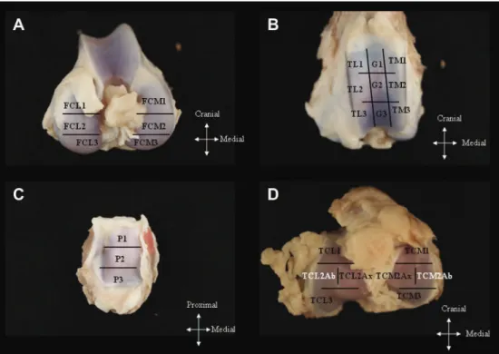
surface; score 1 for surface roughening; score 2 for deeper defects (fibrillation, fissures) not involving the SB; score 3 for erosions down to SB (less than 5 mm diameter); score 4 for large erosions down to SB (more than 5 mm diameter). Scoring of articular sur-faces was performed in 26 anatomic areas of each knee (Fig. 1). In a second phase, two other investigators reviewed the specimens and compared their scoring to the first list of lesions. Discrepancies were discussed between all investigators till a consensus was reached. For macroscopic observation, the most severe lesion observed in one region was used to score the articular surface of that region. Grading of cartilage defects within each knee (“knee grade”) was obtained by the addition of the scores of all regions in that knee. A “sheep grade” was also calculated by addition of the grades from the paired legs.
Histolopathology of cartilage
Three to four millimetre thick osteochondral slabs were ob-tained for every lesion, centred on the articular surface defect, and were processed for histology. Samples from sites with no macro-scopic abnormality (macromacro-scopic scoring of 0) were also obtained for control. These control samples were randomly selected among the articular surfaces which were diagnosed healthy at macro-scopic observation, and in a number similar to the total number of the lesions identified in the 65 sheep (N ¼ 65). Samples were fixed in 10% (v/v) neutral buffered formalin for 48 h. Following fixation, samples were transferred to 70% (v/v) ethanol for storage or further processing. They were decalcified in DC3 (non-ionic surfactants, chlorhydric acid, EDTA) for 2 days and embedded in paraffin. Four-micron sections were cut. Sections were de-paraffinised with xylene and graded ethanol, and then stained with toluidine blue.
Histological analysis was carried out independently by two in-vestigators. Discrepancies were discussed till a consensus was reached. Sheep, age, limb identity and macroscopic score were unknown by the histological scorers. Scoring was performed using the OARSI recommendations for histological evaluation of AC in sheep5. Histological abnormalities seen in osteoarthritic cartilage include structural defects, chondrocyte cloning and death, loss of proteoglycan, calcified cartilage layer (CCL) thickening and tide-mark duplication, penetration of blood vessels through SB plate and tidemark, sclerosis of the SB5,17.
The objectives of histological analysis were (1) to assess carti-lage structure and to evaluate the accuracy of macroscopic obser-vation (for example, to confirm whether a score 2 lesion at gross observation correlated with a partial thickness defect [fibrillation, fissures] not involving the SB at microscopy), (2) to identify any evidence of other abnormalities (other than structural changes) present in OA, and (3) to evaluate whether histological features of OA cartilage pathology were present in normal (non-lesion) cartilage.
Statistical analysis
Data for analysis included 65 (N ¼ 65) “sheep grades”, 130 (n ¼ 130) “limb grades” and CT observations, 100 (n ¼ 100) SF and synovium analyses, 153 (n ¼ 153) macroscopic scores of lesions and 268 (n ¼ 268) histological scores (cases and controls). Kolmo-goroveSmirnov and ShapiroeWilk tests were used to examine the normality of data. Due to the non-normality of data, non-para-metric tests were used. Correlations between variables were assessed by using the Spearman correlation coefficient. The Wil-coxon signed-rank test and the Friedman test were used to
compare observations that were not independent (“limb grades”, anatomical locations). The KruskaleWallis test was used to compare the mean ranks of “sheep grades” in different categories of age. Data were collected in Microsoft Excel and were analysed using IBM"
SPSS"
Statistics, version 15 for Windows. A P-value less than 0.05 was considered to indicate a statistically significant difference. Results
Macroscopic findings
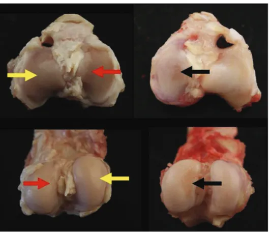
One hundred and fifty three cartilage defects were detected over 43 (n ¼ 43) of the 65 (N ¼ 65) sheep in 13 anatomical locations (Table I). Twenty two (n ¼ 22) sheep were macroscopically normal. They were 70 score 1 (45.8%), 76 score 2 (49.7%), and seven score 3 (4.6%) lesions. Twenty eight sheep (n ¼ 28; 43%) presented at least one score 2 or score 3 lesion. Most frequent localizations of lesions were: axial aspect of the central third of the medial tibial condyle (TCM2Ax; 32.7% of the lesions), middle third of the medial femoral condyle (FCM2; 29.4%), middle third of the articular surface of the patella (P2; 9.8%), and axial aspect of the central third of the lateral tibial condyle (TCL2Ax; 9.8%). Mean macroscopic scores were 1.50 (SD 0.08) for TCM2Ax, 1.58 (SD 0.08) for FCM2, 1.93 (SD 0.15) for P2, and 1.53 (SD 0.13) for TCL2Ax. Examples of gross anatomic lesions are shown inFig. 2.
Age, weight and “sheep grades” are reported inTable II. Mean “sheep grade” was 3.83 (SD 4.99; range 0e23). There was no sig-nificant difference in “limb grades” between left and right limbs. There was no correlation between weight and “sheep grade”, while age and sheep grades were well correlated (r 0.675; P 0.000). “Sheep grade” was significantly (H (3) ¼ 29.31, P 0.000) affected by categories of age (1e3 year old, 4e6, 7e8, 9e11), with mean ranks of 16.21, 28.09, 39.54 and 50.79 respectively.Table IIshows that no lesion was found in the eleven (N ¼ 11; 100%) sheep less than three
year old, while at least one score 2 or 3 lesion was found in 15 (N ¼ 15) of the 17 (N ¼ 17), i.e., 88%, sheep aged between nine and 11. Computed tomography did not identify any signs of SB sclerosis or osteolysis. Osteophytes were found in only two animals (N ¼ 2). One sheep was 11 year old and had a “sheep grade” of 23, while the other was seven and had a “sheep grade” of 18. Osteophytes were bilateral (left and right limb), small and present on the medial aspect of the tibial plateau. CT identified SB sclerosis only in this 11-year-old sheep.
Fig. 1. Anatomic subregions assessed for AC abnormalities. (A) Cranial, middle and caudal thirds of the articular surface of the medial and lateral femoral condyles (FCM1, FCM2, FCM3, FCL1, FCL2, FCL3); (B) proximal, middle and distal thirds of the articular surface of the medial ridge, groove and lateral ridge of the trochlea of the femur (TM1, TM2, TM3, G1, G2, G3, TL1, TL2, TL3); (C) proximal, middle and distal thirds of the articular surface of the patella (P1, P2, P3); (D) cranial, middle and caudal thirds of the articular surface of the medial and lateral tibial condyles, where each middle third was divided into axial and abaxial aspects (TCM1, TCM2Ax, TCM2Ab, TCM3, TCL1, TCL2Ax, TCL2Ab, TCL3).
Table I
Distribution of localizations of lesions (n ¼ 153) in this series of 65 sheep (N ¼ 65). Proximal, middle and distal thirds of the articular surface of the patella (P1, P2, P3); proximal and middle thirds of the groove of the trochlea of the femur (G1, G2); middle third of the lateral ridge of the trochlea of the femur (TL2); middle and caudal thirds of the articular surface of the medial femoral condyle (FCM2, FCM3); cranial and middle thirds of the articular surface of the lateral femoral condyle (FCL1, FCL2); middle third of the articular surface of the medial tibial condyle, divided into axial and abaxial aspects (TCM2Ax, TCM2Ab); middle third of the axial aspect of the articular surface of the lateral tibial condyle (TCL2Ax)
Localization n % of n total TCM2Ax 50 32.7% FCM2 45 29.4% P2 15 9.8% TCL2Ax 15 9.8% G2 8 5.2% FCM3 5 3.3% FCL2 5 3.3% G1 3 2.0% TL2 2 1.4% FCL1 2 1.3% TCM2Ab 1 0.7% P1 1 0.7% P3 1 0.7% Total 153 100.0%
Microscopic findings
SF analysis and synovium histopathology
No synovial effusion was identified in all limbs. In 93 (n ¼ 93) of the 100 knees (n ¼ 100) where arthrocentesis was performed, no SF could be obtained. The volume of SF collected from the seven (n ¼ 7) remaining knees (from five different sheep) varied between 0.5 ml and 1.2 ml (0.93 ' 0.26 SD). All samples were transparent, clear to pale yellow, and highly viscous, except one which was slightly discoloured by blood. Total protein concentration was within normal limits (2.5e5.0 g/100 ml) and varied from 1.69 to 3.41 g/100 ml (mean 2.45 ' 0.73 SD). Red blood cell (RBC) count was within normal limits (<10,000/mm3) except in the contami-nated sample (50,000/mm3). White blood cell (WBC) count was
within normal limits (<5,000/mm3) and varied from 30 to 300 (116.67 mean ' 110.03 SD). Histological analysis of the synovium that was sampled in the 100 limbs (n ¼ 100) did not identify any characteristics of inflammation.
Cartilage histopathology
A total of 153 (n ¼ 153) anatomic sections were obtained at the level of the 153 (n ¼ 153) lesions identified. Random selection of control sites produced respectively 130 (n ¼ 131) one slices. On a total of 284 (n ¼ 284) histological slices, 16 (n ¼ 16) presented ar-tefacts or inadequate colouration and were not used for analysis. Correlation between histological structural score and macro-scopical score was very good (Spearman coefficient, 0.83; P 0.000). Table III summarizes the histological findings (structure, chon-drocyte density, cell cloning, inter-territorial blue, tidemark and extension of lesion) found in every level of macroscopic score Fig. 2. Examples of score 0 (yellow arrow), 1 (red arrow) and 2 (black arrow) defects.
Table II
Distribution of ages (years) for the observed animals (N ¼ 65). Mean weight and “sheep grade” (with their SD) are reported per age (years). The number of animals with at least one score 2 or 3 lesion (N2, 3) and their corresponding percentage in the study sample, are shown
Age Number of animals Weight Sheep grade N2, 3 %
0.5 5 46.40 (0.92) 0.00 (0.00) 0 0.0 1.0 4 41.00 (2.91) 0.00 (0.00) 0 0.0 2.0 2 55.00 (0.00) 0.00 (0.00) 0 0.0 3.0 3 57.33 (2.33) 2.33 (0.88) 1 1.5 4.0 6 63.00 (2.17) 2.50 (1.05) 2 3.0 5.0 10 56.90 (3.52) 0.90 (0.37) 1 1.5 6.0 6 54.83 (3.96) 2.50 (0.67) 3 4.6 7.0 6 59.00 (3.91) 7.50 (2.41) 5 7.6 8.0 6 60.42 (0.27) 2.33 (1.20) 1 1.5 9.0 6 62.67 (3.40) 8.00 (1.23) 6 9.2 10.0 7 57.86 (2.40) 7.14 (2.69) 5 7.6 11.0 4 54.50 (3.59) 11.50 (3.84) 4 6.1 Table III
One hundred and fifty three (n ¼ 153) anatomic sections at the level of score 1 (n ¼ 70), score 2 (n ¼ 76), score 3 (n ¼ 7) lesions, and 131 (n ¼ 131) control sites (score 0), were examined by histology. Two hundred and sixty eight histological slices were analysed (16 slices had artefacts). The mean scores (with SD) for several parameters are summarized for each level of macroscopical score (0e4)
Score 0 1 2 3 Structure 0.68 (0.12) 3.43 (0.32) 6.86 (0.23) 9.7 (0.18) Chondrocyte density 0.36 (0.07) 1.15 (0.14) 2.78 (0.18) 3.86 (0.14) Cell cloning 0.50 (0.08) 1.04 (0.15) 2.93 (0.18) 4.00 (0.01) ITB 0.31 (0.06) 1.12 (0.16) 2.56 (0.16) 3.57 (0.30) Tidemark 0.53 (0.08) 1.18 (0.14) 1.64 (0.13) 2.14 (0.34) Extension 0.26 (0.10) 1.34 (0.20) 2.69 (0.17) 3.86 (0.70) Total 2.65 (0.38) 9.25 (0.95) 19.4 (0.86) 27.14 (0.80)
(0e3). Total histological score was significantly different between localizations of interest (TCM2Ax, TCL2Ax, FCM2, P2) (
c
2¼ 53.87, df1, P 0.000) with mean scores of 14.33 (SD 1.23), 11.61 (SD 2.00), 9.80 (SD 1.28), 9.63 (1.78) respectively.
Discussion
The population of sheep in the “Centre de Recherche du Mouton” includes 450 ewes producing about 1,000 lambs a year. Besides research in reproduction, retired animals are used for or-thopaedic research and teaching. Retirement is usually due to non-orthopaedic diseases (such as mastitis, metritis, loss of fertility) and occurs between five and 11 years. In this study, younger animals were also used so that a larger range of ages was considered to assess the prevalence of cartilage defects, and the effect of age. This study demonstrated that score 2 and 3 cartilage defects were prevalent in 43% of animals in this population of sheep, and they were correlated with histological changes that can be found in OA. However, no clinical signs of OA were present, such as joint effusion and lameness. SF and synovium analysis did not reveal any sign of inflammation. In addition, changes like osteophytes and SB scle-rosis were nearly never identified by CT.
The clinical significance of those AC defects remains to investi-gate. In man, it may seem intuitive that, due to the limited healing ability of AC, the knee defects can only worsen over time; however their natural progress and their clinical significance has not yet been clearly established18. The severity of symptoms attributable to
chondral defects does not correlate with the size of the defect: small defects can be as disabling as end-stage arthritis19. Recent data also suggest that prevalent focal AC defects are a risk factor for future progression of OA20. In Thoroughbred racehorses also, the significance of prevalent cartilage defects in the metacarpo-phalangeal joint has yet to be clarified2.
Our cross-sectional study does not indicate whether the lesions may progress in severity, or whether they can resolve. Longitudinal studies investigating the progress of those lesions will be necessary to understand their clinical significance. The absence of clinical signs (synovial effusion, lameness) and histological abnormalities of the synovium suggest that those defects were not associated with an active inflammatory process of the joints at the time they were assessed.
In humans, one common factor underlying osteochondral pa-thology is an increase in biomechanical stress leading to cartilage deterioration. From an epidemiologic perspective, several biome-chanical risk factors have been demonstrated: overloading of joints due to obesity21, angular deformity22, joint dysplasia23, meniscal tears24, or occupational activities25. In horses, the cartilage degen-eration index increases from lateral to medial and from central to dorsal in the metacarpo-phalangeal joint: this specific distribution pattern strongly supports the important role for biomechanical loading in the distribution of pathology across the joint26. In sheep, almost constant distribution of the joint force (about 1.7 body weight [BW]) is loaded on the medial tibial plateau during the maximum loaded phase of ovine gait, while only about 0.6 BW is carried laterally27. A significantly larger contact area of 107.7 ' 28.7 mm2on the medial plateau than on the lateral plateau
(60.8 ' 56.3 mm2) was reported28. Furthermore, cartilage thickness is greater on the medial plateau by a ratio of 2:129,30. In the current study, the defects were more frequently identified in FCM2 and TCM2Ax. Those sites correspond to the medial femoro-tibial compartment, suggesting that cartilage defects may occur where higher loads are expected. This reinforces the hypothetical link between cartilage defects and mechanical loadings.
Weight and obesity have been associated with a higher preva-lence of knee OA in humans21. In this study, no correlation was
observed between weight and “sheep grade”. Though the number of animals was significant, they were not pure Texel and it is possible that this induced variations of conformations (size and weights) limiting objective evaluation of the impact of weight. Body mass index would be a more useful variable.
Topographical differences between the left and right knee have been reported in the sheep31. The authors concluded that the medial tibial slabs of the right knee had thinner AC than the left medial tibial slabs. This difference is difficult to explain. In our study, no significant differences between right and left limbs were identified.
Exercise and type of exercise also play a role in the development of cartilage lesions in sheep32. Lambs that were circled on an 8.5 m circle showed greater macroscopic evidence of lesion development compared to straight-line exercised and non-exercised control sheep. Palmer4 showed that concerted efforts by producers to change conformation, reduce exercise, and maximize growth rates should eventually precipitate clinical case of osteochondrosis. In our study, all sheep had a normal life in pasture and the same natural level of exercise.
The protective effect of sex hormones on cartilage has been demonstrated in sheep33. In our study, only females were investi-gated. The reason is that usually only males used for breeding are kept; others are sold at young age on the food market. Interestingly, though not included in this series of animals, one 8-year-old male was dissected (for teaching purposes) and had score 3 lesions in the knee. It would be interesting to test the association between sex and prevalence of cartilage defects in future studies. However, higher level of activity in males associated with breeding (copula-tion) might constitute a confounder.
Macroscopic score correlated well with histological score of OA. It was significantly associated with age in this population. For example, while no lesion was found in sheep less than three year old, at least one score 2 or 3 lesion was found in 88% of sheep aged between 9 and 11. Our data seem to indicate that OA exist in ageing sheep. However, the identified morphologic changes were associ-ated neither to clinical signs of OA, nor to cytological and histo-logical signs of inflammation. In research, there is a perceived advantage in using naturally occurring models of OA in that they are more like human OA with slower onset and progression34. OA is a disease of ageing, but age alone does not cause OA; rather, the vulnerabilities of the joint that occur as part of ageing make the joint susceptible to disease35. Thus, to obtain the most meaningful
insights into human OA, it might be interesting to consider the use of old animals (after 8 year old for example) as well as to evaluate whether exercise could exacerbate the development of subclinical and clinical OA in adults between 4 and 8 year old. As macroscopic and histological scorings of AC abnormalities are essential outcome measures in research studies using ovine induced models5, our results suggest that, in a population of crossed-bred Texels used for research, it might be useful to take into account prevalent cartilage defects at baseline.
Author contributions
The initiation, conception and planning of this study was by J.-M.V., J.-F.N. and P.G. Its execution was by J.-M.V., J.-F.N, P.G., F.H., N.K. and statistics by J.-M.V. The paper was written by J.-M.V., J.-F.N, F.H. and P.C. Critical revision of the article for important intellectual content was by P.C.
Competing interests
None of the authors of this paper has a financial or personal rela-tionship with people or organizations that could inappropriately influence or bias the content of the paper.
Role of the funding source
This study was supported by the University of Namur (UNamur), NARILIS (Namur Research Institute for Life Science) and the Uni-versity of Liège (ULg).
Acknowledgements
We acknowledge the technical team of Mont-Godinne Hospital (Max Tellier, Renaud Vanderstricht, Virginie Coibion, Pascal Despy), and of the Integrated Veterinary Research Unit (IVRU) of the Uni-versity of Namur (Robert Graffin, Yves De Raeve, Vincent Simon). We are grateful to Geneviève Keesemaecker for her precious help. We thank NARILIS (Namur Research Institute for Life Sciences) and the University of Liège for their support.
References
1. Curl WW, Krome J, Gordon ES, Rushing J, Smith BP, Poehling GG. Cartilage injuries: a review of 31,516 knee ar-throscopies. Arthroscopy 1997;13:456e60.
2. Barr ED, Pinchbeck GL, Clegg PD, Boyde A, Riggs CM. Post mortem evaluation of palmar osteochondral disease (traumatic osteochondrosis) of the metacarpo/meta-tarsophalangeal joint in Thoroughbred racehorses. Equine Vet J 2009;41:366e71.
3. Neundorf RH, Lowerison MB, Cruz AM, Thomason JJ, McEwen BJ, Hurtig MB. Determination of the prevalence and severity of metacarpophalangeal joint osteoarthritis in Thor-oughbred racehorses via quantitative macroscopic evaluation. Am J Vet Res 2010;71:1284e93.
4. Palmer N. Diseases of bones. In: Jubb KVF, Kennedy PC, Palmer N, Eds. Pathology of Domestic Animals. Academic Press; 1993:118.
5. Little CB, Smith MM, Cake MA, Read RA, Murphy MJ, Barry FP. The OARSI histopathology initiative - recommendations for histological assessments of osteoarthritis in sheep and goats. Osteoarthritis Cartilage 2010;18:S80e92.
6. Ghosh P, Burkhardt D, Read R, Bellenger C. Recent advances in animal models for evaluating chondroprotective drugs. J Rheumatol Suppl 1991;27:143e6.
7. Little C, Smith S, Ghosh P, Bellenger C. Histomorphological and immunohistochemical evaluation of joint changes in a model of osteoarthritis induced by lateral meniscectomy in sheep. J Rheumatol 1997;24:2199e209.
8. Little CB, Ghosh P, Bellenger CR. Topographic variation in biglycan and decorin synthesis by articular cartilage in the early stages of osteoarthritis: an experimental study in sheep. J Orthopaedic Res 1996;14:433e44.
9. Appleyard RC, Ghosh P, Swain MV. Biomechanical, histological and immunohistological studies of patellar cartilage in an ovine model of osteoarthritis induced by lateral meniscec-tomy. Osteoarthritis Cartilage 1999;7:281e94.
10. Cake MA, Read RA, Appleyard RC, Hwa SY, Ghosh P. The nitric oxide donor glyceryl trinitrate increases subchondral bone sclerosis and cartilage degeneration following ovine menis-cectomy. Osteoarthritis Cartilage 2004;12:974e81.
11. Cake MA, Smith MM, Young AA, Smith SM, Ghosh P, Read RA. Synovial pathology in an ovine model of osteoarthritis: effect of intraarticular hyaluronan (Hyalgan). Clin Exp Rheumatol 2008;26:561e7.
12. Kaplan LD, Lu Y, Snitzer J, Nemke B, Hao Z, Biro S, et al. The effect of early hyaluronic acid delivery on the development of an acute articular cartilage lesion in a sheep model. Am J Sports Med 2009;37:2323e7.
13. Lu Y, Markel MD, Swain C, Kaplan LD. Development of partial thickness articular cartilage injury in an ovine model. J Orthopaedic Res 2006;24:1974e82.
14. Oakley SP, Lassere MN, Portek I, Szomor Z, Ghosh P, Kirkham BW, et al. Biomechanical, histologic and macroscopic assessment of articular cartilage in a sheep model of osteoar-thritis. Osteoarthritis Cartilage 2004;12:667e79.
15. Smith MM, Cake MA, Ghosh P, Schiavinato A, Read RA, Little CB. Significant synovial pathology in a meniscectomy model of osteoarthritis: modification by intra-articular hya-luronan therapy. Rheumatology 2008;47:1172e8.
16. Vandeweerd JM, Kirschvink N, Muylkens B, Depiereux E, Clegg P, Herteman N, et al. A study of the anatomy and in-jection techniques of the ovine stifle by positive contrast arthrography, computed tomography arthrography and gross anatomical dissection. Vet J 2012;193:426e32.
17. Pesesse L, Sanchez C, Henrotin Y. Osteochondral plate angio-genesis: a new treatment target in osteoarthritis. Joint Bone Spine 2011;78:144e9.
18. Henn RF, Gomoll AH. A review of the evaluation and man-agement of cartilage defects in the knee. Phys Sportsmed 2011;39:101e7.
19. Heir S, Nerhus TK, Rotterud JH, Loken S, Ekeland A, Engebretsen L, et al. Focal cartilage defects in the knee impair quality of life as much as severe osteoarthritis: a comparison of knee injury and osteoarthritis outcome score in 4 patient categories scheduled for knee surgery. Am J Sports Med 2010;38:231e7.
20. Roemer FW, Felson DT, Wang K, Crema MD, Marra MD, Nevitt MC, et al. Focal cartilage damage of the knee joint and risk for subsequent cartilage loss: an MRI-based analysis from the MOST study. Proceedings of the 5th International Work-shop on Osteoarthritis Imaging. Salzburg: Osteoarthritis Research Society International; 2011:5.
21. Felson DT, Lawrence RC, Dieppe PA, Hirsch R, Helmick CG, Jordan JM, et al. Osteoarthritis: new insights. Part 1: the disease and its risk factors. Ann Intern Med 2000;133: 635e46.
22. Neogi T, Nevitt M, Niu J, Sharma L, Roemer F, Guermazi A, et al. Subchondral bone attrition may be a reflection of compartment-specific mechanical load: the MOST Study. Ann Rheum Dis 2010;69:841e4.
23. Lane NE, Lin P, Christiansen L, Gore LR, Williams EN, Hochberg MC, et al. Association of mild acetabular dysplasia with an increased risk of incident hip osteoarthritis in elderly white women: the study of osteoporotic fractures. Arthritis Rheum 2000;43:400e4.
24. Chang A, Moisio K, Chmiel JS, Eckstein F, Guermazi A, Almagor O, et al. Subregional effects of meniscal tears on cartilage loss over 2 years in knee osteoarthritis. Ann Rheum Dis 2011;70:74e9.
25. Felson DT, Hannan MT, Naimark A, Berkeley J, Gordon G, Wilson PW, et al. Occupational physical demands, knee bending, and knee osteoarthritis: results from the Framing-ham Study. J Rheumatol 1991;18:1587e92.
26. Brommer H, van Weeren PR, Brama PA, Barneveld A. Quanti-fication and age-related distribution of articular cartilage degeneration in the equine fetlock joint. Equine Vet J 2003;35: 697e701.
27. Taylor WR, Poepplau BM, Konig C, Ehrig RM, Zachow S, Duda GN, et al. The medial-lateral force distribution in the ovine stifle joint during walking. J Orthop Res 2011;29: 567e71.
28. Lee-Shee NK, Dickey JP, Hurtig MB. Contact mechanics of the ovine stifle during simulated early stance in gait. An in vitro
study using robotics. Vet Comp Orthop Traumatol 2007;20: 70e2.
29. Appleyard RC, Burkhardt D, Ghosh P, Read R, Cake M, Swain MV, et al. Topographical analysis of the structural, biochemical and dynamic biomechanical properties of carti-lage in an ovine model of osteoarthritis. Osteoarthritis Carti-lage 2003;11:65e77.
30. Kleemann RU, Schell H, Thompson M, Epari DR, Duda GN, Weiler A. Mechanical behavior of articular cartilage after osteochondral autograft transfer in an ovine model. Am J Sports Med 2007;35:555e63.
31. Armstrong SJ, Read RA, Price R. Topographical variation within the articular cartilage and subchondral bone of the normal ovine knee joint: a histological approach. Osteoarthritis Cartilage 1995;3:25e33.
32. Vernon KL, Riggs L, Coverdale J, Bodine AB, Gibbons J. Forced exercise induces histological evidence of osteoar-thritis on the distal metacarpus of sheep. J Equine Vet Sci 2009;29:305e7.
33. Turner AS, Athanasiou KA, Zhu CF, Alvis MR, Bryant HU. Biochemical effects of estrogen on articular cartilage in ovariectomized sheep. Osteoarthritis Cartilage 1997;5: 63e9.
34. Poole R, Blake S, Buschmann M, Goldring S, Laverty S, Lockwood S, et al. Recommendations for the use of preclinical models in the study and treatment of osteoarthritis. Osteoar-thritis Cartilage 2010;18:S10e6.
35. Felson DT, Zhang Y. An update on the epidemiology of knee and hip osteoarthritis with a view to prevention. Arthritis Rheum 1998;41:1343e55.