International Journal of
Molecular Sciences
ISSN 1422-0067 www.mdpi.com/journal/ijms ReviewEffect of Metals, Metalloids and Metallic Nanoparticles on
Microalgae Growth and Industrial Product Biosynthesis:
A Review
Krystian Miazek 1,*, Waldemar Iwanek 2, Claire Remacle 3, Aurore Richel 4 and
Dorothee Goffin 5
1 AgricultureIsLife Platform, University of Liege-Gembloux Agro-Bio Tech, Passage des Déportés 2,
Gembloux B-5030, Belgium
2 Faculty of Mathematics and Natural Sciences, the Jan Kochanowski University in Kielce,
Swietokrzyska 15, Kielce 25-406, Poland; E-Mail: iwanek@pu.kielce.pl
3 Genetics and Physiology of Microalgae, Institute of Botany, University of Liege, B22, 27,
Bld du Rectorat, Liège B-4000, Belgium; E-Mail: c.remacle@ulg.ac.be
4 Unit of Biological and Industrial Chemistry, University of Liege-Gembloux Agro-Bio Tech,
Passage des Déportés 2, Gembloux B-5030, Belgium; E-Mail: a.richel@ulg.ac.be
5 Cellule Innovation et Créativité, University of Liege-Gembloux Agro-Bio Tech,
Passage des Déportés 2, Gembloux B-5030, Belgium; E-Mail: dorothee.goffin@ulg.ac.be * Author to whom correspondence should be addressed; E-Mail: kmiazek@ulg.ac.be;
Tel.: +48-60-817-8511.
Academic Editor: Christopher Q. Lan
Received: 18 August 2015 / Accepted: 24 September 2015 / Published: 9 October 2015
Abstract: Microalgae are a source of numerous compounds that can be used in many branches of industry. Synthesis of such compounds in microalgal cells can be amplified under stress conditions. Exposure to various metals can be one of methods applied to induce cell stress and synthesis of target products in microalgae cultures. In this review, the potential of producing diverse biocompounds (pigments, lipids, exopolymers, peptides, phytohormones, arsenoorganics, nanoparticles) from microalgae cultures upon exposure to various metals, is evaluated. Additionally, different methods to alter microalgae response towards metals and metal stress are described. Finally, possibilities to sustain high growth rates and productivity of microalgal cultures in the presence of metals are discussed.
Keywords: microalgae; metal stress; industrial products; growth rate; metal resistance
1. Introduction
Microalgae are photosynthetic microorganisms, using solar light to convert CO2 from the atmosphere
into organic carbon. There are eukaryotic microalgae such as green microalgae [1], red microalgae [2], diatoms [3] and dinoflagellates [4] or prokaryotic cyanobacteria [5]. Some of them are capable of growing mixotrophically or heterotrophically because they use sugars, glycerol or organic acids as their carbon source [6]. The optimal temperature for microalgae growth is usually 20–30 °C, but it is also reported that some strains are able to grow at much lower [7] or higher [8] temperature conditions. Microalgae are a source of valuable compounds such as lipids, pigments, carbohydrates, vitamins, and proteins, with potential applications in many branches of industry. Nowadays, research is focused on improving synthesis and maximizing production of valuable compounds from microalgae cultures. Microalgal cells are able to synthetize numerous compounds in higher amounts, as a response to stress conditions such as high temperature, high salinity, nutrient starvation, and also metal stress. However, stress conditions can also have negative effects on microalgae growth [9,10].
Human activity, development of industry and natural Earth processess lead to release of numerous metals (Fe, Zn, Cu, Cd, Cr, Ni, Hg, Pb, La, Li, V), metalloids (As, Te) and metallic nanoparticles (Ag, Pt, TiO2, ZnO, CeO2, NiO, BaTiO3, Y2O3, Al2O3) [11–16] that can act as stressors or modulators
for microalgae growth and metabolism. This review presents advantages and disadvantages of metal stress, as a possible method to produce industrial compounds from microalgae cultures.
2. Effect of Metals on Microalgae: Growth Inhibition vs. Growth Enhancement
Metals at small concentrations are indispensable for microalgae cells to perform cellular functions. They act as components for photosynthetic electron transport proteins (Cu, Fe) and photosynthetic water oxidizing centres (Mn) or are constituents of vitamins (Co) [17]. They also serve as cofactors for enzymes participating in CO2 fixation (Zn in carbonic anhydrase) [18], DNA transcription (Zn in RNA
polymerase) and phosphorus acquisition (Zn in alkaline phosphatase) [19] or N2 assimilation (Mo, Fe,
V in nitrogenase) [20] and nitrate reduction (Mo in nitrate and Fe in nitrite reductase) [21]. However, high concentrations of these metals, and other non-essential heavy metals (Hg, As, Cd, Pb, Cr) cause negative effects (impairment of photosynthetic mechanism, blockage of cell division, inhibition of enzyme activity) in microalgae cells [12]. Metals also influence the morphology of microalgal cells. Accumulation of cadmium (Cd) in Chlamydomonas acidophila cells resulted in the increase in cell size and decomposition of polyphosphate bodies [22]. The presence of lead (Pb) in Chlorella sorokiniana culture resulted in the formation of colonies of Chlorella cells possessing cytoplasm lipid droplets and misshaped chloroplasts [23]. Fragmentation of thylakoid membranes was observed in Synechocystis sp. cells upon exposure to thallium (Tl) [24]. Mitochondria in Desmidium swartzii cells became enlarged and bloated, upon cell exposure to Zn [25]. Synergistic effect of aluminum (Al) and lead on Dunaliella tertiolecta caused cell membrane lysis [26]. Cerium (Ce)-associated cell damage in Anabaena flosaquae, can additionally lead to the release of toxins [27]. Lithium (Li) can alter the length and form of flagella
in Chlamydomonas reinhardtii [28] or affect the structure of polysaccharide sheath around Ankistrodesmus gracilis cells [29], and can also at various concentrations inhibit other microalgae strains [30,31]. Cultivation of diatom Synedra acus in the presence of germanium (Ge), titanium (Ti), zirconium (Zr) or tin (Sn) caused alterations in shape, size and mechanical strength of silica valves in Synedra frustules [32].
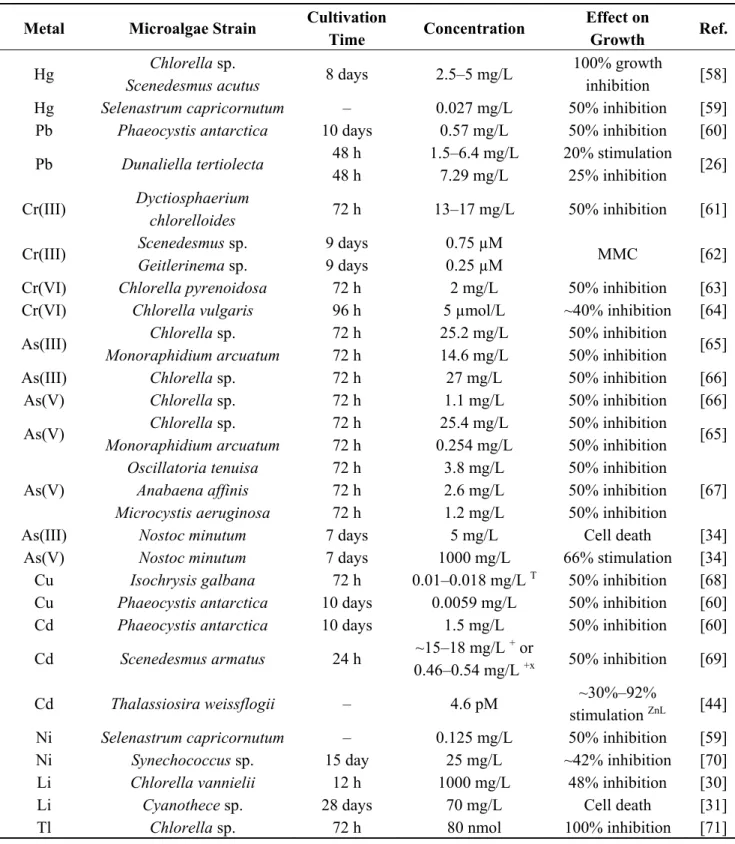
Although heavy metals generally have negative effect on microalgae cultures, some reports suggest also their positive role during microalgae cultivation (Table 1). Lead, aluminum [26] and cobalt [33] at low concentrations had stimulatory effect on growth of Dunaliella tertiolecta [26] and Monoraphidium minutum [33]. Arsenic (As(V)) was reported to improve the growth of cyanobacterium Nostoc minutum [34] and microalgae Chlorella salina [35] and Chlorella sp. [36]. What is more, inorganics can support microalgae growth in case of nutrient deficiency. For instance, 20 µg/L vanadium (VO3−) increased growth of Scenedesmus obliquus grown in iron (Fe3+) deficient medium up to six times.
Vanadium was almost entirely consumed by Scenedesmus cells under photoautotrophic cultivation conditions [37]. In another study, addition of 0.01–1 µg/L vanadium (VO3−) resulted in up to 67% growth
enhancement in photoautotrophic Chlorella pyrenoidosa culture, even with iron (Fe3+) supplementation
in the growth media [38]. However, vanadium (VO3−) at concentrations above 1 mg/L was inhibitory
for Chlorella pyrenoidosa [38]. Vanadium, in a form of VO43− [39] and V2O5 [40], was also reported to
be inhibitory to Haematococcus lacustris [39] and Scenedesmus quadricauda [40].
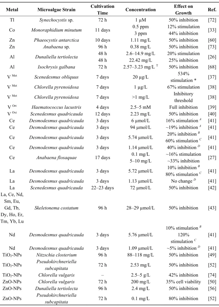
Furthermore, elements from the lanthanide group such as lanthanum (La), cerium (Ce), neodynium (Nd), europium (Eu) or gadolinium (Gd) were reported to constitute a good replacement for calcium deficiency in Desmodesmus quadricauda culture, with Gd, La or Nd supplementation leading to nearly the same culture dry weight when compared to Ca supplemented media. Moreover, addition of cerium at low concentration to standard medium increased Desmodesmus cell number in culture. However, lanthanide elements increased growth suppression of Desmodesmus, when added into manganese deficient medium [41]. Also lanthanum at higher concentration inhibited growth of Scenedesmus quadricauda [42] or Sceletonema costatum [43], and inhibitory concentration of La was the same as for other lanthanides: cerium (Ce), neodymium (Nd), samarium (Sm), europium (Eu), gadolinium (Gd), terbium (Tb), dysprosium (Dy), holmium (Ho), erbium (Er), thulium (Tm), ytterbium (Yb) and lutetium (Lu) [43]. Cerium (Ce) was stimulatory at lower concentration and inhibitory at higher concentration towards cyanobacterium Anabaena flosaquae [27].
Cd2+ at small concentrations was reported to stimulate growth and maintain activity of carbonic
anhydrase in Thalassiosira weissflogii cells, cultivated in Zn-limited medium [44]. Recently, a novel carbonic anhydrase naturally possesing Cd2+ as a catalytic metal ion, has been discovered in
Thalassiosira weissflogii [45].
Ni2+ is an essential metal for cultivation of marine diatoms such as Phaeodactylum tricornutum [46],
Cyclotella cryptica [47], Thalassiosira weissflogii and Thalassiosira pseudonana [48], in the presence of urea as a sole nitrogen source. Nickel serves as a cofactor in an enzyme urease, but Ni at higher concentations was inhibitory for diatom growth [47,48]. A lack of Ni can be partially substituted by cobalt [46].
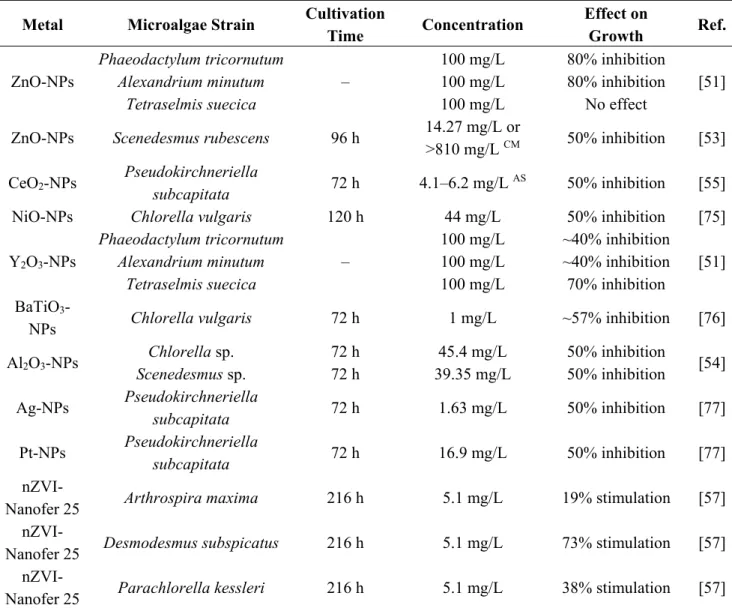
In addition to metals and metalloids, also metallic nanoparticles (NPs) exert activity towards microalgae. Inhibitory effects of TiO2, ZnO, CeO2, NiO, BaTiO3, Y2O3, Al2O3, Ag and Pt nanoparticles
activity was suggested to be due to Reactive Oxygen Species (ROS) generation [49,50] or mechanical damage caused by nanoparticles themselves [51], but also due to metal ions released from nanoparticles [50,52,53], light shading effect [54], interactions with growth media components [55] or simultaneous effect of various factors [56]. Inhibitory activity of nanoparticles also depends on their size [49] and aged suspension [55] or growth medium composition [53]. On the other hand, metal ions released from nanoparticles can also stimulate growth of cyanobacteria and microalgae [57].
Table 1. Effect of metals, metalloids and metallic nanoparticles on growth of microalgae.
Metal Microalgae Strain Cultivation
Time Concentration
Effect on
Growth Ref.
Hg Chlorella sp.
Scenedesmus acutus 8 days 2.5–5 mg/L
100% growth
inhibition [58] Hg Selenastrum capricornutum – 0.027 mg/L 50% inhibition [59] Pb Phaeocystis antarctica 10 days 0.57 mg/L 50% inhibition [60] Pb Dunaliella tertiolecta 48 h 1.5–6.4 mg/L 20% stimulation [26]
48 h 7.29 mg/L 25% inhibition Cr(III) Dyctiosphaerium
chlorelloides 72 h 13–17 mg/L 50% inhibition [61]
Cr(III) Scenedesmus sp. 9 days 0.75 µM MMC [62]
Geitlerinema sp. 9 days 0.25 µM
Cr(VI) Chlorella pyrenoidosa 72 h 2 mg/L 50% inhibition [63] Cr(VI) Chlorella vulgaris 96 h 5 µmol/L ~40% inhibition [64] As(III) Chlorella sp. 72 h 25.2 mg/L 50% inhibition [65]
Monoraphidium arcuatum 72 h 14.6 mg/L 50% inhibition
As(III) Chlorella sp. 72 h 27 mg/L 50% inhibition [66] As(V) Chlorella sp. 72 h 1.1 mg/L 50% inhibition [66] As(V) Chlorella sp. 72 h 25.4 mg/L 50% inhibition [65]
Monoraphidium arcuatum 72 h 0.254 mg/L 50% inhibition
As(V)
Oscillatoria tenuisa 72 h 3.8 mg/L 50% inhibition
[67]
Anabaena affinis 72 h 2.6 mg/L 50% inhibition
Microcystis aeruginosa 72 h 1.2 mg/L 50% inhibition
As(III) Nostoc minutum 7 days 5 mg/L Cell death [34] As(V) Nostoc minutum 7 days 1000 mg/L 66% stimulation [34]
Cu Isochrysis galbana 72 h 0.01–0.018 mg/L T 50% inhibition [68]
Cu Phaeocystis antarctica 10 days 0.0059 mg/L 50% inhibition [60] Cd Phaeocystis antarctica 10 days 1.5 mg/L 50% inhibition [60] Cd Scenedesmus armatus 24 h ~15–18 mg/L
+ or
0.46–0.54 mg/L +x 50% inhibition [69]
Cd Thalassiosira weissflogii – 4.6 pM ~30%–92%
stimulation ZnL [44]
Ni Selenastrum capricornutum – 0.125 mg/L 50% inhibition [59] Ni Synechococcus sp. 15 day 25 mg/L ~42% inhibition [70] Li Chlorella vannielii 12 h 1000 mg/L 48% inhibition [30] Li Cyanothece sp. 28 days 70 mg/L Cell death [31] Tl Chlorella sp. 72 h 80 nmol 100% inhibition [71]
Table 1. Cont.
Metal Microalgae Strain Cultivation
Time Concentration
Effect on
Growth Ref.
Tl Synechocystis sp. 72 h 1 µM 50% inhibition [72] Co Monoraphidium minutum 11 days 0.5 ppm 12% stimulation [33]
3 ppm 44% inhibition
Zn Phaeocystis antarctica 10 days 1.11 mg/L 50% inhibition [60] Zn Anabaena sp. 96 h 0.38 mg/L 50% inhibition [73] Al Dunaliella tertiolecta 48 h 2.6–14.9 mg/L 20% stimulation [26]
48 h 22.42 mg/L 25% inhibition
Al Isochrysis galbana 72 h 2.57–3.23 mg/L T 50% inhibition [68]
V Met Scenedesmus obliquus 7 days 20 µg/L 534%
stimulation * [37] V Met Chlorella pyrenoidosa 7 days 1 µg/L 67% stimulation [38]
V Met Chlorella pyrenoidosa 7 days >1 mg/L Inhibitory
threshold [38] V Ort Haematococcus lacustris 4 days 2.5–5 mM Full inhibition [39]
V Oxi Scenedesmus quadricauda 12 days 2.23 mg/L 50% inhibition [40]
Ce Desmodesmus quadricauda 3 days 6 µmol/L 16% stimulation A [41]
Ce Desmodesmus quadricauda 3 days 94 µmol/L ~19% inhibition A [41]
Ce Desmodesmus quadricauda 3 days 5.74 µmol/L 20% inhibition
B
[41] 60% stimulation C
Ce Desmodesmus quadricauda 3 days 1.14 µmol/L 40% inhibition D [41]
Ce Anabaena flosaquae 17 days 0.1 mg/L ~16% stimulation [27] 5–10 mg/L ~33% inhibition
La Desmodesmus quadricauda 3 days 5.72 µmol/L 10% inhibition
B
[41] 80% stimulation C
La Desmodesmus quadricauda 3 days 1.13 µmol/L No change D [41]
La Scenedesmus quadricauda 22–23 days 72 µmol/L 50% inhibition [42] La, Ce, Nd,
Sm, Eu, Gd, Tb, Dy, Ho, Er, Tm, Yb, Lu
Skeletonema costatum 96 h 28–29 µmol/L 50% inhibition [43]
Nd Desmodesmus quadricauda 3 days 5.76 µmol/L
10% stimulation B
[41] 120%
stimulation C
Nd Desmodesmus quadricauda 3 days 1.09 µmol/L ~5% inhibition D [41]
TiO2-NPs Nitzschia closterium 96 h 88–118 mg/L 50% inhibition [49]
TiO2-NPs Pseudokirchneriella
subcapitata 72 h 2.53 mg/L 50% inhibition [52]
TiO2-NPs Chlorella vulgaris – 2.5–5 g/L 42% inhibition [74]
ZnO-NPs Chlorella vulgaris 72 h 200 mg/L 35% cell viability [50] ZnO-NPs Dunaliella tertiolecta 96 h 2.4 mg/L 50% inhibition [56] ZnO-NPs Pseudokirchneriella
Table 1. Cont.
Metal Microalgae Strain Cultivation
Time Concentration Effect on Growth Ref. ZnO-NPs Phaeodactylum tricornutum – 100 mg/L 80% inhibition [51]
Alexandrium minutum 100 mg/L 80% inhibition
Tetraselmis suecica 100 mg/L No effect ZnO-NPs Scenedesmus rubescens 96 h 14.27 mg/L or
>810 mg/L CM 50% inhibition [53]
CeO2-NPs Pseudokirchneriella
subcapitata 72 h 4.1–6.2 mg/L
AS 50% inhibition [55]
NiO-NPs Chlorella vulgaris 120 h 44 mg/L 50% inhibition [75]
Y2O3-NPs
Phaeodactylum tricornutum
–
100 mg/L ~40% inhibition
[51]
Alexandrium minutum 100 mg/L ~40% inhibition
Tetraselmis suecica 100 mg/L 70% inhibition BaTiO3
-NPs Chlorella vulgaris 72 h 1 mg/L ~57% inhibition [76] Al2O3-NPs Chlorella sp. 72 h 45.4 mg/L 50% inhibition [54] Scenedesmus sp. 72 h 39.35 mg/L 50% inhibition Ag-NPs Pseudokirchneriella subcapitata 72 h 1.63 mg/L 50% inhibition [77] Pt-NPs Pseudokirchneriella subcapitata 72 h 16.9 mg/L 50% inhibition [77]
nZVI-Nanofer 25 Arthrospira maxima 216 h 5.1 mg/L 19% stimulation [57]
nZVI-Nanofer 25 Desmodesmus subspicatus 216 h 5.1 mg/L 73% stimulation [57]
nZVI-Nanofer 25 Parachlorella kessleri 216 h 5.1 mg/L 38% stimulation [57]
MMC, Minimum Metal Concentration significantly affecting Chlorophyll a intensity; T, depending on
temperature applied; +, depending on Cd salt used; x, including complex abilities of media mineral elements;
*, when compared to Scenedesmus growth in Fe deficient medium; ZnL, at low Zn concentrations; Met, added as
metavanadate; Ort, added as orthovanadate; Oxi, added as vanadium pentoxide; A, in standard medium and
compared to a control in standard medium without Ce; B, in Ca deficient medium and compared to a control in
standard medium without tested metal; C, in Ca deficient medium and compared to a control in Ca deficient
medium without testedmetal; D, in Mn deficient medium and compared to a control in Mn deficient medium
without tested metal; NPs, nanoparticles; CM, depending on culture medium; AS, depending on aged suspension;
nZVI, zero-valent iron nanoparticles; Ref., Reference.
3. Metal Stress as a Method for Stimulation of Bioproduct Synthesis
Accumulation of metals in microalgae cells consists of two mechanisms: metal adsorption on the cell wall surface containing functional groups (carboxyl, hydroxyl, phosphate, amino, sulfhydryl) and absorption of metals inside cells via metal transport systems [12,19,78]. Metals in microalgae cells can cause formation of reactive oxygen species (ROS) such as hydroxyl radical (·OH), superoxide anion (O2·−), singlet oxygen (O2*) and hydrogen peroxide (H2O2) that interact with lipids, proteins and nucleic
microalgae cells synthetize chelating agents such as phytochelatin or exopolymers in higher amounts [12,79,80]. Chelating agents are organic compounds that form two or more bonds with a metal ion, thereby creating a coordination complex chelate–metal and preventing metal ions from interaction with biological macromolecules [81]. Another defense mechanism againsts oxidative stress is the synthesis of antioxidant compounds (pigments, glutathione, ascorbate) or enzymes (superoxide dismutase, catalase) that are responsible for quenching reactive oxygen species (ROS) and also reducing metal ions into their less reactive forms [12,79,80]. Therefore, oxidative stress can be considered as a trigger mechanism to induce production of target compounds by metal-exposed microalgae cells, under conditions where the detrimental effect of metals on microalgal culture is avoided.
3.1. Pigments
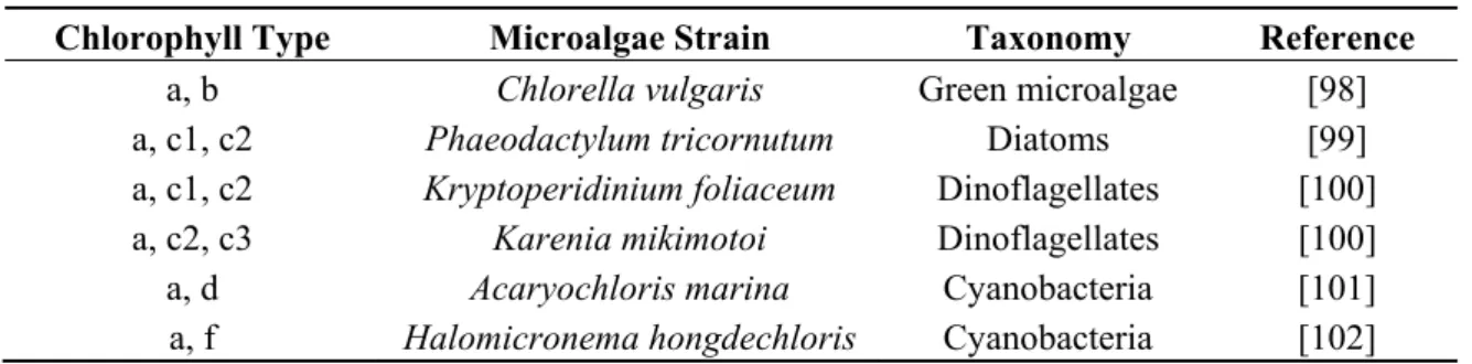
Chlorophylls, carotenoids and phycobilins are microalgal pigments that harvest light in the process of photosynthesis. Chlorophylls are primary photosynthic pigments that contain tetrapyrrole macrocycle rings and are present in various forms (a, b, c1, c2, c3, d, f), in different microalgae or cyanobacteria species (Table 2). Green microalgae possess chlorophyll content up to 6.7% [82], and upon chemical modifications, to phaeophytin [83] or Cu2+-chlorophyllin [84], can be used as a biomordant [83] to
enchance the dyeing process of textile products or as a textile dye [84] with antimicrobial properties. Additionally, an Mg2+ ion in a chlorophyll centre can be substituted with Zn2+, Ni2+, Cd2+, Pb2+, Co2+ or
Pt2+ [85–90]. Carotenoids–accessory photosynthetic pigments, are fat-soluble tetraterpenoid molecules
that are divided into no oxygen-containing carotenes (β-carotene) and oxygen-containing xanthophylls (lutein, astaxanthin, zeaxanthin) [91]. Phycobiliproteins are water-soluble proteins that serve as accessory pigments in blue-green or red microalgae, giving a blue (c-phycocyanin, allophycocyanin) [34,92] or pink, red (b-phycoerythrin, c-phycoerythrin) [93,94] colour. Chlorophylls, carotenoids and phycobiliproteins can find applications in food, cosmetic and pharmaceutical products as coloring, antioxidant, food additive or therapeutic agents [95–97].
Table 2. Types of chlorophyll present in eukaryotic microalgae and cyanobacteria.
Chlorophyll Type Microalgae Strain Taxonomy Reference
a, b Chlorella vulgaris Green microalgae [98]
a, c1, c2 Phaeodactylum tricornutum Diatoms [99]
a, c1, c2 Kryptoperidinium foliaceum Dinoflagellates [100] a, c2, c3 Karenia mikimotoi Dinoflagellates [100]
a, d Acaryochloris marina Cyanobacteria [101]
a, f Halomicronema hongdechloris Cyanobacteria [102]
The presence of metals can have an enchancing effect on pigment content in microalgae or cyanobacteria cells. Copper (Cu2+) at concentration between 0.05–0.2 g/L induced β-carotene production
in Chlamydomonas acidophilla [103]. The change in iron (Fe2+) medium concentation resulted in a
growth improvement and an increase in lutein, zeaxanthin and β-carotene content in Coccomyxa onubensis cells [104]. Also, β-carotene content in Dunaliella salina cells was increased seven times in the presence of 450 µM Fe2+ and 67.5 mM acetate, however at the expense of four-fold reduction in
medium containg 1 g/L arsenic(V) was reported to posses chlorophyll, carotenoid and allophycocyanin content higher by 75%, 40% and 25%, respectively, when compared to control culture [34]. Similarly, small concentrations of Ni (0.1–10 µM) increased chlorophyll content and c-phycocyanin production even by 47% and up to 4.35 times, respectively, in Anabaena doliolum culture [92]. The content of c-phycocyanin, phycoerythrin and allophycocyanin in cyanobacterium Phormidium tenue culture increased considerably in the presence of As, but the uplift profiles were strongly dependent on As dosage (0.1–100 ppm) and exposure time [106]. In other studies, cultivation of Synechocystis sp. in the presence of Pb and Cd, and Spirulina platensis in the presence of Pb, showed a decrease in biomass and pigment (chlorophyll, carotenoid, phycocyanin) concentration, in the culture volume. Nevertheless, pigment content in cyanobacteria biomass increased at some metal concentrations and cyanobacteria growth was stimulated at low Pb concentrations [107,108]. Lead (Pb) and cadmium (Cd) at concentrations up to 10 mg/L increased chlorophyll concentration in cultures of metal resistant Scenedesmus quadricauda and Pseudochlorococcum typicum [109]. Tellurium (TeO32−), added into
Spirulina platensis growth media, was accumulated and incorporated into peptides in Spirulina cells. As a result, production of Te-phycocyanin and Te-allophycocyanin possessing enhanced antioxidant activity, was reported in Spirulina platensis cells [110].
3.2. Lipids
Microalgal cells are a source of lipids including triacyloglycerols (TAGs) and fatty acids [111], but also phytosterols [112] and sphingolipids [113], with potential applications as biofuels, nutraceuticals and food additives. It is reported that nutrient deficiency such as nitrogen deprivation results in oxidative stress and lipid accumulation in microalgal cells [114]. Cultivation of Chlorella minutissima in the presence of Cd (0.2–0.4 mM) or Cu (0.2–1 mM) leads to the increase in both biomass density and cell lipid content, providing lipid productivity improved 2.17-fold with 0.4 mM Cd or by 34% with 0.4 mM Cu [115]. Euglena gracilis cultivated photoautotrophically or mixotrophically in the presence of low chromium (Cr6+) concentration exhibited higher total lipid content, although lipid stimulation
(10%–100%) was dependent on Euglena strain used and medium composition tested [116]. Addition of 0.1 g/L TiO2 nanoparticles with UV-A irradiation applied, slightly increased production of fatty acids
in Chlorella vulgaris cells, without growth reduction [74]. Recently, zero-valent iron nanoparticles (5.1 mg/L) were reported to increase lipid productivity in Arthrospira maxima, Desmodesmus subspicatus andParachlorella kessleri cultures, respectively by 40%, 2.75-fold and by 66% [57]. Metal stress also causes the alteration of fatty acid profile in microalgae cells. The effect of As(III) on Nannochloropsis sp. cells resulted in a slight increase in cell lipid content and a change in lipid profile, as the decrease in polyunsaturated fatty acids and the increase in short-chain saturated (C16:0, C18:0) and monounsaturated (C16:1, C18:1) fatty acids, was depicted [117]. Nickel at 0.5 mg/L caused a shift of fatty acid profile towards saturated fatty acids (C14:0, C16:0, C20:0) in Dunaliella salina and Nannochloropsis salina cells, also with the upshift of saturated C18:0 and unsaturated C18:2 for Nannochloropsis and C22:0 behenic acid for Dunaliella [118]. Composition of fatty acids (chain length, number of double bonds) defines the biodiesels produced from corresponding triglycerides in terms of their quality and properties (including cetane number, density, viscosity, lubricity, calorific value, NOx emissions) [119–121].
biodiesel of desirable quality and properties [117]. As a contrary, cultivation of Nannochloropsis limnetica and Trachydiscus minutus in the presence of zero-valent iron nanoparticles (nZVI) caused the decrease in saturated fatty acids (C14:0, C16:0, C18:0) and the increase in eicosapentaenoic acid (C20:5ω3) content in Nannochloropsis and Trachydiscus biomass [57]. Eicosapentaenoic acid (EPA) can be used as a nutraceutical or pharmacological agent for the treatment of heart and inflammatory diseases [122].
3.3. Exopolymers
Extracellular polymeric substances (EPS), consisting of exopolysaccharides and exoproteins, are excreted by microalgae and cyanobacteria upon exposure to stress factors such as nutrient (N, P) imbalance, but the release mechanism can also depend on cultivation conditions (light intensity, temperature, salinity, microelement availability) and the stage of microalgal growth [123–128]. Exopolysaccharides can be of linear or branched structure and contain C6 (glucose, galactose, fructose, rhamnose, fucose) and C5 (xylose, arabinose) sugars, as well as uronic (glucuronic, galacturonic) acids, aromatic, pyruvate, acetate, sulphate and halide groups. Additionally, extracellular polysaccharides can be also coupled with peptides, lipids and nucleic acids [129,130].
Metals were reported to stimulate the release of exopolymers by microalgal cells. A considerable increase in the release of exopolysaccharides and extracellullar proteins was observed in the culture of cyanobacterium Lyngbya putealis, as a response to the presence of Cu and Co [131]. Increased release of extracellular polymers from Thalassiosira weissflogii [132], and Thalassiosira pseudonana [133] in the presence of Ag [132] and Cd [133] ions released from engineered nanoparticles (ENPs), was also reported. Extracellular polymeric substances possess antiviral, antitumor, anticoagulant, antiinflammatory and immunostimulant activity, but they can also serve as biosurfactants, biolubricants, bioemulsifiers [130] and a source of sugars for biofuels [134].
3.4. Phytochelatin
Phytochelatins are (oligo)peptides synthetized in plants, yeast, algae and cyanobacteria for detoxification of heavy metals. The structure of phytochelatin is (γ-Glu-Cys)n-Gly with γ-Glu-Cys
n being between 2 to 11. Phytochelatin is synthetized by phytochelatin synthase (glutathione-γ-glutamylcysteinyltransferase), by firstly adding γ-Glu-Cys from glutathione (γ-Glu-Cys-Gly) to another glutathione molecule forming (γ-Glu-Cys)2-Gly (PC2) and further incorporates new γ-Glu-Cys units into
PC2 [135]. Synthesis of short chain phytochelatins (2 to 6 of γ-Glu-Cys units) was reported in cells of microalgae (Table 3) such as Scenedesmus vacuolatus [136], Phaeodactylum tricornutum [137–139], Scenedesmus armatus [140], Stichococcus bacillaris [141], Micrasterias denticulata [142] and cyanobacterium Anabaena doliolum [143] exposed to increasing concentration of Cd, Pb, Cu and/or As. Phytochelatin content in Scenedesmus armatus and Stichococcus bacillaris cells exposed to constant (Const.) concentration of Cd and As respectively can be also further elevated, with the upshift of CO2
supplementation for Scenedesmus [140] and decrease of pH for Stichococcus [141]. Also synthesis of iso-phytochelatins such as Cys(GluCys)nGly and (GluCys)nAla was reported in Chlamydomonas
component for biosensors, designed for detection of heavy metals in samples of environmental, biological or pharmaceutical origin [145,146].
Table 3. Synthesis of phytochelatin in microalgae exposed to heavy metals.
Strain Metal Metal
Uplift Phytochelatin Uplift PCN A Growth Rate C Reference
Scenedesmus vacuolatus Cd 0.3→ 79 nM ~3→25 amol/cell PC2 Reduced by 37% [136] ~1→44 amol/cell PC3 ~0→17 amol/cell PC4 Phaeodactylum tricornutum Cd 0→ 0.45 µM ~0.16→3.6 amol/cell PC2 No change [137] ~0.5→1.3 amol/cell PC3 ~0.05→1.5 amol/cell PC4 Phaeodactylum tricornutum Cu 0.068 pM→ 0.4 µM ~0.16→1.7 amol/cell PC2 No change [137] ~0.5→1.5 amol/cell PC3 ~0.05→0.8 amol/cell PC4 Phaeodactylum tricornutum Cd 0→10 µM ~0→12.5 amol/cell PC2 Toxic effect avoided [138] ~0→25 amol/cell PC4 ~0→5 amol/cell PC5 Phaeodactylum tricornutum Pb 0→10 µM ~0→50 amol/cell PC2 Toxic effect avoided [138] ~0→13 amol/cell PC3 ~0→3 amol/cell PC5 Phaeodactylum tricornutum Cu 0→10 µM ~2→18 amol/cell PC2 – [139] ~0→38 amol/cell PC3 ~0→5 amol/cell PC6 Scenedesmus armatus Cd Const. 93 µM * ~40→200 nmol-SH/g PC2 Reduced by 26% [140] ~80→1300 nmol-SH/g PC3 ~20→280 nmol-SH/g PC4 Stichococcus bacillaris As(III) Const. 100 µM ** 0.07→0.15 µmol-SH/g PC2 Reduced by 20% [141] As(V) Const. 100 µM ** 0.14→0.38 µmol-SH/g PC2 Reduced by 30%
A Phytochelatin with N number of γGlu-Cys units; C when compared to control; * increase of CO 2
supplementation from 0.1% to 2%; ** pH shift from 8.2 to 6.8.
3.5. Phytohormones
Zeatin, indoleacetic acid and abscisic acid are phytohormones that can be used as growth regulators for plants [147,148], and yeast [149], but also as anti-aging agents [150] and potential drugs for neural [151] or cancer [152] diseases. Phytohormones can be found in microalgae [153] and their content can be amplified in the presence of heavy metals. The content of indoleacetic acid, zeatin and abscisic acid increased in Chlorella vulgaris cells grown in the medium containing 10−4 M Cd, Pb or Cu, however
at the expense of decreased cell number in the culture. Interestingly, addition of 10−8 M brassinolide into
metal-containing Chlorella culture enabled to achieve cell number comparable to control culture, together with further stimulation of zeatin, indoleacetic acid and abscisic acid production [154].
3.6. Organoarsenical Compounds
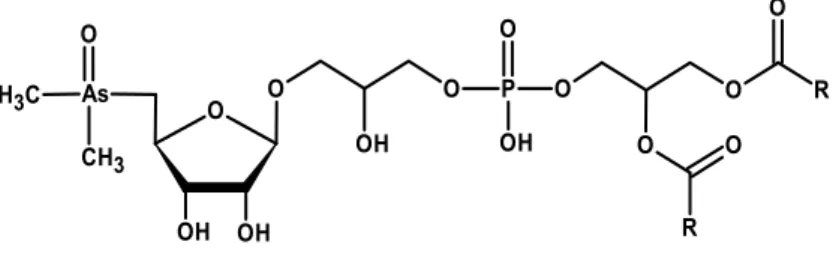
Accumulation of As in microalgae cells has been recently extensively summarized [155]. In essence, the uptake of As(V) from surroundings into microalgae cells is accomplished by means of phosphate transport system, while As(III) is transported by aquaglyceroporins and hexose permeases [155]. Subsequently, As(V) is reduced to As(III) via As reductase action, with simultaneous oxidation of glutathione (GSH). As(III) undergoes methylation via As methyltransferase action into monomethylarsonate (MMA) and dimethylarsinate (DMA). Arsenic(III) can also undergo bio-oxidation to As(V) or be extruded from cells [156–158]. Arsenic(V) can be incorporated into cellular components such as sugars and lipids. In microalgae, dimethylarsinate (or its reduced form: dimethylarsinous acid) can combine with the adenosyl group from S-adenosyl methionine, leading to formation of a dimethylarsinyladenosine, which further undergoes glycosidation to dimethylarsenoribosides [159,160]. In cyanobacteria, dimethylarsinate undergoes reduction, ribose-coupling and glycosidation [161]. Some varieties of arsenosugars containing glycerol, sulphate, sulphonate and phosphate groups were detected for microalgae [160,162]. Arsenolipids in microalgae were determined as dimethylarsenoriboside phospholipids (Figure 1), although phospholipids containing single As(V) or DMA groups were also reported [163]. Content and compositions of arsenoorganics formed in microalgae Chlorella and Monoraphidium [65], Dunaliella and Phaeodactylum [163], Chlamydomonas [160] or cyanobacteria Synechocystis [157,161] and Nostoc [161] cells depends on microalgae strain used, as well as on arsenic(V) concentration applied, exposure time and phosphate availability. Arsenolipids and arsenosugars are currently evaluated as possible therapeutic agents [164]. However, application of As-containing compounds is limited due to high toxicity and so far, only derivatives of arsenolipids have been reported to possess any medical applications [159].
Figure 1. Chemical structure of dimethylarsenoriboside phospholipids (R—a carbon chain of fatty acid).
3.7. Nanoparticles and Nano-Needles
Nanoparticles are particles with sizes ranging between 1–100 nm [165]. Nanoparticles possess antiviral, antibacterial, antifungal, anticancer and antiparasite activity. They also find application in the field of catalysis or photonics or can serve as drug carriers and components of chemical sensors [166]. Methods applied for manufacturing nanoparticles range from mechanical, laser and UV irradiation treatment to microemulsion system, hydrothermal process, sol–gel process, chemical vapor condensation, sonochemical treatment and microbial biosynthesis [165,167]. Synthesis of nanoparticles by microorganisms (bacteria, yeast, fungi and microalgae) can constitute a green and environmentally friendly method for nanoparticles production [168,169]. Formation of nanoparticles: Au, Ag or Pd
O OH OH O OH O P O OH O O O R R O O As O CH3 C H3
(Table 4) from metal ions solutions takes place inside microalgae cells (intracellularly) or in the media (extracellularly) via interactions with molecules of microalgal cell metabolism (NADH, pigments, peptides, proteins and polysaccharides) [170–176]. The size of synthetized nanoparticles depends on microalgal strain and metal type used, but place of synthesis, initial metal loading, light and temperature are also crucial factors influencing formation of nanoparticles. Additionally, synthesis of Cd nanoparticles in a form of CdS [177] or Ni nanoparticles as a product of reduction of other nanoparticles (NiO) [75], was also reported. Besides nanoparticles, biosynthesis of nanoneedles by microalgae also occurs; such nanoneedles, composed of zinc and phosphorous, were detected in Scenedesmus obliquus cells as a result of exposure to high Zn concentration [178].
Table 4. Synthesis of nanoparticles (NP) in microalgae and cyanobacteria cultures.
Element NP Source Strain Place of Synthesis
Average Particle
Size (nm) Reference
Gold (Au) HAuCl4·3H2O Chlorella vulgaris Intracellularly 40–60 [170]
Gold (Au) KAuCl4 Eolimna minima Intracellularly 5–100 [171]
Silver (Ag) AgNO3 Parachlorella
kessleri Extracellularly 9, 14 or 18 [172]
Silver (Ag) AgNO3 Botryococcus
braunii Extracellularly 15.67 [173]
Silver (Ag) AgNO3 Scenedesmus sp. Intracellularly 15–20 [174]
Palladium (Pd) Na2(PdCl4) Chlorella vulgaris Microalga
culture 7 [175]
Palladium (Pd) PdCl2 Chlorella vulgaris Intracellularly 5–12 [170]
Palladium (Pd) PdCl2 Plectonema
boryanum Extracellularly ≤30 [176]
Cadmium
sulphide (CdS) Cd(NO3)2·4H2O Scenedesmus Intracellularly
120–175 (described as nanoparticles)
[177] Nickel (Ni) NiO–NPs Chlorella vulgaris Microalga
culture – [75]
4. Influence of Growth Conditions on Microalgal Resistance Towards Metals
Metals at low concentration can be stimulatory for growth and production of target compounds, but metal overdose has detrimental and lethal effects on microalgae cultures. Hence, microalgal cultivation in metal polluted wastewaters should be designed in such a way to limit cell–metal interactions to the level at which metal concentration exerts only beneficial effects on microalgae growth and biosynthesis of crucial products. Microalgal cell response to metal presence depends on many factors such as conditions of cultivation, nutrient availability, presence of organic compounds and tolerance ability of particular strains.
4.1. Growth Media Composition and Cultivation Conditions
Composition of growth media is a crucial factor regarding microalgae response towards heavy metals, such as arsenic, cadmium or nickel.
Arsenate (AsO43−) and phosphate (PO43−) are mutual competitors for the uptake by microalgal
cells [155]. A 10–fold increase in phosphate concentation resulted in a 18 times higher resistance of Monoraphidium arcuatum against As(V). On the other hand, a 10-fold decrease in medium nitrate NO3− content at ordinary (PO43−) concentation, decreased by 28% Monoraphidium resistance towards
arsenic [65]. In another study, a 131-fold phosphate uplift improved 516 times resistance of Chlorella salina against As(V) [35]. Indeed, increasing concentration of As(V) stimulated growth of arsene tolerant Chlorella sp. at low phosphate (P) concentration, although cell yields obtained were lower than in experiments with high P concentration [36]. Concentration of PO43− in medium in
relation to dissolved lead content can be also important, as Pb2+ can precipitate in a form of
Pb3(PO4)2, thereby removing available phosphate from solution and inhibiting growth of
Chlamydomonas reinhardtii [179].
Sulphur is a component of cysteine that participates in the defense mechanisms against heavy metals. The resistance of Chlamydomonas moewusii exposed to 4 mg/L cadmium can be improved five times and cysteine cell content can be raised 10 times, when sulphate (SO42−) concentration in medium is
increased 100 times [180]. In another study, a 10-fold increase in SO42− supply resulted in a
Chlamydomonas reinhardtii resistance improved by up to 77% towards Cd. Improved Chlamydomonas resistance was accompanied with an increased activity of cysteine desulfhydrase, an enzyme responsible for the cleavage of cysteine into pyruvate, NH3 and sulfide, the latter one reported to react with Cd to
form CdS [181].
A 20-fold increase in ammonium (NH4+) concentration increased five times the accumulation of
PO43− in Chlorella vulgaris cells and caused a 50% alleviation in inhibition of Chlorella growth exerted
by chromium (Cr) [182]. Increase in magnesium (Mg2+) and hydrogen (H+) concentration reduced nickel
toxicity towards Pseudokirchneriella subcapitata, as Mg2+ and H+ compete with Ni2+ for the uptake by
the cell transport system [183]. In other studies, an increase in H+ concentration was reported to improve,
even up to 23 times [184], Chlorella sp. resistance against Cu.
Zn alleviated detrimental effects of Cr on the photosynthetic mechanism in Micrasterias denticulata cells and Fe ameliorated inhibitory effect of Cd and Cr on Micrasterias cell development. Ca and Gd were reported to prevent alterations in cell morphology caused by Pb and Cd, thereby nullifying negative effects of Pb and Cd on Micrasterias cells [185].
Finally, toxicity of thallium towards Chlorella sp. was completely alleviated, when concentration of K+ in media was increased 20 times, presumably due to competive uptake in Chlorella cell transport
systems [71].
Cultivation parameters such as light intensity and CO2 concentration are also important factors
affecting microalgae response towards metals. Alterations in ligh irradiance had influence on inhibition or stimulation of Chlamydomonas reinhardtii growth under different Cu concentrations, and also affected accumulation of Cu in Chlamydomonas cells [186]. Increase of CO2 supply enabled the
alleviation of the inhibitory effect of Cd towards Scenedesmus armatus, although growth inhibition was not entirely overcome [140].
4.2. Supportive Compounds
Another modulating approach could be supplementation of microalgae cultures with organic compounds such as phytohormones or chelating agents.
4.2.1. Phytohormones: Modulating Effect
Phytohormones—spermidine (polyamine), gibberellin and many representatives of auxin and cytokinin groups—were reported to prevent inhibition of Chlorella vulgaris culture exposed to cadmium (Cd), copper (Cu) or lead (Pb) at a concentration of 0.1 mM. What is more, addition of compounds from the cytokinin group such as benzyladenine, zeatin, kinetin, 2-isopentenyladenine, diphenylurea, forchlorphenuron and thidiazuron not only enabled restoration of the Chlorella culture, but also increased cell number by up to 77%, when compared to control. Supplementation of spermidine, gibberellin, auxins or cytokinins generally increased not only the content of chlorophyll, carotenoid, protein, ascorbate and glutathione in Chlorella cells, but also activity of superoxide dismutase and catalase [187]. In earlier studies, it was stated that the inhibitory effect of 0.1 mM Cd, Cu and Pb on Chlorella vulgaris culture can be also nullified in the presence of brassinolide [154].
4.2.2. Chelating Agents: Modulating Effect
Chelating agents are synthetized by microalgae for intracellular (phytochelatin, glutathione) or extracellular (exopolymers) detoxification of metals, but can also be added artificially into growth media to bind metals and modulate cell–metal interactions. Such agents can be low-molecular organic acids (ethylenediamine tetraacetic acid, nitrilotriacetic acid, citrate) or humic substances: humic acid or fulvic acid (Table 5).
Addition of 34 µM ethylenediamine tetraacetic acid (EDTA) into Scenedesmus subspicatus culture enabled a ~55% reduction in growth inhibition exerted by ~40 µM Cu [188]. Also EDTA, as well as nitrilotriacetic acid (NTA) and citrate (Cit), were reported to prevent accumulation of lanthanum (La), gadolinum (Gd) and yttrium (Y) in Chlorella vulgaris cells, with reduction in accumulation around 10- to 30-fold higher for EDTA, when compared to NTA and Cit [189]. On the other hand, citrate was reported to enhance Cd (0.25 µM/L) accumulation and growth inhibition of Selenastrum capricornutum, due to the occasional uptake of Cd-citrate by cells [190]. With the absence of EDTA in growth medium, cadmium (Cd) exerted much stronger inhibitory effects on Scenedesmus armatus, when compared to the growth in EDTA-containing medium [69]. Growth of Scenedesmus quadricauda or Microcystis aeruginosa in the presence of lanthanum (0.72–72 µM) and EDTA (0.269–26.9 µM) was inhibited or enhanced, depending on La and EDTA concentrations. EDTA (2.69–13.4 µM) vastly alleviated the inhibitory effect of La on Microcystis growth, although EDTA alone and at higher concentration had strong inhibitory effects towards Microcystis [42]. EDTA [37,191,192] or citrate [37,193] increased Fe availability to microalgae, although high concentration of chelating agent can have opposite effects [42,191]. Additionally, EDTA that fails to maintain availabilily of Fe at high pH during Spirulina cultivation, can be replaced by alternative chelating agents such as Fe complexes of N,Nʹ-bis(2-hydroxybenzyl)ethylenediamine-N,N'-diacetic acid
(HBED), bis((2-hydroxyphenyl)acetic acid) (EDDHA) or ethylenediamine-N,N'-bis((2-hydroxy-4-methylphenyl)acetic acid) (EDDHMA) [194].
Humic acid was reported to protect Dunaliella salina and Nannochloropsis salina cells against Ni2+
stress, by means of forming humic acid–Ni2+ complexes and/or by adsorbing on cell surface and thus,
creating an additional barrier for Ni2+ uptake [118]. Similarly, humic acids reduced toxicity of Cd2+ and
Zn2+ towards Pseudokirchneriella subcapitata [195], Hg2+ towards Isochrysis galbana [196] and ZnO
nanoparticles towards Anabaena sp [197]. Humic acid itself at 7 and 2.5–10 mg/L stimulated growth of Isochrysis galbana [198] and Stichococcus bacillaris [141], presumably due to improved nutrient uptake via humic acid–cell membranes interaction [198]. However, an opposite effect, enhanced toxicity of Pb towards Isochrysis in the presence humic acid, was also observed [198], because the formation of a ternary complex between Pb, humic acid and microalga cell surface, enhances internalization of Pb [199]. Humic acid was also reported to be inhibitory (0.3 mg/L) and lethal (1 mg/L) for Anabaena circinalis, probably due to its chelating activity towards Fe3+, leading to the decrease in
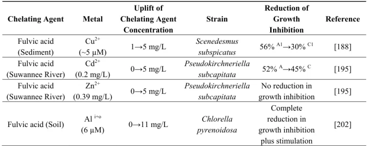
availability of Fe necessary for Anabaena growth [200]. It is also noteworthy, that humic acid can undergo degradation under high light irradiance, leading to the decreased capacity for metal complexation [201]. Fulvic acid contributed to protection of Scenedesmus subspicatus against Cu2+ [188],
but no protective effect against Cd2+ and Zn2+ was found for Pseudokirchneriella subcapitata [195].
Fulvic acid was also reported to serve as a source of phosphorus to nullify toxic effects of aluminum (Al) on P-metabolism in Chlorella pyrenoidosa [202].
Table 5. Effect of humic and fulvic acids on microalgae response towards metals.
Chelating Agent Metal
Uplift of Chelating Agent Concentration Strain Reduction of Growth Inhibition Reference
Humic acid (Soil) Ni2+
(0.5 mg/L) 0→0.2 mg/L Dunaliella salina Nannochloropsis salina 40% A→25% C 30% A→15% C [118]
Humic acid (Soil) Cd2+
(0.2 mg/L) 0→5 mg/L
Pseudokirchneriella
subcapitata 52%
A→28% C [195]
Humic acid (Soil) Zn
2+
(0.39 mg/L) 0→5 mg/L
Pseudokirchneriella
subcapitata 55% A→4% C [195]
Humic acid (Peat) Cd
2+
(0.2 mg/L) 0→5 mg/L
Pseudokirchneriella
subcapitata 52%
A→8% C [195]
Humic acid (Peat) Zn
2+
(0.39 mg/L) 0→5 mg/L
Pseudokirchneriella
subcapitata 55%
A→30% C [195]
Humic acid As(III)
(100 µM) 0→10 mg/L Stichococcus bacillaris 52% A→33% C [141] Humic acid (Sediment) Hg2+ (10 ppb) 0→10 ppm Isochrysis galbana Complete reduction in growth inhibition plus stimulation [196]
Humic acid ZnO–NPs
(1 mg/L) 0→3 mg/L Anabaena sp. 70%
Table 5. Cont.
Chelating Agent Metal
Uplift of Chelating Agent Concentration Strain Reduction of Growth Inhibition Reference Fulvic acid (Sediment) Cu2+ (~5 µM) 1→5 mg/L Scenedesmus subspicatus 56% A1→30% C1 [188] Fulvic acid (Suwannee River) Cd2+ (0.2 mg/L) 0→5 mg/L Pseudokirchneriella subcapitata 52% A→45% C [195] Fulvic acid (Suwannee River) Zn2+ (0.39 mg/L) 0→5 mg/L Pseudokirchneriella subcapitata No reduction in growth inhibition [195] Fulvic acid (Soil) Al i+o
(6 µM) 0→11 mg/L Chlorella pyrenoidosa Complete reduction in growth inhibition plus stimulation [202]
A growth inhibition in the absence of chelating agent; A1, growth inhibition in the presence of decreased amount
of chelating agent; C growth inhibition in the presence of chelating agent; C1, growth inhibition in the presence
of increased amount of chelating agent; i+o, a sum of inorganic and organic aluminum
4.2.3. Nanoparticles: Modulating Effect
The presence of metallic and non-metallic nanomaterials can alter the effect of metals on microalgae. For instance, the presence of graphene oxide (GO) increased toxicity of Cd towards Microcystis aeruginosa [203], while Cd toxicity towards Chlamydomonas reinhardtii was reduced in the presence of titanium dioxide engineered nanoparticles (ENPs) [204]. TiO2 nanoparticles and Zn ions in the
mixture exerted the enhanced or decreased toxicity towards Anabaena sp., depending on mutual interactions between different concentrations of TiO2 and Zn [73]. Finally, the presence of engineered
nanoparticles was reported to decrease intracellular content of Cu and Pb in Chlorella kesslerii and wall-possessing Chlamydomonas reinhardtii, as metal binding to nanoparticles reduces availability of Cu and Pb to these microalgal strains [205].
4.2.4. Macrocycles: Modulating Effect
Supramolecular water soluble compounds such as cyclodextrins, calixarenes and resorcinarenes can possibly change interactions between microalgae and metals.
Cyclodextrins (CDs) are macrocyclic oligosaccharides composed of six, seven, or eight (α 1–4) glucosidic units and called: α,β and γ-CDs, respectively. They are produced from enzymatic hydrolysis of starch, with cycloglycosyl transferase amylases (CGTases) [206,207]. CDs are ring molecules, either toroidal or cone shaped, but not cylindrical [208]. The primary hydroxyl groups are situated on the narrow side while, the secondary groups are located on the wider side. The central cavity of CDs is hydrophobic, while the outer part is hydrophilic due the presence of hydroxyl groups [209]. β-cyclodextrins can possess methyl, carboxymethyl or hydroxypropyl moieties [210,211] and form complexes with metals [212], phytosterols [213] and carotenoids [214]. Carboxymethyl-β-cyclodextrin (3.3 mM) was successfully harnessed for reduction of metal (Cd, Co, Cu) toxicity towards naphthalene-degrading bacterium Burkholderia sp. [215]. On the other hand, alhough hydroxypropyl-β-cyclodextrin
up to 20 mM did not itself cause inhibition of microalga Selenastrum capricornutum growth, it failed to protect this microalga strain against Zn toxicity [216], because hydroxypropyl-β-cyclodextrin does not possess metal-binding substituents [215].
Calix[n]arenes and resorcin[4]arenes are macrocyclic compounds consisting of phenol or resorcinol units, respectively, which are cyclically linked by aliphatic bridges [217]. Calix[n]arenes (n = 4, 5, 6, 7 and 8) are obtained as a result of condensation of p-tert-butylphenol with formaldehyde under alkaline catalysis [218–220], whereas resorcin[4]arenes are formed as a result of acid-catalysed reaction between resorcinol and aliphatic or aromatic aldehydes [221]. Water-soluble calix[4]arenes and resorcin[4]arenes possess charged groups (ammonium, sulphonium, carboxylate, phosphate) or hydrophilic fragments [222–225]. Derivatives of calix[n]arenes such as p-sulphonate or methoxycarboxylic derivatives form stable complexes with Zn2+, Cu2+, Ni2+ under neutral or alkaline conditions [226–228].
Water soluble resorcin[4]arene derivatives are able to form complexes, not only with the metal ions, but also with amino acids, sugars, and nucleosides [229–231]. It was demonstrated that p-sulfonatocalix[4,6,8]arene and C-nonylresorcin[4]arene possess antimicrobial activity against fungal and bacterial microorganisms [232]. Additionally, C-methylcalix[4]-resorcinarene containing pyridinium salt, was reported to exhibit a selective inhibitory effect on Gram-positive bacteria [233].
Water soluble supramolecular molecules have the potential to modify interactions between metals and microorganisms such as microalgae, but their application in this field is highly unexplored.
4.3. Development of Strain Tolerance to Metals
Some microalgae are able to inhabit environments contaminated by heavy metals. Such microalgal strains possess uplifted tolerance towards heavy metals [104,234–237]. Increased tolerance can be also induced on laboratory scale by applying proper metal dosages [238,239] or metal-containing wastes [240]. It results in development of physiologically adapted strains [61,239,241] or metal resistant mutants due to rare spontaneous mutations that occur before metal treatment [61,238,239]. Microalgae with improved tolerance can become promising microbes for cultivation in metal polluted growth media and for production of target compounds [104]. However, it should be taken into consideration that increased tolerance can be strictly strain–metal specific [235] and a lack of inducing metal in the cultivation medium can have a negative effect on growth of metal resistant mutants [238].
5. Strategy for Microalgal Production in the Presence of Metals
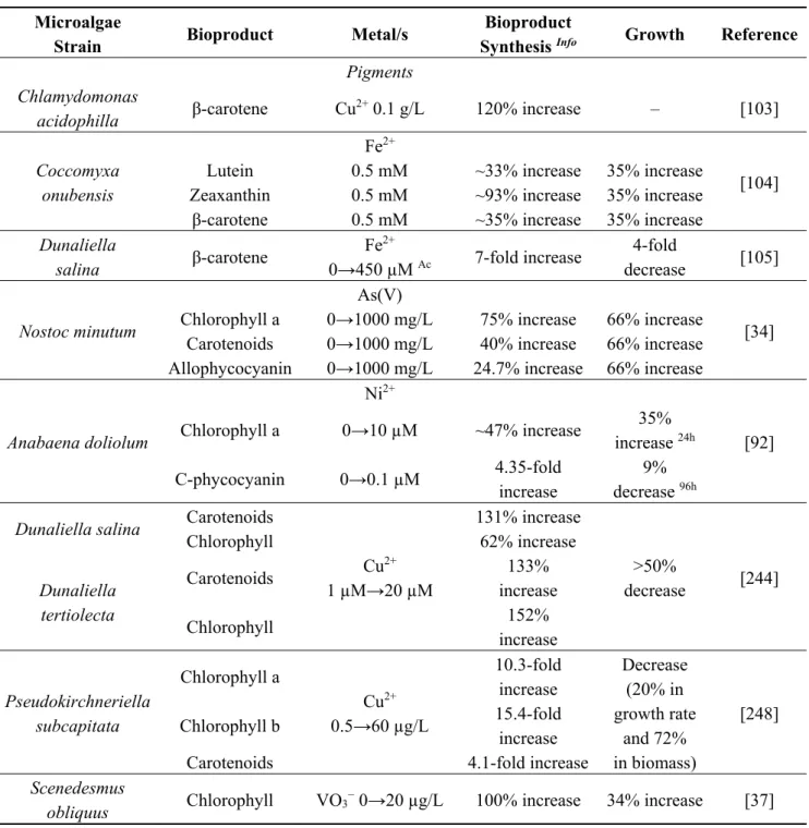
It has been widely reported that microalgae cultures, due to their ability for metal accumulation, can be used for bioremediation of heavy metal contaminated water/wastewater streams [80,242,243]. In this review, other aspects of microalgae exposure to metals, such as production of numerous industrially important compounds from metal-exposed microalgae (Table 6) and stategies to alter microalga–metal interactions for industrial microalgae productions, are discussed. As a result of metal exposure, microalgae are able to synthesize a range of target compounds: pigments, lipids, peptides, exopolymers, phytohormones, arsenoorganics or nanomaterials, as a defense mechanism against metal stress. Although metals induce synthesis of compounds by microalgae cells, they may also have detrimental effects on cell number, growth rate, cell dry weight, thereby diminishing productivity of target compounds in a metal-trigger system. For instance, an elevated copper (Cu) concentration increased
chlorophyll and carotenoid content in Dunaliella cells [244] and stimulated release of polysaccharides from Cylindrotheca fusiformis [245] and phenolics from Dunaliella tertiolecta [246] cells, though at the expense of a reduced number of cells in the culture. In other studies, the content of chlorophyll, protein and lipids in Chlorella vulgaris [247], proline and total amino acids in Chlorella pyrenoidosa [63] and chlorophyll and carotenoid in Pseudokirchneriella subcapitata [248] increased in the presence of cadmium (Cd), chromium (Cr) and copper (Cu) respectively, but the growth in these cultures was considerably suppressed [63,247,248]. A possible strategy to overcome this problem could be cultivation of microalgae under non-stressed conditions in order to obtain higher cells densities, with subsequent addition of metals for inducing stress and synthesis of target products in microalgae cells [10]. Metals at higher concentration are toxic to microalgae, but at lower concentration can be stimulatory for growth (Table 1). Additionaly, it was concluded that growth media might contain nutrients (Ca, Mg) in amounts that are not sufficient for some microalgal strains to achieve desirable growth [249] and therefore some metal-containing effluents could also serve as a nutrient replacement for Ca [41], Fe [37] or Zn [44] deficiency in growth media. Microalgae cultivation systems require large amounts of water [250] and production of target compounds with metal polluted industrial water streams, instead of exploiting clean water sources, could be an additional advantage. Growth of cyanobacteria Nostoc linckia and Nostoc rivularis was stimulated at low loadings of (Zn, Cd)-containing sewage waters, but suppressed at high sewage water loadings [251]. Industrial wastes/wastewaters contain not only metals, but also numerous organic pollutants (pesticides, pharmaceuticals, personal care products etc.) [252] that can be harmful for microalgae cultures. Furthermore, although metal uptake occurs in microalgal cultures, high dosage wastes can strongly decrease productivity of microalgal cultivation [251,253]. Therefore, precautions should be taken to control concentration of metals and/or organic toxicants, so that optimal microalgal growth and product biosynthesis could be obtained.
An integrated process for metal (Al, Fe, Mn, Ba, Ce, La) remediation and lipid production in cultures of marine microalgae (Nannochloropsis, Pavlova, Tetraselmis, Chaetoceros) has already been proposed [254]. Recently, a combination of heavy metal (Zn, Mn, Cd, Cu) removal to increase up to 2.17-fold lipid production from Chlorella minutissima has been described [115]. Further, it was concluded that small concentrations of metal mixtures (As, Cd, Co, Cr, Cu, Hg, Ni, Pb, Se, Zn) present in coal fired flue gas could increase lipid yield in Scenedesmus obliquus cultures by 61% [255]. It was also suggested that uptake of lead (Pb) from textile dyeing industry effluent by Neochloris sp. could be accompanied with accumulation of cell neutral lipid content with increased levels of oleic (C18:1) acid [256]. Additionally, metal exposure can lead to modifications in fatty acid profiles in microalgal cells, thereby improving quality of biodiesel [117]. Finally, the uptake of metals (Cr, Mn, Fe, Co, Ni, Cu, Mo, Cd, Pb) from landfill leachate combined with hydrogen production in Chlamydomonas reinhardtii cultures, has been discussed [257]. It should be noted that products, synthesized by microalgae cells in response to metal stress, can be contaminated by metals. The presence of metals in final products might not be appropriate in terms of application for food or medical purposes. Therefore, desorption methods (EDTA, diethyl dithiocarbamate, carbonate, dicarbonate) should be applied to obtain a metal free product, without causing the degradation of the product structure. Moreover, monitoring to maintain metal concentration in a final product below allowable thresholds must be considered.
Microalgae are capable of absorbing heavy metals under photoautotrophic [12,80,242,243] and heterotrophic conditions [234], and hence biocompound production under metal stress possibly could be
achieved in open ponds, photobioreactors, but also in fermentation tanks [258]. Strictly controlled media compositions can modulate microalgal sensitivity towards heavy metals also during a chemostat-based continuous cultivation [59]. Additionally, an amount of microalgae biomass in relation to metal concentration should be taken into consideration, as high biomass densities can alleviate detrimental effect of metal ions on microalgae cells in culture [259,260]. The use of older culture inocullum also improved resistance of Scenedesmus quadricauda against Ag nanoparticles [239]. Synergistic effects of different heavy metal ions [261] or metal ions with nanoparticles (see Section 4.2.3) on microalgae cells, should be also taken into consideration. Additionally, although nanoparticles can be synthetized by microalgae cells (see Section 3.7), the presence of nanoparticles can have negative effects on microalgae (Table 1).
Composition of growth media and cultivation parameters have significant influence on microalgae resistance towards metal induced stress (see Chapter 4). Moreover, a modification of cultivation media with the change of metal concentration and/or composition can enhance not only growth, but also biosynthesis of target compounds. For instance, an alteration in Fe, Mn, Mo concentration and addition of Ni, caused the increase in biomass and hydrocarbon productivity in Botryococcus braunii culture [262]. Also supplementation of growth medium for Chlorella vulgaris with 12 µM chelated Fe3+,
resulted in an increase in Chlorella cell number by 27% and lipid content by 625%, when compared to the culture without Fe3+ added [263]. In another study, a six-fold uplift in Fe3+ concentration enabled an
increase of 22% lipid productivity in Nannochloropsis oculata culture [264]. Anabaena variabilis, cultivated in a new vanadium (VO3−)-containing growth media, produced 550% more hydrogen, and
VO3− was suggested as a microelement responsible for amplification of H2 synthesis [265]. Addition of
20 µg/L VO3− into growth medium increased dry weight by up to 34%, and cell chlorophyll content by
up to 100% in heterotrophically cultivated Scenedesmus obliquus [37]. Further, 20 µg/L VO3− stimulated
production of zeaxanthin, lutein and β-carotene in Chlorella fusca cultivated at standard Fe medium concentration or Fe deficient conditions, and the stimulatory effect of VO3− was more pronounced at
standard Fe concentration [266]. Vanadium, added as 1.25 mM Na3VO4 to Haematococcus lacustris
culture, increased carotenoid synthesis in cells and carotenoid productivity in culture respectively by 120% and 25%, after a two-day exposure. However, in a prolonged cultivation time, caronenoid productivity decreased drastically if compared to control, presumably due to inhibitory activity of Na3VO4 towards protein tyrosine phosphatase (PTPase) [39].
Supplementation of organic compounds into microalgal culture can be an additional protection in order to diminish interactions of metals from wastes to a level that enables metal-trigger production of target compounds, together with sufficient microalgal growth rate, even in high metal-level environment. Organic compounds such as phytohormones or various chelating agents inducing resistance mechanisms inside cells or creating a resistance barrier outside cells, can serve as a defense for cultivation of microalgae in high dose-metal contaminated systems. Interestingly, phytohormones can not only protect microalgae against metal stress [154,187], but can also improve growth [267] and increase the content of saturated [268] or unsaturated [267] fatty acids in microalgae cells. Therefore, a proper design of media composition (micro/macro-elements, phytohormones, chelating agents, macrocycles) and cultivation conditions (CO2, light, temperature, pH) seems to be necessary in order to avoid detrimental
effects of heavy metal ions and to obtain sufficient growth and productivity of target compounds in metal-exposed microalgae cultures. Finally, microalgae strains isolated from heavy metal polluted areas
or developed in the laboratory, are able to tolerate increased metal concentrations and can become promising candidates for cultivation under metal stress [104,235,236,240,241]. Such strains are more resistant against detrimental effects of metal exposure and could also be suitable for cultivation and synthesis of target products in outdoor open systems, as metal-stress conditions can prevent contamination by competitive or predatory micro and higher organisms [9,269].
Table 6. Some examples of metal effects on microalgae growth and bioproduct synthesis.
Microalgae
Strain Bioproduct Metal/s
Bioproduct
Synthesis Info Growth Reference
Pigments Chlamydomonas
acidophilla β-carotene Cu2+ 0.1 g/L 120% increase – [103]
Coccomyxa onubensis
Fe2+
[104] Lutein 0.5 mM ~33% increase 35% increase
Zeaxanthin 0.5 mM ~93% increase 35% increase β-carotene 0.5 mM ~35% increase 35% increase
Dunaliella salina β-carotene Fe2+ 0→450 µM Ac 7-fold increase 4-fold decrease [105] Nostoc minutum As(V) [34] Chlorophyll a 0→1000 mg/L 75% increase 66% increase
Carotenoids 0→1000 mg/L 40% increase 66% increase Allophycocyanin 0→1000 mg/L 24.7% increase 66% increase
Anabaena doliolum Ni2+ [92] Chlorophyll a 0→10 µM ~47% increase 35% increase 24h C-phycocyanin 0→0.1 µM 4.35-fold increase 9% decrease 96h
Dunaliella salina Carotenoids
Cu2+ 1 µM→20 µM 131% increase >50% decrease [244] Chlorophyll 62% increase Dunaliella tertiolecta Carotenoids 133% increase Chlorophyll 152% increase Pseudokirchneriella subcapitata Chlorophyll a Cu2+ 0.5→60 µg/L 10.3-fold increase Decrease (20% in growth rate and 72% in biomass) [248] Chlorophyll b 15.4-fold increase
Carotenoids 4.1-fold increase
Scenedesmus
obliquus Chlorophyll VO3
Table 6. Cont.
Microalgae
Strain Bioproduct Metal/s
Bioproduct
Synthesis Info Growth Reference
Chlorella fusca Lutein VO3− 0→ 20 µg/L SFeC 18% increase – [266] β-carotene 400% increase Zeaxanthin 130% increase Chlorella fusca Lutein VO3− 0→ 20 µg/L FeDC 17% increase – [266] β-carotene 200% increase Zeaxanthin 40% increase Haematococcus lacustris Carotenoids VO43− 0→1.25 mM 125% increase 2DE 45% decrease 2DE [39] Haematococcus lacustris Carotenoids VO43− 0→1.25 mM No increase 4DE 40% decrease 4DE [39] Lipids Chlorella minutissima Lipids Cd2+ 0→0.4 mM ~94% increase ~12% increase [115]
Euglena gracilis Lipids Cr6+ 0→1.3 µM 40%,1
44% increase 40%,1
IC50 for
3.2 µM 1 [116]
Euglena gracilis Lipids Cr
6+ 0→9.84 µM 40%,2 28.5% increase 40%,2 IC50 for 24.6 µM 2 [116]
Euglena gracilis Lipids Cr
6+ 0→36.16 µM 40%,3 100% increase 40%,3 IC50 for 90.4 µM 3 [116]
Euglena gracilis Lipids Cr
6+ 0→48.2 µM 40%,4 10% increase 40%,4 IC50 for 120.5 µM 4 [116]
Chlorella vulgaris Lipids TiO2-NPs
0→0.1 g/L 10% increase No change [74] Arthrospira maxima Lipids nZVI-Nanofer 25 0→5.1 mg/L 21% increase 15% increase [57] Desmodesmus subspicatus Lipids nZVI-Nanofer 25 0→5.1 mg/L 58% increase 73% increase [57] Parachlorella kessleri Lipids nZVI-Nanofer 25 0→5.1 mg/L 17% increase 41% increase [57] Nannochloropsis limnetica Eicosapentaeno ic acid C20:5 nZVI-Nanofer 25 0→5.1 mg/L 58 % increase 19% increase [57] Trachydiscus minutus Eicosapentaeno ic acid C20:5 nZVI-Nanofer 25 0→5.1 mg/L 34% increase 31% increase [57] Scenedesmus obliquus Lipids
(As, Cd, Co, Cr, Cu, Hg, Ni, Pb, Se, Zn)
as a mixture
61% increase 1x 12%
increase 1x [255]
Neochloris sp. Oleic acid
C18:1
Effluent from textile dyeing industry containing Pb Ut Neutral lipid accumulation Oleic acid accumulation – [256]
Table 6. Cont.
Microalgae
Strain Bioproduct Metal/s
Bioproduct
Synthesis Info Growth Reference
Chlorella vulgaris Lipids Fe3+/EDTA 0→12 µM 7.25-fold increase ~27% increase [263] Nannochloropsis oculata Lipids Fe3++EDTA 3.16→ 18.96 mg/L 22% increase in production – [264] Exopolymers Lyngbya putealis Cu 13% decrease [131] Exopolysaccharides 0→2 mg/L 2.43-fold increase
Exoproteins 0→2 mg/L 3.65-fold increase
Lyngbya putealis
Co
21% decrease [131] Exopolysaccharides 0→2 mg/L 2.09-fold increase
Exoproteins 0→2 mg/L 2.64-fold increase
Thalassiosira
weissflogii Polysaccharides EPF Ag RENP
~3.5-fold increase NL if: Ag 0.03→0.11 nM 50% decrease NL if: Ag 0.01 nM [132] Thalassiosira weissflogii Polysaccharides EPF Ag RENP ~6-fold increase NE if: Ag 0.01→6.14 pM 50% decrease NE if: Ag 2.16 pM [132] Thalassiosira
pseudonana Proteins EPF
Cd RENP 0→0.05 nM 50% increase CM,NE No change NE [133] Thalassiosira pseudonana Carbohydrates EPF Cd RENP 0→0.05 nM 2-fold increase CM,NE No change NE [133] Cylindrotheca fusiformis Exopolysaccharides Cu2+ 0→0.5 mg/L 100% increase RC 57% decrease [245] Phytohormones Chlorella
vulgaris Indole-acetic acid
Cd [154] 0→10−4 M ~147% increase Ct ~35% decrease Ct 0→10−4 M +B 3.6-fold increase Ct ~8% decrease Ct Chlorella vulgaris Zeatin Pb [154] 0→10−4 M ~35% increase Ct ~40% decrease Ct 0→10−4 M +B ~85% increase Ct ~16% decrease Ct Chlorella
vulgaris Abscisic acid
Cu [154] 0→10−4 M ~45% increaseCt ~45% decrease Ct 0→10−4 M +B ~65% increaseCt ~24% decrease Ct