Non-covalent formulation of active principles with dendrimers: current
state-of-the-art and prospects for further development
Authors:
Igor Elkin1,3, Xavier Banquy2‡, Christopher J. Barrett3, Patrice Hildgen1
Affiliations :
1 Laboratoire de Nanotechnologie Pharmaceutique,
Jean-Coutu Building, Faculty of Pharmacy, Université de Montréal, 2900 Édouard-Montpetit, Montreal, Quebec, H3T 1J4, Canada
2 Canada Research Chair in Bio-inspired materials
Jean-Coutu Building, Faculty of Pharmacy, Université de Montréal, 2900 Édouard-Montpetit, Montreal, Quebec, H3C 3J7, Canada
3 Otto Maass Chemistry Building, Office 419
Chemistry Department, McGill University
801 Sherbrooke St. W., Montreal, Quebec, H3A 0B8, Canada ‡ Corresponding author: xavier.banquy@umontreal.ca
Abstract
During the last three decades, dendrimers, nano-sized highly-branched fractal-like symmetrical macromolecules, have been intensively studied as promising candidates for application as drug-delivery carriers. Among other important characteristics arising from their unique and highly-controlled architecture, size and surface properties, the possibility of hosting guest molecules in internal voids represents a key advantage underlying the potential of dendrimers as non-covalent drug-encapsulating agents. The impressive amount of accumulating experimental results to date allows researchers to identify the most important and promising theoretical and practical aspects of the use of dendrimers for this purpose. This review covers the main factors, phenomena, and mechanisms involved in this drug-vectorization approach, including mechanisms of non-covalent dendrimer-drug association, dendrimer-dendrimer interactions, as well as biological properties relevant to the host dendrimers. A discussion is then provided to illustrate some successful existing formulation strategies as well as to propose some new possible ones to optimize further development of the field.
Keywords: dendrimer, active principle, non-covalent dendrimer-association,
Abbreviation list
AFM atomic force microscopy AP active principle
BBB blood-brain barrier
BCSF blood-cerebrospinal-fluid barrier BHBA 2,2-bis(hydroxyl methyl) butyric acid BTCA butane-1,2,3,4-tetracarboxylic acid
Caco2 heterogeneous human epithelial colorectal adenocarcinoma cells CF cystic fibrosis
DHBA 3,5-dihydroxybenzoic acid
DNA deoxyribonucleic acid DLS dynamic light scattering DOX doxorubicin
DPMA poly(N,N-dimethylaminoethyl methacrylate EPR enhanced permeability and retention (effect) FA folic acid
FDA Food and Drugs Administration Agency of the US government FTIR Fourier transform infrared spectroscopy
5-FU 5-fluorouracil
G generation number of dendrimer GSH glutathione
HAIYPRH transferrin T7 receptor specific ligand HCEC human corneal epithelial cells
HUVEC human umbilical vein endothelial ITC isothermal titration calorimetry ITZ itraconazole
KB human epidermoid carcinoma cells LFC131 peptide Tyr-Arg-Arg-Nal-Gly MTX methotrexate
NMR nuclear magnetic resonance spectroscopy PAMAM poly(amidoamine) dendrimers
PFD® mark of dendrimers produced by Polymer factory (Stockholm, Sweden) PEG poly(ethylene oxide)
PEI polyethyleneimine dendrimer PEPE polyester-co-polyether dendrimer PPI poly(propyleneimine) dendrimer
pORF-hTRAIL human tumor necrosis factor produced by InvivoGen (San Diego, CA, USA) PTX paclitaxel
RBC red blood cells (erythrocytes)
RGD arginine-glycine-aspartic acid peptide ROS reactive oxygen species
SEM scanning electron microscopy siRNA small interfering ribonucleic acid t-BOC tert-butoxycarbonyl
TRIS tris(hydroxymethyl)-aminomethane UV ultraviolet radiation
TABLE OF CONTENTS 1. Introduction
2. Current state-of-the-art: main factors involved in non-covalent drug association to dendrimers
2.1. Dendrimer-drug interactions
2.1.1. Mechanisms of non-covalent dendrimer-AP association
2.1.1.1 Mechanisms of AP-encapsulation by PAMAM, PPI dendrimers and their close derivatives
2.1.1.2 Mechanisms of AP-encapsulation by other types of dendrimers
2.1.2. Influence of medium properties on the stability of AP-dendrimer complexes 2.2. Dendrimer-dendrimer interactions
2.3. Biological properties of dendrimers
2.3.1. Bio-distribution and biosafety of dendrimers
2.3.1.1. Bio-distribution 2.3.1.2. Biosafety
2.3.2. Proposals of encapsulating FDA-approved APs for specific therapies
2.3.2.1. Encapsulating APs for antineoplastic chemotherapy 2.3.2.2. Encapsulating APs for other chemotherapies
3. Main hurdles and prospects for further development 3.1. Aspects of chemical synthesis and purification 3.2. Aspects of AP-retention efficacy
3.3. Aspects of dendrimer-mediated targeting of pathological sites 3.4. Aspects of controlled AP-release in vivo
3.5. Toxicological aspects 4. Conclusions
1. Introduction
By and large, the use of inexpensive excipients capable of solving bioavailability problems for FDA-approved active principles (APs) is considered to be the most economical approach over the development of new more-efficient drug entities [1-3]. Therein, particular attention has been paid to excipients acting as encapsulating agents at the nano and sub-micron scales, able to improve the delivery of APs to specific pathological sites.
Figure 1. Schematic representation of the general molecular architecture of dendrimers. A typical molecule of dendrimer consists of a plurifunctional central core and dendrons that determine its overall globular hyper-branched structure. The dendrons attached to the core are composed of spacer groups and branching points covalently linked in a regular arborescent geometry, increasing geometrically the number of the structural elements within each generation, G (each structural layer between the core of G0 and the next branching point).
In this context, dendrimers, highly branched fractal-like symmetrical macromolecules (FIG. 1), described first in 1978 by the group of F. Vögtle [4] deserve more attention. Currently, thousands of different dendrimer structures are known, grouped into more than 50 families [5] and are sometimes also less commonly identified as arborols, cascade molecules, cauliflowers, starburst polymers, star shaped nanomolecules, and molecular asterisks [6]. Advantages include,
firstly, that dendrimers are characterized by well-defined architectures that potentially insure a very high level of reproducibility in both preparing AP-formulations, and in their pharmaceutical effects after administration. Secondly, being monomolecular structures, dendrimers should not present the problems of low colloidal stability, especially, in biological media commonly observed with nano-formulations. Thirdly, the presence of multiple functional groups at the periphery allows the fine-tuning of surface properties, e.g., chemically grafting substituents capable of improving the solubility in selected media, or using molecular markers to facilitate detection and bio-distribution mapping, or inclusion of biologically active entities such as APs and/or ligands to target specific receptors. Fourthly, depending on their generation (G) and chemical structure, dendrimers can carry internal cavities capable of hosting guest molecules of appropriate size and nature, thereby functioning as a ‘unimolecular box’ [7-14].
The methods of association of dendrimers with APs fall into two main approaches: (i) the covalent grafting of APs to the dendrimer’s periphery, and (ii) non-covalent AP-dendrimer interactions. The first approach of covalent grafting is characterized by a higher control over AP-loading as well as by a higher stability of resulting AP-dendrimer hybrid macromolecules. The absence of a spontaneous AP-release in systemic blood circulation is often viewed as the pivotal factor in favoring this approach over the non-covalent association [15, 16]. In addition, AP-covalent grafting allows the transport of large APs, such as siRNA [17], ribosome-inactivating proteins [18], as well as the combinatory insertion of APs and ligands specific to cell-receptors [19-23]. An adequate selection of linker groups between APs and dendrimers, providing sensitivity to pathological microenvironments, can potentially lead to a well localized AP-release [24-28].
Despite these seemingly obvious advantages, effective AP-covalent grafting is still, however, very challenging. Above all, there are difficulties in identifying a physical parameter (pH, temperature, chemically active substance, etc.) specific to the pathological site only, which, at the same time, will be sufficiently active to break the strong bond between the AP and the dendrimer surface. This often leads to a non-targeted or too-slow drug release severely affecting in vivo efficacy [15, 29-32]. The use of linkers can also cause the release of less active AP-derivatives [24, 33]. Being covalently attached to the dendrimer surface, APs are also more exposed to external factors, increasing the possible risks of transformations before reaching their site of action [33]. An economic aspect is also not to be overlooked since covalent conjugation
requires additional chemical transformation and purification steps, which inevitably increase the cost of the final product [15]. Finally, once chemically modified, the AP becomes a New Chemical Entity (NCE) that requires an additional approval process [34], so perhaps one of the most promising opportunities for non-covalent systems is that the AP, while bound for transport and release, is still chemically identical to the free AP, and free from any side reactions of breaking a covalent bond to release These non-covalent ‘soft-bonds’, such as ionic attachments, hydrogen bonds, or halogen bonds, are also bound by far lower energy interactions- ideally sufficient for loading, transport, and stable to premature release, but then requiring only much more gentle conditions to release, more mild or subtle shifts in local equilibria, or via triggers such as visible light. In many aspects, the philosophical context of such a ‘soft-bonded’ approach mimics many natural transport systems preferred by biology, so can be thought of as a bio-inspired design approach, and a ‘more natural’ motif.
Comparatively speaking, the approach of non-covalent association of APs with dendrimers excludes any NCE formation [15], and is generally characterized by the simplicity of the formulation processing and can lead to an increased solubility of hydrophobic APs through the formation of affinity complexes [35-49], to the reduction in systemic cytotoxicity [46], as well as to an improvement in the bio-distribution in vivo after parenteral administration [43]. In some cases, the increase in bioavailability of oral drug formulations was also reported [29, 38]. The major drawbacks of this non-covalent formulation approach are mainly limited to a relatively low stability of AP-dendrimer complexes in the systemic circulation, which often results in too-rapid and uncontrolled release (often called ‘burst release’) of the AP-payload, and consequently, in an improper drug-targeting ratio [15, 16]. Despite this limitation, recent progresses in dendrimer chemistry and pharmacology [6], as well as some considerations that will be developed below, give hope that in the near future this non-covalent approach will represent one of the most viable future AP-vectorisation strategies.
The primary goal of this review is to provide a general overview of the use of dendrimers in non-covalent AP-encapsulation, by presenting existing formulation strategies and proposing new ones in order to improve performance. To this end, we will first identify the most important factors influencing the efficacy of dendrimer-based AP-nano-vectors, and then we will describe some possible strategies, both experimental and theoretical, to optimize their further development and application.
2. Current state-of-the-art: main factors involved in non-covalent drug association to dendrimers
Historically, the theoretical concept of the physical uptake of small molecules by highly branched macromolecular architectures was suggested first by M. Maciejewski in 1982 [50]. The first publication reporting the use of dendrimers in APs formulation (i.e., of aspirin with poly(amidoamine) (PAMAM) dendrimers) dates back to 1989 [51]. Since then, the successful encapsulation of dozens of different APs by dendrimers has been reported, and the number of studies focused on this topic continues to grow. The impressive amount of accumulated data permits analysis of the most important theoretical and practical aspects of the use of dendrimers for this purpose. With this in mind, the following section of this review will cover the main factors, phenomena, and mechanisms involved in this AP-vectorization approach, including the mechanisms of non-covalent dendrimer-AP association, dendrimer-dendrimer interactions, as well as biological properties relevant to the host dendrimers.
2.1. Dendrimer-drug interactions
2.1.1. Mechanisms of non-covalent dendrimer-AP association
An understanding of the function of dendrimers as AP-encapsulating agents can be approached by an effective combination of physico-chemical interactions such as electrostatic forces, hydrophobicity, π-π stacking, hydrogen bonds, and the effects of physical steric immobilization [52-55], none of which lead to the formation of covalent bonds with the AP. The presence of these interactions is determined by the chemical structure of the dendritic skeleton and the AP. Conceptually, an optimal combination of dendrimer’s structural features should facilitate the attraction of the AP-molecules at the dendrimer’s periphery, then promote their retention inside the macromolecular matrix and ultimately allow releasing them under appropriate conditions, acting as a fine-controllable molecular container.
2.1.1.1 Mechanisms of AP-encapsulation by PAMAM, PPI dendrimers and their close derivatives
The most-studied dendritic structures proposed as AP-nanoencapsulation agents are mainly represented by poly(amidoamine) and poly(propyleneimine) dendrimers (PAMAM, PPI; FIG. 2a
and 2b, respectively) and their close derivatives. This is primarily due to the relatively facile chemical synthesis conditions, the long-standing commercial availability of PAMAM and PPI, and to the possibility of modifying their peripheral primary amine groups, in order to change surface properties, if needed. The skeletons of PAMAM and PPI contain aliphatic primary and tertiary amine functional groups, which can foster the entrapment of APs through electrostatic (or ionic) forces, hydrogen bonds, or hydrophobic interactions [15, 52, 56, 57], among which the energy of interaction via ionic forces is generally the highest [58, 80]. Consequently, the presence of positively charged amino groups in the dendritic structures and of negatively charged ones in the structures of the APs can greatly facilitate the process of dendrimer-AP association. Examples of successful formulation of acidic APs with PAMAM have been extensively reported, including with methotrexate (MTX) [37], chlorambucil [47], indometacin [59, 60], ibuprofen [56], flurbiprofen [61], ketoprofen [62, 63], diflunisal [63], enoxaparin [64], as well as mycophenolic [57], acetylsalicylic [51], nicotinic [65], benzoic and salicylic [66] acids, etc. In these cases, the encapsulation occurs in a two-step process, involving first the dendrimer’s surface primary amines, and subsequently the inner tertiary ones [15, 52, 56, 57]. The higher the G-number of the dendrimers, the stronger the electrostatic interactions which result in higher AP-encapsulation rates [68]. In this context, even bulky negatively charged siRNA and DNA have been effectively encapsulated in dendrimers of this type, including asymmetric PAMAM with supplementary polymeric chains grafted to the central core segment [67], as well as hybrid pseudo-dendritic structures of 5 to 25 kDa assembled by grafting PPI and PAMAM dendrons to a central core represented by the polyethyleneimine dendrimer (PEI, analogue of PPI) of pseudo-G3 [69]. Noteworthy here is that the encapsulation processes based on acid-base interactions are very sensitive to changes in pH. PAMAM and PPI dendrimers are evenly suitable for encapsulating non-acidic APs, as demonstrated with camptothecin [68], piroxicam [70], nifedipine [71], sulfamethoxazole [72], phenobarbital [73]. As in the above case of acidic APs, AP-encapsulation rates tend to increase with the G-number, suggesting that non-ionic interactions, such as hydrophobic attraction, steric immobilization, hydrogen bonds and dipole-dipole effects (van der Waals interactions), can also play an important role in AP-dendrimer complexation [68, 70-73].
The AP-dendrimer-association mechanisms are influenced by chemical modification of the PAMAM and PPI periphery. In particular, the presence of peripheral hydroxy groups favours the
formation of hydrogen bonds [15, 16, 44, 66, 74]. The peripheral esters, depending on the structures of organic substituents, tend to support hydrophobic interactions [15, 71, 75], while the carboxylic groups maintain interionic effects (i.e., with basic APs [74]). The grafting of poly(ethylene glycol) (PEG) chains provides an increase in the solubility in aqueous media and enables improved biological properties (see more in Section 2.3.1). The PEGylation can also lead to an increase in AP-payload, compared to the unmodified macromolecules, as shown for MTX and doxorubicin (DOX) in the case of PAMAM G3 et G4 [76], and for 5-fluorouracile in the case of PAMAM G5 [46]. It should however be noted that the AP-encapsulation rates are not directly correlated to the PEG length. Several comparative studies have demonstrated an increase in drug loading from PEG550 to PEG2000 for PAMAM G3-G7 [76, 77], while the use of bulkier PEG5000 leads to the opposite effect compared to PEG2000 [77, 78]. In the last case, two possible mechanisms were proposed. The first one considers the possibility of agglomerates formation via the penetration of PEG5000 chains inside neighbouring dendrimers, thereby reducing the overall volume of the internal cavities available for encapsulation [78]. The second one, evidenced by in silico simulations, involves the self-folding of PEG5000-chain towards the dendrimer core [77]. Therefore, it can be reasonably expected that the critical length of peripheral PEG-chains for the AP-encapsulation efficiency will be strongly limited to the molecular weights of 1000-5000 Da, a more accurate value of which is still to be clarified. The use of less common bulky substituents such as poly(N,N-dimethylaminoethyl methacrylate) (DPMA) chains, allowed to decrease the release rate of chlorambucil compared to non-substituted PAMAM G3, even under partial modification of the dendrimer periphery [47].
Similar results were observed with DOX and DOX-pRF-hTRAIL conjugate encapsulated in PAMAM, the peripheries of which were modified with polysaccharides, (60-kDa dextran) [39], and with the peptides RGD (arginine-glycine-aspartic acid peptide) [79] and HAIYPRH (T7, transferrin receptor specific ligand) [80, 81], respectively. The observed effects were explained by the increase in volume of the resulting dendrimers [39] without, however, proposing any detailed PA-retention mechanisms. The grafting of much less bulky molecules of mannose (up to 33) onto a PPI G5 led to a moderate encapsulation of lamivudine (8%) and to a significant 6-folds decrease in release rate, due to the increase in the number of groups available for complexation and the steric hindrance preventing the AP from leaving the dendrimer capsule freely [82]. In the same line, folic acid (FA) molecules at the periphery of PAMAM G4[43, 60],
hydroxylated PAMAM G5 [16], and PPI G5 dendrimers [40] led to a slower release of indomethacin, 5-FU, MTX and DOX, respectively, through the increased hydrophobic interactions and steric effects [60]. Improved sustained release was observed with 5-FUwhen FA residues were grafted on PAMAM G4 via PEG4000-spacers [43], suggesting that in some cases, combinations of molecular segments with different physicochemical properties can be beneficial. At the same time, an attempt to attach N-tert-butoxycarbonyl (t-BOC) functions using a PEG5000 spacer on PAMAM G4 dendrimers had very little impact on the MTX release parameters, even by varying the number of t-Boc-NH-PEG5000-NHS units [37]. Especially noteworthy, the prevention of uncontrolled AP-release by steric hindrance maximization at the dendrimer surface should not be considered universal for drug delivery. For example, when the PPI-G4 periphery was modified with phenyl and t-BOC groups, the release of Rose Bengal stain was only observed after hydrolysis at reflux in 12 N HCl [75], which is not realistic for therapeutic applications. To be effective, this strategy necessitates more elaborated approach, taking into account not only the relative size of the AP molecules and the cavities available in the dendritic architecture, but also opportunities to effectively manage the process of capture and release. One clever approach is to use dendrimers with a periphery bearing substituted azobenzene groups. Sensitive to UV light, pH and to some enzymes as well, the azobenzene units enable PPI [83-86] and PAMAM [87] to effectively release model anionic and hydrophobic molecules (i.e., organic dyes such as eosin) via a trans-cis geometric isomerization that can be effected readily and reversibly with low intensity light This release motif triggered by external stimuli represents a recent and emerging class of ‘release-on-demand’ encapsulation systems that can provide many advantages over traditional release dependent on thermodynamic equilibrium shifts.
Figure 2. Examples of different dendrimer families proposed as non-covalent AP-encapsulating agents: (a) poly(amidoamine) dendrimer (PAMAM) elaborated by D. Tomalia [6]; (b) poly(propyleneimine) dendrimer (PPI) obtained independently by research groups of E. W.
Meijer and R. Mulhaupt [6]; (c) idealized structure of commercially available pseudo-dendrimer Boltorn H40 assembled from BMPA [107]; (d) dendrimer with the interior composed of p-phenylene aromatic rings linked by aliphatic short chains and the methoxy-terminated PEG-periphery, elaborated by M. Liu et al. [92]; (e) polyglycerol dendrimer synthesized by T. Ooya et al. [41]; (f) dendrimer assembled from melamine, piperazine and piperidine units, developed by the group of E. Simanek [96].; (g) dendrimer composed of glycerol and succinic acid units, reported by M. Morgan et al. [48]; (h) polyester-co-polyether (PEPE) dendrimer elaborated by R. Dhanikula et al. [98], combining different hydrophilic molecular segments, including PEG, DHBA, BTCA and aspartic acid; (i) dendrimer composed of triazine and diethanolamine units, elaborated by K. Bansal et al. [104].
2.1.1.2 Mechanisms of AP-encapsulation by other types of dendrimers
Dendritic macromolecules other than PAMAM and PPI represent a very large and heterogeneous structural family (just the number of known polyester dendrimers alone exceeds several thousand [88]). We will therefore only provide a general overview of existing architectures as well as highlight the most important structural aspects directly related to drug delivery. Of note, some of these architectures were developed long before PAMAM and PPI, i.e., the dendrimers of F. Vögtle reported in 1978 [4] and those of R. Denkewalter in 1981 [89]. Nevertheless, given that these dendrimers generally are not readily available, their experimental use as AP-encapsulating agents dates back only after 2000 and is still quite limited [90, 91]. Recent years have seen the appearance of commercially available products at relatively large scale (more discussion below) providing hope of more development opportunities. In this section, more attention will be paid to the internal macromolecular structure of dendrimers, given that the role of peripheral functional groups in AP-encapsulation has been detailed in the previous section. The internal structure of dendrimers governs the size of inner cavities available to accommodate AP-molecules, as well as the types of physicochemical interactions with them, so can be used to rationally tailor and tune AP-specific loading and release.
In 2000, M. Liu et al. [92] reported the encapsulation of indomethacin by a G3 dendrimer, whose interior was composed of p-phenylene aromatic rings linked by aliphatic short chains and the periphery was decorated with hydrophilic chains of methoxy-terminated PEG700 (FIG. 2d). The authors observed slower in vitro release profiles versus the naked drug, which was explained
as arising from strong hydrophobic interactions between the dendrimer interior and the AP [92]. In 2003, T. Ooya et al. [41] studied the solubilisation of paclitaxel (PTX) in 10 and 80-% aqueous solutions of polyglycerols dendrimers of G3, G4 and G5 (FIG. 2e), reaching a 102-104 -fold increase in PTX-solubility, compared to the naked AP, and a nearly 10--fold increase versus formulations with linear PEG400 and PEG2000. The improved effects were interpreted as a direct consequence of the higher density of ethylene glycol units in the dendritic architectures [41]. A year later, using NMR data, the same authors showed that the polyglycerol dendrimers were able to interact with the aromatic rings and methylene groups of PTX, thereby suggesting that the presence of hydrophobic segments in the dendrimers is not a determining factor to act as effective solubilising agents of hydrophobic molecules [42]. Interestingly, the polyglycerol dendrimer of G3 was found to transform the lactam structure of 5-fluorouracil (5-FU) into its lactim form [93], illustrating the possibility of chemical transformations induced by the a priori “inert” structure. Further to this, the entrapment of the hydrophobic APs diclofenac, 5-aminosalicylic and mefenamic acid, by highly polar polyester dendrimers of G1-3 composed of linear PEG600-core and two dendrons formed by polycitric acid was reported by H. Namazi and M. Adeli [94]. The increase in AP-payload with each increased dendritic generation was linked with steric effects and dipole-dipole interactions. These dendrimers were also proposed as nanovectors for cisplatin by complexation of APs with polyanionic surface [95].
Another family of effective dendrimers has been developed by Simanek, based on melamine, piperazine and piperidine units (FIG. 2f) [96]. The high encapsulation rates of methotrexate and 6-mercaptopurine in these dendrimers were probably due to the presence of aromatic rings and polar and ionizable groups. M. Morgan et al. reported the encapsulation of hydrophobic camptothecins in a dendrimer of G4.5 assembled from natural metabolites, glycerol and succinic acid (FIG. 2g). Despite the 10-fold increase in solubility of the APs in water, the system showed a very rapid drug release in PBS buffer (up to 80% after 2h), indicating the insufficient AP-dendrimer affinity for long-circulating formulations [48]. D. Bhadra et al. [97] carried out the encapsulation of chloroquine phosphate into dendrimers exhibiting a PEG1500- or PEG4000-core and poly-L-lysine dendrons. Both the AP-loading and the AP-release time increased with increasing dendrimer generation (G4 to G5), probably due to both steric effects and complexation affinity. Dhanikulaet al. [98] effectively combined different hydrophilic molecular segments to obtain novel polyester-co-polyether (PEPE) dendrimers (FIG. 2h) capable of
encapsulating hydrophobic β-carotene and rhodamine. The observed relatively slow and sustained release of the payloads (~90% after 120 h) was mostly explained by steric effects caused by the dendritic structures, due to the absence of π-π interactions, as shown by UV-spectral studies [98]. Different dendrimers of this family were employed to encapsulate MTX. The MTX-loading rates were found to increase with the PEG length and presence of aromatic branching points by both hydrophobic and aromatic interactions [35]. The AP-release profile in PBS buffer could be divided into two clearly distinguishable phases: firstly, a strong ‘burst release’ over 6 hours, and secondly a sustained release extending for 170 hours. Of note here, no significant change in drug encapsulation and retention efficiency was observed after modifying the dendrimer’s peripheries by glycosamination [36, 99]. Other PEPE families obtained recently by Hildgen’s group were specially designed to encapsulate itraconazole (ITZ), a highly hydrophobic antifungal agent [100-102]. These structures differ by their central parts formed by organic residues possessing different rigidity while having the same tetra(ethylene glycol)-based periphery [101, 102]. Encapsulation studies of ITZ including both in silico simulations and in vitro testing revealed an increase in AP-loading when the central part of the dendrimers was composed of more flexible hydrophobic segments [102]. The effect was explained by the possibility of effective bending of dendrimer segments around AP molecules, facilitating the retention by steric effects. For example, O-acylated glyceryl tris[9,10-(threo)-dihydroxyoctadecanoate] with three free alkyl chains was the most efficient, despite the absence of dipole-dipole or aromatic staking effects presented by other more rigid core structures [102]. Remarkably, similar conclusions about the importance of including flexible central segments in unimolecular nanocapsules were recently drawn by Lukowiak and al. [103]. Particularly, the encapsulation rates of hydrophobic molecules were higher in hybrid core-shell nanoparticles with a more flexible polyethylene-dendrimer core versus a core represented by nano-diamond. Another family of dendrimers composed of triazine cyclesanddiethanolamine spacers (FIG. 2i), proposed by Bansal et al. [104], increased by 100 times the solubility of PTX in water as well as allowed control over its release time by hydrophobic interactions. In parallel, dendrimers having a 1,3,5-tricarboxybenzoïc-acid core and dendrons assembled from tris(hydroxymethyl)-aminomethane (TRIS), 3-hydroxypropionic acid, ethylene diamine and guanidine (FIG. 3) were elaborated by Durairaj et al. [105] as ophthalmic surfactants increasing the bioavailability of gatifloxacin. The mechanisms of interaction between the AP and the dendrimers were studied by
FTIR, 1H NMR and isothermal titration calorimetry (ITC), allowing the identification of ionic, hydrophobic and hydrogen-bond binding sites [105] (FIG. 3). A number of recent dendrimer architectures can be defined as new combinations of structural elements which are already well-known in dendrimer chemistry. Such is the case of dendrimers composed of pentaerythritol, 1-thioglycerol, BMPA et PEG, elaborated by Ma et al. for encapsulating DOX [106]. In this context, it is worth mentioning that systematic comparative studies aimed to identifying the most efficient arrangements for the macromolecules assembled from the same molecular residues are still, however, very limited.
Figure 3. Schematic representing the available sites for various physico-chemical interactions between the Durairaj dendrimer and gatifloxacin. Adapted with permission from Durairaj et al., Invest. Ophth. Vis. Sci. 51 (2010) 5804-5816 [105]. Copyright 2017 IOVS Online by the Association for Research in Vision and Ophthalmology.
In the framework of non-covalent drug-vectorization, some pseudo-dendritic architectures (‘imperfect’- or pseudo-dendrimers) also deserve to be mentioned. Conceived as regular dendrimers, pseudo-dendrimers are mixtures of macromolecular homologues synthesized using the same initial monomers. Their ‘imperfection’ characterized by lower skeleton symmetry stems mostly from the incomplete chemical transformation of peripheral groups as well as from
limitations inherent in purifying macromolecular products by common methods. Even so, the possibility of avoiding complicated and very expensive steps of purification required to obtain perfect dendrimer structures can offer a cost-effective option (often the decisive factor) when moving from small- to large-scale manufacturing. In this regard, recently, a Swedish company Polymer Factory [107] proposed a few series of different pseudo-dendritic products, such as polyesters Boltorn® and polyester-polyamides Hybrane®, and polyamide-polyamine Helux®. In terms of AP encapsulation, the most interesting candidates are BMPA-based Boltorn® (more on the biological characteristics of polyester macromolecules to follow in Section 2.3), whose physicochemical properties were deeply studied by E. Zagar, M. Zigon [108, 109] and U. Domanska et al. [110]. These pseudo-dendrimers differ in the molecular weight and nature of the groups grafted to the periphery. For example, Boltorn H40 (of ~7.3 kDa) is a pseudo-G4-dendrimer, assembled only from BMPA, e.g., FIG 2c (idealized structure), while Boltorn H2004 (~3200 Da), Boltorn U3000 (~6500 Da) and Boltorn W3000 (~9000 Da) are BMPA-based architectures obtained by surface post-modification with C6- and C14-chain fatty acids, and PEG chains, respectively. R. Reul et al. [111] reported the encapsulation of PTX by Boltorn H40, U3000 and W3000. The presence of fatty acids on the surface of Boltorn H40 and U3000 led to a higher AP-payload due to the possibility of additional hydrophobic interactions with PTX. Interestingly, the AP-release profiles showed no difference between the samples. In addition, the presence of 70-170-nm particles in solution indicates the formation of supramolecular systems. Boltorn H40 can also be used as a starting reagent in syntheses of other hyperbranched architectures, i.e., via their polyhydroxyl periphery modification. For example, S. Aryal et al. [112] applied the grafting of poly(ɛ-caprolactone) obtained in situ, using Boltorn H40 as a multifunctional initiator, followed by the esterification of terminal hydroxyl groups with MeO-PEG2000-COOH. The product was tested as an encapsulating agent for 5-FU, demonstrating a 26-% AP-payload. In PBS buffer, the ‘burst release’ of 5-FU (up to 40% of the initial charge after 8 h) was observed, followed by a sustained liberation for about 130 hours [112]. X. Zeng et al. [113]reported the encapsulation of DOX, using Boltorn H30 and Boltorn H40 modified with PEG5000 and PEG10000. Longer PEG chains tended to reduce AP-loading (from 4 to 3%), which is in accordance with conclusions drawn in Section 2.1.1.1. Nevertheless, the AP-release in PBS buffer was more prolonged in the case of the bulkier PEG10000-perifery. Of note here, formulating with DOX resulted in 6-to-16-fold increase in the particle size, indicating
aggregation, and consequently the possibility of an additional PA-release mechanism, not only from the internal cavities (see also Section 2.2).
2.1.2. Influence of medium properties on the stability of AP-dendrimer complexes
In addition to the structural characteristics of dendrimers and APs, the process of formation of complexes between them may also be greatly affected by the properties of the dispersion medium (i.e., pH, toC, presence of electrolytes, proteins, etc.). The sensitivity to pH is generally determined by the presence of ionizable acidic or basic functional groups [15]. In this case, the pH may favour interactions of dendrimers either with APs or with the external medium, by firming or destabilizing complexes, respectively. For example, the stability of systems composed of polycationic dendrimers (PAMAM and PPI) and acidic APs (i.e., MTX and all-trans-retinoic acid [49], chlorambucil [47], nifedipine [71], flurbiprofen [61], indomethacin [114]) is higher in neutral and/or basic conditions than in acidic environments, confirming that the AP-encapsulation is chiefly induced by ionic forces. Similar effects were observed with modified PAMAM and PPI exhibiting non-ionic acetylated [71], azobenzene [83] or FA [40] functional groups at their peripheries, involving the internal tertiary amine functions of dendrimers. The higher stability of the inclusion complexes in neutral and basic media is also observed when APs are basic (e.g., famotidine [114]) and amphoteric (e.g., amphotericin B [114]) molecules, suggesting that both, polycationic dendrimers and APs, are associated by non-ionic mechanisms, i.e., hydrophobic and van der Waals forces. In the case of non-ionisable dendrimers, the sensitivity to pH hinges on the possibility of AP-ionization, as, for example, shown for the basic DOX encapsulated in PEPE architectures [106]. Temperature is another important factor influencing dendrimer-AP association. PEGylated and polyamino-PAMAM dendrimers with ibuprofen [56], 5-FU [46] or piroxicam [70] payloads, showed a significant AP-loss and the occurrence of precipitation and degradation products with the rise of temperature to 50-60oC. Furthermore, the AP-loss was much higher at simultaneous light exposure [46, 70]. The necessity of taking into consideration the presence of electrolytes is supported by the fact that a faster AP-release in buffered saline is often observed versus that for simple aqueous solutions [15]. For example, at pH 7.4, PAMAM G3-G4 modified with MeO-PEG550 and MeO-PEG 2000 at their peripheries demonstrated a more rapid release of MTX in isotonic 150 mM NaCl compared to 1 mM TRIS-HCl buffer. This is due to weaker ionic interactions between
dendrimers and AP with the increasing concentration of electrolytes [76]. The multifunctional structure of plasma proteins can also interfere with the AP-dendrimer association process, leading to non-targeted or early AP-release [15, 57]. The presence of albumin even at 1% concentration causes the accelerated release of a piroxicam payload encapsulated into PAMAM G3-4, compared to that in protein-free buffer [70]. Fluorescence measurements revealed that the interactions of PAMAM G3.5-4.0 having -NH2, -OH and -COOH peripheral groups with negatively charged albumin are mostly limited to strong electrostatic effects and can induce conformational changes (denaturation) of the protein [115]. In addition, APs can demonstrate more affinity for albumin than for the vectors, probably, due to the better arrangement of molecular segments responsible for electrostatic and hydrophobic interactions, as shown with 1-anilinonaphthalene-8-sulfonate and PAMAM [116]. Similar results were obtained after adding serum to solutions of PTX encapsulated in polyglycerol dendrimers [41]. Thus, the above outcomes are consistent with data obtained in vivo demonstrating the significantly higher AP-release after administration to animal models versus simple aqueous solutions. Despite evident progress, the exact mechanisms of AP-release in vivo are still poorly understood, given that the in vivo environments represent very complex and dynamic media, which are extremely difficult, if not impossible, to model and study accurately. In addition to the above factors, changes in dendrimer’s chemical structure induced by biological agents should also be considered (see more on bio-degradable dendrimers in Section 2.3.1.2).
2.2. Dendrimer-dendrimer interactions
In most studies related to dendrimer-based nano-vectors, dendrimer-dendrimer interactions are largely overlooked, yet are often quite relevant and important. One of the after-effects of such interactions would however be readily detectable; in particular, it is a question of a spontaneous increase in size of dendrimer particles in solution due to direct dendrimer-dendrimer aggregation. The aggregation processes can, in turn, alter important formulation parameters such as the AP-encapsulation rate, the AP-release profile, size-dependent bio-distribution and bio-absorption (see more about size limitations at the biological level in Section 2.3). As a matter of fact, such dendrimer-based aggregates should not normally be considered as unimolecular encapsulation systems [117], and therefore, supplementary studies should be undertaken to establish the optimal particle size distribution and mechanisms of AP-encapsulation/release.
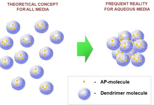
In this context, it is interesting to note that to date, there is no experimental evidence supporting the concept of the ‘unimolecular behaviour’ of dendrimers in aqueous media. In the large majority of the reported cases, e.g., [45, 98, 111, 113, 118-120], results of the size measurements (by, e.g., DLS, SEM and AFM techniques), indicate the presence of dendrimer aggregates in suspension rather than unimolecular solutions (FIG. 4).
Figure 4. Schematic representation of the theoretical concept of dendrimer unimolecular behaviour in solution proposed for all media and the formation of dendrimers aggregates frequently observed in real aqueous media.
Generally, it is reasonable to expect that the aggregation mechanisms depend on surface properties of dendrimers and involve similar interactions to those discussed in Section 2.1. The most recent attempts to study the aggregation mechanisms using in silico modeling showed that the attraction between macromolecules is due to van der Waals interactions between uncharged regions of the dendrimer surfaces [121]. Therefore, it is reasonable to assert that ‘unimolecularity’ in aqueous solutions as a distinctive property of dendrimers can be achieved only at a sufficiently high surface charge density, which obviously depends on the nature of the terminal groups and the G value, as well as on the parameters of the media and the dendrimer concentration. At this point however, the optimal surface properties are still to be clarified, since the aggregating behaviour has been observed not only for highly hydrophilic noncharged
peripheries composed, for e.g., of PEG groups [35, 98, 101, 102, 112], but also for the case of highly charged COONa-surfaces [122]. In particular, in the last example [122], a preliminary filtration of dendrimer solutions was not sufficient to prevent the presence of aggregate particles, the size of which was exceeding the filter porosity, suggesting that the aggregation process was spontaneous and energetically favourable. Dendrimer-dendrimer interactions can also be affected by the presence of AP-molecules. The increase in size of particles formed by AP-dendrimer complexes [45, 98, 101, 102, 111, 113, 118-120] could be explained by the ‘bridging effect’ exerted by the AP, as demonstrated by in silico studies on model systems [123]. Consequently, the concept of dendrimer-based-nano-vectors proposed as ‘unimolecular nano-boxes’ must be carefully used and demands strong experimental evidence.
Instead of avoiding the aggregation of dendrimers, several researchers have proposed to take advantage of it to form regular supramolecular assemblies. Giles et al. showed that a guest molecule could be used as a template at neutral pH to generate a trimolecular inclusion complex, by testing a large range of hydrophobic guest substances (FIG. 5), including biologically active estradiol [124]. Additionally, recent data have shown the possibility of obtaining bilayer spherical structures (dendrosomes) of 2 to 50 μm in size, composed solely of dendritic molecules [125]. A promising avenue of dendrimer development is the design of non-symmetric branched architectures presenting amphiphilic properties, leading to the formation of multi-layered polyfunctional dendrimeric polyplex micelles for overcoming various physiological barriers [126], or light-induced gene transfer [127, 128]. Such an approach could open up new perspectives in developing novel submicronic dendritic AP-vectors benefiting from their supramolecular behaviour.
Figure 5. Formation of a nanocapsule from two dendrimers, each carrying an internal hydrophobic hemisphere, around a hydrophobic molecule in water at neutral pH. Reprinted with permission from Giles et al., J. Am. Chem. Soc. 130 (2008) 14430-14431 [124]. Copyright 2017 American Chemical Society.
2.3. Biological properties of dendrimers
2.3.1. Bio-distribution and biosafety of dendrimers
The use of dendrimers as AP-delivery vectors requires one to consider the interactions of these macromolecules with the organism. This should take into account the factors which include not only the potential therapeutic benefits of the formulation but also the potential biological risks that future patients could run. In this context, biological properties of dendrimers have been intensively studied over the last few decades [129, 130] and can be briefly summarized into two main characteristics, namely, bio-distribution and biosafety, the main aspects of which will be discussed below in detail.
2.3.1.1. Bio-distribution
The bio-distribution of dendrimers is determined by their surface properties, such as the size, the surface charge, and the presence of hydrophilic and/or hydrophobic groups. Polycationic dendrimer peripheries (i.e., due to the ionized amino groups) are known to interact by strong electrostatic effects with anionic functional groups of cell membranes [15]. An effective use of dendrimers as AP-vectors for oral administration, except a few cases [29, 38, 141, 146], is still limited, probably, due to many biological barriers to overcome. After intravenous (iv) or intraperitoneal (ip) administration in mice, polycationic PAMAM and poly-lysines, tend to be rapidly absorbed at the surface of blood vessels, that prevents their effective bio-distribution throughout the body [131-133]. Depending on the G-value and animal model, the polyamine PAMAM, poly-lysines and polymelamine dendrimers can distribute differently in the liver, kidneys and pancreas [131, 133, 134]. The modification of the dendrimer surface with anionic (acidic) groups can increase their residence time in the bloodstream, as reported for polycarboxylated PAMAM [133]. The nature of the surface acidic groups can also impact the bio-distribution. Poly-lysine dendrimers with polysulfonate peripheries were opsonised quickly and retained mainly by the reticuloendothelial system (liver and spleen), while dendrimers
bearing succinate groups were rapidly eliminated by renal clearance [135]. Non-ionized hydrophilic polyhydroxy terminated PAMAM provide site-specific delivery of small molecular drugs across the blood-brain and blood-cerebrospinal-fluid barriers (BBB and BCSF, respectively), specifically targeting the injured brain in canine [134] and rabbit [136] models. In rabbit models, polyxydroxylated PAMAM are also found to be more effective in crossing the fetal blood-brain barrier, and targeting foetus activated microglia than polycarboxylated PAMAM [137]. The bio-distribution studies in rats with PEGylated PAMAM and poly-lysine dendrimers demonstrated a marked increase of the bloodstream residence time and a significant lowering of the dendrimer accumulation in the liver a decrease in elimination by renal filtration and an increase in reaching the lymphatic system for structures having higher molecular weights [133, 138-140]. Similar conclusions were drawn for the dextran conjugated PPI G5 [39].
Interestingly, the presence of the peripheral hydrophobic tetradecane groups may impart a moderate increase in AP-concentration in the bloodstream after oral administration [141].
Coating dendrimers with phospholipids may also increase the uptake by the lymphatic system [119]. The 125I-labelled non-ionized polyester dendritic scaffolds (up to 23 kDa) presenting different arrangements of BMPA and PEG showed that the rapid excretion in urine after iv administration in mice is mostly dependent on the molecular weight of the macromolecules and not on their degree of ramification [142]. On the other hand, the larger dendrimers (40 kDa and greater) with a more pronounced branched architecture, demonstrated an increase in circulation time and a decrease in renal clearance, probably due to their lower flexibility and consequently, to the difficulties in passing through glomerular pores [143]. Remarkably, no specific organ accumulation was observed for these macromolecules, despite a significant portion of the dose found in the carcass after 48 hours[143].
In the context of bio-distribution, it is worth mentioning ex vivo and in vitro studies dealing with the interactions of dendrimers with biological membranes at the tissue and cell levels respectively. The ex vivo studies are mostly limited to the general membrane penetration tests, in some cases, demonstrating selectivity to specific tissues, depending on the dendrimer structure. For example, the extravasation duration of polycationic PAMAM G0-4 labeled with fluorescein isothiocyanate through the microvascular endothelium in hamster cremaster muscles increases with G [144]. Concomitantly,PAMAM G4-NH2 demonstrated only very limited transfer rates to the fetus side of human placenta [145]. The 125I-labelled polyanionic PAMAM (G 2.5, 3.5, and
5.5, -COONa) showed a better penetration capacity through the intestinal sac of adult rat compared to dendrimers with polycationic periphery (G3 and G4, -NH2) [146]. In porcine ear skin, the infiltration enhancement of PAMAM with different peripheral functionalization (-COOH, -OH, -NH2) was found to follow: G4-NH2 > G4-OH > G3.5-COOH. Skin penetration resistance was also found to increase with G for G2-G6-NH2 [44]. Similar results were observed in an in vivo study on rabbit eye corneas, using PAMAM G2- and G4-NH2, G2- and G4-OH, G1.5- and G3.5-COOH [74].
The dendrimer surface charge and area also play an important role in interactions between dendrimers and cell walls membranes, as demonstrated by in vitro assays on different cell lines. In particular, the enhanced cell membrane permeability observed with dendrimers having polycationic peripheries can be associated with their capability to non-selectively disrupt the negatively charged cell walls (in some cases, creating pores of 15-40 nm [147]), as shown on the human corneal epithelial (HCEC) [105], human epidermoid carcinoma (KB) [147], rodent fibroblast Rat-2 [147] and L929 [148], heterogeneous human epithelial colorectal adenocarcinoma (Caco2) [149], human umbilical vein endothelial (HUVEC) [150] cells, etc. Accordingly, the absence of positively charged surface groups considerably restrains interactions of dendrimers with the cell membrane. For example, the fluorescently labeled PPI and PAMAM with neutral polyacetyl or PEG peripheries showed a substantial loss of polymer adhesion to the plasma membrane and then damage, even at longer incubation times (>60 min), compared to their polyamino precursors [147, 150]. There is no association between the polyacetilated PAMAM dendrimers and the folate down-regulated KB cells [151]. Analogously, a pronounced decrease in transfection efficiency was observed after transforming PAMAM G4-NH2 into its G4-OH derivative, suggesting that the polyhydroxy structure is significantly less capable of binding electrostatically to the surface cell-matrix and cell-cell anchoring proteins, such as heparan sulfate proteoglycans [152]. It should however be noted that in the absence of electrostatic effects, other dendrimer-transfection mechanisms are also possible. For example, energy-dependent fluid-phase endocytosis was evidenced by fluorescence microscopy in the case of star-shaped PEGs bearing polyesters of G1-4 [129]. Rhodamine-labeled PEPE dendrimers were found to be able to cross the cell membrane by both energy-dependent and clathrin- and caveolin-mediated endocytosis pathways [99].
The bio-distribution studies focused on exploring the ‘active transport’ approaches should also be mentioned. In this case, the surface of dendrimers is partially modified by grafting molecular ligands, in order to provide a high selectivity to the cells possessing the corresponding membrane receptors (e.g., dendrimers decorated with a tripeptide arginine-glycine-aspartate showed an increased selectivity to cells with integrin receptors [153]). Although it is extremely difficult to find the receptor-ligand combinations to insure their targeting specificity, recent advances in this field have shown very encouraging results when treating some affected cells and pathogens (see more in Section 2.3.2). The intercellular fate of dendrimers is another important aspect of their biological properties, especially when it comes to specifying their influence on the host-cell machinery. This aspect represents one of the less studied areas of dendrimer properties and is mostly restricted to a few toxicological studies [129, 154] (see more in Section 2.3.1.2). It is worth also mentioning the recent advances in creating artificial model systems as well as the virtual structures intended for computational (in silico) simulations. Given the tremendous challenge in modeling complex biological systems, such approaches are still limited to elucidating the molecular mechanisms driving the most simple intermolecular interactions. For example, a very good consistency was observed in testing the phospholipid membrane permeability by dendrimers with different surface properties between cell cultures studies, artificial phospholipid bilayer vesicles modeling, and in silico simulations [55, 147]. In silico approaches gain more popularity when studying the physico-chemical interactions of dendrimers with genetic material (DNA and RNA) and proteins [55].
2.3.1.2. Biosafety
Overall, our knowledge of the biosafety of dendrimers needs to include possible adverse effects at the cellular, tissue, and organism levels after both short and long exposures [130]. For example, for any polymeric AP-vectors designed for parenteral administration, it is essential for the formulation to be non-toxic, non-immunogenic, and preferably, bio-degradable [129]. In other words, the biosafety analysis needs to include many multi-parameter tests in vivo, the implementation of which represents a tremendous challenge, given their complexity and high cost. Consequently, in the vast majority of cases, data on the biosafety of dendrimers in humans are still very poorly documented. For example, T. Toyama et al. [155] reported one case of toxic dermatitis developed in a Japanese student of 22 years of age, as a possible response of his
immune system to the topical contact with a dendrimer. However, the exact nature of the substance that caused these effects has not been specified with certainty. In this regard, it should be noted that systematic in vivo toxicity tests in humans, using dendrimers for AP-delivery, are still nonexistent. This is probably due to the insufficient level of development in this area as well as to unsatisfactory results obtained during preliminary tests in cell cultures and animals. Therefore, so far, the general toxicological profile of dendrimers is mostly limited to data collected using different in vivo and in vitro model systems.
As a rule of thumb, dendrimers presenting polycationic groups should be expected to provoke increased toxicological effects. In vivo tests showed that after ip administration of polyamino PAMAM in mice, G7-NH2 caused 40%-mortality, while G3-NH2 and G5-NH2 did not exhibit any short-term acute complications. In 7- and 30-day multiple dose experiments, the histopathology analysis revealed some vacuolization of cytoplasm in all liver samples [131] indicating possible lysosomal storage problems [129], however, no significant liver damage (assessed via the expression of pyruvic glutamic transaminase) was found after iv administration in mice of 10 mg/kg doses [139]. In addition, no evidence of immunogenicity was established, after administrating PAMAM-NH2 in rabbits [131]. Similarly, other polyamino-terminated dendrimers, i.e., composed of melamine [156] and lysine [140] units, did not show hepatotoxic effects in mice at a 10 mg/kg iv dose, while the its increase to 40 mg/kg and 160 mg/kg dose led to an important hepatic necrosis and to the animal death, respectively [156]. On the other hand, dendrimers with neutral or acidic peripheries are found to be much better tolerated. For example, the PEG-grafting (typically of 0.2-40 kDa [157]) allowed the dendrimer tolerability to increase dramatically to the g/kg-dosing range, i.e., up to 2.56 g/kg (ip) et 1.28 g/kg (iv) as demonstrated with the G2 polymelamine-piperazine-pyrimidine structures in mice [158]. No negative changes had been reported in the anatomy of mouse organs such as the liver, kidneys, spleen, heart and lungs after iv-administration of 10-mg/kg doses of a PEGylated polylisne G6 [140]. Blood parameters such as hemoglobin level, RBC, WBC, differentiated monocytes, lymphocytes and neutrophils, measured after 14 days following administration of PEGylated PAMAM-G4 (12 mg/kg) in rats, did not show significant changes compared to the control group [46]. No gross toxicity, either acutely or chronically up to 99 days (including a study for behavioural and histopathology abnormalities), was observed after a single 10 mg/kg iv injection of acetyl and folate PAMAM-G5 conjugates [159]. G3-triazine-based polyhydroxy (diethanolamine)
terminated dendrimers showed no hemolytic effects and toxicity to the kidneys and the liver in mice up to 200 mg/kg ip injection [104]. No side effects were also reported in mice after repeated ip daily 95-mg/kg administration of PAMAM-G3.5-COONa [25]. A study on zebrafish embryos showed that PAMAM-G4-NH2 affected the embryonic growth and development at sub-lethal concentrations, while PAMAM-G3.5-COONa was practically harmless [160].
Biosafety assessment performed in vitro on different cell lines also suggests that the nature of the surface groups influences the short-term toxicological profile of dendrimers. As discussed in Section 2.3.1.1, these groups are responsible for the capability of crossing the epithelial cell membrane, due to electrostatic attraction. Consequently, dendrimers with polycationic peripheries are able to disrupt the cell membrane integrity, affecting the cell viability. For example, dendrimers bearing primary amine groups exhibit hemolytic properties [46, 61, 70, 133] associated with some important visually detectable changes in the morphology of erythrocytes (RBC), as well as with the release of haemoglobin induced by cell membrane leakage. Strong decreases in survival rates were also reported for many different cell lines, such as rat liver Clone-9 [156], HCEC [105], KB [147], rodent fibroblasts Rat-2 [147] and L929 [148], Caco2[149], HUVEC [150], etc. It is noteworthy, however, that a comparative study [29] on L929 and RBC showed that dendrimers with a polyamine periphery (i.e., PAMAM-G3-NH2) were less toxic than linear polyamine polymers, in the following ranking: poly(ethylenimine) = poly(l-lysine) > poly(diallyl-dimethyl-ammonium chloride) > diethylaminoethyl-dextran > poly(vinyl pyridinium bromide) > PAMAM-G3-NH2 > cationized albumin > native albumin. Also of importance, the mechanisms underlying the cytotoxicity effects caused by amino-terminated dendrimers are not limited to their interactions with the cell membrane. For example, the exposure of U-937 human macrophages to PPI G2-NH2 and G3-NH2 at concentrations greater than 90-% survival led to significant fluctuations of the reactive oxygen species (ROS) content and of the mitochondrial membrane potential, both related to the apoptotic activation of this organelle [161]. In addition, even without affecting the integrity of the cell membrane, dendrimers can dramatically affect the cell functions and provoke DNA cleavage [161], suggesting their very complex adverse impact on the cell machinery. Compared to this, dendrimers with neutral and/or negatively charged peripheries (e.g., -OC(O)Me, -OMe, -PEG-OMe, -OH, -COOH, -SO3H, -PO(OH)2 are considerably less cytotoxic at short-term exposure, as reported in many cases [23, 39, 61, 104, 133, 147, 150-153, 158, 162]. The beneficial effect is
generally explained by the decrease in capacity of interacting electrostatically with the external cell-membrane matrix (See in Section 2.3.1.1).
Dendrimers must also be safely transformed and/or removed from the organism to avoid long-term undesirable effects. In this connection, non-bio-degradable hydrophilic linear polymers that cannot be eliminated by renal clearance (i.e., with Mw > 18 kDa) tend to accumulate inside the body, causing deposit formation in different organs, especially, in the skin and muscles [163]. The accumulation of dendrimers having relatively stable structures (PPI, PAMAM, poly-lysines, polyethers) mostly occurs in the liver [15, 129]. In addition, the intracellular accumulation of non-bio-degradable macromolecules can cause lysosomal disease [129, 164]. To avoid this problem, a promising strategy is to develop dendrimers containing bonds that can be broken over time under biologically relevant conditions, leading to the formation of non-toxic by-products. One of the most hopeful solutions is to employ polyester scaffolds that can be relatively easily degraded by carboxyesterases [15, 165-167], as well as under weakly acidic or basic conditions [143, 165]. Bio-degradability can also be achieved by inserting disulfide bridges sensitive to glutathione (GSH), a tripeptide produced by the majority of mammalian cells [15, 28, 168, 169] (see also Section 2.3.2). The advantages of ester and disulfide groups can be effectively combined in the same structures, as carried out in a series of commercial dendrimers, PFD® [107]. Among other degradable macromolecules, some polycarbamate [170, 171] and poly(phenethyl carbonate) [172] have also be reported, but a more thorough toxicological assessment is, however, still needed.
2.3.2. Proposals of encapsulating FDA-approved APs for specific therapies
A successful clinical use of dendrimers as non-covalent AP-encapsulating agents in specific therapeutic treatments would represent the quintessence of the significant basic work in materials development accomplished over preliminary stages. So far yet, no commercially available AP-formulations of this type are present on the pharmaceutical market, as well as no data on ongoing clinical trials with dendrimer excipients were publicly reported. At the same time, some dendrimer-based APs have already begun to gain recognition in medical applications, e.g., VivagelTM is now used topically in preventing and treating sexually transmitted viral and bacterial infections [173]. Another compound, a multiantigenic peptide PHSCN-lysine dendrimer, is currently undergoing preclinical study as a very potent inhibitor of human breast
cancer cell invasion, extravasation, and lung colony formation [174]. Therefore, it is safe to assume that in the near future some promising dendrimer based excipients, i.e., intended for AP-encapsulation and delivery (see Table 1), will be available for wide application.
Table 1. Proposals of using dendrimers as non-covalent AP-encapsulation agents in specific therapies Aimed therapeutic activity Encapsulated FDA-approved AP
Type of dendrimer structure Type of test performed*
Antibacterial and antifungal
amphotericin B [114]
polyamino terminated PPI [114] in potus [114]
benzoic acid [66] polyhydroxy (Tris) terminated PAMAM [66] in potus [66]
gatifloxacin [105]
polyguanidyl terminated dendrimers composed of guanidine, ethylenediamine, amides of 1,3,5-tricarboxybenzoïc and 2-hydroxypropanoïc acids [105] in potus [105], in vitro [105], in ex vivo [105], in vivo [105] itraconazole [101, 102]
PEPE dendrimers composed of O-acylated glyceryl tris[9,10-(threo)-dihydroxyoctadecanoate] core, succinic acid spacers and PEG-peripheries [101, 102]; PEPE dendrimers with tetra(ethylene oxyde) (TEG) peripheries, succinic acid, TEG and 1,10-decanediol spacers, and cores represented by pentaerythritol, tetraphenylporphine [102]
in potus [101],
[102], in vitro [101]
salicylic acid [66] polyhydroxy (Tris) terminated PAMAM [66] in potus [66]
sulfamethoxazole [72, 73]
polyamino terminated PAMAM [72, 73] in potus [72,
73], in vitro [72]
triclosan [175] polycarboxy terminated PAMAM [175] in potus [175], in vitro [175], in ex vivo [175] Antiepileptic (Sedation) phenobarbital [73]
polyamino terminated PAMAM [73] in potus [73]
Anthelmintic niclosamide
[185]
polyamino terminated PAMAM [185] in potus [185]
Antihypertensive nifedipine [71] polyamino terminated PAMAM [71] in potus [71]
Antiinflammatory and analgesic
aspirin [51] methyl ester terminated PAMAM [51] in potus [51]
cortisol [52] dendrimers composed of PPI dendritic core, TEG peripheries, urea and hexamethylene spacers [52]
in potus [51]
diflunisal [63] polyamino terminated PAMAM [63] in vivo [63]
ibuprofen [56] polyamino terminated PAMAM [56] in potus [56]
[59, 60, 114] terminated PAMAM [60]; polyamino terminated PPI [114]
60, 114], in
vivo [59, 60]
flurbiprofen [61] polyamino terminated PAMAM [61] in potus [61], in vitro [61], in vivo [61]
ketoprofen [62, 63]
polyamino terminated PAMAM [62, 63] in potus [62], in vivo [62, 63]
mefenamic acid [186]
polyamino terminated PAMAM [186] in silico [186]
piroxicam [70, 187]
polyamino terminated PAMAM [70, 187] in potus [70], in vitro [70], in vivo [70], in silico [ 187]
sulfasalazine [188]
PPI with polyfucosylate periphery[188] in potus [188], in vitro [188], in vivo [188]
Antimalarial artemether [176] polylisine dendrimers with PEG core having either
polylisine or chondroitin peripheries [176]
in potus [176], in vitro [176], in vivo [176]
chloroquine phosphate [97]
polylisine dendrimers with PEG core having either polylisine or chondroitin peripheries [97]
in potus [97], in vitro [97], in vivo [97]
primaquine phosphate [177]
polyamino and polygalactose PPI [177] in potus [177], in vitro [177], in vivo [177]
Antineoplastic camptothecins
[48, 68]
polycarboxy terminated dendrimers composed of glycerol units as branching points and succinic acid core, spacers, and periphery [48]; polyamino terminated PAMAM [68]
in potus [48,
68], in vitro [48]
carboplatin [178] polycarboxy terminated PAMAM [178] in vivo [178]
chlorambucil [47]
dendrimers composed of PAMAM core and PDMA periphery [47]
in potus [47]
cisplatin [25, 95, 179]
polycarboxy terminated PAMAM [25]; PPI with diaminobutyrate periphery [179]; polyester dendrimers with polycitric acid periphery composed of PEG-core and polycitric acid dendrons [95] in potus [25, 179], in vitro [95], in vivo [25] DOX [38-40, 79-81, 189-192] and DOX- pORF-hTRAIL conjugate [81]
polyamino terminated PAMAM [38]; polyamino terminated PPI with the periphery partly modified by grafting dextran [39]; PPI dendrimers with polyamino and polyfolate peripheries [40]; polyamino terminated PAMAM with the periphery
in potus [38-40,
79-81, 189-192], in vitro [38-40, 79-81, 189-192], in
partly modified by grafting the peptides HAIYPRH [80, 81] or LFC131 [189], or YLFFVFER (H6) [190], or RGD [79] via PEG spacers [80, 81, 190]; assymetric polyamino terminated PAMAM, one of the dendrons of wich is composed of C18 alkyl chains [191]; PAMAM with the periphery sequentially modified with fluorescein isothiocyanate (FI) and LA and polyethylene glycol (PEG)-linked LA, PEG-LA), followed by acetylation [192]
vivo [38, 39,
80, 81, 191]
etoposide [193] PAMAM with poly(ε-caprolactone)-co-PEG periphery [193]
in potus [193], in vitro [193]
5-FU [43-46, 180, 194]
Polyamino terminated PAMAM [44-46]; PAMAM with FA and FA-PEG-N-hydroxy-succinimide peripheries [43]; polycarboxyl and polyhydroxy terminated PAMAM [44], PEGylated PAMAM [46]; polyamino terminated PAMAM partly modified by grafting poly(N-isopropylacrylamide) [180]; PAMAM modified by grafting both
poly(2-(N,N-diethylamino)ethyl methacrylate) (PDEA) and MeO-PEG chains [194]
in potus [43, 45, 46, 180, 194], in vitro [43, 45, 46, 180] in vivo [43, 46, 194], in ex vivo [44] 6-mercaptopurine [96]
polyamino terminated dendrimers composed of melamine, piperazine and piperidine [96]
in vivo [96]
MTX [16, 35-37, 49, 96]
polyhydroxy and poly-FA terminated PAMAM [16]; PEPE dendrimers composed of PEG spacers and peripheries, 3,5-dihydroxybenzoic acid
(DHBA), 2,2-bis(hydroxyl methyl) butyric acid (BHBA) and gallic acid used as branching points and cores comprising butane-1,2,3,4-tetracarboxylic (BTCA) and aspartic acids [35]; PEPE dendrimers composed of PEG spacers and glucosamine-PEG peripheries, DHBA and gallic acid used as branching points and cores comprising BTCA and aspartic acids [36]; PAMAM dendrimers with tBoc-NH-PEG periphery [37]; polyamino terminated PAMAM [49]; polyamino terminated dendrimers composed of melamine, piperazine and piperidine [96] in potus [16, 35-37, 49], in vitro [16, 35-37, 49], in vivo [37, 96]