Large Scale Identification of Protein SUMOylation by
Mass Spectrometry in HEK293 Cells
par
Louiza Mahrouche
Département de Biochimie, Institut de recherche en immunologie et cancérologie Faculté de Médecine
Mémoire présenté à la Faculté des études supérieures et postdoctorales en vue de l’obtention du grade de Maîtrise ès science (M.Sc.)
en Biochimie
décembre 2010 ©Louiza Mahrouche, 2010
Faculté des études supérieures et postdoctorales
Ce mémoire intitulé :
Large Scale Identification of Protein SUMOylation by Mass Spectrometry in HEK293 cells
Présentée par : Louiza Mahrouche
a été évalué par un jury composé des personnes suivantes :
Dr. Muriel Aubry, président-rapporteur Dr. Pierre Thibault, directeur de recherche
Résumé
Une large gamme d’événements cellulaires est régulée par la SUMOylation des protéines. Cette modification post-traductionnelle est impliquée dans le cancer notamment dans la leucémie promyélocytaire aigue. À ce jour, peu d’études à grande échelle ont porté sur l’identification des sites de modification. Ce mémoire présente une approche protéomique quantitative unique qui combine une double purification par affinité au niveau des protéines cibles ainsi que des peptides modifiés.
L’approche la plus répandue de purification des protéines SUMOylés implique l’utilisation d’une forme de SUMO modifié avec une étiquette (His6-SUMO). A ce jour, les
approches permettant l’enrichissement au niveau peptidique nécessite une forme mutante de SUMO.
Notre analyse consiste à premièrement enrichir en protéines SUMOylés dans les cellules humaines vierges ou sur exprimant His6-SUMO-1/3 en présence ou pas de trioxyde
de diarsenic, un traitement de leucémie promyélocytaire aigue. Par la suite, les échantillons sont digérés et les peptides obtenus des protéines SUMOylés conservent un branchement caractéristique. Les peptides sont soit immunoprécipités avec un anticorps spécifique au branchement SUMO ou directement analysés par nano LC/LC-MS/MS par un spectromètre de masse LTQ-Orbitrap. Une analyse manuelle des données révèle des fragments caractéristiques correspondant à la chaîne latérale de SUMO. L’originalité de l’approche réside dans l’identification quantitative et sans ambigüité des sites de SUMOylation. Cette approche a permis l’identification de 17 et 3 sites de SUMO-3 et SUMO-1 respectivement dans les cellules HEK293. Finalement, la SUMOylation de PML est induite suite au traitement d’arsenic.
Mots-clés : SUMOylation, spectrométrie de masse, purification par affinité, identification de sites de SUMOylation
Abstract
A wide range of cellular events are regulated by protein SUMOylation. This posttranslational modification was involved in APL (acute promyelocytic leukemia). Only a few large scale studies in mammalian cells have focused on identifying the conjugation sites. This thesis presents a unique quantitative proteomics approach that combines double affinity purification at the protein and peptide level.
A common approach to purification of SUMOylated proteins involves the use of a tagged SUMO (His6-SUMO). To date, the SUMO peptide isolation is addressed using an
engineered SUMO.
In presence or absence of arsenic trioxide, a treatment of APL, mock and His6
-SUMO1/3 expressing cells are lysed and the SUMOylated proteins are isolated under denaturing conditions. Subsequently, these samples are digested and the peptides bearing the modification site bear a specific SUMO stub. They are either immunoprecipitated with an anti SUMO stub antibody or directly analyzed by nano LC coupled to an LTQ-Orbitrap mass spectrometer. Manual analysis of the data reveals characteristic fragmentation corresponding to the side chain of SUMO. The originality of the approach lies in the quantitative and unambiguous identification of SUMOylation sites in vivo. This approach allowed the identification of 17 and 3 sites of SUMO-3 and SUMO-1, respectively, in HEK293 cells. Finally, PML was identified as the major SUMOylation target following arsenic treatment.
Keywords: SUMOylation, mass spectrometry, affinity purification, identification of
Table of Contents
RÉSUMÉ ... I ABSTRACT ... II TABLE OF CONTENTS ... III LIST OF TABLES ... VI LIST OF FIGURES ... VII REMERCIEMENTS ... X ABBREVIATIONS ... XI
1. INTRODUCTION ... 1
1.1 SUMOYLATION PROCESS IN THE CELL ... 2
1.1.1 Discovery of SUMO ... 2
1.1.2 SUMO conjugation, processing and deconjugation ... 3
1.1.2.1 SUMO conjugation ... 4
1.1.2.2 SUMO deconjugation ... 6
1.1.2.2.1 Endopeptidase activity ... 6
1.1.2.2.2 Isopeptidase activity ... 7
1.1.2.3 Poly-SUMO chain formation ... 7
1.1.3 Consensus sequence ... 8
1.1.4 Molecular consequences of SUMOylation ... 9
1.2 MASS SPECTROMETRY ... 10
1.2.1 Ionization ... 11
1.2.2 The Mass Analyzer ... 13
1.2.2.1 The Orbitrap mass analyzer ... 14
1.2.2.2 Linear ion trap mass analyzer ... 15
1.2.3 Tandem MS ... 15
1.2.5 Database Searching ... 18
1.2.6 Quantitative Proteomics ... 22
1.3 SUMO MS BASED PROTEOMICS ... 24
1.3.1 Enrichment of SUMOylated proteins ... 24
1.3.2 Enrichment strategies of SUMO-modified peptides ... 25
1.3.3 Stress-induced SUMOylation ... 27
1.3.4 Spectral interpretation strategies ... 27
1.4 PROJECT’S GOAL ... 31
1.5 REFERENCES ... 33
2. A NOVEL PROTEOMICS APPROACH TO IDENTIFY SUMOYLATED PROTEINS AND THEIR MODIFICATION SITES IN HUMAN CELLS ... 41
2.1 ABSTRACT ... 42 2.2 INTRODUCTION ... 43 2.3 EXPERIMENTAL SECTION ... 47 2.4 RESULTS ... 53 2.5 DISCUSSION ... 64 2.6 ACKNOWLEDGEMENTS ... 69 2.7 REFERENCES ... 81 3. DISCUSSION ... 88 3.1 GENERAL APPROACH ... 89 3.2 IN VITRO SUMOYLATION ... 89
3.3 THE HIS-SUMO MUTANT FUNCTIONALITY IN HEK293 CELLS ... 92
3.4 OVEREXPRESSION OF HIS-SUMO PROTEINS IN HEK293 CELLS ... 93
3.5 SUBCELLULAR DISTRIBUTION OF SUMO ... 95
3.6 NI-NTA ENRICHMENT ... 96
3.7 DIGESTION AND PEPTIDE SEPARATION ... 97
3.8 SUMO PEPTIDE IDENTIFICATION BY MASS SPECTROMETRY ... 99
3.8.2 Spectral interpretation strategies ... 99
3.8.3 Fragmentation methods ... 102
3.9 IMMUNOPRECIPITATION AND SUMO-1 SITE IDENTIFICATION ... 102
3.10 EFFECT OF ARSENIC TRIOXIDE ... 104
3.11 IDENTIFIED TARGETS ... 106
3.12 REFERENCES ... 109
4. CONCLUSION ... 116
4.1 THE SUMO CHALLENGE ... 117
4.2 FUTURE PERSPECTIVES ... 118
4.3 REFERENCES ... 119 ANNEX I: SUPPLEMENTARY FIGURES 2.1 -2.9 FOR CHAPTER 2 ... I ANNEX II: SUPPLEMENTARY FIGURE 2.10 FOR CHAPTER 2 ... XIX ANNEX III: SUPPLEMENTARY FIGURE 2.11 FOR CHAPTER 2 ... XLVI ANNEX IV: SUPPLEMENTARY TABLES I-III FOR CHAPTER 2 ... LI
List of Tables
Table I : List of confirmed SUMOylated peptides from NTA-enriched of nuclear proteins from HEK293 His6-SUMO-3 mutant ... 70
Table II :List of confirmed SUMOylated peptides from the immunoprecipitation of nuclear proteins from HEK293 His6-SUMO-1 mutant ... 71
Supplementary Table I: Peptide and protein identification from NTA affinity-purified mock HEK293 and HEK293 His6-SUMO3 mutant nuclear extracts with and without As2O3.
Table on CD ROM ... lii Supplementary Table II: Peptide and protein identification from dual affinity purification (NTA and immunoprecipitation) of mock HEK293 and HEK293 His6-SUMO1 mutant
nuclear extracts with and without As2O3. Table on CD ROM ... lii
Supplementary Table III : Identification of SUMOylated peptides with Mascot, ChopNSpice and SUMmOn from NTA affinity-purified HEK293 His6-SUMO3
List of Figures
Figure 1.1: Overlay SUMO-1 (black) and ubiquitin (red) ... 3
Figure 1.2: SUMO pathway: activation, conjugation and deconjugation ... 4
Figure 1.3: Isopeptide link between SUMO and its target protein ... 5
Figure 1.4: Reactions catalyzed by SENPs ... 6
Figure 1.5: Use of ESI in an LC-MS coupled instrument ... 12
Figure 1.6: Peptides are ionized in the ESI and fragmented in either the LTQ or in the HCD collision cell. The Orbitrap is used to detect parent m/z ... 14
Figure 1.7: Nomenclature of fragment ions observed for the dissociation of peptide ions . 17 Figure 1.8: The MS/MS spectrum is compared against a theoretical MS/MS spectrum generated in silico. A score is given to each peptide based on the similarity of the two spectra ... 21
Figure 1.9: Contour map created by Proteoprofile... 23
Figure 1.10: Purification strategy of His-SUMO conjugated proteins using the Ni-NTA pull down ... 24
Figure 1.11: Theoretical MS/MS CID spectra of a) unmodified peptide, b) phospho-peptide and c) SUMO-modified peptide. ... 29
Figure 2.1: Overview of proteomics approach to the identification of SUMOylated peptides. ... 72
Figure 2.3 : Comparison of His-SUMO WT and mutants to SUMOylate PML and to colocalize within nuclear bodies ... 74 Figure 2.4: Immunoblots of NTA purified extracts from control HEK293 and
HEK293-His-SUMO cells ... 75 Figure 2.5: Mass spectrometry analyses of SUMOylated proteins from extracts of control HEK293 and HEK293 His6-SUMO-3 in As2O3-stimulated and non-stimulated cell .. 77
Figure 2.6: Identification of new SUMOylation sites in the protein promyelocytic leukemia (PML) ... 79 Figure 2.7: Identification of proteins modified by SUMO-1 using a combined
NTA/immunoaffinity enrichment approach ... 80
Supplementary Figure 2.1: MS analyses of in vitro digestion products of GST-RanGAP1 (418-587) confirmed the SUMOylation site K524 for all SUMO paralogs. ... iii Supplementary Figure 2.2: MS analyses of in vitro SUMOylation enabled the identification of sites of polySUMOylation. ... vii Supplementary Figure 2.3 : in vitro deSUMOylation assay of E2-ligase and RanGAP1
using SENP1 ... ix Supplementary Figure 2.4 : Comparison of His-SUMO1 WT and His-SUMO1 mutant to
SUMOylate PML and to colocalize within nuclear bodies ... x Supplementary Figure 2.5: Analysis of Gene Ontology terms associated with biological
processes ... xi Supplementary Figure 2.6: NTA purified extracts from control HEK293 and
Supplementary Figure 2.7: Tandem mass spectra of synthetic peptides to confirm SUMOylation of sites K380 and K400 ... xiv Supplementary Figure 2.8 : Design of an immunoprecipitation experiment using a
centrifugal filter device and Recovery yields of SUMOylated peptides ... xvi Supplementary Figure 2.9: Mass spectrometry analyses of SUMOylated proteins from
Immunoaffinity purified NTA extracts of mock HEK293 and HEK293 His6-SUMO1
in As2O3-stimulated and non-stimulated cells... xviii
Supplementary Figure 2.10: Tandem Mass Spectra of All Identified SUMO-3 Peptides Following Database Search and Manual Validation. ... xix Supplementary Figure 2.11: Tandem Mass Spectra of All Identified SUMO-1 Peptides
Remerciements
D’abord, je remercie mon directeur de recherche le Dr Pierre Thibault pour son accueil ainsi que les connaissances scientifiques qu’il m’a transmises. Son grand intérêt ainsi que la confiance qu’il m’a témoignée ont permis à ce projet de progresser
Je souhaite également remercier vivement Frédéric Galisson pour sa très grande contribution au projet SUMO. Son excellent travail, son esprit critique et sa grande expérience ont permis au projet SUMO de voir le jour mais aussi de se développer. Merci à Dr Mounira Chelbi-Alix pour sa collaboration et son accueil.
Je désire remercier le Dr Eric Bonneil pour son expertise sur la spectrométrie de masse et le partage de ses connaissances, Mathieu Courcelles pour son assistance aux outils bioinformatiques ainsi que Christelle Pomiès pour son aide dans les différentes techniques de biologie. De plus, je remercie sincèrement Chantal Durette, Robert Gomma et Jean-Michel Laprise pour leur aide lors de l’écriture de ce mémoire. Merci Chantal de prendre le relais du projet. Mes remerciements s’adressent également à tout mes collègues de laboratoire notamment Gaëlle Bridon, Marlene Gharib et Tara Muratore pour avoir créé un environnement d’entraide et amical.
Un grand merci à ma sœur Lilia et à Nicolas ainsi que tous mes proches qui m’ont soutenue durant ces deux dernières années.
Pour terminer, je remercie le FQRNT ainsi que la Faculté des études supérieures de l’Université de Montréal pour leur soutien financier.
Abbreviations
ACN Acetonitrile
ADP Adenosine diphosphate
APL Acute Promyelocytic Leukemia
As2O3 Arsenic trioxide
ATP Adenosine-5'-triphosphate
CI Chemical ionization
CID Collision induced dissociation Daxx Death domain-associated protein DMEM Dulbecco’s modified eagle medium E2-25K E2 ubiquitin ligase – 25 kilo dalton
EDTA Ethylenediaminetetraacetic acid
EI Electron ionization
ESI Electrospray ionization
ETD Electron transfer dissociation
FPR False positive rate
FTMS Fourier transform mass spectrometer
GST Glutathione S-transferase
HCD Higher-energy C-trap dissociation
HEK Human embryonary kidney
hnRNP Heterogeneous nuclear ribonucleoprotein HPLC High-performance liquid chromatograph IMAC Immobilized metal affinity chromatography IP Immunoprecipitation
kDa Kilo dalton
LC Liquid chromatography
LC-MS/MS Liquid chromatography tandem mass spectrometry
LTQ Linear trap quadrupole
m/z Mass/charge
MALDI Matrix-assisted laser desorption ionization
Mdm2 Murine double minute-2
MS Mass spectrometry
MS/MS Tandem mass spectrometry
MudPit Multidimensional protein identification technology
MW Molecular weight
NB Nuclear body
NDSM Negatively charged amino acid-dependent SUMOylation motif NEM N-Ethylmaleimide
NFkB Nuclear factor kB
Ni-NTA Nickel-nitriloacetic acid
NLS Nuclear localization signal
PARP1 Poly (ADP-ribose) polymerase 1 PCNA Proliferating cell nuclear antigen
PDSM Phosphorylation-dependent sumoylation motif PIAS Protein inhibitor of activated STAT
PML Promyelocytic leukemia protein
ppm Parts per million
PTM Post-translational modification
RanBP2 Ran Binding Protein 2
RanGap Ran GTPase activating protein RAR-α Retinoic acid receptor alpha RBCC Ring finger B-box coiled-coil RING Really interesting new gene RNF4 RING finger 4 protein
RSF1 Remodeling and spacing factor 1
SAFB2 Scaffold attachment factor B 2 SENP SUMO-specific isopeptidase
SIM SUMO interacting motif
STAT Signal transducer and activator of transcription protein SUMO Small ubiquitin-like modifier
TAP Tandem Affinity Purification
TBS Tris buffered saline
TIF1b (TRIM28) Transcription intermediary factor 1-beta (TRIM28) Ub Ubiquitin
Ubc9 Ubiquitin-like protein SUMO-1 conjugating enzyme
Ubl Ubiquitin-like protein
WT Wild type
1.1 SUMOylation process in the cell
Post-translational modifications (PTMs) represent universal and fundamental mechanisms by which protein function, activity, stability and localization can be regulated. These modifications extend significantly the diversity of the proteome. PTMs are divided into two groups: proteolytic cleavages and covalent modifications. There are over 150 different covalent modifications on a variety of amino acid side chains (Voet and Voet 1995), among these are small chemical group modifications such as acetylation, phosphorylation or larger macromolecules attachment such as ubiquitylation and SUMOylation, the latter being the primary focus of the present study.
1.1.1 Discovery of SUMO
In the 1970’s, the first protein acting as a ubiquitin-like (UBL) modifier was discovered : ubiquitin (Hochstrasser 2009). However, the first documented report of a related small ubiquitin modifier (SUMO) was only made 20 years later by Mhajan et al. for Ran GTPase 1 (RanGap1) covalently modified by SUMO-1 in mammals (Mahajan, Delphin et al. 1997). At the present day, at least nine UBLs are shown to covalently modify their targets, and it is suspected that additional UBLs are likely to be discovered in the future (Hochstrasser 2009). Regardless of their low sequence similarity to ubiquitin, all UBLs share a common 3D structure: the ubiquitin fold. For instance, although SUMO-1 and ubiquitin share only 18% sequence identity, SUMO has the characteristic ββαββαβ fold of the ubiquitin protein family (Bayer, Arndt et al. 1998) as observed in Figure 1.1.
Figure 1.1: Overlay SUMO-1 (black) and ubiquitin (red) (Bayer, Arndt et al. 1998)
SUMO is expressed in all eukaryotic cells and in different cell types in multicellular organisms. Yeasts have a single isoform of SUMO, while in vertebrates, four paralogs of SUMO (SUMO1-4) are expressed (Saitoh and Hinchey 2000), and in plants eight versions of SUMO have been identified (Kurepa, Walker et al. 2003).
In humans, SUMO-1 is the most studied paralog and hundreds of SUMO1-2-3 conjugates have been identified. SUMO1 and SUMO2/3 share about 50% similarity, but SUMO-2 and SUMO-3 differ only by 4 residues. SUMO1 and SUMO2/3 seem to have different functions since they conjugate different substrates in vivo and have different responses to stress (Saitoh and Hinchey 2000). The role of SUMO-4 is still unknown, no conjugates have been detected in vivo and its in vivo maturation into a conjugation-competent form still remains unclear (Bohren, Nadkarni et al. 2004).
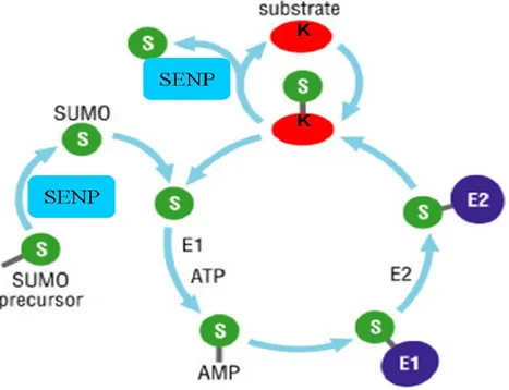
1.1.2 SUMO conjugation, processing and deconjugation
UBLs not only share a common structure, but they use similar conjugation mechanisms requiring a multi-stage ATP-dependent enzymatic cascade (Hochstrasser 2009). Specific and highly dynamic machinery is responsible for SUMO conjugation: the formation of an isopeptide bond between the C-terminal glycine of the mature SUMO and the amino ε group of the target’s specific lysine residue. The specificity of
the reversible SUMO conjugation is regulated by a specialized set of enzymes (Figure 1.2).
Figure 1.2: SUMO pathway: activation, conjugation and deconjugation adapted from (Petsko and Ringe 2004)
1.1.2.1 SUMO conjugation
The SUMO conjugation proceeds in three steps requiring a cascade of enzymes: E1, E2 and E3 which results in the formation of an isopeptide bond (Figure 1.3).
First, the SUMO-Activating enzyme (E1), a dimer composed of Sae1 and Sae2 in mammals, forms a thioester bond with the carboxy group of the SUMO protein and the cys173 of Sae2 (Ulrich 2008). This step requires the hydrolysis of one ATP molecule.
Then, SUMO is transferred to an E2 enzyme. Contrary to ubiquitin, the SUMO pathway has only one E2 enzyme, namely UBC9 (Saitoh, Sparrow et al. 1998). The
SUMO protein is transferred from Sae2 to UBC9 and forms a thioester link with cys93 of UBC9. Note that UBC9 contains a SIM (SUMO-interacting motif) domain capable of recognizing the SUMO protein. Also UBC9-SUMO complex is capable of substrate specificity through the consensus SUMOylation motif found on the target protein (Tatham, Jaffray et al. 2001). At this point, UBC9 can either directly transfer SUMO protein to its substrate or optionally in vivo this step may require the cooperation of an E3 ligase.
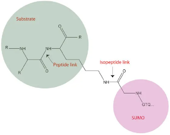
Figure 1.3: Isopeptide link between SUMO and its target protein
A certain number of E3 ligases have been identified for the SUMO pathway. They are believed to facilitate the formation of the isopeptide bond between the C-terminal SUMO protein and the acceptor’s lysine by forming a complex with UBC9 and the SUMO protein. E3 enzymes do not seem to form thioester linkage with SUMO, but function as scaffold proteins bringing UBC9-SUMO complex in close contact with the substrate. E3 ligases have two roles: i) enhance the SUMO conjugation, and ii)
participate in the specificity of the substrate. Most of the identified E3 ligases are nuclear, although new studies have reported the existence of a mitochondrial SUMO E3 (Braschi, Zunino et al. 2009). Examples of E3 ligases include PIAS family, HDAC4 and RanBP2 (Wilkinson and Henley 2010).
1.1.2.2 SUMO deconjugation
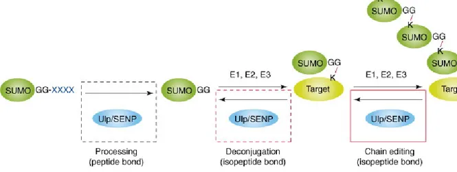
SUMO is removed from its substrate by a family of enzymes known as the sentrin-specific proteases (SENPs). Seven SUMO-sentrin-specific proteases have been identified in the human genome (Marcin Drag 2008), showing different specificities towards SUMO isoforms. SENPs are cysteine proteases and have two main enzymatic activities: endopeptidase and isopeptidase as described in Figure 1.4.
Figure 1.4: Reactions catalyzed by SENPs. Adapted (Mukhopadhyay and Dasso 2007)
1.1.2.2.1 Endopeptidase activity
Newly synthesized SUMO is in an immature form and prior to conjugation, it needs to be activated by SENPs (see Figure 1.2). The propeptide is cleaved by SENP to reveal the essential diglycine motif which is conserved among all ubiquitin-like proteins
(Mukhopadhyay and Dasso 2007). The free diglycine motif is essential for the conjugation reaction.
1.1.2.2.2 Isopeptidase activity
The second role of SENP is the cleavage of the isopeptide bond (Figure 1.3), which is formed between the target and the SUMO protein. Therefore not only do SENPs control the pool of available SUMO, but they are also responsible for the half life of the conjugated species (Mukhopadhyay and Dasso 2007). Moreover, SENPs also control the poly-SUMO chain formation, by hydrolyzing the SUMO-SUMO bond. Due to their central roles, SENPs are believed to be highly regulated.
In summary the SUMO pathway is highly regulated, first by its conjugation using a cascade of enzymes comprising an E1, an E2 and multiple E3 enzymes, but also by its deconjugation pathway.
1.1.2.3 Poly-SUMO chain formation
The concept of a modifier being modified adds a new layer of complexity, but also provides versatility to protein function. The formation of poly-ubiquitin chains has been extensively studied and it became clear that a specific poly-Ub chain structure provides a specific biological outcome (Pickart and Fushman 2004). The wide array of ubiquitylation outcome can be explained by the diversity of ubiquitin chain structure: the chain length but most importantly the different cross linkages that could be formed.
Poly-SUMO chain formations have not been extensively studied due to important technical limitations: the half life and low abundance of the SUMO conjugates, as well as the absence of fast and straightforward method for the identification of SUMOylation sites. So far, SUMO2/3 has been shown to form poly-SUMOylation sites in vivo (Tatham, Jaffray et al. 2001), through the internal K11 that lies in a consensus sequence that is missing in SUMO-1 (Matic, Van Hagen et al. 2008). Although SUMO-1 is also
incorporated into the SUMO2/3 chains, it can’t be further modified and seems to work as a capping modifier. However, in vitro SUMO-1 has been shown to form polymers through K7,16,17,37,39,46 (Cooper, Tatham et al. 2005; Pedrioli, Raught et al. 2006). It is important to note that in vitro conditions are artificial (high concentrations of E1 and E2, absence of SENPs or E3 ligase) and can introduce artifacts.
1.1.3 Consensus sequence
Through the use of known SUMOylation sites of RanGap1, PML, p53 and SP100 a SUMOylation acceptor motif has been identified as ψ-K-X-E/D (Rodriguez, Dargemont et al. 2001) (where K is the acceptor lysine and ψ is a hydrophobic residue). No consensus sequence has been identified for ubiquitylation that might be due in part to the presence of a single E2 enzyme for SUMO (namely UBC9) that recognizes the consensus sequence and can directly SUMOylate the target. In contrast, the ubiquitin pathway contains about 20 E2 ligases and hundreds of E3 ligases (Semple 2003).
Although 75% of known SUMO substrates are modified at the ψ-K-X-E/D motif (Xue, Zhou et al. 2006), this proportion is probably overestimated. Recently, Matic et al have proposed a new consensus motif: the inverted motif (E/DxK) (Matic, Schimmel et al. 2010) that seems to be less common.
Two extended motifs have been discovered PDSM and NDSM. PDSM is a SUMOylation motif (ψ-K-X-E/D-X-X-pS-P) where downstream phosphorylation enhances SUMOylation (Hietakangas, Anckar et al. 2006). NDSM is characterized by the presence of the core consensus sequence followed by negatively charged residues at the C-terminus of the acceptor lysine (Yang, Galanis et al. 2006). A hydrophobic cluster SUMOylation motif was identified on 16 sites and its presence seems to greatly increase the efficiency of SUMOylation (Matic, Schimmel et al. 2010). The presence of a consensus is not indicative that the protein is modified. As revealed by crystal structure studies, the SUMOylation consensus sequence has been shown to be recognized
specifically by UBC9 when found in an unstructured region of a protein (Bernier-Villamor, Sampson et al. 2002).
Since 25% of known SUMOylation sites are non-consensus sites, the mechanism through which these substrates are recognized by UBC9 is still unknown.
Because of the difficulty in identifying the SUMOylation site on a given potential substrate, in silico prediction tools that make use of consensus motifs have emerged such as SUMOsp (Xue, Zhou et al. 2006).
1.1.4 Molecular consequences of SUMOylation
Protein SUMOylation is associated with numerous cell functions. In contrast to ubiquitylation that is best known to target protein for degradation, it is not possible to predict the biological outcome of SUMOylation on a given target. As for other PTMs, SUMO has been shown to be implicated in diverse and multiple biological mechanisms: intracellular transport, regulation of transcription and protein degradation. In the present section, I will cover one of the most studied functions of SUMO: Promyelocytic Leukaemia Protein Nuclear Bodies (PML-NBs) regulation.
Nuclear bodies (NBs) are discrete protein aggregates where PML functions as the main scaffold protein. NBs have been implicated in multiple cellular functions such as transcriptional regulation and apoptosis, they are highly dynamic and a large number of SUMOylated proteins lie within this structure (Van Damme, Laukens et al. 2010). Because they are associated with a high number of cellular disorders (Lallemand-Breitenbach and de The 2010), multiple studies in the past years have been conducted in understanding the composition as well as the function of NBs. More than 50 different proteins have been shown to shuffle in and out of NBs (Reineke, Liu et al. 2009), and in order to gain a better understanding of these structures, great efforts were made to characterize the complex regulation of PML. NBs were proposed to act as SUMOylation platforms, since a high proportion of NBs’ proteins are either SUMOylated or contain a
SIM domain. Moreover, the SIM-SUMO interaction might account for protein recruitment into the NBs (Lallemand-Breitenbach and de The 2010).
PML is essential to nuclear body formation and seems to be the main recruiter of all the components, although it mainly interacts indirectly with NB proteins. PML is highly regulated at the transcriptional level but also by post-translational modifications such as SUMOylation. PML has been shown to be modified by all SUMO paralogs at K65,160 and 490 (Kamitani, Nguyen et al. 1998; Ayaydin and Dasso 2004) and it contains a SIM domain. It has been shown that SUMOylation of PML promotes its subsequent ubiquitylation and subsequent degradation, and this process is enhanced by arsenic trioxide (As2O3), a drug that is used in treating APL (Lallemand-Breitenbach, Jeanne et
al. 2008).
1.2 Mass Spectrometry
Mass spectrometry is an analytical technique used to determine the elemental composition, structural information as well as the amount of analyte. The mass spectrometer separates and measures gas-phase ion’s m/z (mass to charge ratio). Mass spectrometry qualitative and quantitative applications are diverse and numerous and the focus in this thesis will be on MS based proteomics.
The mass spectrometer can only detect molecules that are ionized; therefore the first step is the ionization and the vaporization of the molecule. Once the ion is in the gas phase it is separated by the mass analyzer based on its m/z. The observed ion can then be fragmented in order to obtain structural information. Finally, the abundance of the separated ion is recorded by the detector.
In a typical large scale proteomics experiment, the analyte is the peptide mixture. The general procedure usually starts with cells lysis and can be followed by sub-proteome fractionation. The proteins of choice are then enzymatically digested into peptides, usually by trypsin. The peptides are then chromatographically separated based
on their charge and hydrophobicity and analyzed by MS/MS. The raw data (MS and MS/MS spectra) are submitted to database search engines and a list of peptides along with their PTMs is obtained.
1.2.1 Ionization
Mass spectrometry (MS) has long been used mainly to analyze small volatile organic molecules, mostly because of the limitations of the classical ionization systems: electron ionization (EI) and chemical ionization (CI). With the introduction of electrospray ionization, ESI (Fenn, Mann et al. 1989), and matrix-assisted laser desorption ionization, MALDI (Karas and Hillenkamp 1988), the mass spectrum of intact peptides and proteins could be obtained. ESI and MALDI both present two main characteristics that make it possible to analyze biomolecules: i) soft ionization that does not disrupt the molecule, and ii) vaporization of non-volatile compounds.
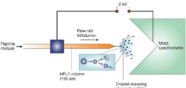
One of ESI’s main advantages is the possibility of using MS coupled to a liquid chromatography (LC) system (Figure 1.5). The LC-MS combination represented two main challenges. First the removal of all the solvent coming from the LC, because the MS operates under vacuum, and second, the production of gas-phase ions from the normally non-volatile LC’s analytes (Watson and Sparkman 2007). Thus, ESI became the most successful interface for LC/MS, as the majority of the solvent is evaporated.
Figure 1.5: Use of ESI in an LC-MS coupled instrument (adapted from (Steen and Mann 2004))
In ESI the droplets containing the analyte are formed after the solution has been forced through a very small capillary (20 μm internal diameter) in the presence of an electric field. The electric field is fundamental for the ionization, but also for the nebulization of the analyte solution into fine droplets. As the volatile solvent (water and acetonitrile mixture) evaporates from the droplets, the ratio of charge to droplet size increases leading to charge repulsion. This process ends with a coulombic explosion, where smaller droplets are produced and analyte ions are ejected. The droplets eventually desolvate completely after multiple cycles of coulombic explosions. ESI can be used in positive and negative modes, by switching the potential, but it is mostly used in positive mode, and ions that are produced are [M+H]+ type ions. Acidified solvents
are used to promote the protonation of the analyte. Nano-ESI, a miniaturized version of ESI, has the advantage of consuming very little sample, and its uses low flow rates to increase the ionization efficiency (Karas, Bahr et al. 2000).
1.2.2 The Mass Analyzer
The mass analyzers are used to separate ions by their mass-to-charge ratio. Because of their charges, the ions’ position and trajectory in gas phase can be manipulated with magnetic and electric fields (Watson and Sparkman 2007). The mass spectrometer operates in a reduced pressure environment. This vacuum is mandatory to maintain the focusing capability of the analyzer, as collision with neutral molecules will lead to ion diffusion.
A diverse and versatile range of analyzers exists on the market, and the important characteristics are the resolving power and the mass range. Resolving power (R) is the capacity for an instrument to separate two peaks and is defined by the following equation. M is the m/z of the peak and Δm is the difference in m/z between two adjacent peaks: m M R Δ =
Equation 1: Resolving Power
High resolution instruments have the advantage of performing accurate mass measurement, and therefore enable the determination of potential elemental formulae within specific mass tolerances. High mass accuracy is achieved when the instrument is capable of well separating neighboring peaks (Gross 2004). Note that an increase in resolution is usually made at the cost of the sensitivity. Mass accuracy is typically expressed in ppm (parts per million) which corresponds to the difference between the observed and calculated mass divided by the molecular mass of the analyte. Proteomic analysis takes advantage of high resolution instruments by reducing the false positive rate in peptide assignments (Mann and Kelleher 2008). For instance, an accuracy of +/- 1ppm reduces by 99 % all possible peptide assignments compared to using nominal mass measurements (Zubarev, Hakansson et al. 1996).
1.2.2.1
The Orbitrap mass analyzer
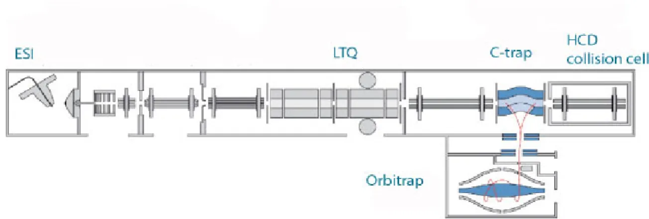
The mass analyzer used in this study is the LTQ-Orbitrap (Figure 1.6), a hybrid instrument comprising a linear ion trap combined with a high resolution orbital trap (Scigelova and Makarov 2006). This hybrid instrument contains two analyzers that can detect ions: the LTQ most commonly is used to generate tandem mass spectra (MS/MS) while the orbitrap detects all ions of the survey MS scan at high resolution. The linear ion trap or linear trap quadrupole (LTQ) are capable of generating MS and MS/MS at high sensitivity, but with low mass accuracy. In the Orbitrap mass analyzer, the ion oscillates around an electrode, resulting in a frequency which is a function of its m/z. The Orbitrap’s main advantage is high resolution (~100,000 at m/z 400) enabling high mass accuracy measurements (1-2ppm) (Scigelova and Makarov 2006) while the LTQ offers high speed and sensitivity. By using high resolution MS, it is possible to detect the isotopic pattern of a given ion. The isotopic pattern provides information about the charge state (z) of the ion.
Figure 1.6: Peptides are ionized in the ESI and fragmented in either the LTQ or in the HCD collision cell. The Orbitrap is used to detect parent m/z (adapted from(Olsen,
Schwartz et al. 2009))
The orbitrap is an ion trap, where ions are trapped around an electrode. It consists of 2 electrodes (inner and outer) between which an electric potential is imposed. Because of their shape, the electric field is non-homogenous between the electrodes; leading the
ions to oscillate along the inner electrode (Watson and Sparkman 2007). These oscillations are m/z dependent and this feature of the orbitrap makes it a mass analyzer.
This concept had existed for a long time, but the main challenge was to introduce ions in the orbitrap. The C trap was designed to focus ions before injecting them into small pockets and this process is coordinated with the increase in the electric field in the orbitrap (Watson and Sparkman 2007).
1.2.2.2
Linear ion trap mass analyzer
The linear ion trap (LIT) is composed of a linear quadrupole and its function is to trap ions, select those of interest and fragment them (Watson and Sparkman 2007). High potential is applied at the front and the end of the quadrupole thus enabling the trapping. A potential well is created and the ions are stored in defined boundaries. To achieve a scan, the ions are ejected sequentially by applying an RF voltage and once ejected, the ions hit the detector. The ejection of the ion occurs when the RF voltage matches the frequency of the ion of interest which is m/z dependent (Gross 2004). The selection of a particular ion is achieved when all the ions of lower and higher m/z are ejected. When the ion of interest is isolated, collision induced dissociation can occur in the trap using helium gas, producing fragments that can be detected (Gross 2004).
1.2.3 Tandem MS
MS/MS also referred to as tandem MS enables the acquisition of product ion spectra from precursor ions selected by the first mass analyzer. The second mass analyzer is thus scanned to transmit in turn fragment ions generated in a collision cell located between the two mass analyzers. The MS/MS spectrum reveals further structural information about the molecule and is the basis for peptide sequencing. Different algorithms currently exist to search the experimental MS/MS spectra and correlate observed fragment ions against those predicted from peptide candidates derived from a protein
database. Peptide fragmentation can be achieved using various techniques; some of which will be reviewed in this chapter.
Typically proteomics experiments involve the use of an LC-MS, and therefore ions are only observed at a given elution time in a short time window (30-60 s). Ideally all ions that are detected in a given MS scan should be fragmented and sequenced. Despite the short period for the acquisition of MS/MS spectra (~300 ms ns for CID), only a small fraction of all detected peptide ions can be sequenced in a given LC-MS/MS analysis. This method of collection is called data-dependent acquisition, where only a pre-selected number of the most intense ions are being selected for MS/MS (Mann, Hendrickson et al. 2001).
1.2.3.1 CID
CID (collision induced dissociation) is by far the most popular fragmentation technique in proteomics. In CID, ions are first accelerated by an electrical potential, and then made to collide with neutral gas molecules such as nitrogen or helium. The collision between the incoming precursor ions and the target gas converts kinetic into potential energy that is distributed into the different oscillators of the ions. Bonds that have the lowest energy requirements (typically the amide bond of peptides under low energy CID) will be dissociated first. This technique is optimal when the precursor is doubly or triply charged, leading to singly charged fragments. The preferred fragmentation for peptides occurs at the amide bond, yielding b and y type fragments (refer to Figure 1.7). By looking at the mass difference of neighboring peaks, each of which corresponds to the mass of an amino acid, it is possible to deduce the peptide sequence. In the LTQ-Orbitrap instrument the CID fragmentation occurs in the LTQ.
Figure 1.7: Nomenclature of fragment ions observed for the dissociation of peptide ions . Figure from: http://www.ionsource.com/tutorial/DeNovo/full_anno.htm
1.2.3.2 ETD
ETD (electron transfer dissociation) is a dissociation technique that is based on adding a low energy electron to a multiply charged species (Watson and Sparkman 2007). The fragmentation is very specific to the N-Cα bond (Boersema, Mohammed et al. 2009) first converting the peptide to a radical and forming c and z type fragment ions (see Figure 1.7). As electron source, ETD employs a radical anion (Syka, Coon et al. 2004) such as fluoranthene radical and the ETD-reaction is held in the LTQ. The fluoranthene has a low electron affinity; therefore it readily passes its electron to a peptide (Boersema, Mohammed et al. 2009). The main advantage of ETD is to fragment large peptides and those with labile PTMs (Syka, Coon et al. 2004). Contrary to CID, ETD gives best results with multiply charged species with z>2.
1.2.3.3 HCD
The fragmentation and detection in the ion trap is efficient and sensitive, however it lacks the mass resolution for the fragment ions and low m/z ions are not trapped (Olsen, Macek et al. 2007). In high energy collisional dissociation (HCD), the fragmentation occurs in an octopole collision cell located at the far end of the C-trap. The fragmentation mechanism and pattern is very similar to the LTQ CID fragmentation, leading to y and b ions. One advantage in HCD is the capability of detecting low m/z
fragments. Since the detection occurs in the Orbitrap, high mass resolution is obtained on the product-ion spectra. Consequently, high mass resolution on fragment ions leads to higher confidence on the peptide identity.
1.2.4 Detectors
The last component of the mass spectrometer is the detector. Once the ions are separated, it is the detector that measures their respective signal. In the Orbitrap two detectors are present: a dynode detector after the LTQ and the image current measured from the motion of ions cycling in theOrbitrap analyzer. The dynode detector amplifies the signal by increasing the difference in potential in multiple steps. Once the ion hits the first electrode, multiple electrons are emitted which then hit the next electrode and so on (Gross 2004). In the Orbitrap the ions oscillate around a central electrode and surrounding plates record the frequency of the current followed by a Fourier transform that converts the frequency into m/z data (Makarov 2000).
The dynamic range is an important factor when considering the choice of a detector. The dynamic range is the ratio of the most intense peak over the least intense peak in the same spectrum (Gross 2004). In proteomics analysis the two main challenges are the sample complexity and the very high dynamic range of protein abundance in the cell. For comprehensive proteomic analysis, a high dynamic range is required; however, the present day dynamic range of 103-104 is insufficient to cover the entire proteome (Mann and Kelleher 2008).
1.2.5 Database Searching
Following a single LC-MS run of 70 minutes, the instrument acquires up to 20 000 spectra. In a typical 2 condition proteomics experiment performed in this laboratory, about 800 000 spectra are generated. This large amount of data cannot be analyzed manually and computational methods have been developed to process the raw data.
The mass of the peptide is obtained from the MS while the tandem MS contains masses of fragments relevant to the sequence. It is possible to determine the peptide’s sequence by calculating the mass difference between fragments and this procedure is called de novo sequencing. However, often MS/MS spectrum will only contain partial
information and the entire sequence cannot be determined. In the 1990’s, following genome sequencing, the peptide sequencing became a database-search problem. In nature, only a small number of combinations of amino acid sequence exists compared to all the possibilities when dealing with de novo sequencing. Nowadays, the most popular
approach is through database searching, where MS and MS/MS scans are submitted to search engines such as MASCOT (Perkins, Pappin et al. 1999) or SEQUEST (Eng, McCormack et al. 1994). Thanks to the genome sequencing project, it is possible to virtually digest all the proteins present in an organism and create a peptide database. First, a database of peptides is generated for an entire genome, using the sequence of the proteins and the enzymatic cleavage rules (for instance, trypsin cuts at the N-terminus of lysine and arginine). The theoretical MS/MS spectrum for a specific fragmentation method is obtained for each of those peptides with different PTMs. The precursor mass (from the MS spectrum) and the fragment mass (from the MS/MS spectrum) of each theoretical peptide are compared to the experimental result. When submitting raw data to search engines, the first step is to use the peptide mass from the MS spectrum to obtain a list of all possible peptides with all allowed post-translational modifications respecting the enzyme cleavage rules that correspond to this mass within the allowed mass deviation. The second step is to generate mock MS/MS spectrum for those peptides, and a matching score is calculated for each every possibility. When a peptide is modified, the mass of the modified residue is considered when interpreting MS/MS spectrum. The comparison is done based on the allowed mass deviation for the precursor and the fragments’ mass. For instance when acquiring data in the LTQ-Orbitrap the allowed mass deviation is around 15ppm for the precursor mass and 0.5Da for the fragments. Database-searching approaches can only be used for organisms’ whose genome is sequenced and for which all the theoretical peptides are known (Steen and Mann 2004).
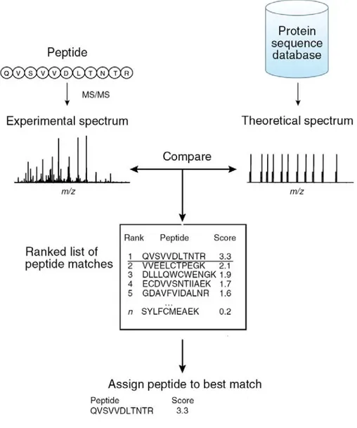
Each candidate peptide that matches to the experimental spectrum is assigned a score and a rank (Figure 1.8). The score is based on the quality of the match, for instance the number of fragments that were matched. Therefore, the score is the main parameter that is used to discriminate between right and wrong assignments. MASCOT, a widely used search-engine, uses the MOWSE algorithm to evaluate the match between the peptide and the spectrum (Pappin, Hojrup et al. 1993). MASCOT uses a probability-based scoring where the probability that a given match occurs randomly is calculated and its negative logarithm used as the score. The higher the score, the lower the probability that a given match is a random event. It is worth noting that the search engines assume modifications are unfragmentable during the CID or ETD process, which is true for small chemical modifications such as phosphorylation and acetylation; however this is not the case for the SUMO-modified peptide.
Figure 1.8: The MS/MS spectrum is compared against a theoretical MS/MS spectrum generated in silico. A score is given to each peptide based on the similarity of
the two spectra. Adapted from (Nesvizhskii, Vitek et al. 2007).
The matching between the theoretical and the experimental peptide’s MS/MS is not an ideal process and some errors occur (Elias and Gygi 2007). The low intensity of the precursor peptide, the poor quality of fragmentation, the fragmentation of two peptides in a single MS/MS all can lead to mistakes. In order to calculate the false discovery rate (FDR), the data is searched against a decoy database (Elias and Gygi 2007). The decoy database is created by reversing the protein sequences used in the database, and the MS/MS spectra are searched against a composite database: the forward and the reverse
database. At a given score cut-off, the FDR is calculated with the number of reverse-database matched peptides and therefore gives an estimate of the probability that a given match is a false positive.
Equation 2: Calculating the false discovery rate
peptides Total peptides matched database reverse FDR # # 2 100× × − =
1.2.6 Quantitative Proteomics
Until recently, MS-based proteomics was mostly a qualitative technique that resulted in a list of proteins found under a given condition. In order to gain better insight into the biological relevance of the proteins present, the relative abundance of the proteins and of their respective modifications is necessary (Schulze and Usadel 2010). MS is not inherently quantitative: the ion’s intensity not only depends on the ion’s abundance, but also on the chemical properties of the peptide (charge, length, amino acid composition, etc.) and its environment (salts present during ionization). Absolute quantification cannot be performed on a given peptide simply based on its intensity. As a result, most of the large-scale MS quantification approaches always involve a comparison between two or more samples. Comparisons can only be made based on the same specie (the same peptide with the same m/z) since different peptides have different ionization efficiencies.
A typical experimental design compares two conditions: a stressed versus a control
status. Different quantification strategies have been developed in recent years and are divided as label-free and stable-isotope-labeling approaches (Schulze and Usadel 2010). In this laboratory, a label-free approach has been developed based on the correlation of peptide coordinates (m/z, time charge, intensity) across replicates and conditions. ProteoProfile is an in-house label-free bioinformatics software that quantifies peptides and proteins across different samples.
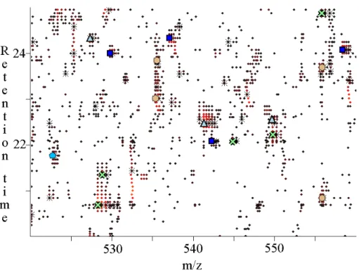
ProteoProfile creates peptide maps from LC-MS raw data and then clusters maps from different sample sets. First it creates contour maps that include retention time, charge, intensity and m/z for all detection peptide ions (see Figure 1.9).
Figure 1.9: Contour map created by Proteoprofile. Symbols circle, cross and
triangles are represent singly, doubly and triply charged species respectively, while the range of intensities is represented with color from dark red to yellow being the most intense.
Once maps are created for each sample, ProteoProfile then clusters the different samples together by aligning maps using linear dynamic correlation. Since the overlap in MASCOT identification between two LC-MS/MS of the same sample is around 60% due to the random process of selection when using data-dependent acquisition, the ions are not aligned based on their MASCOT identification, but rather accordingly to their retention time, m/z as well as the surrounding environment. Mass accuracy becomes even more important when dealing with label free quantification, since the clustering
relies on mass accuracy. Therefore, high-mass resolution instruments made label-free quantification even more appealing in recent years (Schulze and Usadel 2010).
1.3 SUMO MS based proteomics
In this section, different enrichment techniques of SUMO-conjugated proteins and SUMO-modified peptides bearing the modification sites will be described, followed by spectral interpretation strategies.
1.3.1 Enrichment of SUMOylated proteins
In order to identify Ub/Ubl modified proteins and their sites, the need for an enrichment strategy became apparent due to the low abundance and high turnover of those species. A strategy was successfully employed to identify ubiquitin-conjugated proteins and their modification sites in yeast: around 1000 potential conjugates and 100 ubiquitylation sites were identified (Peng, Schwartz et al. 2003).
Figure 1.10: Purification strategy of His-SUMO conjugated proteins using the Ni-NTA pull down. Contaminants are identified using the WT control and the SUMO-conjugated proteins are uniquely present in the His-SUMO sample.
The approach described in Figure 1.10 and developed by Gygy laboratory is based on the expression of His6-ubiquitin in yeast and its subsequent purification. His6
-ubiquitin-conjugated proteins are isolated using Ni-NTA (nickel nitriloacetic acid) resin in denaturing conditions. In parallel, a control sample (wild type) expressing no His6-Ub
is purified in the same manner and the isolated proteins are the non-specific contaminants that bind the resin. The success of this proteomic study inspired multiple groups to study SUMO-conjugates in a similar manner in yeast and in mammals (Matic, Schimmel et al. 2010), (Blomster, Imanishi et al. 2010), (Wohlschlegel, Johnson et al. 2004), (Wohlschlegel, Johnson et al. 2006). The denaturing conditions are extremely important, since they first assure the inactivity of SENPs, and secondly assure the exclusive purification of SUMO-conjugates proteins and not their associated proteins. The proteins that are uniquely identified in the His6-SUMO expressing strain are
assumed to be bona fide SUMO substrates. Up to now hundreds of putative SUMO
conjugated proteins have been identified.
1.3.2 Enrichment strategies of SUMO-modified peptides
Mass spectrometric analysis of SUMOylation sites is technically very challenging and only a few sites have been identified until recently (Wilson and Heaton 2008). The majority of identified SUMO targets are only putative targets and their modification sites are still unknown. The identification of the conjugation sites can be important in understanding the regulation of SUMOylation.
The digestion of purified SUMO conjugates leads to a mixture of peptides, most of which do not contain the modification site. This mixture is too complex for efficient identification of the modification site. Since SUMO-modified proteins are very low in abundance, this is even more problematic.
Secondly, following trypsin digestion, the modified peptide is a branched peptide that contains a large C-terminal SUMO sequence. The absence of arginine and lysine residues makes the chromatographic separation and the MS analysis challenging.
Different strategies have been designed to alleviate the MS interpretation and SUMO peptide enrichment.
In order to purify SUMO1-modified peptides, Bloomster et al engineered the human SUMO-1 protein so that following LysC digestion, the resulting peptide contains two cysteines followed by an arginine on the SUMO1 moiety (Blomster, Imanishi et al. 2010). The SUMO1-modified peptides are then enriched using thiopropyl sepharose beads that bind through disulfide linkage with the SUMO1-modified peptides. The peptides of interest are then released from the beads using trypsin digestion that leaves a di-glycine tag on the lysine. This method allowed the identification of 14 SUMOylation sites in HeLa cells (Blomster, Imanishi et al. 2010). However, this method presents several drawbacks, such as the difficulty of distinguishing SUMO1-modified from Ubiquitin-modified peptides. The second drawback is the possible enrichment of non-SUMO1-modified peptides that contain cysteine residues.
A second purification strategy was developed using an engineered SUMO2/3. A LysC resistant His-SUMO protein was engineered where all lysines residues were mutated to arginines (Matic, Schimmel et al. 2010). The first step involves a LysC digestion followed by the Ni-NTA enrichment of SUMO protein linked to Lys-C digested target peptide. Following the Ni-NTA enrichment, the mixture is digested by trypsin. Using this method, 103 SUMO-2/3 acceptor sites were recently identified in mammals (Matic, Schimmel et al. 2010). Since all lysine residues are substituted by arginine, this mutant SUMO2 cannot form polySUMOylation and thus changes the function of the corresponding protein.
All modified peptide based enrichment strategies present a highly modified form of the SUMO protein and therefore can introduce some artifacts.
1.3.3 Stress-induced SUMOylation
Numerous studies observed an accumulation of SUMO-conjugates upon cellular stress (Saitoh and Hinchey 2000; Kurepa, Walker et al. 2003; Bohren, Nadkarni et al. 2004; Manza, Codreanu et al. 2004; Golebiowski, Matic et al. 2009). Various external stress such as heat shock, oxidative and genotoxic chemicals have been tested on the SUMO system in many cell types, and SUMO4 expression has only been reported in extreme stress conditions (Wei, Yang et al. 2008).This effect is mostly observed with SUMO2/3 conjugates, following stress, the free SUMO2/3 is highly conjugated (Saitoh and Hinchey 2000), however SUMO1 does not seem to be affected in the same manner.
When exposed to arsenic trioxide, PML and PML-RARα are SUMOylated and subsequently degraded by the ubiquitin-proteasome system (Lallemand-Breitenbach, Jeanne et al. 2008). PML-RARα is a fusion protein expressed in APL (acute promyelocytic leukaemia) and As2O3 is an agent used to treat APL (Zhu, Chen et al.
2002).
1.3.4 Spectral interpretation strategies
So far, the common strategy in identifying SUMOylation sites is through site-directed mutagenesis of consensus site lysines. However, it has been shown that non-consensus lysines can be SUMOylated as for E2-25K (Pichler, Knipscheer et al. 2005) and the opposite is also true: not all consensus site lysine are SUMOylated. In order to gain a better insight on the biological role of SUMOylation, the need for a new and unbiased strategy for SUMO site identification has become important in the last decade. Mass spectrometry is the method of choice in identifying the modification sites for numerous PTMs.
Most PTMs, such as ubiquitylation, methylation or acetylation, do not fragment during the CID or the ETD process in the MS. As an example, phosphorylation sites can
be readily identified using standard sequencing softwares by including phosphorylation as a variable modification and simply altering the fragment’s mass by the mass of the modification (Figure 1.11b). Following trypsin digestion, the large SUMO tags produce multiple fragments that lead to complex fragmentation patterns, and although this adds confidence to the identification of the SUMO moiety, the MS/MS spectra is highly complex (Figure 1.11c) and renders spectral interpretation by MASCOT more prone to errors. Currently, two softwares were developed to specifically address this issue: SUMmOn (Pedrioli, Raught et al. 2006) and ChopNSpice (Hsiao, Meulmeester et al. 2009). SUMmOn algorithm was developed to interpret complex fragmentation patterns of PTMs such as SUMO (Pedrioli, Raught et al. 2006). The CID spectra are scanned for the specific fragments that originate from the modifier. SUMmOn calculates two scores one for the target peptide and one for the modification, and although this system seemed to be successful for in vitro by identifying RanGap SUMOylation site, no conclusive
Figure 1.11: Theoretical MS/MS CID spectra of a) unmodified peptide, b) phospho-peptide and c) modified phospho-peptide. Complex fragmentation pattern of
The branched SUMO-modified peptides fragment in a manner similar to that of the linear peptide with a lysine missed cleavage and the SUMO moiety on the N-terminus (Hsiao, Meulmeester et al. 2009). Based on this observation, ChopNSpice algorithm was developed. A database is constructed where every possible lysine is modified by SUMO and the resulting peptide is linearized. The MS and MS/MS spectra are searched against the ChopNSpice generated database and using this strategy 18 SUMOylation sites were identified in vivo for SUMO-1 in HeLa cells (Hsiao, Meulmeester et al. 2009).
Although new bioinformatics strategies are more suitable for studying SUMOylation by MS, further technological advances would be required to provide more comprehensive SUMOylome analyses.
1.4 Project’s goal
SUMO are a group of ~10kDa proteins that form an isopeptide linkage with the ε amino group of the target lysine. SUMOylation is a post-translational modification required for cell viability (Johnson, Schwienhorst et al. 1997; Hayashi, Seki et al. 2002) and shares great similarity with ubiquitin (Bayer, Arndt et al. 1998). At present, around a hundred SUMO targets have been identified and the molecular consequences of protein SUMOylation in vivo such as reciprocity between ubiquitylation and SUMOylation,
degradation and transcription regulation are unpredictable (reviewed in (Geiss-Friedlander and Melchior 2007)). This modification affects protein function in a diverse, complex and sometimes opposite way.
Large scale proteomics studies have been successful in identifying a high number of potential SUMO targets (Li, Evdokimov et al. 2004; Vertegaal, Ogg et al. 2004; Wohlschlegel, Johnson et al. 2004; Denison, Rudner et al. 2005; Gocke, Yu et al. 2005; Ganesan, Kho et al. 2007; Flick and Kaiser 2009). However, SUMOylation site identification is still a challenge, due to the very low abundance of SUMO targets in vivo
as well as its rapid turnover upon cell lysis. Until now, SUMOylation site identification has mainly relied on site-directed mutagenesis, a time-consuming and burdensome task. To get a better insight into the molecular function of SUMOylation and a confidence in the target’s identity, the number and the location of the exact site is essential.
In this context, the goal of this project is to develop a large scale MS based method to identify SUMOylation targets and their respective sites. Because of the very low abundance of SUMOylated proteins in the cell, and its highly dynamic nature (Geiss-Friedlander and Melchior 2007), the first part of this thesis will focus on target enrichment.
Following trypsin digestion of SUMOylated proteins, the peptides containing the modification site are branched. The analysis of branched peptide by MS is very limited,
partly due to the complexity of the MS-MS spectrum and the absence of efficient sequencing software (Pedrioli, Raught et al. 2006; Hsiao, Meulmeester et al. 2009). Hence, the second goal of this project is based on the interpretation of SUMO peptide MS/MS spectra.
1.5 References
Ayaydin, F. and M. Dasso (2004). "Distinct in vivo dynamics of vertebrate SUMO paralogues." Mol Biol Cell 15(12): 5208-18.
Bayer, P., A. Arndt, et al. (1998). "Structure determination of the small ubiquitin-related modifier SUMO-1." Journal of Molecular Biology 280(2): 275-286.
Bernier-Villamor, V., D. A. Sampson, et al. (2002). "Structural basis for E2-mediated SUMO conjugation revealed by a complex between ubiquitin-conjugating enzyme Ubc9 and RanGAP1." Cell 108(3): 345-56.
Blomster, H. A., S. Y. Imanishi, et al. (2010). "In vivo identification of sumoylation sites by a signature tag and cysteine-targeted affinity purification." J Biol Chem 285(25): 19324-9.
Boersema, P. J., S. Mohammed, et al. (2009). "Phosphopeptide fragmentation and analysis by mass spectrometry." Journal of Mass Spectrometry 44(6): 861-878.
Braschi, E., R. Zunino, et al. (2009). "MAPL is a new mitochondrial SUMO E3 ligase that regulates mitochondrial fission." EMBO Rep 10(7): 748-754.
Cooper, H. J., M. H. Tatham, et al. (2005). "Fourier transform ion cyclotron resonance mass spectrometry for the analysis of small ubiquitin-like modifier (SUMO) modification: identification of lysines in RanBP2 and SUMO targeted for modification during the E3 autoSUMOylation reaction." Anal Chem 77(19):
6310-9.
Denison, C., A. D. Rudner, et al. (2005). "A proteomic strategy for gaining insights into protein sumoylation in yeast." Mol Cell Proteomics 4(3): 246-54.
Elias, J. E. and S. P. Gygi (2007). "Target-decoy search strategy for increased confidence in large-scale protein identifications by mass spectrometry." Nat
Methods 4(3): 207-14.
Eng, J. K., A. L. McCormack, et al. (1994). "An approach to correlate tandem mass spectral data of peptides with amino acid sequences in a protein database."
Journal of the American Society for Mass Spectrometry 5(11): 976-989.
Fenn, J. B., M. Mann, et al. (1989). "Electrospray ionization for mass spectrometry of large biomolecules." Science 246(4926): 64-71.
Flick, K. and P. Kaiser (2009). "Proteomic Revelation: SUMO Changes Partners When the Heat Is On." Sci. Signal. 2(81): pe45-.
Ganesan, A. K., Y. Kho, et al. (2007). "Broad spectrum identification of SUMO substrates in melanoma cells." Proteomics 7(13): 2216-21.
Geiss-Friedlander, R. and F. Melchior (2007). "Concepts in sumoylation: a decade on."
Nat Rev Mol Cell Biol 8(12): 947-56.
Gocke, C. B., H. Yu, et al. (2005). "Systematic Identification and Analysis of Mammalian Small Ubiquitin-like Modifier Substrates." J. Biol. Chem. 280(6):
5004-5012.
Golebiowski, F., I. Matic, et al. (2009). "System-wide changes to SUMO modifications in response to heat shock." Sci Signal 2(72): ra24.
Guo, D., M. Li, et al. (2004). "A functional variant of SUMO4, a new I kappa B alpha modifier, is associated with type 1 diabetes." Nat Genet 36(8): 837-41.
Hayashi, T., M. Seki, et al. (2002). "Ubc9 Is Essential for Viability of Higher Eukaryotic Cells." Experimental Cell Research 280(2): 212-221.
Hietakangas, V., J. Anckar, et al. (2006). "PDSM, a motif for phosphorylation-dependent SUMO modification." Proc Natl Acad Sci U S A 103(1): 45-50.
Hsiao, H. H., E. Meulmeester, et al. (2009). ""ChopNSpice," a mass spectrometric approach that allows identification of endogenous small ubiquitin-like modifier-conjugated peptides." Mol Cell Proteomics 8(12): 2664-75.
Kamitani, T., K. Kito, et al. (1998). "Identification of three major sentrinization sites in PML." J Biol Chem 273(41): 26675-82.
Karas, M., U. Bahr, et al. (2000). "Nano-electrospray ionization mass spectrometry: addressing analytical problems beyond routine." Fresenius J Anal Chem
366(6-7): 669-76.
Karas, M. and F. Hillenkamp (1988). "Laser desorption ionization of proteins with molecular masses exceeding 10,000 daltons." Anal Chem 60(20): 2299-301.
Kerscher, O., R. Felberbaum, et al. (2006). "Modification of proteins by ubiquitin and ubiquitin-like proteins." Annu Rev Cell Dev Biol 22: 159-80.
Kurepa, J., J. M. Walker, et al. (2003). "The small ubiquitin-like modifier (SUMO) protein modification system in Arabidopsis. Accumulation of SUMO1 and -2 conjugates is increased by stress." J Biol Chem 278(9): 6862-72.
Lallemand-Breitenbach, V. and H. de The (2010). "PML nuclear bodies." Cold Spring
Harb Perspect Biol 2(5): a000661.
Lallemand-Breitenbach, V., M. Jeanne, et al. (2008). "Arsenic degrades PML or PML-RARalpha through a SUMO-triggered RNF4/ubiquitin-mediated pathway." Nat
Cell Biol 10(5): 547-55.
Mahajan, R., C. Delphin, et al. (1997). "A small ubiquitin-related polypeptide involved in targeting RanGAP1 to nuclear pore complex protein RanBP2." Cell 88(1):
97-107.
Makarov, A. (2000). "Electrostatic axially harmonic orbital trapping: a high-performance technique of mass analysis." Anal Chem 72(6): 1156-62.
Mann, M., R. C. Hendrickson, et al. (2001). "Analysis of proteins and proteomes by mass spectrometry. " Annual Review of Biochemistry 70(1): 437-473.
Mann, M. and N. L. Kelleher (2008). "Precision proteomics: The case for high resolution and high mass accuracy." Proceedings of the National Academy of
Sciences 105(47): 18132-18138.
Manza, L. L., S. G. Codreanu, et al. (2004). "Global shifts in protein sumoylation in response to electrophile and oxidative stress." Chem Res Toxicol 17(12):
1706-15.
Marcin Drag, G. S. S. (2008). "DeSUMOylating enzymes - SENPs." IUBMB Life 60(11): 734-742.
Martin, S., K. A. Wilkinson, et al. (2007). "Emerging extranuclear roles of protein SUMOylation in neuronal function and dysfunction." Nat Rev Neurosci 8(12):
948-59.
Matic, I., J. Schimmel, et al. (2010). "Site-Specific Identification of SUMO-2 Targets in Cells Reveals an Inverted SUMOylation Motif and a Hydrophobic Cluster SUMOylation Motif." Mol Cell 39(4): 641-652.
Matic, I., M. van Hagen, et al. (2008). "In Vivo Identification of Human Small Ubiquitin-like Modifier Polymerization Sites by High Accuracy Mass Spectrometry and an in Vitro to in Vivo Strategy." Mol Cell Proteomics 7(1): 132-144.
Mukhopadhyay, D. and M. Dasso (2007). "Modification in reverse: the SUMO proteases." Trends Biochem Sci 32(6): 286-95.
Nesvizhskii, A. I., O. Vitek, et al. (2007). "Analysis and validation of proteomic data generated by tandem mass spectrometry." Nat Meth 4(10): 787-797.
Owerbach, D., E. M. McKay, et al. (2005). "A proline-90 residue unique to SUMO-4 prevents maturation and sumoylation." Biochem Biophys Res Commun 337(2):
517-20.
Pappin, D. J. C., P. Hojrup, et al. (1993). "Rapid identification of proteins by peptide-mass fingerprinting." Current biology : CB 3(6): 327-332.
Pedrioli, P. G., B. Raught, et al. (2006). "Automated identification of SUMOylation sites using mass spectrometry and SUMmOn pattern recognition software." Nat
Peng, J., D. Schwartz, et al. (2003). "A proteomics approach to understanding protein ubiquitination." Nat Biotech 21(8): 921-926.
Perkins, D. N., D. J. Pappin, et al. (1999). "Probability-based protein identification by searching sequence databases using mass spectrometry data." Electrophoresis 20(18): 3551-67.
Petsko, G. A. and D. Ringe Protein Structure and Function, New Science Press.
Pichler, A., P. Knipscheer, et al. (2005). "SUMO modification of the ubiquitin-conjugating enzyme E2-25K." Nat Struct Mol Biol 12(3): 264-9.
Pickart, C. M. and D. Fushman (2004). "Polyubiquitin chains: polymeric protein signals." Current Opinion in Chemical Biology 8(6): 610-616.
Reineke, E. L., Y. Liu, et al. (2009). "Promyelocytic leukemia protein controls cell migration in response to hydrogen peroxide and insulin-like growth factor-1." J
Biol Chem 285(13): 9485-92.
Rodriguez, M. S., C. Dargemont, et al. (2001). "SUMO-1 Conjugation in Vivo Requires Both a Consensus Modification Motif and Nuclear Targeting." Journal of
Biological Chemistry 276(16): 12654-12659.
Saitoh, H. and J. Hinchey (2000). "Functional heterogeneity of small ubiquitin-related protein modifiers SUMO-1 versus SUMO-2/3." J Biol Chem 275(9): 6252-8.
Schulze, W. X. and B. r. Usadel (2010). "Quantitation in Mass-Spectrometry-Based Proteomics." Annual Review of Plant Biology 61(1): 491-516.