Université de Montréal
Influence of Fetal Tissue Transplant on
the Morphology of the Neuromuscular Junctions of
Tibialis Anterior and Medial Gastrocnemius
Following Spinal Transection in the Rat
par
Catherine Chan
Département de Kinésiologie
Mémoire présenté à la Faculté des études supérieures
en vue de l’obtention du grade de
Maître en sciences de l’activité physique
août 2005
054
r’
2o06
n
Direction des bibliothèques
AVIS
L’auteur a autorisé l’Université de Montréal à reproduire et diffuser, en totalité ou en partie, par quelque moyen que ce soit et sur quelque support que ce soit, et exclusivement à des fins non lucratives d’enseignement et de recherche, des copies de ce mémoire ou de cette thèse.
L’auteur et les coauteurs le cas échéant conservent la propriété du droit d’auteur et des droits moraux qui protègent ce document. Ni la thèse ou le mémoire, ni des extraits substantiels de ce document, ne doivent être imprimés ou autrement reproduits sans l’autorisation de l’auteur.
Afin de se conformer à la Loi canadienne sur la protection des renseignements personnels, quelques formulaires secondaires, coordonnées ou signatures intégrées au texte ont pu être enlevés de ce document. Bien que cela ait pu affecter la pagination, il n’y a aucun contenu manquant.
NOTICE
The author of this thesis or dissertation has granted a nonexclusive license allowing Université de Montréal to reproduce and publish the document, in part or in whole, and in any format, solely for noncommercial educational and research purposes.
The author and co-authors ifapplicable retain copyright ownership and moral rights in this document. Neither the whole thesis or dissertation, nor substantial extracts from it, may be printed or otherwise reproduced without the author’s permission.
In compliance with the Canadian Ptivacy Act some supporting forms, contact information or signatures may have been removed from the document. While this may affect the document page count, it does flot represent any loss of content from the document.
Université de Montréal
Faculté des études superieures
Ce mémoire intitulé:
Influence of Fetal Tissue Transplant on
the Morphology of the Neuromuscular Junctions of
Tibialis Anterior and Medial Gastrocnemius
Following Spinal Transection in the Rat
Présenté par:
Catherine Chan
A été évalué par un jury composé des personnes suivantes:
M. François Péronnet
—Président-rapporteur
Dr. Phïllip Gardiner
—Directeur de recherche
Dr. Jean-Marc Lavoie
—Membre du jury
Mémoire présenté à la Faculté des études supérieures
en vue de l’obtention du grade de
Maître en sciences de l’activité physique
août 2005
Summary
The present work examined the adaptations of the neuromuscularjunction occuring in the tibialis anterior (TA) and the medial gastrocnemius (MG) following fetal tissue transplants in a rat model following spinal transection at T1O-11. The cholinesterase-contaïning endplate was quantified using a modification of the method used by Pestronk and Drachman (1978) which also allows staining of the nerve terminais. The parameters measured included the endplate area, endplate longitudinal length, number of branch points and muscle fiber width, under conditions of a
controiled state (CNTRL group, n=3), transected state (TRANS group, n=3) and transection wïth transplant state (TRNPL group, n=3). Both of these muscles were chosen on the basis that they elicit different actions but are of similar fiber-type composition. Another reason for the selection was to examine whether the function of the muscle plays a role in the resuits of transection or transplants. The present study reported no significant findings to embryonic tissue transplants post transection. Statïstical analysis uncovered no significant difference between the TA and MG in terms of endplate area, endplate longitudinal length, number of branchpoints and muscle fiber width. Most mean results, excluding mean resuits for muscle fiber width, were found to be highest in the transection and transplant groups for both the TA and MG. Spinal transection did flot cause muscle fibers to significantly atrophy in this experiment. Transection alone as well as the transplant condition did not result in any differences in the parameters measured.
to consider the effect of near-total inactivity on the motor endplate which is n problem
in other models where inactivity is less complete. With a spinal transection there is no
direct damage to the muscle and innervation of muscles is flot physically disrupted; therefore, the intemption of transfer of electrical activity through the motoneurons likely stimulates changes that are observed (Salmons & Henriksson, 1981).
Spinal transection is of particular interest because it more closely mimics the human condition of spinal cord injury than spinal isolation and intact reflexes aÏlow the study of the role of reflex-generated locomotor activity in attenuating the atrophic responses (Gardiner, 2001). Animais in the transplant group had a 4 week period of
transplantation. The transplant aspect of the present model is of interest because fetal spinal cord tissue produces several growth and trophic factors during its development
and it is possible that some of these may be released to have an influence upon the
muscle directly or indirectly through action of the motorneurons (Houle et al.,1999).
Dr. J. Houle bas found that passive exercise seems to attenuate many muscle effects such as the decrease in gross muscle size and atrophy of individual muscle fibers
following a spinal cord injury. His findings indicate that cycling exercises or fetal tissue transplantation can effectively limit the decrease in muscle size observed following a complete spinal cord lesion but neither approach affects, changes in the expression of proteins important for muscle contractility. Though the present study uncovered no significant resuits, it does set the stage for future research eventually looking at the effects of passive exercise on any spinalectomy-induced changes in the
NMJ.
Résumé
Cette expérience examine les adaptations des jonctions neuromusculaires du muscle antérieure (tibiale) et du (gastroc) médiale suivant la transplantation des cellules embryonnaires chez le rat transecté. La plaque motrice contenant de la cholinesterase était quantifiée par une modification de la méthode de Pestronk et Drachman (1978). Cette méthode permet de noircir les plaques motrices ainsi que les terminaisons nerveuses. Les paramètres mesurées étaient: l’aire de la plaque motrice, la longueur des plaques motrices, la largeur des fibres musculaires, et le nombre de points de bifurcations selon trois conditions. La première était pour un état contrôlé, la deuxième dans un état transecté et la troisième dans un état transecté avec une transplantation. Ces deux muscles a été choisies parce qu’ils provoquent des actions différentes et parce qu’ils sont d’une composition de fibres musculaires semblables. Ces muscles permettent à determiner si leur actions différents influencent les effets d’une transection ou d’une transplantation. La transplantation des tissus
embryonnaires à la suite d’une transection a effectué aucun changement significatif pour cette expérience. Les resultats indiquent aucune différence statistiquement significative entre l’antérieure tibiale et le gastroc médiale pour les valeurs mesurées de l’aire de la plaque motrice, la longeure des plaques motrices, la largeure de la plaque motrice et le nombre de bifurcations. La plupart des valeurs, sauf celles pour la largeure des fibres musculaires, étaient plus élevées pour les états de transection et de transplantation pour l’antérieur tibiale et le gastroc médiale. La transection expérimentale n’a pas effectué l’atrophie des fibres musculaires pour cette étude. La modèle de l’éxperience est important à cause de l’inactivité presque complète de la
plaque motrice à la suite d’une transection, ce qui permet d’autre rechercheurs de
l’examinercard’autre modèles d’inactivité moins complète sont problématiques. fl
n’y a pas d’endommagement directe au muscle ni d’interruption physique à
l’innervation du muscle dans un cas de transection donc la transferation d’activité électrique à travers le motneurone n’est pas interompue. Ceci probablement stimule
des changements (Salmons & Henriksson, 1981). La transection et sa vraisemblance à
la condition d’une blessure de la moelle épinière chez 1’ humaine permet l’étude de
locomotion géneré par les reflexes pour diminuer l’atrophie des muscles (Gardiner,
2001). La transplantation est important parce que l’extrait de la moelle épinière
foetale produit plusieurs facteurs de la croissance au cours du développement alors il
est possible que le déchargement de ces facteurs influence les muscles directement ou
indirectementparl’action des motoneurones (Houle et al., 1999). Selon les
recherches de Dr. J. Houle, l’exercice passive semble attenuer les effets musculaires comme la réduction de la taille du muscle (en entier) et l’atrophie des fibres
musculaires individuelles à la suite d’une blessure de la moelle. Les résultats de ses études indiquent que l’exercice du cyclisme ou la transplantation embryonnaire peut effectivement limiter la réduction en taille du muscle à la suite d’un transection complète. Ni l’un ni l’autre affectent les changements de l’expression des protéines nécessaire pour la contraction. Même si cette expérience a montré aucun résultat significatif elle est d’importance pour d’autres rechercheurs à la poursuite d’étudier
les lésions spinales et les effets d’exercice passive sur les jonctions neuromusculaires
à l’avenir.
Mots-clés Jonction neuro-musculaire, blessure de la moelle épinière, transplantation de cellules
Table of Contents
Summary iv
Résumé vi
Table of Contents viii
List of Abbreviations x
List of Tables xi
List of Appendices xii
List of Figures xiii
Acknowledgments xiv
Review of Literature
General 1
Historical Perspective-The Neuromuscular Junction 1
Muscle fiber types and their recruitment during movement 3
Morphology of NMJs of different fiber type 5
Effects of increased and decreased activity on the neuromuscular system 8
Sprouting of motor nerve terminais 14
Spinal cord injury 17
Studies on regeneration in spinal cord injuries 20
Methodology: Subjects 22
Surgical Procedures- Transection of Spinal Cord 23
-Embryonïc Tissue Transplant 24
Tissue Preparation 25 Histochemical Procedures 26 Statistical Analysis 30 Resu its 31 Discussion 36 Conclusion 45 References 46 Appendices 52
List of Abbreviations
AChE Acetylcholinesterase
AchR Acetylcholine Receptors
ATPase Adenosine trïphosphatase
C Celsius
ChE Cholineserase
CNS Central nervous system
CNTRL Control
EDL Extensor Digitorem Longus
EP Endplate
FDL Flexor Digitorem Longus
FG Fast-twitch glycolytic
FOG Fast-twitch oxidative glycolytic
fSC Fetal Spinal Cord
HRP Horseradish Peroxidase
IP Intraperitoneal
MG Medial Gastrocnemius
MIIC Myosin Heavy Chain
NMJ Neuromuscular Junction
PDQ Peroneus Digiti Quinti
PL Plantaris
PN Peripheral Nerve
RF Rectus Femoris
SD Standard Deviation of the mean
$0 Slow-twitch oxidative Sol Soleus TA Tibialis Anterior TRANS Transected TRNPL Transection +Transplant micrometer(s) squared micrometer(s) VE Vastus Lateralis VM Vastus Medialis
List of Tables
Table I: Muscle Fiber Widths (pm)± SD 32
Table II: Endplate Lengths (im)± SD 33
Table III: Endplate area (tm2)±SD 34
List of Appendices
APPENDD( A: Staïning for quantitative measurement of neuromuscularjunction 52 Cholinesterase-staining procedure
List of Figures
Figure 1: Endplate area outlined by cholinesterase staining 27 Figure 2: Longitudinal length of endplate along fiber length 27
Figure 3: Endplate branchpoints 28
Acknowledgements
First and foremost, I would like to extend my deepest respect and appreciation to Dr. Phillip Gardiner for bis supervision, guidance, patience and perseverance throughout my studies at Université de Montréal.
I would also like to greatly acknowledge Pierre Corriveau for lis excellent technical contribution.
Thanks to my parents for their constant faith in me. They have aiways fostered my fascination with science and provided me with unending support for whichever path I chose to follow.
A special thanks to my classmate Peter Tzavares for his efforts and help with my studies. Lastly, I would like to thank my husband, Remy Pilon, whose pursuit of higher education bas encouraged my own. His presence has aiways lead to great things.
Thefiture influences the present just as much as the past.
Review of literature
General
Adaptation to skeletal muscle fibers to increased and decreased use has been extensively investigated (Deschenes et al., 1994). However, little is known about the adaptations that occur to other components of the neuromuscular system (Deschenes et al 1994). The link of communication between motoneurons and muscle fibers is the
neuromuscular junction (NMJ). The neuromuscular junction is specifically defined as the synaptic site between a motoneuron and muscle fibers belonging to a motor unit. It is the essential element of neuromuscular control of skeletal muscle (Shenington, 1929). Thïs dynamic structure undergoes both morphological and functional changes throughout the course of a lifetime (Sieck & Prakasli, 1997).
Historical Perspective-THE NEUROMUSCULAR JUNCTION
Histologists in the early 1840’s agreed with both Valentin and Emmert in 1836, as cited in Couteux, 1973, that nerves boasted bow-shaped endings in striated muscles with the nerve branches joining up and continuing in one another. This opinion, however, negated the existence of truc nerve endings with direct connections between the motor nerve and each muscle fiber.
Wagner’s observations in 1847, cited in Couteaux, 1973, projected doubt for the first time on the existence of terminal bows in striated muscles of vertebrates stated by Valentin and Emmert. Through lis studies on the hyoidean muscles of the frog, Wagner was the first to reveal two fundamental features of the NMJ of vertebrates; lie concïuded that the motornervefiber, after branching, loses its myelin sheath and closely connects with the muscle fiber.
Subsequently discovered were two other important characteristics in various types of NMJs found in striated muscles. The first, described by Kuhne in 1862, showed that after piercing the sarcolemma on frog muscles, the motor nerve fiber branches again and provides a terminal arborization. The second, imparted by Rouget in 1862, reported the presence of a flattened heap of granular nucleated substance at the NMJ level, in the muscles of reptiles, birds, and mammals. He interpreted it as the spreading of the axis cylinder substance at the surface of the myofibrils and called it the endplate.
The following year Krause in 1863, described the terminal branching at the level of the endplate on the retractor muscle of the cat’s eye, later termed it the motor endplate. The first researchers to really detail observations of the structure of the motor endplate were Ranvier and Kuhne. further morphological researcli was based on the work of these two authors. Ranvier identified structural and functional differences between fast-twitch (type II) and slow-twitch (type I) mammalian muscles and Kuhne discovered the
Muscle fiber types and their recruitment during movement
Muscles of the rat hindlimb are composed of three distinct muscle fiber types in varying proportions. High proportions of slow-twitch oxidative fibers and fast-twitch glycolytic fibers are typically in the deepest regions of the hindlimb muscles. More superficially found are the fast-twitch glycolytic fibers (Armstrong & Phelps, 1984).
Most skeletal muscles are flot homogeneous in their muscle fibre composition. The heterogeneity of muscle fibers within a muscle was first identified by the
histochemical detectïon of different staining intensities of myosin ATPase and of oxidative and glycolytic enzymes in human and animal studies performed by Dubowitz and Pearce in 1960 and by Engel in 1962. Engel first classified muscle fibers into two types: type I and type II which correspond with slow twitch and fast twitch respectively. Muscle fibers have been classified according to myosin heavy-chain (MHC) protein that they possess. Adult mammalian limb muscles myosin heavy-chain species include types I, lia, lix (also termed IId) and 11h (Gardiner, 2001).
Structural and functional differences between slow tonic and fast twitch
mammalian muscles were identified more than a hundred years ago by Ranvier in 1874 and Grutzner in 1884. Since most mammalian muscles contain fast and slow twitch muscle fibers there is considerable heterogeneity of muscles and motor unit properties. This heterogeneity is functionally important for grading and controlling muscle force during normal movement (Vrbova, Gordon and Jones, 1995).
When movement is carried out whether it is running or walking, it is necessary that the appropriate muscles are activated for the specific movement and that the force
generated by those muscles is controlled. Henneman and Oison (1965) emphasized the functionai matching of the slow and fast twitch muscle properties to their mode of recmïtment during movement and made the astute observation that ‘three heads are better than one ‘,in their description of the differences between the three heads of the triceps surae. The three muscles inserting into the Achille’s tendon, act at the ankle
j
oint, however the point of insertion at the ankle may be sufficiently different for the muscles possibly to control the rotation of the ankle joint differentiaiiy (Nichols et al., 1993). The more obvious difference is the fibers, the gastrocnemius muscles inserting on the femur and acting on knee joint to flex the limb during movement. The soleus muscle ïs a single joint muscle which stabilizes only the ankle joint. Finally the architecture of thesynergistic ankle extensor muscles is quite different.
Many motor units varying widely in contractile force, speed and endurance and in the excitability, size and firing properties of their motoneurones, is the basis for the fine degree of controi of force during movement. In a muscle which has a mixture of fiber types, motor units that have slow contractions and low susceptibility to fatigue are readily recmited and maintain force for long periods. In general, the large, fast motor units, particularly the fatiguable units, are recruited for brief, ïntermittant activities such as jumping, running or lifting (Burke 1981). 11e largest motor units, which arethe most fatiguable, are recruited at high levels of force when the blood supply may be occluded. During repetitive activity, the discharge rate in motoneurones fails significantly
concurrent with a slowing of the motor unit contractions and a decline of the fusion frequency.
Morphology of NMJ’s of different fiber types
It has been found that NMJ’s of different muscle fiber types manifest different
structural and functional characteristics. Padykula & Gauthier (1970) were among the first investigators to detail muscle fiber type differences of NMJ’s. Using the rat
diaphragm they classified the muscle fibers according to cytochemical properties such as myoglobin and mitochondrial content, Z band width, and fiber diameter. These 3 criteria
were the basis from which the 3 different fiber types were established: red, white and
intermediate. These correspond to the slow-oxidative, fast-glycolytic, and fast-oxidative glycolytic classification generally used. They reported, through the use of electron microscopy that motor endplates on red (type I or Ha) fibers are smaller and less flat compared to endplates on white (type Hx or llb) fibers. The junctional folds of the NMJ from red fiber were shorter and less abundant than those of white fiber. For the
intermediate fiber the features were in-between those of the red and white fibers.
In 1957, CoÏe reported that some structural variation occurs in the endplate pattern
of different muscles of the rat. He found the endplates of the diaphragm to be most irregular and difficuit to classify. Other investigators observed that muscle fibers of larger diameter tend to have larger motor endplates. Gruber (1966) indicated that the length of a motor endplate, in the rat model, was directly related to the diameter of its motor nerve fiber (Padykula & Gauthier, 1970).
In the findings of Eccles et al. (1958), the axon diameter ofmotoneurons
innervating fast-twitch muscle fibers was greater that that of motoneurons innervating slow-twitch muscle fibers. In addition, larger motoneuron fibers discharge 6 times faster
than smaller motoneurons. Considering this, Padykula & Gauthier (1970) then
hypothesized that the difference in NMJ morphology of different muscle fiber types were related to structural and functional differences in the associated motoneurons. In studies of the diaphragm, spontaneous and evoked transmitter release per endplate increases with age according to Smith (1979), accompanied by an increase in the number of nerve terminal branches per endplate. However, there is an age-related decrease in brandi number at the endpiates of hindlimb muscles which was noted by other authors (Pestronk et al., 1980; Rosenheimer & Smith, 1985; Tuffery, 1971). The decrease is usually more pronounced in fast-twitch muscle than in slow-twitch muscle.
Padykula and Gauthier (1970) found in all three fiber types in the rat diaphragm branches of the motor nerve fibers terminated in an ultrastructural arrangement which is in general typical of neuromuscular junctions. The axonal ending lies in a depression of the muscle fiber surface (“primary synaptic cleft’ or “synaptic gutter”). The sarcolemma of this region extends inward to form an elaborate system of infoldings (“secondary synaptic clefts” or “junctional folds”). The plasma membranes bounding the axon and muscle fiber are separated at ail points along the primary synaptic cleft by a single basal lamina which presumeably represents a fusion of basal laminae from the two ceils. An extension of this structure enters each secondary synaptic cleft as a single layer and continues along each wali of the cleft.
In Padykula and Gauthier’s study it was demonstrated that the NMJ’s of red, white and intermediate fibers could be distinguished by differences in shape and size of axonal endings and numbers of axoplasmic vesicles, by the distribution and spacing ofjunctional folds, and by the appearance of both axoplasmic and sarcoplasmic mitochondria.
Ogati & Yamaska (1985), using the more advanced technoiogy of electron
scannÏng microscopy, examined 3-dimensional characteristics of NMJTs of muscle fibers from intercostal muscle. Findings were in agreement to Padykula & Gauthier that different motor endplates characteristics were found on different fiber types.
In a more recent study by Sieck & Prakash (1997) a 3-color immunofluorescence
technique was used to label motor axons, nerve terminais, motor endpiates and MHC isoform expression. Clear images through microscopic inspection showed differences in
the morphology of both nerve terminais and motor endplates on different fiber types of
the rat diaphragm. It was found that the motor axons innervating type I fibers are the
smaflest and become progressiveiy larger for the innervation of type fia, lix and 11h
respectiveiy. Such differences in the axonal diameter might be predicted based on the size principie of Henneman in 1957, whereby muscles composed of different motor unit types
in varying proportions are capable of a number of functions involving seiective
Effects of increased and decreased activity on the neuromuscular system
Skeletal muscles are well designed and matched for highly specific functions and, although they may be grouped as physïological flexors and extensors around any one joint, their architecture specialization is taken ïnto account in the orchestration of any movement by the central nervous system.
Mammalian skeletal muscles with their hïgh degree of specialization have a remarkable capacity for accommodating changes in demand. Muscles can acquire physiological and biochemical characteristics befitting the new functional requirements.
A diminishment in functional demand can be a result of joint immobilization, bed rest,
weightlessness, lesions of the spinal cord and dorsal roots, and neuromuscular or peripheral nerve block (Salmons & Henriksson,19$1).
An increase in functional demand can be created either by the central nervous
system (as in exercise or under hypergravity conditions) or by electrical stimulation of the peripheral nerve (Salmons & Henriksson, 1981). Using electrical stimulation as a means
to subject selected muscles to an increased level of use bas the advantage that the pattern
of impulse can be closely defined by the experimenter. Chronic low-frequency
stimulation of fast-twitch muscles bas been achieved by implantable devices, externally mounted devices and external Iead systems. Under these conditions a fast-twitch muscle undergoes an orderly sequences of change which uÏtimately bring about a complete transformation to a slow-twitch muscle by ah criteria which have so far been applied. After the first week of stimulation there appeared to be increasing evidence of metabolic changes, consisting of an increase in the activity of enzymes of aerobic metabolism and a
decrease in the activity of enzymes of anaerobic metabolism.
In a number of respects, including contractile speed, specific activity of myosin ATPase, and calcium uptake by fragmented sarcoplasmic reticulum, the stimulated fast muscle is ?slower? than the soleus, a typical slow muscle. A partial explanation for this overshoot lies in the total homogeneity of the transformed fast muscle: a normal rabbit soleus contains a small but finite number of fast fibers. Whether individual transformed fibers can acquire a normally slow combination of properties is a question which awaits the application of single-fiber techniques. Continuous low-frequency stimulation is accompanied by signifïcant reductions in wet weight and cross-sectional area (Pette et al.,
1976). The fibers seem to acquire diameters typical of those found in the slow soleus muscle. Since such a change would presumably facilitate the diffusion of oxygen from the capilllaries to the centers of the fibers, it can be viewed as an integral part of the adaptive process. The response to stimulation appears to be a reversible phenomenon.
Investigations have established that many of the normal properties of muscle fibers aremaintained at least inpart by muscle activity. A fali in resting membrane
potential, an increase in input resistance, and spread of acetycholine receptors to
extrajunctional sites can ail be induced by eliminating muscle activity and prevented by direct stimulation of denervated muscle fibers (Snider & Harris, 1979).
Different models of disuse have been employed in investigating the muscles response. Spinal isolation results in an alteration of neuromuscular activity with virtually no electrical or mechanical activity. Spinal transection reduces both electrical and
mechanical activity. A reduction in mechanical and no change in electrical activity can be generated through limb immobilization and most likely, space flight. In spinal isolation,
ail supraspinal, infraspinal, and peripheral input is eliminated to the motoneurons isolated
in the lumbar region of the spinal cord. Spinal transection is an experimental method allowing the study of neuromuscular activity where there is no direct damage to the muscle and there is no physical disruption to the innervation of the muscle. The spinal cord is cut transversely whereby ail sensations and voluntary movements arelost below
the lesion. Motoneurons caudal to the lesion lose neural input which affects both their tonic and phasic firing pattems, and influences the electrophysiological and metabolic activities (Houle, 1988).
Spinal transection generally results in an atrophic response below the level of the lesion in a variety of muscles in humans (Grimby et aL,1976). The degree of atrophy is muscle-specific such that extcnsors atrophy more than flexors, and predominantly slow extensors areaffected more than fast extensors (Roy & Acosta, 1986). Regardless of
whether the transection occurs at an early age of development or as an aduit atrophy will occur. According to a study by Roy and Acosta, the Sol and MG atrophy by 40% and 30%, respectively in cats transected at 2 weeks of age and by 45% and 15% respectively in cats transected as aduits. In both of these groups, the TA, an ankle flexor, was
minimally affected. Most muscles showed an increase in the percentage of fast-twitch fibers and a decrease in the number of slow-twitch fibers following spinal cord
transection. A complete transection of the spinal cord in cats reduces the contraction time, relaxation time of the Sol but does flot affect predominantly fast muscle at least up to several months after the lesion, this was observed by Buller et al. 1960, Edgerton et al.1980, and Gallego et al. 1978. Mayer et al. (1984) reported that decreases in wet weight were somewhat greater in Sol than in MG after spinal transection in the cat.
The limb immobilization model resuits in the elimination of weight-support activity without any surgical intervention which wouid compromise the nervous system. This model has been used extensively as a ground-based model in studying the effects of weightiessness on skeletal muscle properties. In a study carried out by Fahim (1989), the immobilization of the soleus was achieved by unilateral pinning of the ankie and the knee joints at right angles. His resuits suggest that immobilization can modulate the morphology of the NMJ, characterized by continuai sprouting, retraction, and degeneration of presynaptic nerve terminais.
The hindlimb suspension modei of altered activity uses a tau suspension
technique whereby a tau cast (for rats) can be fastened to a harness and attached to the top of the cage by a swivei ailowing 360 degrees of rotation. The suspension height can be adjusted aiiowing the rats to.support their weight and move freely on their foreiimbs, whiie the hindiimbs do flot make contact with any surface. Hindlimb suspension
progressively decreases the mass of some of the muscles in the hindiimb. The most rapid decrease occurs within the first 2 weeks of suspension (Despianches et ai., 1987). fi other reiated experimental models in which atrophy occurs, the magnitude of the atrophic response us greater in predominantly slow extensors, which is greater than in
predominantly fast extensors, which us greater than in predominantly fast flexors. The atrophy appears to be due oniy to a decrease in fiber size, flot fiber number (Darr & Shultz, 1989). Hindiimb suspension is accompanied by a progressive decrease in the percentage of $0 fibers in the Soi, whereas there appears to be minimal changes in fiber type composition of predominantiy fast muscles such as the MG and TA (Graham et al.,
1989).
Weightlessness is a mode! of decreased use where the skeletal muscles are chronically unloaded due to the elimination of gravitational effects. During spaceflight, for the human, the tonic activity of the Sol is reduced, where as the tonic activity of the TA is enhanced during postural adjustments (Clement & Lestienne, 1988).
Weightlessness results in a rapid atrophy of rat skeletal muscle, particularly those muscles that are comprised of predominantly slow fibers. Al! fiber types show some degree of atrophy following spaceflight (Baldwin et al., 1990).
The application of an exercise program would be a more physiological way of subjecting muscles to increased use. Ail forms of training involve both endurance and explosive aspects which are flot easily separated. Exercise provides only an intermittent stimulus to the muscles. Another important consideration is in the way in which an increase in the norma! level of activity is distributed along motor units within the muscle. Since motor unïts are recruited in an orderly manner the major effects of endurance exercise are !ikely to be concentrated in those motor units which are activated only infrequently under resting conditions but whose threshold is traversed repeatedly during exercise. A stimulus such as chronic exercise induces development of growth
configurations without increasing the rate of fiber degeneration, could potentia!ly be a positive intervention reducing the rate of (senile) muscular atrophy. The end result of exercise could increase the capacity of the muscle fibers to withstand age-related degeneration. A study investigating early events involved in regulating the muscles response to spinal transection and passive hindlimb exercise indicated that passive exercise can improve muscle atrophy after spinal cord transection and that ccli activation
may play a role in muscle plasticity in response to transection and exercise (Houle & Reier, 1998).
Total disuse of the mammalian NMJ rapidly affects both pre- and post-synaptic characteristics, producing sproutÏng of terminais, enhanced synaptic transmission, and atrophy of post-synaptic foids of the perijunctional raised area’ within 3-7 days (Fahim & Robbins, 1986). A well-documented form of neuromuscular disuse which lasts 2-3 weeks, produces about 90% disuse and does flot entail use of drugs or nerve injury, is obtained by limb immobilization. This procedure causes changes in synaptic transmission within 3-5 days. A rapid morphological response to subtotal disuse occurs within 5 days and consists of sprouting and longitudinal distortion of nerve terminais, flattening of primary grooves and partial loss of perï-junctional surface features. A brief period of partial disuse induces considerable and rapid synaptic plasticity in the aduit nervous system.
The rate at which the process of atrophy develops can vary both within and among muscles. With this in mmd, muscle-specific preferential fiher-type atrophy (Tuffery, 1971) and fiber-type conversion have been observed, suggesting that the fïber type composition of muscle may influence the process and development of atrophy.
Sprouting of Motor Nerve Terminais
Motor nerve sprouting was first observed in response to partial denervation, and the possible consequences that foiiow partial denervation have ail been postulated at one time or another as the sprouting stimulus. It has been possible to induce denervation-like changes in a muscle by simply blocking nerve-induced activity, when this is done
sprouting is observed (Brown et al., 1980).
Motor nerve terminals have the ability to sprout under a variety of normal and pathologicai conditions. Continuai sprouting occurs at the normal NMJ providing renewal of nerve terminals. Partial denervation of muscle tends to evoke growth of the nerve terminals. Accidentai or experimental nerve injury resuit in sprouting of the intact motor nerves (Pestronk & Drachman, 197$).
Muscle activity exerts a trophic influence on motoneurones. li may be a factor in the regulation of sprouting (Snider & Harris, 1979). Brown and fronton (1977) found fine, ultra terminal sprouts emanating from the endplates of muscles rendered inactive by chronic conduction block of the muscle nerve. Pestronk and Drachman (197$) observed increased motor nerve terminal branching and a consequent increase in endplate size in similar conditions.
The extent and nature of the motoneuron sprouting response have been shown to differ between slow and fast muscles (Brown et al., 1980). Duchen (1970) observed
dramatic differences in the sprouting response of soleus and gastrocnemius motor units and even amongst motor units from different regions of the same muscle (gastrocnemius and plantaris) when mouse motoneurons sprouted in muscles that had been paralyzed by botulinum toxin. His data showed extensive sprouting in the soleus, somewhat less extensive in the deep regions of the gastrocnemius and plantaris, and absent in the
superficial regions, which were composed of almost exclusively of large type II b muscle fibers. These differences persisted for up to 4 weeks at which point the deep portion of the fast mixed muscles (plantaris and gastrocnemius) resembled soleus in regard to the number of sprouts, terminal morphology and cholinesterase distribution.
Axonal withdrawal has been implicated as a causation factor for fiber atrophy associated with aging muscles. The peripheral nervous system is also active in the process that reduces fiber atrophy. This process known as terminal sprouting, occuring throughout the life-span, is believed to be a mechanism involved in end-plate growth and
reconstruction. Outgrowths (sprouts) on the motor axons migrate toward and eventually innervate the parent end-plate.
The process of terminal sprouting and withdrawal may act to sustain and reorganize motor end-plate morphology in response to changes in functional demand during growth, aging or disease (Cardasis & Padykula, 1981). Increases in neuromuscular contact through terminal sprouting helps to maintain integrity of the NMJ.
In a study by Cuppini et al., (1993), muscle reinnervation, after motor nerve lesion, is a reparative process model that can be reproduced in the peripheral nervous system. Proximal stumps of axotomized axons can regenerate and growing axons reach the muscles to reinnervate them. On average, branchpoints per endplate was higher in
reinnervated muscles than in normally innervated muscles. Terminal sprouting is stimulated by muscle inactÏvity or, altematively, by some nerve degeneration products while nodal sprouting necessarily requires nerve degeneration products (Brown et al. 1981). When muscle, which has been inactive for several days, is reached by
regenerating axons, it most likely stimulates terminal sprouting. As reinnervation proceeds, the stimulus for terminal sprouting decreases.
Spinal cord injurv
The spinal cord and the brain make up the CNS. The spinal cord coordinates the body’s movement and sensation. Unlike nerve celis, or neurons, of the PNS, which carry signals to the limbs, torso and other parts of the body, neurons of the CNS do flot
regenerate after injury (Fawcett, 1992).
An injury to the spinal cord used to be considered a fatal condition. if one did not die as a direct resuit of the injury he/she would die within a few weeks or months from complications, such as a kidney infection, respiratory problems or badly infected skin sores (National Spinal Cord Injury Association, 2000).
A spinal cord injury disconnects the major conduits through whïch sensory and motor signais pass from the body to the brain and vice versa (Li et al. 1994). It was believed this
condition was irreversible because it was thought that the environment of the central nervous system was inhibitory to neuron growth. The potential for regeneration and extension of the CNS axons was recognized by the turn of the century (Ramon y Cajal,
1928). if a permissive environment is provided, axons are able to grow for long distances following a CNS lesion.
Repair of the injured mammalian spinal cord by neurotranspiantation procedures bas been the subject of considerable studies. Neural tissue transplant experiments bave
been devised to investigate the capacity for promoting regrowth and integration after injury to the CNS (Houle, 1992). Resuits from neural tissue transplantation studies indicate that both central and peripheral neurons have the capacity for regrowing their axonal process after injury and that PN grafts can promote and guide axonal regrowth for long distances toward specific target regions. Mature nerve celis cannot divide to heal a wound as skin cells can. Replacement of nerve celis requires transplantation of new nerve ceils into the site of injury with the hope that they will mature and integrate into the host nervous system.
Researchers have been in agreement that transplantation of aduit nerve tissues does not work, while embryonic or fetal transplantation can be quite successful. fetal spinal cord tissue implants can rescue neurons from injury-induced ceil death as well as provide a source of tissue capable of supporting neogenic axonal growth (Houle, 1991). Attempting to repair injured mammalian spinal cord through neurotransplantation procedures lias been under considerable study recently.
Houle (1992) conducted a study wliereby fetal spinal cord tissue was transplanted into a hemisection lesion site of the adult rat spinal cord that had been produced 3, 6 or 11 weeks prior to grafting. Glial scar tissue formation following surgical manipulation was excised prior to transplantation of FSC tissue. Results support the liypothesis that FSC tissue will affect the persistence of glial scar tissue in a chronic lesion site as well as limit the extent to which a new scar ïs formed, thereby facilitating axonal contact between the transplant and liost spinal cord (Houle and Reier, 1988). This establishes that FSC tissue transplantation grafts can survive and mature within this potentially histopathological environment.
Regeneration of dorsal root axons could be enhanced if their proximal cut end was immediately apposed to an intraspinal transplant of fetal spinal cord tissue. Transplanted fetal CNS tissue can provide an environment conducive to promoting structural
reorganization of the injured spinal cord. Extensive axonal growth could be initiated even after a considerable delay between the time of injury and the placement of fetal tissue into the lesion site. A potential exists for regrowth of certain axonal types from within the chronically injured spinal cord (Houle & Reier, 1989).
Houle (1991) designed an experiment to determine if neurons associated with a chronic spinal cord injury retain the capacity to regenerate their axonal process for an extended time period after injury. True blue was injected into the adult rat lumbar spinal cord to label neurons with axons coursing through this region. Spinal cord tissue
surrounding the injection sites was removed 7 days later, creating a hemisection cavity. four weeks later scar tissue lining the cavity was removed prior to grafting segments of autologous tibial nerve to the rostral and caudal surfaces of the cavity wall, leaving the distal end of each peripheral nerve graft ligated and unapposed to spinal cord tissue. four weeks later the distal end was exposed to nuclear yellow to retrogradely label neurons that had grown an axon into the graft. Neurons containing both TB and NY were deemed capable of axonal regeneration while in a chronically injured state. Most neurons were located within lOmm of the lesion with the majority caudal to the lesion. Such results indicate that certain neurons associated with a chronic spinal cord injury have the potential to regenerate their axonal process for at least 4 weeks after sustaining a direct injury
(
Houle, 1991).Studies on regeneration in spinal cord injury
Researchers are applying new knowlcdge to approach regeneration in animal models of spinal cord injury. Some strategies include grafting peripheral nerve segments and fetal tissue into the damaged area of the spinal cord, administration of growth factors, genetic manipulation of cell death, and bypassing or neutralizing natural growth
inhibiting substances (National Institute of Neurological Disorders and Stroke).
Considering that demyelination plays an important role in the pathology of spinal cord injury, a new strategy of restoring function in a spinal cord injury has been suggested by transpianting myelin forming guai ceils to demyellnated parts of the injured spinal cord. Remyelination can enhance conduction in demyelinated axons (Honmou et al.,1996; Imaizumi et al.,199$).
Animal studies have suggested that some trophic factors may also play a role in stimulating axons to regenerate (Schnell et al., 1994). Neurotrophin-3, when combined with an antibody J-1 that blocks myelin inhibitory proteins, facilitates increased axonal growth or sprouting in axotomized corticospinal fibers.
The probiem of CNS response to injuiy is incredibly complex. No one theory or approach will overcorne ah of the effects ofSCI, and many scientists now believe that the “cure” will flot be found in a single approach, but rather in a combination of techniques
(National Institute of of Neurological Disorders and Stroke, 2000). Consequently, it is important for ail possible research areas to be addressed so overali knowledge about how the system works may eventually lead to a cure for SCI.
Rationale for the current study
When the spinal cord is damaged and loss of motor function occurs, it becomes almost inevitable for muscles to undergo atrophy. Slow-twitch muscles, which under normal conditions are recruited more frequently, seem to atrophy more than fast-twitch muscles (Roy & Acosta 1986). It bas been shown in studies that slow-twitch muscle fibers tend to take on more of a fast-twitch character in SCI (Graham et al., 1989).
This study measured the morphological parameters of nerve terminais from control, transected and transection-transplanted groups. Both the MG and TA are fast muscles. They were selected for the following reasons:
i) The MG and TA perform different actions (flexion and extension of the lower limb);
ii) The MG and TA have a similar fiber-type make up but the TA bas a slightly higher percentage of fast-twitch fibers;
the function play a role in the results of transectïon or transplants?
MEIHODOLOGY
SUBJECTS
Experiments were performed conforming to the guidelines of the Canadian Council on Animal Care and were sanctioned by the animal ethics committee of
Université de Montréal. Nine adult (16 to 24 weeks old) male Sprague-Dawley rats (from University of Arkansas) were given a standard rat diet (ProLab RMh 401$) and water ad libitum. They were housed in controlled surroundings (12 hours light-12 hours dark cycle, 22°C). This experiment comprised of 3 groups of subjects: a control group (group
CNTRL, n=3), a spinal transection group (group TRANS, n=3) and a spinal transection with fetal transplant group (TRNPL, n=3).
SURGICAL PROCEDURES
Rats from the University of Arkansas were subjected to the following surgical pro cedures.
Transection of Spinal Cord
An intraperitoneal injection of ketamine/xylazine was given to anesthetize the rats. A dorsal laminectomy of vertebral bone was then performed exposing thoracic level T9-T1O. Upon opening the dura mater the dorsal spinal vein was cauterized at two sites, separated by about 6 mm, preventing extensive bleeding. An incision through the pia mater was made. Spinal cord tissue was aspirated using a glass micropipette until a cavity of about2mmin length was produced. The cavity was enlarged extending to the
lateral and ventral borders of the meninges. Gelfoam was placed in the cavity to control bleeding and left there in preparation for transplantation (group TRNPL). Suturing of the dura mater was done using 10-O silk thread. Subjects received an W injection of glucose-saline post-op and antibiotics for one week post-op. The urinary bladder was manually depressed 2-3 times per day for 2 weeks. Daily monitoring was done to detect urinary tract infections or self-mutilation of the limbs.
Embryonic tissue transplant
A 4% solution of chlorai hydrate (1 ml/lOO gram body weight
)
was used toanesthetize a pregnant dam. Once removed from the uterus, embryos were immersed in Hank’s balanced salt solution. The embryo was pinned to a wax plate so as to expose the dorsal side. The spinal cord was excised from the embryo with microforceps and sharp tungsten wires. Spinal cord tissue was sectioned into small segments and passed through a graded series of smaller hypodermic needles, up to about 2$ gauge. This created a mixture of tissue easily transferred by micropipette to the cavity created in the adult rat spinal cord. It is crucial that no air bubbles be present in the tissue after the
transplantation procedure. Suturing of the dura mater was done using 10-O silk thread. As with TRANS rats, the TRNPL rats were given an W injection of glucose-saline post-op and received antibiotics for 1 week post-op.
The urinary bladder was manually depressed 2-3 times per day for about 2 weeks, in order to eliminate accumulation of urine. Daily monitoring was done to detect urinary tract infection or self- mutilation of limbs. Animals in the TRNPL group had a transplant for 4 weeks, +I three days.
TISSUE PREPARATION
Electrophysiological recordings ftom motoneurones were performed under ketamine/xylazine anesthetic for 12 hours following the 4- week treatment period. Rats were then sacrificed using an overdose of anesthetic. After the excision of the MG and TA, these muscles were placed on cork sheeting and submerged in isopentane for freezing. Muscles were then wrapped in aluminum foil and kept in a freezer at 8O C.
HISIOCUEMICAL PROCEDURES
Muscle samples were allowed to warm up for 30 minutes in a cryostat (-15° C). Sections were cut 50 im in thickness longitudïnally, placed on glass slides and dipped in a 3% solution of EDTA cooling in an ice bath. Siides dried at room temperature.
Staining of the endplates was establïshed using a procedure modified from Pestronk and Drachman’s technique (1978) (refer to appendix A). Nerve terminais were stained by a technique using a silver nitrate solution (refer to appendix B). The
combination of these two methods allowed the demonstration of both the endpiate and its contrasting nerve terminais.
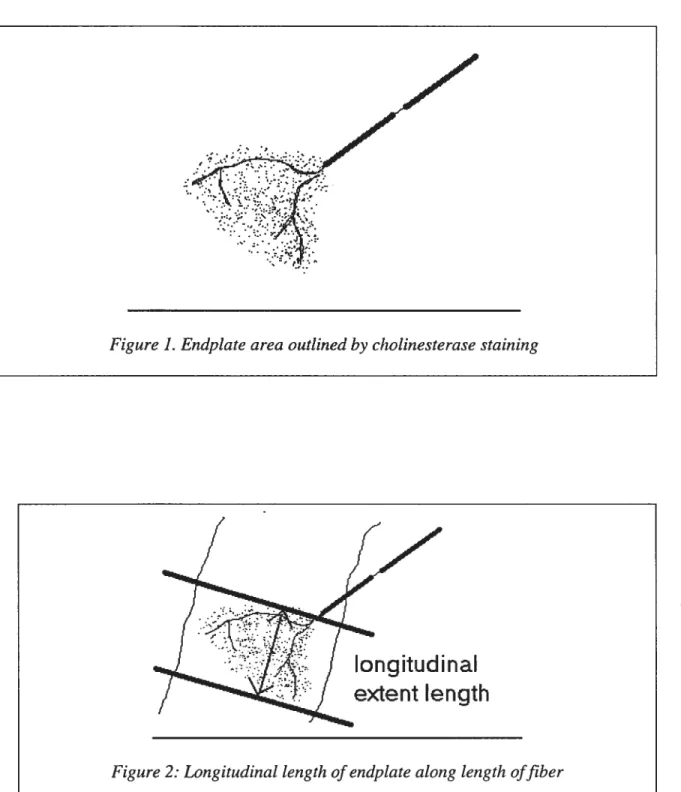
The following measurements were taken:
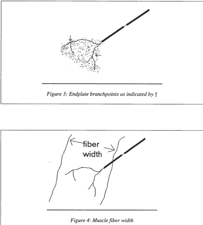
I. Area of the end-plate, outiined by the blue cholinesterase stain (Figure 1); II. The longitudinal length of each endplate, outlined by the cholinesterase stain,
paraiiel to the length of the muscle fiber (Figure 2);
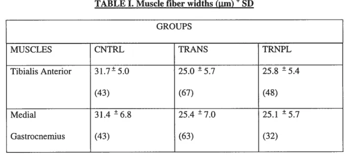
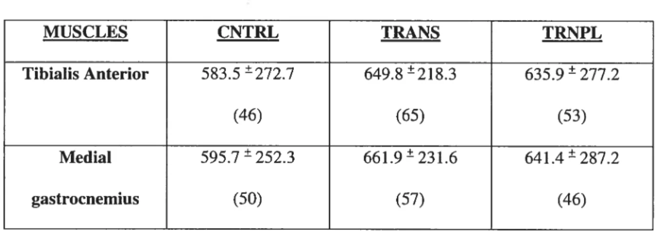
ifi. The number of nerve terminal branch points within each end plate (Figure 3); W. Muscle fiber width (Figure 4), demonstrating atrophy of the muscle in question.
Figure]. Endplate area outÏined by chotinesterasestaining
longitudinal
extent length
figure 3: Endplate branchpoints as indicated by
T
width
Data were collected and recorded for each parameter (ïe: EP area, EP length, branch points and fiber width) until 145 entries for each muscle were obtained. A maximum of 4 endplates were measured from each image.
An endplate was selected if a single parameter was clearly measurable.
A Nikon Optiphot-2 light microscope, equipped with a JVC TK-5210 video camera (TK-A240 power unit supply) was connected to a video monitor (Javelin). The screen was linked to a PC-based 83386 microcomputer (Jaripel) equipped with a digital image processing softward (Image Pro II version 2.0). The PhotoShop software permitted the measurement of end-plate area, longitudinal length and nerve terminal branch points. Images were captured on hard drive and disk for later analysis at varying magnifications.
STATISTICAL ANALYSIS
Data was analyzed using one-way ANOVA, to determine the effects cf transection alone and transection with transplant. When a significant main effect was found, a post hoc test was used to determine significance of the difference between specific means. A probability level below .05 was considered significant.
RESULTS
FIBER WIDTH
MUSCLE DIFFERENCE
Statistical analysis uncovered no significant main effect (p>O.05) between the fiber widths of the TA and MG. Mean muscle fiber widths of TA were only siightly higher than those of MG in ail groups except for the TRANS group (Table I).
TREATMENTS
Statistical analysis uncovered no significant main effect (p>0.05) between the different treatments. Spinal transection did not cause the muscle fibers to atrophy significantly in this experiment. Transected TA muscle fiber widths were shown to be 21% lower than control TA muscles. Muscles undergoing transection plus fetal tissue transplant were shown to have muscle fiber lengths 18% lower than controls. Tt was expected that fetal tissue transplants would delay the onset of muscle atrophy, however, a difference of 3% in terms of muscle fiber width for TA between the TRANS group and the TRNPL group is insignïficant. Fiber widths of transected MG muscles were found to be 20% lower than those of the control group. Those of the TRNPL group were shown to be 20% lower than the control group as well.
TABLE I. Muscle liber widths (m)+ SD GROUPS MUSCLES CNTRL TRANS TRNPL . . . . + + + TibialisAnterior 31.7—5.0 25.0 —5.7 25.8 —5.4 (43) (67) (48) Medial 31.4 6.8 25.4 7.0 25.1 57 Gastrocnemius (43) (63) (32)
EP LONGITUDINAL LENGTH: MUSCLE DIFFERENCE
Statistical analysis uncovered no significant main effect (p>0.05) between the end-plate lengths of the TA and MG. Very littie difference between endplate lengths of TA muscle and the MG muscle was detected within each condition group. (Table II).
TREATMENTS
Statistical analysis uncovered no significant main effect (p>O.05) between the different treatments. For the TA muscle, endplate lengths of the transected group and the transplant group were 5.6% longer than the control group. Endplates of the MG for both the transected and transplant group were also 5.6% longer than the control group.
TABLE II: EP lengths (Lun) ÷ SD GROUPS
MUSCLES CNTRL TRANS TRNPL
Tibialis Anterior 28.9 ±3.4 30.4 ±3.8 30.8 ±3.4
Medial 2$.46.Y 30.1 4.6 30.1 73
EP AREA:
MUSCLE DIFFERENCE
Statistical analysis uncovered no significant main effect (p>0.05) between the endplate areas of the TA and MG (Table ifi).
TREATMENTS
Statistical analysis uncovered no significant main effect (p>O.05) between the different treatments. According to the data obtained, endplate area is greater in the transected and the transplant group for both the TA and the MG. Since there was no significant main effect discovered it is questionable whether transection causes the endplate to enlarge. Although endplates in the transplant group seem to be smaller than the transected group, it is stiil indefinite whether a transplant can counter the effects of transection.
TABLE III. Endplate area (Ltm2) + SD
MUSCLES CNTRL TRANS TRNPL
Tibialis Anterior 583.5 ±272.7 649.8 ±2 18.3 635.9±277.2
(46) (65) (53)
Medial 595.7 252.3 661.9±231.6 641.4±287.2
gastrocnemius (50) (57) (46)
NUMBER 0F BRANCH POINTS MUSCLE DIFFERENCE
Statistical analysis uncovered no significant main effect (p>O.O5) between the number of branch points of the TA and MG (Table W).
TREAIMENTS
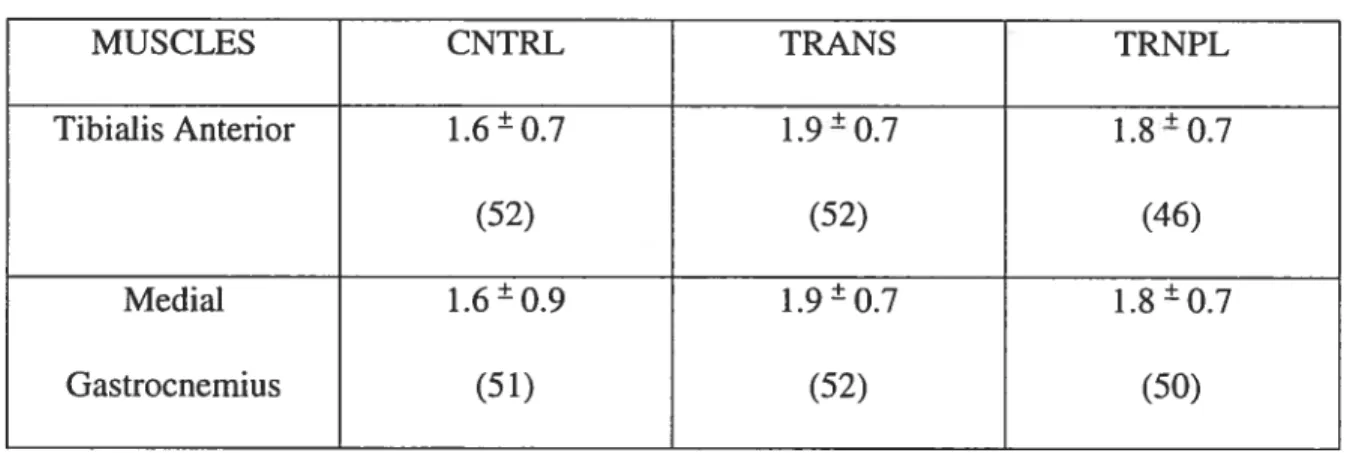
Statistical analysis uncovered no significant main effect (p>0.05) between the different treatments. The number of branchpoints did flot differ between the TA and MG. There was a 16% increase in the number of branchpoints in the transected group in comparison to the control group. The transplant group showed an increase of 11% in number of branchpoints compared to the control group for both the TA and MG muscles.
TABLE IV. Brandi points + SD
MUSCLES CNTRL TRANS TRNPL . . . . + + + Tibialis Anterior 1.6—0.7 1.9—0.7 1.8 —0.7 (52) (52) (46) Medial 1.6O.9 1.90.7 1.80.7 Gastrocnemius (51) (52) (50)
DISCUSSION
The purpose of this study was to determine whether the different functionality of similar fiber type muscles plays a role in the resuits of transection or transplantation on the morphology of the nerve terminal. The anterior tibialis and the medial gastrocnemius muscles were chosen in particular for this study because they are responsible for different functions of the lower limb. The TA muscle contracts in order to flex the anide whereas the MG muscle contracts to extend the ankle and flex the knee (Gordon & Mao, 1994). Both muscles are considered to be predominantly fast muscles with at least 60% of the muscle fibers being type II. Theyare considered to be phasic muscles, which are mainly
recmited during strenuous activities (Armstrong & Phelps,1984). Fast twitch muscles are usually active less often than slow muscles but respond with greater magnitude to
exercise training. Because of this, they seem to be least affected by conditions of inactivity (Eldridge et aL, 1981). In a case where the leg lias a loss of function due to spinal transection the extent of muscle atrophy of predominantly fast twitch muscles is less than the muscle atrophy in slow-twitch muscles (Roy et al., 1986). This may be due partly to the fact that slow twitch muscle is recruited more often since it is a tonic muscle. When transection dismpts the impulse transmission, tonic muscles are no longer recruited
leading to muscle atrophy. Phasic muscles, on the other hand, can undergo a longer period of disuse or inactivity before showing any muscle atrophy.
It lias been noted in previous studies (Buller et al. 1980 Mayer et al. 1984, Roy et al. 1986) that fast extensors atrophy more than fast flexors. A tendency toward this result occurred in this study although means were flot significantly different. In terms of
muscle fiber width, transected muscles had lower fiber widths (24.9 ±5.7 jim) than the
control muscles (31.7±5.0 tm) for TA. The transected MG also had lower fiber widths
(25.4±7.0 jim) compared to the control group (31.4±6.8 11m). fiber width resuits
showed no significant difference between the TA and MG. The average mean of muscle fiber widths showed no significant differences among the three groups. (Refer to TABLE I).
In a study involving spinal transection of cat muscles, Mayer et al. (1984) noted decreases in muscle fiber size of the MG in comparison to control values. lis resuits were based upon 2 1-23 weeks post transection. Less overall atrophy in type I fibers than type II fibers was also noted.
Motor endplate area, which is demonstrated by cholinesterase staining, is related to muscle fiber size. There is a linear relationship between muscle fiber diameter and endplate size (Harris, 1954). Larger fibers tend to have larger endplates (Crockett et al.
1976). Endplates in the TA of the rabbit mode! seemed to have very littie alteration after 3 weeks of tenotomy compared to controls. The gastrocnemius, as well, showed no visible atrophy according to a study conducted by Dias (1979). Fibers of fast twitch muscles are generally larger. TA muscle fiber width was larger than MG in control groups. The TA has more fast twitch fibers than the MG (Armstrong & Phelps, 1984). The size of an individual endplate within a muscle group is correlated with the diameter of the fiber on which it is located. Different muscle groups have their own ratios of endplate size to fiber diameter, which might be regulated by the motor neurons
by finkelstein et al. the fiber cross-sectional area of MG decreased by 25% for
denervations up to 7 days. When compared to longer periods of denervation there was a 5O6O% decrease in fiber area. In the present study there were no significant findings when comparing the endplate area of TA
(
649.8± 2 18.3 tm2 )and MG(
66 1.9±23 1.7Jim2) in the transected group. Although average mean values for endplate area were slightly higher for MG for ail three groups (Refer to TABLE ifi).
Muscles undergoing transplantation of embryonic ceils had lower muscle fiber widths than the control. No significant findings were obtained when comparing results of transected muscles and transplant muscles (Refer to TABLE I). According to Howland et al. (1995), intraspinal transplants of fetal spinal cord enliance the
development of locomotor performance after the spinal cord of newborn animals lias been eitlier completely transected or hemi-sected. In adult recipients limited recovery is
observed when transplants were placed into the site of a complete spinal cord transection (Miya et al.1994). The mechanisms by whicli transplants enhance locomotor
performance are unknown. It was expected that transplantation of embryonic ceils would bave an impact on diminishing the extent of muscle atrophy in the condition of
transectïon, unfortunatelythis did flot occur in the present experiment. Intraspinal
transplant of fetal spinal cord tissue affects myofiber size following complete transection. In a study conducted by Houle et al. 1999 it was noted that fast twïtch fibers were
significantly larger in rats receiving transplants in comparison to transection alone. It remains to be determined whether fetal tissue transplants can reverse the loss in muscle fiber size following SCIor whether it can prevent the reduction in size.
Spinal cord transplants in aduit rats may serve as a relay to convey supraspinal input to spinal cord levels caudal to the transplant. It lias been shown in previous studies that after spinal cord lesions in either newborn or adult rats, the presence of a transplant of fetal spinal cord tissue increases the extent of recovery of locomotor function beyond that seen in lesion-only control animals.
Nerve terminals respond to the presence of denervated or paralyzed muscle fibers by sprouting. Following the loss of even a portion of the motor supply, sprouting
maintains muscle strength through the innervation of denervated fibers (Son et al., 1996). In the rat adult mode!, stress presented in the form of exercise training resulted in
endpiates becoming more branched in hindlimb muscles (Rosenheimer & Smith, 1985; Gardiner et al., 1984). Rosenheimer and Smith found that age-related changes in endplate architecture of sedentary control animals seem to be quite different in
functionally diverse muscles. At 25 months of age both fast-twitch phasic EDL and slow twitch tonic soleus muscles, which were recmited less often with age, exhibited a
significant decrease in terminal brandi number compared with younger (lO-month) animais. Brandi number, in contrast, increased with age in endplates of the fast-twitch diaphragm, which remains constantly active (Rosenheimer, 1985).
The brandi number (number of brandi points) provides an index of nerve terminal complexity. One would assume that more branching would occur as a result of
transection or transplantation. In a study conducted by Rosenheimer (1985) brandi number within endplates was either elevated or reduced depending on the functional characteristics of different muscles. However, there is an age-related increase in terminal branch number at the endplates within the hindlimb muscles. The decrease generally being more pronounced in fast twitch phasically activated muscles (Pestronk et al.,1980). Changes in terminal branch numberarelinked with corresponding alterations in releasing acetylcholine. It may be assumed that a 4-week period of transplantation may flot be long enough to manifest a counter effect to transection. Average branch points were highest in the transection group for both muscles. For the MG the average branch point for
transected muscle was 1.9 ±0.7 whereas the control group the average brandi point was
1.6±0.9. for the TA the average branch point for the transected group was 1.9±0.7
whereas for the control it was 1.6±0.7. (Refer to TABLE W). In comparison, control
branch point values from Rosenheimer (1985) in which he looked at the EDL and Sol his findings were 1.25 0 .004 and 1.63 ±0.008 respectively. According to Tomas et al.
(1992) the percentage of brandi points in controlled muscles were higher in the fast twitch EDL compared to the Sol.
following spinal cord lesions and transplants of fetal spinal cord tissue at birth, there is extensive growth of host descending axons into the transplants (Bregman et al.,1997). Host axons extend throughout the territory provided by the transplant, and both regenerating axons and sprouting axons contribute to this growth. Although descending axons project into the a transplant of fetal spinal cord tissue after injury in the aduit, despite the fetal CNS environment within the transplant, host axons terminate within a
few hundred microns of the hostltransplant border. After spinal cord lesions in aduit rats, for each of the pathways examined, exogenous application of BDNF, NT-3, or NT-4 increased both the distance of axonal growth and the density of axonal growth within transplants of fetal spinal cord tissue placed at the lesion site. It is suggested that after injury in the mature CNS, neurotrophic factors can exert a neurotrophic influence on mature CNS fleurons in vivo, increasing the extent of axonal growth. As the age of an animal at lime of injury increases, the capacity for axonal growth decreases and the extent of axonal growth becomes restricted.
Why did neither transection nor transplant have any effect on the parameters measured?
In assuming that both muscles are composed ofsimilarfiber types, the TA being
composed of a slightly greater percentage of fast twitch, glycolytic fibers than the MG (Armstrong & Phelps, 1984), it seems very likely that the muscles would show no distinct differences in terms of muscle atrophy. Muscle fiber width may be a less accurate
measure of muscle fiber atrophy because contractile properties of predominantly fast muscles are relatively unaffected by a decrease in activity
(
Herbert et al., 1988). In a study by Finkelstein et al. (1993) mean fiber area of MG decreased by 25%followingdenervations of up to 7 days. Although some studies found that slow twitch muscle shows grater atrophy in a case of muscle disuse or inactivity
t
Gordon & Mao, 1994), there was no significant difference found in this experiment between the TA and the siower MG. Both muscles underwent the same conditions of transection and transplantation without showing any significant differences in muscle fiber width or endplate area. It is likely that a 4-week period of tissue transplant is flot enough time to produce any morphological changes. Another reason as to why differences were flot observed with transplantation may be explained by the findings of Das (1981). He reported that spinal cord tissue from16-18-day-old fetuses did not persÏst in the injured spinal cords of adult rats. Reïer et al. (1983) determined that the best growth and long-time survival of fetal spinal cord
transplants were obtained with donor tissue taken from fetuses ranging in age from 12-15 days of gestation. It would be expected that since the TA and MG are responsible for
different functions that they would elicit different responses to transection and to
transplantation however this was not observed in this experiment. Gordon & Mao (1994) found that in animal models with spinal cord injuries there is more severe atrophy of extensor muscles, especially slow—twitch muscles that cross a single joint. In a study by Roy & Acosta (1986) there was a differential atrophic effect on the muscles below the level of lesion, 6- 12 months after transection, ie., extentensors atrophied more than flexors. Tt was noted that the TA was minimally affected by transection whereas the MG atrophied by 15% in comparison to the control group. Jiang et al. (1991) also noted that the MG of the cat atrophied by 12% 6 months following spinal transection. When neuromuscular activation is reduced the relative atrophy of muscles is as follows:
atrophy in the slow extensor is greater than the fast extensor which is greater than the fast flexor (Roy et al. 1991). Considering that these muscles arefast- twitch they areusually recruited lcss often during rest as oppçsed to slow twitch muscles. During activity these fast-twitch muscles arerecruited more. Regardless of their difference in functionality these two muscles in a condition of inactivity responded similarly. Seeing as though both the MG and TA are predominantly fast muscles, the change in activity pattern through transection equally affected the muscles. A general observation is that atrophy is related more to the function of the muscle than to its fiber composition (Roy & Acosta, 1986). if this holds truth then the expected outcome for this experiment would be for the transected MG (siower extensor) fibers to atrophy more than those of the TA (fast flexor), however, the length of disuse or inactivity needs to be taken into account.
According to Buller et al., (1960) complete transection does flot affect predominantly fasi muscles up until several months after the lesion in the cat model. In a
study by Duchen et al. (1970) muscle fibers of the gastrocnemius became progressively atrophied six weeks or more following botulinum toxin injection in the mouse model. Repairing damaged human nerves can be more complex than inflictions used in experimental procedures. Damage may not aiways be clearÏy defined and infection or extensive crushing of the nerve can further complicate recovery.
CONCLUSION
Research lias been advancing towards very promising directions in one day regenerating the spinal cord. Transplantation of fetal tissue presents an important method in which to study problems related to spinal cord damage and regeneration. It lias been established in other studies that such transplants survive, differentiate and become integrated with the CNS of neonatal and adult rat recipients (Reier et al, 1983). Although the present study reported no significant findings to embryonic tissue transplants post transection, several limitations may have been present. These limitations include: a small sample size, assumptions in the duration of transection and transplants because of off-site procedures, unknown age of donor fetus, human inaccuracies in the quantification of endplates from computer screen resolution and microscope magnification. Most mean resuits, exciuding mean resuits for muscle fiber width, were fouiid to be highest in the transection and transplant groups for both the TA and MG. The study however provides valuable information in recreating conditions promoting regeneration experiments. With rapidly expanding information, researchers may have better understanding of growth factors, inhibitory factors and roles that transplants can play in improving the environment of an injured spinal cord.