Letter to the Editor
Behavioural and brain responses in cognitive trance: A TMS-EEG case study
Cognitive trance is defined as a volitional, purposeful and self-induced modified state of consciousness (inherited from shamanic traditional practice), characterized by lucid but narrowed aware-ness of external surroundings with hyper-focused immersive expe-rience of flow, expanded inner imagery, modified somatosensory processing, and an altered sense of self and time (Flor-Henry et al., 2017). The underlying neurophysiology of this particular state of consciousness remains poorly understood. We here report a case study of a highly trained expert in cognitive trance using transcranial magnetic stimulation combined with electroen-cephalography (TMS-EEG), aiming to probe trance-induced changes of electrical reactivity of cortical circuits to magnetic field perturbations. The study was approved by the Ethics Committee of the Faculty of Medicine of the University of Liège in Belgium, and the subject gave her written informed consent.
The participant (C.S.), a 56 year-old right-handed female, origi-nally trained in Mongolia, has been practicing trance for 17 years and is able to self-induce a trance state without external help. Neu-ropsychiatric conditions were excluded. TMS-evoked EEG poten-tials (TEPs) were recorded in eyes-closed conditions during (i) normal resting wakefulness (baseline) and (ii) cognitive trance. Cognitive trance was induced using a standardized protocol (Flor-Henry et al., 2017) employing body movements and vocaliza-tions for about 2 minutes, after which the participant remains in trance without moving throughout the recordings. After each TMS-EEG session, C.S. provided a free recall of her subjective expe-rience and scored her time perception (i.e., subjective duration of the experience, in minutes), level of arousal (i.e., wakefulness), absorption (i.e., become fully involved in the experience), and dis-sociation (i.e., mental separation from the environment) using 0– 10 VAS scorings (Vanhaudenhuyse et al., 2019).
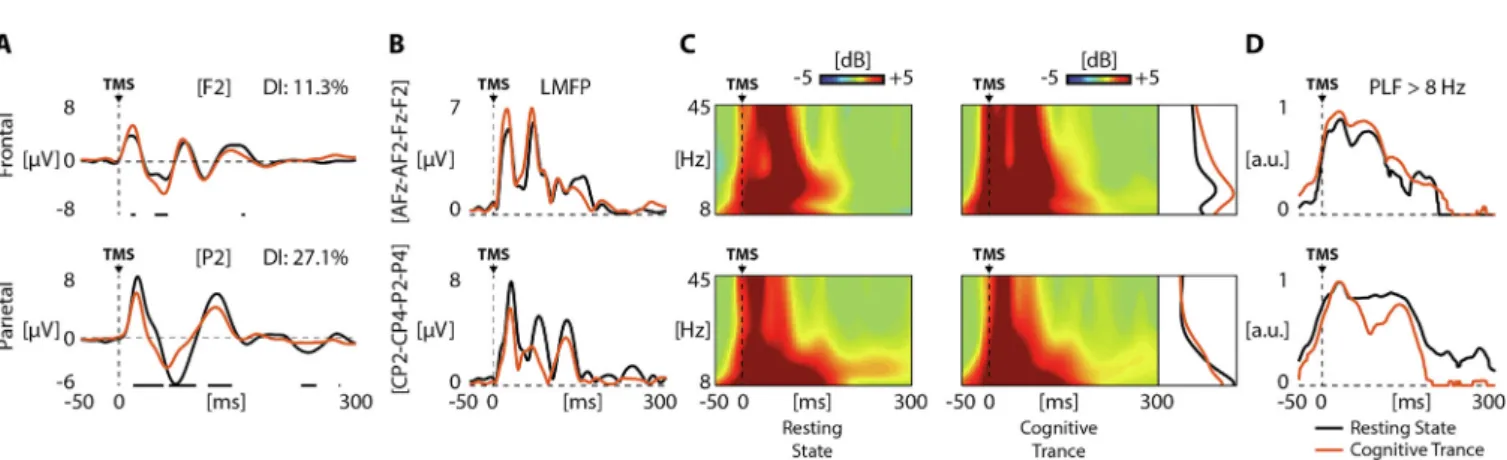
TMS-EEG was performed as previously described (e.g., Bodart et al., 2018), with a TMS compatible 60-channels EEG amplifier and a neuronavigation system (Nexstim Plc, Finland). We targeted one frontal area (premotor cortex) and one parietal area (posterior parietal cortex) on the right hemisphere, using the subject’s T1-weighted structural MRI. At least 150 TMS pulses were delivered at randomly jittered frequencies between 0.4 and 0.5 Hz. TEPs were obtained by averaging a minimum of 130 artifact-free trials for each session. We first calculated the Divergence Index (DI) to evaluate differences between resting state and cognitive trance. DI was computed on TEPs filtered between 1 and 45 Hz, as the per-centage of samples that significantly differ across all channels and latencies, and compared them to normative test-retest variability (Casarotto et al., 2010). Then, we characterized the differences between rest and trance by means of three local measures calcu-lated across the four channels located under the stimulation coil
(AFz, AF2, Fz, and F2 for frontal cortex; CP2, CP4, P2, and P4 for parietal cortex): (i) the amplitude of local mean field power (LMFP, averaged between 8 and 250 ms), as index of reactivity (Fecchio et al., 2017); (ii) the event-related spectral perturbation (ERSP) to evaluate the power and frequency of the oscillations induced by TMS at different cortical sites (Fecchio et al., 2017); and (iii) the local phase-locking factor (PLF) computed on all TEPs filtered above 8 Hz to estimate the impact of the trance state on local cau-sal interactions (Nieminen et al., 2016).
At the descriptive phenomenological level, the subject reported during trance (compared to rest) to feel more awake (fully awake for both sessions during trance vs. normal wakefulness during rest), with higher absorption (8 for frontal and 10 for parietal ses-sion in trance vs. 6 for rest), higher dissociation (8 for frontal and 10 for parietal session in trance vs. 0 for rest), and a time-scale dis-tortion (perceived duration of 8 min for frontal and 2 min for pari-etal session in trance vs. 15 min in rest – real duration was 15 min). The free recall of the trance while TEPs were recorded at the frontal site was the following: ‘‘I had the vision of a samurai with a well-anchored song. Then I saw a harmonious female char-acter, who seemed to be from Thailand with a high-pitched song. After, there were movements of harmonization and there was a spiral that tried to catch the little woodpecker that was on my head (note: the TMS). Some other sounds arrived, with a sensation that they work on the body to restore harmony”. The subjective recall of the trance while targeting the parietal site was the follow-ing: ‘‘I saw a little ant and then I was this ant. I climbed in a tree and I fell from it. After, I had visions of insects and big lizards. I experienced a transformation again, with the feeling of becoming something else, like an iguana. Then my tongue started to come out with the sensation of a turtle’s tongue. After, there were the hisses of snakes, I went through all the reptiles. I had a feeling of joy, I wanted to laugh. Then my breathing changed, and it became very slow. I understood that my tongue was in the perfect place and I was thinking ‘‘the trance is teaching me how to put my ton-gue to slow down the exhalation to be able to induce a feeling of ecstasy”. Then it was pure joy, total happiness and a huge expan-sion of my perception of self”.
For TMS-EEG, the DI computed between resting state and cog-nitive trance was higher than the empirical cut-off of 1.7% (Casarotto et al., 2010), with 11.3% for the frontal session and 27.1% for the parietal session. As an additional control, we also split each resting condition in two and we compared the first half with the second half of the trials, which provided a DI of 0.7% for the frontal and 0.9% for the parietal sessions. These results indicate that the observed changes between rest and trance were signifi-cantly larger than the physiological variability of TEPs (Fig. 1A). This finding is similar to the one recently observed in an expert meditator (Bodart et al., 2018). Since the DI only provides a basic evaluation of overall changes in the brain response, we next exam-https://doi.org/10.1016/j.clinph.2019.11.011
1388-2457/Ó 2019 International Federation of Clinical Neurophysiology. Published by Elsevier B.V. All rights reserved.
Clinical Neurophysiology xxx (xxxx) xxx
Contents lists available atScienceDirect
Clinical Neurophysiology
ined local reactivity changes for the two stimulated targets. As seen inFig. 1, during trance, TEPs amplitude was increased for the frontal stimulation but decreased for the parietal stimulation, as also indicated by the LMFP (Fig. 1B). For frontal stimulation, we observed a broad-band enhancement of significant PLF and power (ERSP) in trance compared to rest, while for the parietal stimulation we observed an early drop of PLF and no difference in power (Fig. 1C and D). These target-specific changes in TEPs amplitude are thus characterized by an enhancement in reactivity while stimulating the frontal cortex and a reduction of local causal interactions while stimulating the parietal cortex during trance.
Altogether, these TMS-EEG results suggest a target-specific dis-sociation: all TMS-EEG metrics (i.e., TEP, LMFP, ERSP, and PLF) increased during cognitive trance when stimulating the frontal cortex, while most measures decreased with parietal stimulation, compared to resting state. This marked increase in brain responses during frontal stimulation could be related to the narrowness of trance experience with focused attention on relevant internal stimuli, vivid senses and monitoring of internal states, as reported by the participant. This could also be associated with increased activation in the premotor regions induced by the mental imagery of movements, sounds, and visual scenes. Selective decrease in brain responses during parietal cortex stimulation might be linked to a lower consciousness of the environment (‘external aware-ness’), and thus a weaker involvement of parietal areas during a state in which cognitive and thought-like activity is increased.
In conclusion, the present results, although limited to a single highly trained practitioner, show that cognitive trance induces a modified state of consciousness characterized by changes in phe-nomenology (e.g., more dissociation) and neurophysiological pro-cesses (e.g., global and local changes in cortical reactivity, synchrony and phase locking) that can be willfully modulated. Fur-ther studies on a larger sample of subjects are needed to better understand the neural basis of cognitive trance. Noteworthy, it seems that this state could also possibly be reached by any trained individual using specific (self-)induction technique, which opens new avenues for neuroscientific studies and potential novel thera-pies with self-exploration processes. This proof-of-concept case report also highlights that it is possible to remain in trance while
being immobile, and while receiving TMS on the brain. Finally, these results extend the use of TMS-EEG to the study of non-ordi-nary states of consciousness, and complement previous results obtained during meditation practice (Bodart et al., 2018). Acknowledgments
This research was supported by BIAL Foundation, Belgian National Funds for Scientific Research (F.R.S-FNRS), EU Horizon 2020 Grant # 785907 Human Brain Project (SGA2), Luminous project (EU-H2020-fetopenga686764), Fondazione Europea di Ricerca Biomed-ica, Benoit Foundation, and Belgian Cancer Foundation (2017-064). We thank Aminata Bicego, Marcello Massimini’s research group at the University of Milan, in particular Mario Rosanova, the Radiodi-agnostic Department of the University Hospital of Liege, and TranceScience Research Institute. LRDS is research fellow and SL is research director at F.R.S-FNRS.
Declaration of Competing Interest
None of the authors declared any conflict of interest. References
Flor-Henry P, Shapiro Y, Sombrun C. Brain changes during a shamanic trance: Altered modes of consciousness, hemispheric laterality, and systemic psychobiology. Cogent Psychol 2017;4(1313522). https://doi.org/10.1080/ 23311908.2017.1313522.
Bodart O, Fecchio M, Massimini M, Wannez S, Virgillito A, Casarotto S, et al. Meditation-induced modulation of brain response to transcranial magnetic stimulation. Brain Stimul 2018;11(6):1397–400. https://doi.org/10.1016/j. brs.2018.08.018.
Casarotto S, Romero Lauro LJ, Bellina V, Casali AG, Rosanova M, Pigorini A, et al. EEG responses to TMS are sensitive to changes in the perturbation parameters and repeatable over time. PLoS One 2010;5(4):e10281. https://doi.org/10.1371/ journal.pone.0010281.
Fecchio M, Pigorini A, Comanducci A, Sarasso S, Casarotto S, Premoli I, et al. The spectral features of EEG responses to transcranial magnetic stimulation of the primary motor cortex depend on the amplitude of the motor evoked potentials. PLoS One 2017;12(9):e0184910.https://doi.org/10.1371/journal.pone.0184910. Nieminen JO, Gosseries O, Massimini M, Saad E, Sheldon AD, Boly M, et al. Consciousness and cortical responsiveness: a within-state study during non-rapid eye movement sleep. Sci Rep 2016;6:30932. https://doi.org/10.1038/ srep30932.
Fig. 1. TMS-EEG reveals modulation of brain activity during cognitive trance (red traces) compared to resting state (black traces) while stimulating frontal and parietal cortex.A. TMS-evoked EEG potentials recorded at F2 and P2 electrodes during rest and trance while targeting the frontal and parietal cortex. The Divergence Index (DI) values were computed over the post-stimulus period (250 ms) between rest and trance conditions for both stimulation targets, and were much larger (indicated in the upper right corners) than the expected cut-off of 1.7%. For each plot, time-points where the two traces significantly differ (p < 0.05) are underlined by horizontal black lines. Vertical dashed lines mark the time of TMS occurrence.B. Local mean field power (LMFP) averaged over the 4 channels closest to the stimulation site (AFz, AF2, Fz, F2 for frontal and CP2, CP4, P2, P4 for parietal) during rest and trance.C. Averaged event-related spectral perturbation of the 4 channels closest to the stimulation site (between 8 and 45 Hz) and the corresponding power spectrum profile evoked during the first 250 ms after TMS during rest and trance. Significant activation was calculated compared to pre-stimulus activity (from 400 to 100 ms) by means of bootstrap statistics (p < 0.05) and is colored in red, while the absence of any significant activation is colored in green. D. Phase-locking factor (PLF) averaged over the 4 channels closest to the stimulation site during cognitive trance are superimposed to PLF profiles calculated for the resting state. By applying a statistical analysis assuming a Rayleigh distribution of the pre-stimulus values (from 400 to 100 ms), PLF time points that were not significantly different (p < 0.05) from pre-stimulus activity were set to zero.
Vanhaudenhuyse A, Ledoux D, Gosseries O, Demertzi A, Laureys S, Faymonville ME. Can subjective ratings of absorption, dissociation, and time perception during ‘‘neutral hypnosis” predict hypnotozability?: An exploratory study. Int J Clin Exp Hypn 2019;67(1):28–38.https://doi.org/10.1080/00207144.2019.1553765.
O. Gosseries1,⇑ Coma Science Group, GIGA Consciousness, University and University Hospital of Liège, Liège, Belgium ⇑ Corresponding author at: Olivia Gosseries, Avenue de l’hôpital, 1, 4000 Liège, Belgium. E-mail address:[email protected] M. Fecchio2 Department of Biomedical and Clinical Sciences ‘‘L. Sacco”, University of Milan, Milan, Italy A. Wolff Coma Science Group, GIGA Consciousness, University and University Hospital of Liège, Liège, Belgium L.R.D. Sanz Coma Science Group, GIGA Consciousness, University and University Hospital of Liège, Liège, Belgium C. Sombrun TranceScience Research Institute, Paris, France A. Vanhaudenhuyse3 Algology Department & Sensation & Perception Research Group, GIGA consciousness, University and University Hospital of Liège, Liège, Belgium S. Laureys4 Coma Science Group, GIGA Consciousness, University and University Hospital of Liège, Liège, Belgium Accepted 16 November 2019 Available online xxxx
1
Contributed equally as first author.
2Contributed equally as first author. 3
Contributed equally as last author.
4
Contributed equally as last author.