HAL Id: dumas-00626806
https://dumas.ccsd.cnrs.fr/dumas-00626806
Submitted on 17 May 2012HAL is a multi-disciplinary open access archive for the deposit and dissemination of sci-entific research documents, whether they are pub-lished or not. The documents may come from teaching and research institutions in France or abroad, or from public or private research centers.
L’archive ouverte pluridisciplinaire HAL, est destinée au dépôt et à la diffusion de documents scientifiques de niveau recherche, publiés ou non, émanant des établissements d’enseignement et de recherche français ou étrangers, des laboratoires publics ou privés.
Artère pulmonaire et ventricule droit dans
l’hypertension pulmonaire : corrélation entre la TDM
synchronisée à l’ECG et le cathétérisme cardiaque droit
Élodie Abel
To cite this version:
Élodie Abel. Artère pulmonaire et ventricule droit dans l’hypertension pulmonaire : corrélation entre la TDM synchronisée à l’ECG et le cathétérisme cardiaque droit. Médecine humaine et pathologie. 2011. �dumas-00626806�
0
UNIVERSITÉ JOSEPH FOURIER
FACULTÉ DE MÉDECINE DE GRENOBLE
Année 2011
Artère
pulmonaire
et
ventricule
droit
dans
l’hypertension pulmonaire:
corrélation entre la TDM
synchronisée à l’ECG et le cathétérisme cardiaque droit
THÈSE
PRÉSENTÉE POUR L’OBTENTION DU DOCTORAT EN MÉDECINE
DIPLÔME D’ÉTAT
Elodie ABEL
Née le 22 Mars 1980 à Metz
Thèse soutenue publiquement à la faculté de médecine de Grenoble Le 29 avril 2011
Devant le jury composé de :
Monsieur le Professeur Gilbert FERRETTI, Président du Jury et Directeur de thèse Monsieur le Professeur Christophe PISON
Monsieur le Professeur Jean-Luc BOSSON Madame le Docteur Hélène BOUVAIST Monsieur le Docteur Adrien JANKOWSKI
2
UNIVERSITÉ JOSEPH FOURIER
FACULTÉ DE MÉDECINE DE GRENOBLE
Année 2011
Artère
pulmonaire
et
ventricule
droit
dans
l’hypertension pulmonaire:
corrélation entre la TDM
synchronisée à l’ECG et le cathétérisme cardiaque droit
THÈSE
PRÉSENTÉE POUR L’OBTENTION DU DOCTORAT EN MÉDECINE
DIPLÔME D’ÉTAT
Elodie ABEL
Née le 22 Mars 1980 à Metz
Thèse soutenue publiquement à la faculté de médecine de Grenoble Le 29 avril 2011
Devant le jury composé de :
Monsieur le Professeur Gilbert FERRETTI, Président du Jury et Directeur de thèse Monsieur le Professeur Christophe PISON
Monsieur le Professeur Jean-Luc BOSSON Madame le Docteur Hélène BOUVAIST Monsieur le Docteur Adrien JANKOWSKI
3
Année universitaire 2010-2011 Doyen de la faculté : M. le Professeur B. SELE
Vice-doyen : M. le professeur J-P. ROMANET
LISTE DES PROFESSEURS D'UNIVERSITÉS – PRATICIENS HOSPITALIERS
NOM PRENOM ADRESSE
ALBALADEJO Pierre CLINIQUE D'ANESTHESIE
PÔLE 2 ANESTHESIE - REANIMATIONS
ARVIEUX-BARTHELEMY Catherine CLINIQUE DE CHIRURGIE ET DE L'URGENCE POLE 6 DIGIDUNE
BACONNIER Pierre
BIOSTATISTIQUES ET INFORMATIQUE MEDICALE PAVILLON D
POLE 17 SANTE PUBLIQUE
BAGUET Jean-Philippe CLINIQUE DE CARDIOLOGIE / HYPERTENSION ARTERIELLE POLE 4 CARDIO VASC. & THORACIQUE
BALOSSO Jacques RADIOTHERAPIE PÔLE 5 CANCEROLOGIE
BARRET Luc CLINIQUE MEDECINE LEGALE
POLE 8 PLURIDISCIPLINAIRE DE MEDECINE
BAUDAIN Philippe CLINIQUE RADIOLOGIE ET IMAGERIE MEDICALE POLE 13 IMAGERIE
BEANI Jean-Claude
CLINIQUE DERMATOLOGIE-VENEREOLOGIE- PHOTOBIOLOGIE ET ALLERGOLOGIE
POLE 8 PLURIDISCIPLINAIRE DE MEDECINE
BENHAMOU Pierre Yves CLINIQUE ENDOCRINO DIABETO NUTRITION EDUCATION THERAPEUTIQUE/ DIABETOLOGIE - POLE 6 DIGIDUNE
BERGER François ONCOLOGIE MEDICALE POLE 5 CANCEROLOGIE
BLIN Dominique CLINIQUE CHIRURGIE CARDIAQUE POLE 4 CARDIO VASC. & THORACIQUE
BOLLA Michel CENTRE COORD. CANCEROLOGIE POLE 5 CANCEROLOGIE
BONAZ Bruno CLINIQUE HEPATO-GASTRO- ENTEROLOGIE POLE 6 DIGIDUNE
BOSSON Jean-Luc DPT DE METHODOLOGIE DE L'INFORMATION DE SANTE POLE 17 SANTE PUBLIQUE
BOUGEROL Thierry PSYCHIATRIE D’ADULTES - PAVILLON D. VILLARS POLE 10 PSYCHIATRIE & NEUROLOGIE
BRAMBILLA Élisabeth DPT ANATOMIE & CYTOLOGIE PATHOLOGIQUES POLE 14 BIOLOGIE
4
BRAMBILLA Christian CLINIQUE DE PNEUMOLOGIE
POLE 7 MEDECINE AIGÜE & COMMUNAUTAIRE
BRICHON Pierre-Yves CLINIQUE DE CHIRURGIE VASCULAIRE ET THORACIQUE POLE 4 CARDIO VASC. & THORACIQUE
BRIX Muriel CLINIQUE CHIR. MAXILLO-FACIALE POLE 3 TETE & COU & CHIR. REPARATRICE
CAHN Jean-Yves CANCEROLOGIE POLE 5 CANCEROLOGIE
CARPENTIER Patrick CLINIQUE MEDECINE VASCULAIRE POLE 8 PLURIDISCIPLINAIRE DE MEDECINE
CARPENTIER Françoise CLINIQUE URGENCE POLE 1 SAMU SMUR
CESBRON Jean-Yves IMMUNOLOGIE - BATIMENT J. ROGET FAC MEDECINE POLE 14 BIOLOGIE
CHABARDES Stephan Clinique de Neurochirurgie
CHABRE Olivier
CLINIQUE ENDOCRINO DIABETO NUTRITION EDUCATION THERAPEUTIQUE / ENDOCRINOLOGIE
POLE 6 DIGIDUNE
CHAFFANJON Philippe CLINIQUE CHIRURGIE THORACIQUE, VASCULAIRE ET ENDOCRINIENNE
CHAVANON Olivier CLINIQUE DE CHIRURGIE CARDIAQUE POLE 4 CARDIO VASC. & THORACIQUE
CHIQUET Christophe CLINIQUE OPHTALMOLOGIQUE POLE 3 TETE & COU & CHIR. REPARATRICE
CHIROSSEL Jean-Paul ANATOMIE - FACULTE DE MEDECINE POLE 3 TETE & COU & CHIR. REPARATRICE
CINQUIN Philippe DPT D'INNOVATIONS TECHNOLOGIQUES- POLE 17 SANTE PUBLIQUE
COHEN Olivier DELEGATION - HC FORUM
COUTURIER Pascal CLINIQUE MEDECINE GERIATRIQUE POLE 8 PLURIDISCIPLINAIRE DE MEDECINE
CRACOWSKI Jean-Luc Laboratoire de Pharmacologie
DE GAUDEMARIS Régis DPT MEDECINE & SANTE DU TRAVAIL POLE 17 SANTE PUBLIQUE
DEBILLON Thierry CLINIQUE REA. & MEDECINE NEONATALE POLE 9 COUPLE/ENFANT
DEMONGEOT Jacques BIOSTATISTIQUES ET INFORMATIQUE MEDICALE POLE 17 SANTE PUBLIQUE
DESCOTES Jean-Luc CLINIQUE UROLOGIE POLE 6 DIGIDUNE
DYON J.François
ESTEVE François Dir. Équipe 6 U836 - ID17 /ESRF Grenoble Institut des Neurosciences
FAGRET Daniel CLINIQUE DE MEDECINE NUCLEAIRE POLE 13 IMAGERIE
5
FAUCHERON Jean-Luc CLINIQUE DE CHIRURGIE DIGESTIVE ET DE L'URGENCE POLE 6 DIGIDUNE
FAVROT Marie Christine DPT DE BIOLOGIE INTEGREE / CANCEROLOGIE POLE 14 BIOLOGIE
FERRETTI Gilbert CLINIQUE RADIOLOGIE & IMAGERIE MEDICALE POLE 13 IMAGERIE
FEUERSTEIN Claude GIN
FONTAINE Éric CLINIQUE NUTRITION ARTIFICIELLE POLE 7 MED. AIGÜE & COMMUNAUTAIRE
FRANCO Alain CLINIQUE VIEILLISSEMENT ET HANDICAP POLE 7 MED. AIGUE & COMMUNAUTAIRE
FRANCOIS Patrice DPT DE VEILLE SANITAIRE POLE 17 SANTE PUBLIQUE
GARNIER Philippe
GAUDIN Philippe CLINIQUE DE RHUMATOLOGIE
POLE 11 APPAREIL LOCOMOTEUR GERIATRIE CHISSE
GAY Emmanuel CLINIQUE NEUROCHIRURGIE
POLE 3 TETE & COU & CHIR. REPARATRICE
GIRARDET Pierre
GUIDICELLI Henri
HALIMI Serge CLINIQUE ENDOCRINO-DIABETO-NUTRITION POLE 6 DIGIDUNE
HOMMEL Marc CLINIQUE DE NEUROLOGIE
POLE 10 PSYCHIATRIE & NEUROLOGIE
JOUK Pierre-Simon DEPARTEMENT GENETIQUE ET PROCREATION POLE 9 COUPLE/ENFANT
JUVIN Robert CLINIQUE DE RHUMATOLOGIE - HOPITAL SUD POLE 11 APPAREIL LOCOMOTEUR & GERIATRIE CHISSE
KAHANE Philippe CLINIQUE DE NEUROLOGIE
POLE 10 PSYCHIATRIE & NEUROLOGIE
KRACK Paul CLINIQUE DE NEUROLOGIE
POLE 10 PSYCHIATRIE & NEUROLOGIE
KRAINIK Alexandre CLINIQUE NEURORADIOLOGIE & IRM POLE 13 IMAGERIE
LANTUEJOUL Sylvie DEPARTEMENT D'ANATOMIE ET CYTOLOGIE PATHOLOGIQUES PÔLE 14 BIOLOGIE
LE BAS Jean-François CLINIQUE NEURORADIOLOGIE & IRM POLE 13 IMAGERIE
LEBEAU Jacques CLINIQUE CHIR. MAXILLO-FACIALE POLE 3 TETE & COU & CHIR. REPARATRICE
LECCIA Marie-Thérèse
CLINIQUE DERMATOLOGIE-VENEREOLOGIE- PHOTOBIOLOGIE ET ALLERGOLOGIE
6
LEROUX Dominique DEPARTEMENT BIOLOGIE ET PATHOLOGIE DE LA CELLULE POLE 14 BIOLOGIE
LEROY Vincent CLINIQUE D'HEPATO GASTRO ENTEROLOGIE POLE 6 DIGIDUNE
LETOUBLON Christian CLINIQUE CHIRURGIE DIGESTIVE & URGENCE POLE 6 DIGIDUNE
LEVERVE Xavier LABORATOIRE THERAPEUTIQUE UFR BIOLOGIE BAT 72 UJF BP 53X
LEVY Patrick PHYSIOLOGIE
POLE 12 REEDUCATION & PHYSIOLOGIE
LUNARDI Joël BIOCHIMIE ADN- POLE 9 COUPLE/ENFANT
MACHECOURT Jacques CLINIQUE DE CARDIOLOGIE
POLE 4 CARDIO VASC. & THORACIQUE
MAGNE Jean-Luc CLINIQUE CHIRURGIE VASCULAIRE & THORACIQUE POLE 4 CARDIO VASC. & THORACIQUE
MAITRE Anne Médecine du travail EPSP/DPT DE BIOLOGIE INTEGREE - POLE 14 BIOLOGIE - J.ROGET 4e ETAGE
MALLION J. Michel
MASSOT Christian CLINIQUE MEDECINE INTERNE
POLE 8 PLURIDISCIPLINAIRE DE MEDECINE
MAURIN Max DEPARTEMENT DES AGENTS INFECTIEUX / BACTERIOLOGIE POLE 14 BIOLOGIE
MERLOZ Philippe CLINIQUE CHIR. ORTHOPEDIE TRAUMATOLOGIE POLE 3 TETE & COU & CHIR. REPARATRICE
MORAND Patrice DPT DES AGENTS INFECTIEUX / VIROLOGIE POLE 14 BIOLOGIE
MOREL Françoise
MORO-SIBILOT Denis
MOUSSEAU Mireille ONCOLOGIE MEDICALE POLE 5 CANCEROLOGIE
MOUTET François CHIR. PLASTIQUE ET RECONSTRUCTRICE ET ESTHETIQUE
PASQUIER Basile
PASSAGIA Jean-Guy ANATOMIE
POLE 3 TETE & COU & CHIR. REPARATRICE
PAYEN DE LA GARANDERIE Jean-François CLINIQUE REANIMATION POLE 2 ANESTHESIE-REANIMATION
PELLOUX Hervé
DEPARTEMENT DES AGENTS INFECTIEUX PARASITOLOGIE ET MYCOLOGIE POLE 14 BIOLOGIE
7
PEPIN Jean-Louis CLINIQUE PHYSIOLOGIE SOMMEIL & EXERCICE - POLE 12 REEDUCATION & PHYSIOLOGIE
PERENNOU Dominique SERVICE DE REEDUCATION
POLE 12 REEDUCATION & PHYSIOLOGIE
PERNOD Gilles CLINIQUE DE MEDECINE VASCULAIRE-
POLE PLURIDISCIPLINAIRE DE MEDECINE - POLE 8
PIOLAT Christian Clinique de chirurgie infantile
PISON Christophe CLINIQUE PNEUMOLOGIE
POLE 7 MEDECINE AIGÜE & COMMUNAUTAIRE
PLANTAZ Dominique CLINIQUE MEDICALE PEDIATRIQUE POLE 9 COUPLE/ENFANT
POLACK Benoît DEPARTEMENT DE BIOLOGIE ET PATHOLOGIE DE LA CELLULE POLE 14 BIOLOGIE
POLLAK Pierre NEUROLOGIE
POLE 10 PSYCHIATRIE & NEUROLOGIE
PONS Jean-Claude CLINIQUE UNIVERSITAIRE GYNECOLOGIE OBSTETRIQUE POLE 9 COUPLE/ENFANT
RAMBEAUD J Jacques CLINIQUE UROLOGIE POLE 6 DIGIDUNE
REYT Émile CLINIQUE O.R.L.
POLE 3 TETE & COU & CHIR. REPARATRICE
ROMANET J. Paul CLINIQUE OPHTALMOLOGIQUE POLE 3 TETE & COU & CHIR. REPARATRICE
SARAGAGLIA Dominique
CLINIQUE ORTHOPEDIQUE ET TRAUMATOLOGIE POLE 11 APPAREIL LOCOMOTEUR & GERIATRIE CHISSE HOPITAL SUD
SCHAAL Jean-Patrick CLINIQUE UNIVERSITAIRE GYNECOLOGIE OBSTETRIQUE POLE 9 COUPLE/ENFANT
SCHMERBER Sébastien CLINIQUE O.R.L.
POLE 3 TETE & COU & CHIR. REPARATRICE
SEIGNEURIN Daniel DPT ANATOMIE & CYTOLOGIE PATHOLOGIQUES POLE 14 BIOLOGIE
SEIGNEURIN Jean- Marie DPT AGENTS INFECTIEUX POLE 14 BIOLOGIE
SELE Bernard DPT GENETIQUE & PROCREATION POLE 9 COUPLE/ENFANT
SESSA Carmine CHIRURGIE THORACIQUE VASCULAIRE POLE 4 CARDIO VASC. & THORACIQUE
SOTTO Jean-Jacques
STAHL Jean-Paul CLINIQUE INFECTIOLOGIE
POLE 7 MEDECINE AIGÜE & COMMUNAUTAIRE
TIMSIT Jean-François CLINIQUE REANIMATION MEDICALE POLE 7 MED. AIGUE & COMMUNAUTAIRE
8
TONETTI Jérôme CLINIQUE ORTHOPEDIQUE ET TRAUMATOLOGIE POLE 11 APPAREIL LOCOMOTEUR & GERIATRIE CHISSE
TOUSSAINT Bertrand BIOCHIMIE ET BIOLOGIE MOLECULAIRE POLE 14 BIOLOGIE
VANZETTO Gérald CLINIQUE DE CARDIOLOGIE POLE 4 CARDIO VASC. & THORACIQUE
VUILLEZ Jean-Philippe BIOPHYSIQUE ET TRAITEMENT DE L’IMAGE
ZAOUI Philippe CLINIQUE NEPHROLOGIE POLE 6 DIGIDUNE
ZARSKI Jean-Pierre CLINIQUE HEPATO-GASTRO-ENTEROLOGIE POLE 6 DIGIDUNE
LISTE DES MAITRES DE CONFÉRENCES DES UNIVERSITÉS - PRATICIENS HOSPITALIERS
NOM PRENOM ADRESSE
BOTTARI Serge Département de Biologie Intégrée Pôle 14: Biologie
BOUTONNAT Jean Département de Biologie et Pathologie de la Cellule Pôle 14: Biologie
BRENIER-PINCHART M. Pierre Département des agents infectieux Parasitologie Mycologie
Pôle 14: Biologie
BRICAULT Ivan Clinique de radiologie et imagerie médicale Pôle 13: Imagerie
BRIOT Raphaël Pôle Urgence SAMU CALLANAN-WILSON Mary Génétique IAB
CARAVEL Jean-Pierre Clinique de médecine Nucléaire Pôle 13: Imagerie
CRACOWSKI Jean Luc Laboratoire de Pharmacologie CROIZE Jacques Département des agents infectieux
Micro biovigilance Pôle 14: Biologie DEMATTEIS Maurice Clinique de physiologie
sommeil et exercice
Pôle 12: Rééducation et physiologie DERANSART Colin GIN - BATIMENT E. SAFRA
9
DETANTE Olivier Clinique de Neurologie
DROUET Christian Département de Biologie et Pathologie de la Cellule Centre angiodème - Pôle 14: Biologie
DUMESTRE-PERARD Chantal Immunologie - BATIMENT J. ROGET. EYSSERIC Hélène Clinique de Médecine Légale
Pôle 8: Pôle Pluridisciplinaire de Médecine FAURE Anne-Karen Biologie de la procréation / CECOS
Département génétique et procréation Pôle 9: Couple/enfant
FAURE Julien Département génétique et procréation Pôle 9: Couple/enfant
GARBAN Frédéric Unité clinique thérapie cellulaire Pôle 5 : Cancérologie
GAVAZZI Gaëtan Clinique médecine interne gériatrique Pôle 8 : Pôle pluridisciplinaire de Médecine GRAND Sylvie Clinique de Radiologie et Imagerie Médicale
Pôle 13 : Imagerie
HENNEBICQ Sylviane Biologie de la procréation / CECOS Département génétique et procréation Pôle 9: Couple/enfant
HOFFMANN Pascale Clinique Universitaire Gynécologie Obstétrique Pôle 9: Couple/enfant
JACQUOT Claude Clinique d'Anesthésie
Pôle 2 : Anesthésie - Réanimations LABARERE José Département de veille sanitaire
Pôle 17 : Santé Publique LAPORTE François Département de biologie intégrée
Pôle 14: Biologie
LARDY Bernard Département de biologie et pathologie de la cellule - Laboratoire d'Enzymologie
Pôle 14: Biologie
LARRAT Sylvie Département des agents infectieux Pôle 14: Biologie
LAUNOIS-ROLLINAT Sandrine Clinique de Physiologie sommeil et exercice Lab. explor. fonct. cardio-respiratoires Pôle 12 : Rééducation et physiologie MALLARET Marie-Reine Unité d'Hygiène Hospitalière
Pavillon E
MOREAU-GAUDRY Alexandre Département d'innovations technologiques Pôle 17 Santé Publique
MOUCHET Patrick Clinique de Physiologie sommeil et exercice Lab. explor. fonct. cardio-respiratoires Pôle 12 : Rééducation et physiologie
10
PACLET Marie-Hélène Département de biologie et pathologie de la cellule - Laboratoire d'Enzymologie Pôle 14: Biologie
PALOMBI Olivier Clinique de neurochirurgie
Pôle 3 : Tête et cou et chirurgie réparatrice PASQUIER Dominique Département d'anatomie et cytologie pathologiques
Pôle 14 : Biologie
PELLETIER Laurent Centre d'innovation biologique PAYSANT François Clinique de Médecine Légale
Pôle 8: Pôle Pluridisciplinaire de Médecine RAY Pierre Biologie de la reproduction
Département génétique et procréation Pôle 9: Couple/enfant
RENVERSEZ J.Charles Département de biologie intégrée Biochimie et Biologie Moléculaire Pôle 14 : Biologie
RIALLE Vincent Laboratoire TIMC LA TRONCHE SATRE Véronique Génétique chromosomique
Département génétique et procréation Pôle 9: Couple/enfant
STANKE-LABESQUE Françoise Laboratoire de Pharmacologie
STASIA Marie-Josée Département de biologie et pathologie de la cellule Pôle 14: Biologie
TAMISIER Renaud Clinique de Physiologie sommeil et exercice Lab. explor. fonct. cardio-respiratoires Pôle 12 : Rééducation et physiologie WEIL Georges Biostatistiques et Informatique Médicale
11
À notre Maître et Président de thèse,
Monsieur le Professeur Gilbert FERRETTI,
Vous nous faites l’honneur de présider le jury de cette thèse et de nous avoir guidés dans notre réflexion en tant que directeur de thèse. Nous vous remercions de votre enseignement. Veuillez trouver l’expression de notre profond respect et de notre sincère gratitude pour votre confiance et enseignement de grande qualité.
Aux membres du Jury,
Monsieur le Professeur Jean-Luc BOSSON,
Vous nous faites l’honneur de juger ce travail. Veuillez accepter mes sincères remerciements pour votre aide précieuse à l’élaboration de cette thèse.
Monsieur le Professeur Christophe PISON,
Vous nous faites l’honneur de juger ce travail en tant qu’expert et référent en hypertension pulmonaire. Nous avons apprécié votre enthousiasme et vos compétences. Soyez assuré de notre admiration et de notre reconnaissance.
Madame le Docteur Hélène BOUVAIST,
Merci pour votre disponibilité et vos compétences lors de la préparation de cette thèse. Vos remarques préliminaires nous ont beaucoup aidés.
Monsieur le Docteur Adrien JANKOWSKI,
Tes compétences en imagerie cardio-thoracique ne sont plus à démontrer. Tu m’as été d’une grande aide dans la réalisation de ce travail. Merci pour toutes ces heures passées à matérialiser ce projet et pour ta grande disponibilité. Accepte ma profonde gratitude.
12 A mes parents et ma famille, que j’aime.
A mon frère Adrien, tu es tout pour moi.
A tous mes amis d’ici et d’ailleurs, d’aujourd’hui et d’hier, présents ou non à mes côtés ce jour. Merci pour tous ces moments.
A toutes les équipes médicales et paramédicales d’Annecy, Chambéry, Grenoble et Nouméa avec qui ce fut un plaisir de partager ces années.
13
Sommaire
RÉSUMÉ ... 14 ABBREVIATIONS ... 16 SUMMARY ... 17 INTRODUCTION ... 18MATERIELS AND METHODS ... 19
RESULTS ... 23 DISCUSSION ... 29 CONCLUSION ... 32 BIBLIOGRAPHY ... 33 CONCLUSIONS ... 35 SERMENT D’HIPPOCRATE... 37 ANNEXE : PROTOCOLE DE RECHERCHE CECIC ...
14
Artère
pulmonaire
et
ventricule
droit
dans
l’hypertension pulmonaire:
corrélation entre la TDM
synchronisée à l’ECG et le cathétérisme cardiaque droit
Objectifs :
Rechercher rétrospectivement une relation entre des paramètres fonctionnels fournis par TDM synchronisée à l’ECG (fraction d’éjection du ventricule droit (FEVD), débit cardiaque éjecté du ventricule droit (DCVD), distensibilité de l’artère pulmonaire (AP)) et les données hémodynamiques du cathétérisme cardiaque droit (pression artérielle pulmonaire moyenne (PAPm) et débit cardiaque), chez les patients porteurs d’une hypertension artérielle pulmonaire.
Matériels et méthodes
Notre étude rétrospective a été soumise au comité d’éthique de notre établissement. Le consentement éclairé des patients n’a pas été requis.
Vingt sept patients atteints d’hypertension pulmonaire, ayant bénéficié d’une TDM synchronisée à l’ECG et d’un cathétérisme droit ont été inclus. Deux observateurs indépendants, sans connaissance des données du cathétérisme cardiaque droit ont étudié sur la TDM synchronisée à l’ECG différents paramètres : le diamètre de l’AP, le diamètre de l’aorte, la distensibilité de l’AP, la FEVD, le débit cardiaque éjecté du VD, à l’aide de deux méthodes : la segmentation automatique et la technique de Simpson. Le débit cardiaque et la PAPm ont été mesurés par cathétérisme cardiaque droit. L’existence d’une relation entre les données de la TDM synchronisée à l’ECG et le cathétérisme cardiaque droit a été testée par une analyse de régression linéaire.
Résultats
La corrélation inter-observateur est bonne pour toutes les mesures (R>0.7) à l’exception du débit cardiaque mesuré avec la technique de Simpson (R=0.63). Il existe une corrélation significative entre la distensibilité de l’AP et la PAPm (r=-0.426, p=0.027). La FEVD est corrélée à la PAPm (r=-0.417, p=0.034) uniquement lorsqu’elle est mesurée par la technique de Simpson. Nous n’avons pas mis en évidence de corrélation entre la FEVD et le débit cardiaque, tant avec la technique de segmentation que celle de Simpson (p>0.2).
Le débit cardiaque éjecté du VD mesuré avec la technique de Simpson (r=0.487, p=0.010) et la technique de segmentation automatique (r=0.549, p=0.005) est corrélé au débit cardiaque mesuré par cathétérisme cardiaque droit.
15
Conclusion
La FEVD et le débit cardiaque mesurés par TDM synchronisée à l’ECG sont corrélés de façon significative à la PAPm et aux débits cardiaques mesurés par le cathétérisme cardiaque droit et pourraient être utiles dans l’évaluation initiale et potentiellement le suivi des patients atteints d’hypertension pulmonaire.
Mots clés
16 Abbreviations:
CO= Cardiac Output
CT= Computed Tomography CSA= Cross Section Area
ECG-gated-CT= electrocardiographically- gated computed tomography mPAP= Mean Pulmonary Arterial Pressure
MRI= Magnetic Resonance Imaging NYHA= New York Heart Association PA= Pulmonary Artery
PH= Pulmonary Hypertension r= Pearson’s correlation coefficient R= interobserver agreement RHC= Right Heart Catheterization RV= Right Ventricle
RVEF= Right Ventricular Ejection Fraction SD= Standard Deviation
17
Pulmonary artery and right ventricle in Pulmonary
Hypertension: correlation between thoracic
ECG-gated-CT and right-side heart catheterization
Purpose:
To retrospectively investigate whether a relationship exists between ECG-gated-CT functional parameters (right ventricular ejection fraction (RVEF), right ventricular cardiac output (RVCO), pulmonary artery (PA) distensibility) and mean pulmonary arterial pressure (mPAP) or CO measured by right-side heart catheterization (RHC) in patients with pulmonary hypertension.
Materials and Methods:
This retrospective study was submitted to the institutional review board, patient consent was not required. Twenty-seven patients with PH who had undergone both ECG-gated-CT and RHC were included. Two independent observers, blinded to RHC results, measured on ECG-gated-CT: PA diameter, PA distensibility, aorta diameter, RVCO and RVEF with two methods: automatic segmentation and Simpson technique. CO and mPAP were measured on RHC. The relationship between ECG-gated-CT and RHC measurements were tested with linear regression analysis.
Results:
Interobserver agreement was good for all measurements (R>0.7) except for RVCO calculated with Simpson’s technique (R=0.63). PA distensibility was significantly correlated to mPAP (r=-0.426, p=0.027). RVEF was correlated with mPAP only when issued from Simpson technique (r=-0.417, p=0.034). RVEF was not significantly correlated to CO, whether with segmentation or Simpson technique (p>0.2). RVCO measured with Simpson technique (r=0.487, p=0.010) and automatic segmentation (r=0.549, p=0.005) correlated equally with CO measured with RHC.
Conclusion:
RVEF and RVCO measured on ECG-gated-CT are significantly correlated respectively to mPAP and CO measured on RHC and could be useful for staging and follow up of patient with PH.
18
Introduction
Pulmonary hypertension (PH) requires a series of investigations to confirm the diagnosis, clarify the clinical group, specify the etiology within its group 1 and evaluate the functional and hemodynamic impairment of the right heart 2. As right ventricular (RV) function predicts outcome in patients with PH, accurate assessment of right ventricular function is essential to graduate severity, follow-up and assess response to therapy 3.
In PH, echocardiography is the first-line non-invasive examination for screening and evaluating hemodynamic status and global function of the RV 4 as it is widely available, radiation free, and less costly than other techniques. However, suboptimal acoustic window can alter RV evaluation, especially in patients with advanced lung disease. Moreover, echocardiography may frequently be inaccurate in estimating mean pulmonary arterial pressure (mPAP) or cardiac output (CO) and misclassify the severity of the disease 5.
Right-side heart catheterization (RHC) remains the required reference standard to diagnose PH 2, assess hemodynamic impairment, and follow-up patients under treatment, although it is invasive, delivers radiation, and is associated with recognized complications 6.
Cardiac-MRI is considered as the reference standard for accurate and reproducible measurement of ventricular function 7. It allows non invasive assessment of right ventricular ejection fraction (RVEF) and CO 8. Pulmonary artery (PA) diameter, PA /aorta diameter 9, distensibility of PA and relative area change of PA has been correlated with mPAP and prognosis of PH 10. Contrast injection is not necessary and there is no ionizing radiation. Apnea in poor respiratory condition can be a limitation. However, MRI is not recommended in the initial evaluation of PH and is not a routine investigation 2.
Computed Tomography (CT) is part of standard initial diagnosis assessment in patients with PH 11. It is commonly used to depict lung diseases or vascular abnormalities that may be responsible for PH 12 . Technological advances in spatial and temporal resolution, extended coverage per tube rotation give a new interest in the technique 13 and allow performing ECG-gated-CT with acceptable levels of irradiation 14. ECG-gated-CT is accurate for measuring RV function as validated by comparison to MRI 15. PA distensibility assessed with ECG-gated-CT has been evaluated in screening PH 1, but not in evaluation of severity.
Whereas many studies took interest in assessing RV morphologic parameters with ECG-gated-CT, few of them investigated the hemodynamic parameters that can be calculated using ECG-gated-CT data 16.
The aim of the present study was to evaluate whether PH severity could be assessed using ECG-gated-CT parameters. Besides PA diameter and PA/aorta diameter ration, functional parameters such as PA distensibility, RVEF and CO were compared to RHC measurements. A further objective was to evaluate CO using two ECG-gated-CT methods: the reference Simpson technique and the fully automatic technique generated by commercially available cardiac software.
19
Materials and Methods
Study design / patients population
This retrospective study was submitted to our institutional review board. Informed consent was not required as we used the patients’ data retrospectively. This was possible by prospective storage of ECG-gated-CT images of all patients referred for evaluation of clinically suspected PH in our PH reference center. A review was undertaken to select the patients with proved PH (mPAP > 25 mmHg) who had undergone both ECG-gated-CT and RHC between July 2008 and May 2010.
Thirty-two consecutive patients were selected but five patients were excluded because of incomplete data (n=3) or insufficient CT image quality (n=2). Therefore, we included twenty-seven patients aged 30-84 years (mean 61). The time between ECG-gated-CT and RHC was 68 days (range 1- 373 days). All included patients had a normal sinus rhythm and no contraindication to contrast media injection.
Right Heart Catheterization
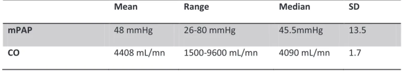
Swang-Ganz right heart catheterization was performed by using a balloon-floating catheter. Pressure measurements and CO were performed by using standard techniques. CO, mPAP, right atrial pressure, pulmonary capillary wedge pressure, pulmonary vascular resistance and mixed venous oxygen saturation were obtained. CO was determined by using the Fick equation. The mean resting PAP was 48 mm Hg ± 13.5 (range 26-80 mm Hg); the median PAP was 45.5 mm Hg.
ECG-gated-CT acquisition
All CT were performed with a 64-section-CT scan (Philips Brilliance 64, Philips Medical Systems, Netherlands) using retrospective ECG-gating technique. The acquisition parameters were as follows: collimation of 64 x 0.625 mm, rotation time of 400 milliseconds, pitch of 0.203, tube voltage 120 kV and tube current 500mAs. The dose-length product in the study population was 1177 mGy.cm (range 853- 1414mGy.cm, SD 144), giving a mean effective dose of 20 mSv (range 14.5-24 mSv).
Contrast media injection of 110 mL of nonionic low-osmolar contrast agent (Iobitriol, Xenetix 350, Guerbet, France) was administered intravenously at a flow rate of 4 mL/s; bolus tracking in the ascending aorta was used to trigger CT-acquisition. No beta-blockers were used.
20
CT Image Post-processing
Row data were reconstructed at section widths of 1 mm, every 10% of R-R’ intervals, with a standard reconstruction filter. We used a workstation (EBW, Philips Medical Systems, Cleveland, OH) with dedicated cardiac software (Comprehensive Cardiac, version 4.5.2.40007).
Image Interpretation
All CT scans were anonymized. Two independent experienced observers (G.F and A.J., with 9 and 7 years of experience in cardio-pulmonary imaging) unaware of the clinical information or RHC results reviewed all images on a workstation with dedicated cardiac software.
PA and PA/aorta diameter, PA distensibility
The widest short-axis diameter of the PA was measured perpendicularly to the long-axis of the PA at 3 cm below the pulmonary artery division. This measurement was obtained on the axial section, reconstructed at 80% of R-R’ interval. The widest short-axis of the ascending aorta was measured on the same axial-section used for measuring the diameter of the main PA. The PA/aorta diameter index is then calculated.
To measure PA surface, we first defined on the 40% RR’ phase a cross sectional plane of PA trunk, perpendicular to its long axis, on sagittal and axial views, 2 cm above the pulmonary valves annulus. We then automatically reproduced the same plane of reformation on the nine other image sets of the cardiac cycle. The PA cross section area (CSA) was measured on each cardiac plane using the semi-automatic software calipers with manual correction if needed. PA distensibility was then calculated from the highest and the lowest of the 10 CSA measurements. We used the following formula: PA distensibility=(CSA max-CSAmin)/CSAmax
17
.
RVEF, RVCO
The evaluation of the volumes of the cardiac cavities was performed using two different techniques.
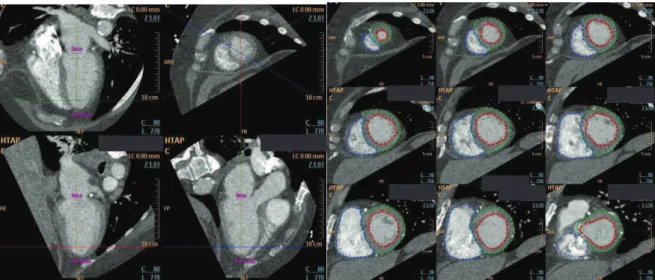
The Simpson segmentation (figure 1) is a two-dimensional short-axis semi-automatic method. Short-axis multiplanar sections were defined, including the whole ventricular chamber, from base to apex. Then, the observers modified if necessary the automatic outlining of the cardiac cavities, on the end-systolic and end-diastolic images. Once all slices were outlined, the software automatically calculated the end-systolic and end-diastolic volumes by Simpson’s rule.
21
Figure 1. Simpson segmentation
Short-axis reformatted slices. Endocardial contour tracing of RV (red) on nine successive short-axis reformatted-slices at end-systole and end-diastole phases.
Figure 2. 3D automatic segmentation
Axial, sagittal and coronal reformatted slices. All pixels taken into account for volume calculations are highlighted (blue: RA, green: LA, violet: RV, red: LV).
The automatic segmentation (figure 2) is a volumetric 3D technique. Evaluation of cardiac cavities volume at end-systolic and end-diastolic phases was calculated using commercial software. This method was based on counting the voxels included in an outlined region of interest between two predefined points so as to calculate the end-systolic and end-diastolic volumes produce of the number of pixels, the pixel area and the slice thickness. The software colored artificially all included pixels so that the observer could check on three orthogonal planes the RV volume. Then, the observer scored quality of computerized segmentation in four groups (excellent, good, acceptable, poor) in order to exclude too poor quality ones. This scoring was done both in end-systolic and end-diastolic phases. The observers did not modify the automatic outlining proposed by the software.
22 We excluded from the analysis two patients that had a poor quality of segmentation (patients 8 and 18) using the automatic segmentation.
Integrating cardiac rhythm during acquisition, the cardiac software calculated the different hemodynamic parameters: RVEF, CO.
Statistical Analysis
Descriptive statistics were applied on all collected variables.
Interobserver agreement on measurements was determined by calculating Pearson’s correlation coefficient (r).
Data are expressed as medians with ranges for none normally distributed data or means with standards deviations (SD) for normally distributed data, or proportions.
Results were considered statistically significant at p<0.05. Linear regression was used to assess the relationships between variables with significant associations (p<0.05).
Interobserver correlation (R) was considered significant above 0.7.
According to Bland and Altman analysis, the reliability and the repeatability of each software method was evaluated after log transformation by Pearson’s correlation coefficient (r), bias estimated by the mean difference and the SD from the mean deviation, interobserver correlation and means of values obtained by short-axis Simpson and 3D automatic segmentation techniques plotted against their differences. We also analyzed the measurements’ error graphically, with the plotting of the individual subjects’ SD against his mean and analytically by r of log-transformed data and within-subject SD.
23
Results:
Patient Characteristics
Patient characteristics are summarized in table 1
Table 1. Patient characteristics
Characteristics Datum Number 27 Age (years) 61 (30-84) Female /male 16/11 mPAP (mmHg) RHC 48±13.5 Heart rate (bpm) CT 77 (40-100) CO (mL/mn) RHC 4408 (1500-9600)
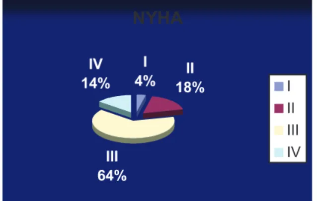
The group consisted of 11 men (mean age 65; 51-81 years) and 16 women (mean age 58; 30-84 years). Of the 27 patients, 64% were NYHA (New York Heart Association) functional class III (figure 3).
Figure 3: NYHA Functional
NYHA I 4% 18%II III 64% IV 14% I II III IV
24 The patients had a wide spectrum of disease associated with PH (table 2). According to the Dana point classification 2008: 37% had pulmonary arterial hypertension (Dana 1), 33% had chronic thromboembolic pulmonary hypertension (Dana 4), 15% had left heart disease (Dana 2), and 11% had hypertension due to lung diseases and/or hypoxia (Dana 3). One patient had haemangiomatosis (Dana 1’). Mean heart rate on ECG-gated-CT was 77 bpm, median 80 bpm, range 40-100 bpm.
Table 2. Clinical classification of pulmonary hypertension (Dana Point, 2008)
1 Pulmonary arterial hypertension (PAH)
1.1 Idiopathic 1.4.1 Connective tissue diseases
1.4.3 Portal hypertension
10 8 1 1 1’ Pulmonary veno-occlusive disease and/or pulmonary capillary haemangiomatosis 1 2 Pulmonary hypertension due to left heart disease
2.1 Systolic dysfunction 2.2 Diastolic dysfunction 2.3 Valvular disease 4 2 1 1
3 Pulmonary hypertension due to lung diseases and/or hypoxia
3.1 Chronic obstructive pulmonary disease 3.2 Interstitial lung disease
3.3 Other pulmonary diseases with mixed restrictive and obstructive pattern
3 1 1 1
4 Chronic thromboembolic pulmonary hypertension 9
Right Heart Catheterization (table 3)
For the study group mPAP was 48mmHg ±13.5 SD, median was 45.5 mmHg, range: 26-80 mmHg. The mean CO was 4408 mL/mn, ± 1.7 SD, range: 1500-9600mL/mn, median: 4090mL/mn.
Table 3. RHC measurements: means, range, median and standard deviation
Mean Range Median SD
mPAP 48 mmHg 26-80 mmHg 45.5mmHg 13.5
25
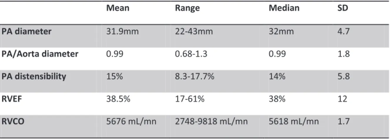
ECG-gated-CT (table 4)
Means range, median and SD of PA diameter, PA/aorta diameter, PA distensibility, FEVD and CO are summarized in table 4.
Table 4. ECG-gated-CT measurements: means, range, median and standard deviation
Mean Range Median SD
PA diameter 31.9mm 22-43mm 32mm 4.7
PA/Aorta diameter 0.99 0.68-1.3 0.99 1.8
PA distensibility 15% 8.3-17.7% 14% 5.8
RVEF 38.5% 17-61% 38% 12
RVCO 5676 mL/mn 2748-9818 mL/mn 5618 mL/mn 1.7
Interobserver Agreement for measurements (table 5)
The two observers showed good agreement for all parameters. The highest agreement was obtained for the PA/aorta diameter (r=0.966) and for the PA diameter (r=0.960). CO measured with Simpson’s technique has the lowest coefficient correlation (r=0.627).
Agreement for CO was higher using the segmentation technique (r=0.910) than with the Simpson technique (r=0.627).
Main pulmonary artery and aorta: correlation with mPAP on RHC (table 5)
Main PA diameter and PA/aorta diameter ratio demonstrated significant positive correlation with mPAP (respectively r=0.537, p<0.01 and r=0.409, p= 0.034) (figure 4a and 4b).
Mean PA distensibility was 15%, ranging from 8.3% to 17.7% (SD: 5.8%). Regarding the PA distensibility, we showed a significant (p=0.027) and linear correlation (r=-0.426) with mPAP (figure 4c).
RVEF: correlation with mPAP in RHC (table 5)
Inverse correlation between RVEF and mPAP was found with the Simpson technique (r=-0.417, p=0.034) whereas absent with RVEF issued from the automatic segmentation (r=-0.353, p=0.091) (figure 4d and 4e).
26
RVEF: correlation with CO in RHC (table 5)
We did not demonstrate significant correlation between RVEF measured neither by the Simpson technique (r=0.242, p=0.224) nor by the segmentation technique (r=0.242, p=0.245) and the CO measured on RHC.
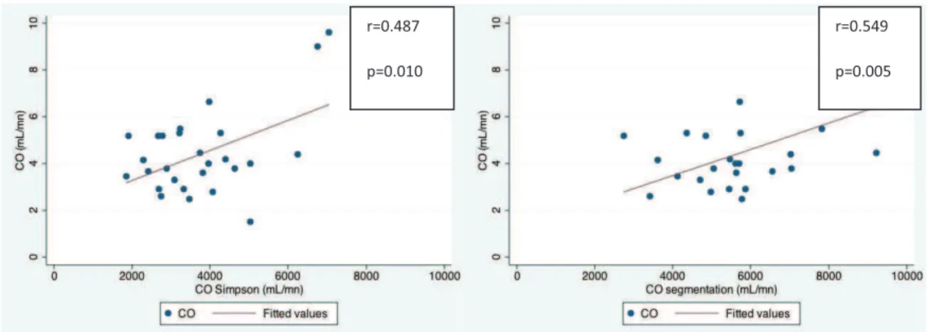
RVCO: correlation with CO in RHC (table 5)
RVCO measured with ECG-gated-CT and CO measured with RHC showed excellent correlation, either when issued from Simpson technique (r=0.487, p=0.010) or from automatic segmentation technique (r=0.549, p=0.005) (figure 4f and 4g).
RVEF, RVCO: correlation between two techniques (figure 5)
Good agreement was found between both methods. Considering RVEF and RVCO, values measured with Simpson techniques are smaller than the ones measured with the automatic segmentation technique.
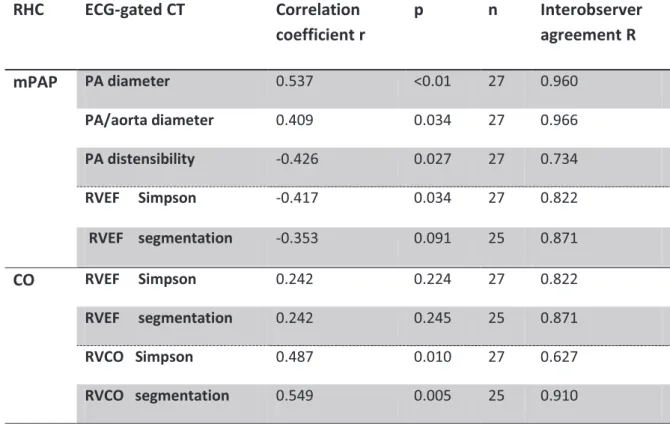
Table 5. Relationship between MDCT signs and RHC measurements
RHC ECG-gated CT Correlation coefficient r p n Interobserver agreement R mPAP PA diameter 0.537 <0.01 27 0.960 PA/aorta diameter 0.409 0.034 27 0.966 PA distensibility -0.426 0.027 27 0.734 RVEF Simpson -0.417 0.034 27 0.822 RVEF segmentation -0.353 0.091 25 0.871 CO RVEF Simpson 0.242 0.224 27 0.822 RVEF segmentation 0.242 0.245 25 0.871 RVCO Simpson 0.487 0.010 27 0.627 RVCO segmentation 0.549 0.005 25 0.910
27
Figure 4. Relationship between MDCT signs (x axis) and RHC measurements (y axis)
a. PA diameter (mm)-mPAP(mmHg) b.PA/aorta diameter-mPAP (mmHg)
c. PA distensibility (%)-mPAP (mmHg)
d. RVEF Simpson (%) - mPAP (mmHg) e. RVEF segmentation (%) - mPAP (mmHg)
r=0.537 p<0.01 r=0.409 p=0.034 r=-0.417 p=0.034 r=-0.426 p=0.027 r=-0.353 p=0.091
28 f. RVCO Simpson (mL/mn)- CO (mL/mn) g. RVCO segmentation (mL/mn)- CO (mL/mn)
Figure 5. Means of CT log transformed measurements between segmentation and Simpson techniques plotted against their differences according to Bland and Altman analysis. a:
RVEF, b: RVCO. Solid lines: mean difference, dashed lines: mean difference plus/minus. All graphs show a good agreement between both methods.
r=0.549 p=0.005 r=0.487
29
Discussion
In PH, we found that CO can be correctly assessed by ECG-gated-CT, and a clear correlation between mPAP and PA distensibility.
The presence of right ventricle dysfunction is a marker for adverse clinical outcome in patients with PH 18. RHC being an invasive test, there is a potential role for a non-invasive exploration to identify the poor prognosis factors due to decrease CO and to follow-up patients under treatment. Echocardiography may frequently be inaccurate in estimating mean pulmonary arterial pressure (mPAP) or cardiac output (CO) and misclassify the severity of the disease 5. Indeed, the up-to-date recommendations are to practice a RHC for any change in treatment 2.
Non ECG-gated-CT is routinely performed on patient with PH to clarify the clinical group and specify the etiology within its group 1, mainly throughout the study of lung parenchyma and pulmonary artery tree. Functional evaluation is however limited. More recently introduced ECG-gated-CT offers anatomic information about the pulmonary arteries and the lung parenchyma as well as functional parameters, thanks to the retrospective reconstruction of cardiac images at different phases of the cardiac cycle. In the present work, we studied morphological and functional parameters obtained in a population with PH and investigated with RHC and ECG-gated-CT.
RVEF and CO
RV function and RVEF estimate the ability of the right heart to compensate or not the consequences of PH. RVEF is the most commonly used and accepted index of RV contractility, but it depends on loading conditions and does not always adequately reflect RV contractility 19.
Several authors have validated the use of ECG-gated-CT, in comparison to echocardiography
16, scintigraphy 20 and MRI 21 for assessing right ventricular function despite the fact that CT
is not the technique of choice because it requires using iodinated contrast agent injection and delivers radiation. However, evaluation of RV function is accurate and CT provides information on the adjacent lung parenchyma.
On ECG-gated-CT, mean normal RVEF is around 60% 22. RVEF under 40% is an important mortality predictor in moderate congestive heart failure 23. In our study, mean RVEF was 37.5%, showing the severity of our patients’ population.
Simon et al. 24 using RHC and ECG-gated-CT compared two groups of patients with PH: one with compensated and one with decompensate RV function. In the decompensate group mPAP was significantly higher, RVEF and RVCO were lower than in the compensate group. In our study, we found a moderate correlation between RVEF and mPAP only with the Simpson’s technique.
30 Moreover, RVEF is linked to heart rate. In few patients, heart rate was elevated; the small errors calculated on the automatic segmentation were even more accentuated by the number of heartbeat.
In PH, increased RV afterload causes RV failure with a decreased RV stroke volume and consequently reduced CO. Low stroke volume is associated with an increase mortality 25 and low CO at diagnosis is linked to a decreased survival 26. Some studies showed that an improved CO after treatment is associated with an improved survival 27. CO is important in staging and follow-up under treatment as it has been proved in MRI.
We studied CO in ECG-gated-CT and RHC and found a statistically significant link between the results of the two techniques. In our small population with a large spectrum of underlying disorders, ECG-gated-CT appears to be powerful in detection of patient with decreased CO.
Simpson vs. automatic segmentation
Differences between automatic and manual tracing are close to differences due to inter-observer reproducibility. Coche et al. 28 studied RVEF using a semi-manual and an automatic segmentation algorithm similar to ours. RV analysis with automatic segmentation was successful in 86% of their patients. Reproducibility of measurement was perfect (R=1) for automatic and good for semi manual. Results from the automatic method were significantly the same compared to the semi automatic for the RVEF. They concluded that the automatic segmentation algorithm enable highly reproducible global heart function to be rapidly obtained.
Several studies have pointed out the advantages of a fully automated method compared to Simpson’s technique for RVEF measurements: better accuracy and time savings 29. Muhlenbruch et al. 30 spared 44.2% of time processing when using automated segmentation vs Simpson’s technique.
In our study, RV segmentation was acceptable for at least twenty-five out of twenty-seven patients (92.6%), and a good correlation was found for RVEF and CO between Simpson and automatic methods. To assess RVCO, using automated RV segmentation instead of Simpson’s technique resulted in better interobserver measurement reproducibility (R=0.910 vs 0.627) and better correlation to RHC CO (p=0.005 vs. 0.010).
31
PA distensibility
Distensibility of right PA has been studied previously on ECG-gated-CT and correlated to RHC. Revel et al. 17 showed a good interobserver agreement (R>0.8) and a significant correlation between distensibility of right PA and mPAP (r=-0.79, p<0.001). These authors concluded that the right PA distensibility had a good diagnosis value for PH and could be useful for risk stratification. Using a cut-off value of 16.5%, they found 86% sensitivity and 96% specificity. Whereas they studied pulmonary distensibility on right PA, we measured it on main PA. In our study we also found a good inverted linear correlation between main PA distensibility and severity of PH (r=-0.426, p=0.027). Seventeen of our twenty-seven patients (63%) with proved PH had a PA distensibility below the 16.5% cut-off value proposed in the study of Revel et al 17.
MRI was used to study PA distensibility which was found lower of 8% in patients with PH as compared to a reference population 31. We did not correlate our results to a reference population; the aim of our study was to assess the correlation of distensibility with the severity of PH.
PA diameter, PA/aorta diameter
The simplest way to estimate the mPAP using CT is the evaluation of the diameter of the main PA. Kuriyama et al. 32 founded a cut-off value at 29 mm. In our study, there was a good correlation between main PA diameter and mPAP (p<0.01, r=0.537).
In chronic pulmonary embolism, PA diameter and ratio PA/aorta has proved a good correlation with mPAP, respectively r=0.42, p<0.001 and r=0.48, p<0.001 33. In patients with PH, the ratio PA/aorta diameter was well (p<0.05) to strongly (p<0.001) correlated with mPAP. The coefficient correlation (r=0.45) was similar to the one we found. The presence of a ratio greater than 1 was correlated to a very high probability of PH 34.
In our study, PA diameter and ratio PA/aorta diameter showed excellent interobserver measurement reproducibility. PA diameter being dependent on cardiac phase, ECG-gated-CT can probably explain our excellent reproducibility and strong correlation for PA diameter.
Limitations
Limitations to this study include its retrospective nature, the few patients and the heterogeneity of the population. Nevertheless, our population reflects the type of patients who undergo RHC in clinical practice in a PH referential center 1.
Larger groups of patient are required to assess the prognostic significance of functional CT findings and to weigh their clinical value. It would be useful to test in a broader number of patients, even more after changes in therapeutics strategies.
32 We also acknowledge that the time interval between ECG-gated-CT and RHC was heterogeneous and sometimes too long. Despites this gap, we found strong correlations because our patients did not change treatment during the study period.
Radiation dose is still a limitation for the use of ECG-gated-CT. In our study, the mean dose-length product was 1177 mGy.cm which was in the range of retrospective ECG-gated-CT using a 64 channel single source detector 35. However, low dose ECG-gated-CT can be obtained in routine clinical practice with 64-slice CT technology without altering the diagnostic value 36. The use of ECG-based tube current modulation does not preclude RVEF calculation and allows a significant reduction in radiation dose 37.
Enhanced temporal resolution in 64-slice CT improves the quantification of volumes enabling reliable assessment of volumes even at increased heart rates. The accuracy of RV function analysis is dependent on the level of contrast medium attenuation achieved in the inferior RV and there should be at least intermediate attenuation to enable accurate functional analysis. We have to keep in mind that, at low attenuation levels, RV function parameters are prone to underestimation 38.
However, clinical outcome studies are required to assess the additional value of ECG-synchronization techniques and for weighing the advantages against possible increased radiation burden.
In conclusion, our study has shown that CO and RVEF could be obtained by a simple, accurate, non-invasive, not time-consuming and accessible method. The automatic segmentation in ECG-gated-CT appears to be useful for identifying patient with decreased CO, as it is known to be linked to poor outcome and one of the main prognostic values in PH. This technique could be useful for evaluation of PH severity and potentially following-up under treatment.
33
References
1. Simonneau G, Robbins IM, Beghetti M, et al. Updated clinical classification of pulmonary hypertension. J Am Coll Cardiol 2009;54(1 Suppl):S43-54.
2. Galie N, Hoeper MM, Humbert M, et al. Guidelines for the diagnosis and treatment of pulmonary hypertension. Eur Respir J 2009;34(6):1219-63.
3. Badesch DB, Champion HC, Sanchez MA, et al. Diagnosis and assessment of pulmonary arterial hypertension. J Am Coll Cardiol 2009;54(1 Suppl):S55-66.
4. Borges AC, Knebel F, Eddicks S, et al. Right ventricular function assessed by two-dimensional strain and tissue Doppler echocardiography in patients with pulmonary arterial hypertension and effect of vasodilator therapy. Am J Cardiol 2006;98(4):530-4.
5. Fisher MR, Forfia PR, Chamera E, et al. Accuracy of Doppler echocardiography in the hemodynamic assessment of pulmonary hypertension. Am J Respir Crit Care Med 2009;179(7):615-21.
6. Hoeper MM, Lee SH, Voswinckel R, et al. Complications of right heart catheterization procedures in patients with pulmonary hypertension in experienced centers. J Am Coll Cardiol 2006;48(12):2546-52.
7. Alunni JP, Degano B, Arnaud C, et al. Cardiac MRI in pulmonary artery hypertension: correlations between morphological and functional parameters and invasive measurements. Eur Radiol;20(5):1149-59.
8. Geva T, Powell AJ, Crawford EC, Chung T, Colan SD. Evaluation of regional differences in right ventricular systolic function by acoustic quantification echocardiography and cine magnetic resonance imaging. Circulation 1998;98(4):339-45.
9. Sanz J, Kuschnir P, Rius T, et al. Pulmonary arterial hypertension: noninvasive detection with phase-contrast MR imaging. Radiology 2007;243(1):70-9.
10. Gan CT, Lankhaar JW, Westerhof N, et al. Noninvasively assessed pulmonary artery stiffness predicts mortality in pulmonary arterial hypertension. Chest 2007;132(6):1906-12.
11. McLaughlin VV, Archer SL, Badesch DB, et al. ACCF/AHA 2009 expert consensus document on pulmonary hypertension a report of the American College of Cardiology Foundation Task Force on Expert Consensus Documents and the American Heart Association developed in collaboration with the American College of Chest Physicians; American Thoracic Society, Inc.; and the Pulmonary Hypertension Association. J Am Coll Cardiol 2009;53(17):1573-619.
12. Hoeper MM. Definition, classification, and epidemiology of pulmonary arterial hypertension. Semin Respir Crit Care Med 2009;30(4):369-75.
13. Bruzzi JF, Remy-Jardin M, Delhaye D, Teisseire A, Khalil C, Remy J. When, why, and how to examine the heart during thoracic CT: Part 2, clinical applications. AJR Am J Roentgenol 2006;186(2):333-41.
14. Remy-Jardin M, Hachulla AL, Pontana F, Faivre JB, Remy J. [CT features of right heart involvement in thoracic diseases]. J Radiol 2009;90(11 Pt 2):1819-29.
15. Lembcke A, Dohmen PM, Dewey M, et al. Multislice computed tomography for preoperative evaluation of right ventricular volumes and function: comparison with magnetic resonance imaging. Ann Thorac Surg 2005;79(4):1344-51.
16. Dogan H, Kroft LJ, Bax JJ, et al. MDCT assessment of right ventricular systolic function. AJR Am J Roentgenol 2006;186(6 Suppl 2):S366-70.
17. Revel MP, Faivre JB, Remy-Jardin M, Delannoy-Deken V, Duhamel A, Remy J. Pulmonary hypertension: ECG-gated 64-section CT angiographic evaluation of new functional parameters as diagnostic criteria. Radiology 2009;250(2):558-66.
18. Gaine SP, Rubin LJ. Primary pulmonary hypertension. Lancet 1998;352(9129):719-25.
19. Dupont MV, Dragean CA, Coche EE. Right ventricle function assessment by MDCT. AJR Am J Roentgenol;196(1):77-86.
20. Delhaye D, Remy-Jardin M, Teisseire A, et al. MDCT of right ventricular function: comparison of right ventricular ejection fraction estimation and equilibrium radionuclide ventriculography, part 1. AJR Am J Roentgenol 2006;187(6):1597-604.
34 21. Guo YK, Gao HL, Zhang XC, Wang QL, Yang ZG, Ma ES. Accuracy and reproducibility of assessing right ventricular function with 64-section multi-detector row CT: comparison with magnetic resonance imaging. Int J Cardiol;139(3):254-62.
22. Lin FY, Devereux RB, Roman MJ, et al. Cardiac chamber volumes, function, and mass as determined by 64-multidetector row computed tomography: mean values among healthy adults free of hypertension and obesity. JACC Cardiovasc Imaging 2008;1(6):782-6.
23. de Groote P, Millaire A, Foucher-Hossein C, et al. Right ventricular ejection fraction is an independent predictor of survival in patients with moderate heart failure. J Am Coll Cardiol 1998;32(4):948-54.
24. Simon MA, Deible C, Mathier MA, et al. Phenotyping the right ventricle in patients with pulmonary hypertension. Clin Transl Sci 2009;2(4):294-9.
25. van Wolferen SA, Marcus JT, Boonstra A, et al. Prognostic value of right ventricular mass, volume, and function in idiopathic pulmonary arterial hypertension. Eur Heart J 2007;28(10):1250-7. 26. Sandoval J, Bauerle O, Palomar A, et al. Survival in primary pulmonary hypertension. Validation of a prognostic equation. Circulation 1994;89(4):1733-44.
27. Sitbon O, Humbert M, Nunes H, et al. Long-term intravenous epoprostenol infusion in primary pulmonary hypertension: prognostic factors and survival. J Am Coll Cardiol 2002;40(4):780-8. 28. Coche E, Walker MJ, Zech F, de Crombrugghe R, Vlassenbroek A. Quantitative right and left ventricular functional analysis during gated whole-chest MDCT: A feasibility study comparing automatic segmentation to semi-manual contouring. Eur J Radiol 2009.
29. Plumhans C, Muhlenbruch G, Rapaee A, et al. Assessment of global right ventricular function on 64-MDCT compared with MRI. AJR Am J Roentgenol 2008;190(5):1358-61.
30. Muhlenbruch G, Das M, Hohl C, et al. Global left ventricular function in cardiac CT. Evaluation of an automated 3D region-growing segmentation algorithm. Eur Radiol 2006;16(5):1117-23.
31. Bogren HG, Klipstein RH, Mohiaddin RH, et al. Pulmonary artery distensibility and blood flow patterns: a magnetic resonance study of normal subjects and of patients with pulmonary arterial hypertension. Am Heart J 1989;118(5 Pt 1):990-9.
32. Kuriyama K, Gamsu G, Stern RG, Cann CE, Herfkens RJ, Brundage BH. CT-determined pulmonary artery diameters in predicting pulmonary hypertension. Invest Radiol 1984;19(1):16-22. 33. Heinrich M, Uder M, Tscholl D, Grgic A, Kramann B, Schafers HJ. CT scan findings in chronic thromboembolic pulmonary hypertension: predictors of hemodynamic improvement after pulmonary thromboendarterectomy. Chest 2005;127(5):1606-13.
34. Ng CS, Wells AU, Padley SP. A CT sign of chronic pulmonary arterial hypertension: the ratio of main pulmonary artery to aortic diameter. J Thorac Imaging 1999;14(4):270-8.
35. Huda W, Rowlett WT, Schoepf UJ. Radiation dose at cardiac computed tomography: facts and fiction. J Thorac Imaging;25(3):204-12.
36. d'Agostino AG, Remy-Jardin M, Khalil C, et al. Low-dose ECG-gated 64-slices helical CT angiography of the chest: evaluation of image quality in 105 patients. Eur Radiol 2006;16(10):2137-46.
37. Salem R, Remy-Jardin M, Delhaye D, et al. Integrated cardio-thoracic imaging with ECG-Gated 64-slice multidetector-row CT: initial findings in 133 patients. Eur Radiol 2006;16(9):1973-81.
38. Aho MR, Gebregziabher M, Schoepf UJ, et al. Impact of right ventricular contrast attenuation on the accuracy of right ventricular function analysis at cardiac multi-detector-row CT. Eur J Radiol;73(3):560-5.
35
Artère pulmonaire et ventricule droit dans l’hypertension pulmonaire : corrélation entre la TDM synchronisée à l’ECG et le cathétérisme cardiaque droit
Thèse soutenue par Elodie ABEL
CONCLUSION
L’hypertension pulmonaire est une pathologie rare mais associée à une morbi-mortalité sévère. Une série d’examens complémentaires permet de confirmer son diagnostic, clarifier son groupe clinique et spécifier son étiologie. L’importance d’une évaluation précise et peu agressive de la fonction cardiaque droite dans le dépistage, le diagnostic, l’évaluation de la sévérité, le suivi et la réponse thérapeutique est cruciale.
Chez les patients atteints d’hypertension pulmonaire, le cathétérisme cardiaque droit est l’examen clé pour l’étude de la fonction cardiaque, avec la mesure de la pression artérielle pulmonaire moyenne (PAPm) et du débit cardiaque. L’échocardiographie et l’IRM ont déjà fait leurs preuves pour une évaluation non invasive, mais avec des limites.
Selon les recommandations internationales, la TDM thoracique fait partie du bilan initial et du suivi. L’amélioration des techniques scannographiques associant augmentation des résolutions spatiale et temporelle et diminution des doses d’irradiation permet l’ajout de la synchronisation à l’ECG sans risque supplémentaire pour les patients. Nous avons formulé l’hypothèse que la TDM synchronisée à l’ECG pouvait apporter un bénéfice pour l’exploration des patients présentant une hypertension pulmonaire, grâce à l’étude de la fonction cardiaque droite.
Notre étude rétrospective inclut 27 patients consécutifs atteints d’hypertension pulmonaire prouvée par cathétérisme (PAPm : 48mmHg ± 13.5) et ayant bénéficié d’une TDM synchronisée à l’ECG. Nous avons identifié par TDM synchronisée à l’ECG les paramètres morphologiques tels que la distensibilité de l’artère pulmonaire et fonctionnels tels que la fraction d’éjection du ventricule droit (FEVD) et le débit cardiaque comme marqueur de sévérité et pronostic chez les patients atteints d’hypertension pulmonaire. De plus nous avons étudié deux techniques de segmentations cardiaques : automatique et selon Simpson.
36 De bonnes corrélations interobservateurs sont retrouvées pour la quasi-totalité des critères TDM mesurés par les deux experts. Nous avons démontré une corrélation linéaire significative entre la diminution de la distensibilité de l’artère pulmonaire (AP) et l’augmentation de la PAPm (r=-0.426, p=0.027). La distensibilité moyenne de l’AP de notre population est de 15%. Selon la technique de Simpson, la diminution de la FEVD est corrélée avec l’augmentation de la PAPm (r=-0.417, p=0.034). Selon la technique de segmentation automatique, aucune résultat significatif n’est mis en évidence (r=-0.353, p=0.09). Enfin, les débits cardiaques droits mesurés par nos deux techniques ont une excellente corrélation avec les débits cardiaques mesurés par cathétérisme pour la segmentation automatique (r=0.549, p=0.005) et pour la technique de Simpson (r=0.487, p=0.01). En ce qui concerne la comparaison des deux techniques de segmentation, nous avons retrouvé une meilleure corrélation avec la méthode de segmentation automatique par rapport à la méthode de Simpson.
En conclusion, notre étude a prouvé que la FEVD et le débit cardiaque pouvaient être calculés à partir d’un examen TDM synchronisé à l’ECG par une méthode simple, précise, non invasive, peu chronophage et accessible. La segmentation automatique en TDM synchronisée à l’ECG apparait utile afin d’identifier les patients au débit cardiaque abaissé, facteur de mauvais pronostic et de survie diminuée dans l’hypertension pulmonaire. Cette technique pourrait s’avérer utile au suivi sous traitement. Des études conduites avec des cohortes plus importantes sont nécessaires.
37
Serment médical
d’Hippocrate
En
présence des Maîtres de cette école, de mes chers condisciples et
devant l’effigie d’Hippocrate, je promets et je jure d’être fidèle aux
lois de l’honneur et de la probité dans l'exercice de la Médecine.
Je donnerai mes soins gratuitement à l’indigent, et n'exigerai jamais
un salaire au-dessus de mon travail. Je ne participerai à aucun
partage clandestin d'honoraires.
Admis dans l'intimité des maisons, mes yeux n'y verront pas ce qui s'y
passe; ma langue taira les secrets qui me sont confiés et mon état ne
servira pas à corrompre les mœurs ni à favoriser le crime.
Je ne permettrai pas que des considérations de religion, de nation, de
race, de parti ou de classe sociale viennent s’interposer entre mon
devoir et mon patient.
Je garderai le respect absolu de la vie humaine. Même sous la
menace, je n'admettrai pas de faire usage de mes connaissances
médicales contre les lois de l'humanité.
Respectueux et reconnaissant envers mes Maîtres, je rendrai à leurs
enfants l'instruction que j’ai reçue de leur père.
Que les hommes m'accordent leur estime si je suis fidèle à mes
promesses.
Que je sois couvert d'opprobre et méprisé de mes confrères si j’y
manque.
1
D
I
‘
B
signes scannographiques morphologiques et fonctionnels
HTAP
Évaluation des signes scannographiques morphologiques et
fonctionnels Hypertension Pulmonaire
Responsable scientifique : Pr Gilbert FERRETTI, PU PH
Clinique Universitaire de Radiologie Imagerie Médicale CURIM
CHU de Grenoble, BP 217 38043 GRENOBLE Cedex 09 Tel : 04.76.76.52.41 Fax : 04.76.76.59.01
Scientifiques Associés :
Dr JANKOWSKI Adrien, PH, CURIM, CHU de Grenoble
Pr BOSSON Jean Luc, PU PH, Département Méthodologie Information Santé Pole Santé Publique, CHU Grenoble
Pr PISON Christophe, PU PH, Clinique de pneumologie, CHU de Grenoble Dr BOUVAIST Hélène, PH, Clinique de cardiologie, CHU de Grenoble
2 RÉSUMÉ
Titre :
Évaluation des signes scannographiques morphologiques et
fonctionnels
HTAP
Responsable du projet : Pr FERRETTI Gilbert CURIM
CHU de Grenoble, BP 217, 38043 GRENOBLE Cedex 09 Tel : 04.76.76.52.41 Fax : 04.76.76.59.01
Objectif principal :
Etudier la corrélation entre :
- de nouveaux critères fonctionnels issus du ECG: débits
- HTAP mesuré par la méthode de référence : le cathétérisme cardiaque.
Objectifs secondaires :
- Déterminer la pertinence signes scannographiques pour évaluer le degré
HTAP VD .
- Déterminer la pertinence des signes scannographiques pour aluation du
HTAP .
Méthodologie : Etude observationnelle, rétrospective
C inclusion : patients atteints HTAP confirmée par cathétérisme cardiaque droit
et ayant bénéficié un angioscanner thoracique ECG
Nombre de patients : 27
D 22 mois
Mots clés : Hypertension artérielle pulmonaire, angioscanner thoracique synchronisé à