Abnormal Hypothalamic Response to Light in Seasonal
Affective Disorder
Gilles Vandewalle, Marc Hébert, Catherine Beaulieu, Laurence Richard, Véronique Daneault,
Marie-Lou Garon, Jean Leblanc, Didier Grandjean, Pierre Maquet, Sophie Schwartz, Marie Dumont,
Julien Doyon, and Julie Carrier
Background: Vulnerability to the reduction in natural light associated with fall/winter is generally accepted as the main trigger of seasonal affective disorder (SAD), whereas light therapy is a treatment of choice of the disorder. However, the relationship between exposure to light and mood regulation remains unclear. As compared with green light, blue light was shown to acutely modulate emotion brain processing in healthy individuals. Here, we investigated the impact of light on emotion brain processing in patients with SAD and healthy control subjects and its relationship with retinal light sensitivity.
Methods: Fourteen symptomatic untreated patients with SAD (34.5⫾ 8.2 years; 9 women) and 16 healthy control subjects (32.3 ⫾ 7.7 years; 11 women) performed an auditory emotional task in functional magnetic resonance imaging during the fall/winter season, while being exposed to alternating blue and green monochromatic light. Scotopic and photopic retinal light sensitivities were then evaluated with electroretinography.
Results: Blue light enhanced responses to auditory emotional stimuli in the posterior hypothalamus in patients with SAD, whereas green light decreased these responses. These effects of blue and green light were not observed in healthy control subjects, despite similar retinal sensitivity in SAD and control subjects.
Conclusions: These results point to the posterior hypothalamus as the neurobiological substrate involved in specific aspects of SAD, including a distinctive response to light and altered emotional responses.
Key Words: Emotion, fMRI, hypothalamus, light, melanopsin, mood, seasonal affective disorder
W
inter seasonal affective disorder (SAD) is a recurrent major depressive disorder occurring in fall/winter with full re-mission in spring/summer (1– 4). Patients with SAD tend to report typical depression complaints such as decreased mood and motivation but also atypical symptoms such as hypersomnia and fatigue, and hyperphagia (particularly for carbohydrates) asso-ciated with weight gain, which implies alteration in sleep/wake regulation (5,6) and possibly in metabolism (7). Despite substantial research efforts, the pathophysiology of the disorder is not estab-lished. Vulnerability to day-length shortening associated with fall/ winter is generally accepted as the main triggering factor of the disorder. Indeed, SAD prevalence varies with latitude (to reach up to 3% in Canada and possibly even up to 10% at higher latitude [2,4]), and light therapy is a treatment of choice for the disorder, with symptom improvements observed within a few weeks of daily (generally morning) light exposures (8). However, the mechanism linking exposure to light and mood regulation is still largely un-known.Retinal light sensitivity was recently reported to be abnormal in patients with SAD during the depressive episode, with normaliza-tion of retinal funcnormaliza-tion after 4 weeks of light therapy in winter (9). Light also regulates circadian rhythms (10) and acutely affects many processes other than vision, such as melatonin secretion, alertness, sleep, performance, and cognition (11–14). These effects of light are mediated by a photoreception system, which recruits intrinsically photosensitive retinal ganglion cells (ipRGCs) expressing the pho-topigment melanopsin (15,16), in addition to rods and cones (17). These melanopsin ipRGCs present a maximal sensitivity to blue light (460 – 480 nm) and confer a shorter wavelength maximal sen-sitivity to nonvisual responses to light, as compared with the pho-topic visual system, which is maximally sensitive to green light (at around 550 nm) (17).
Seasonal changes in the spectral composition of light occur, with relatively less blue light in winter (18), and recent data showed that blue light therapy is effective to treat SAD (19 –22). In addition, blue light therapy requires light levels significantly lower than the recommended 10,000 lux of white light, suggesting that nonclassic photoreception and melanopsin-expressing ipRGCs contribute to the therapeutic effects of light exposure. Functional magnetic res-onance imaging (fMRI) studies in healthy individuals showed that exposure to blue monochromatic light, as compared with green monochromatic light, exerts an acute influence on cerebral activa-tions associated with the processing of auditory emotional stimuli, notably in the hypothalamus and amygdala (23). Because these brain areas—involved in emotional processing—are also impli-cated in mood regulation and mood disorders (23), this acute effect of light could also be involved in the long-term regulation of mood by light, possibly through melanopsin-based photoreception. Win-ter depression in SAD could thus be caused by some abnormal influence of light (or lack of light) on brain responses to emotionally relevant signals.
Here, we studied the acute impact of light on auditory emotional processing in SAD and investigated the role of classical and non-From the Functional Neuroimaging Unit (GV, LR, VD, JD, JC), University of
Montréal Geriatric Institute; Psychology Department (GV, CB, LR, VD, JD, JC), Ecole d’optométrie (M-LG), Psychiatry Department (JL, MD), Univer-sité de Montréal, Montréal; Centre de recherche UniverUniver-sité Laval Robert-Giffard (MH), Québec, Québec, Canada; Geneva Center for Neuroscience and Swiss Center for Affective Sciences (DG, SS), University of Geneva, Geneva, Switzerland; and the Cyclotron Research Centre (PM), University of Liège, Liège, Belgium.
Address correspondence to Gilles Vandewalle, Ph.D., Centre de Recherche de l’Institut Universitaire de Gériatrie de Montréal, Functional Neuroim-aging Unit, 4565 Queen Mary, Montréal, Québec, H3W 1W5 Canada; E-mail:[email protected].
Received Mar 9, 2011; revised Jun 20, 2011; accepted Jun 20, 2011.
BIOL PSYCHIATRY 2011;xx:xxx 0006-3223/$36.00
classical photoreception in the disorder. We measured retinal light sensitivity and examined the effect of blue and green light expo-sures on the brain responses to neutral and emotional auditory stimuli in untreated symptomatic patients with SAD and healthy control subjects in fall/winter. We hypothesized that, during the symptomatic episode of SAD, the impact of light exposure on audi-tory emotional processing would be abnormal, in key brain areas for emotion regulation, such as the amygdala and hypothalamus. We also hypothesized that retinal dysfunction in SAD, which alters the light signal reaching the brain, would be related to the influence of light on these emotional responses.
Methods and Materials
More details can be found in Supplement 1. Subjects
Patients with SAD and control subjects were recruited in the Montreal area (approximate latitude 45°30’N). They were between 18 and 45 years of age (Table 1for complete characteristics) and gave written informed consent. The study was approved by the institutional Regroupement Neuroimagerie/Québec Ethics Com-mittee.
Patients. Candidates had to present a Global Seasonality Score ⱖ 9 on the Seasonal Pattern Assessment Questionnaire (24), per-ceive seasonal changes as at least a “moderate” problem, and pres-ent a scoreⱖ11 on the Beck Depression Inventory II (25). A psychol-ogist then determined the presence of SAD symptoms on the basis of the Structured Interview Guide for the Hamilton Depression Rating Scale, Seasonal Affective Disorder version (SIGHSAD) (total scoreⱖ25; atypical item score ⱖ9) (26). The psychologist also ex-cluded bipolarity (with the Mood Disorder Questionnaire [27]) or other psychiatric or medical disorders (with a semistructured inter-view). Sixteen patients completed the protocol, but two were ex-cluded for technical reasons. All patients participating in the exper-iment were studied in the absence of any treatment (light therapy or drugs).
Control Subjects. Control subjects were matched with pa-tients for gender, laterality, and age (⫾2 years except for one female and one male pair, for which the difference was 4 and 6 years, respectively). Control subjects presented a Global Seasonality Score ⬍8, with “no problem” with seasonal changes, and a semistruc-tured interview established the absence of a medical or psychiatric disorder. No sleep disturbances were reported as determined by the Pittsburgh Sleep Quality Index Questionnaire (28) (scoreⱕ5).
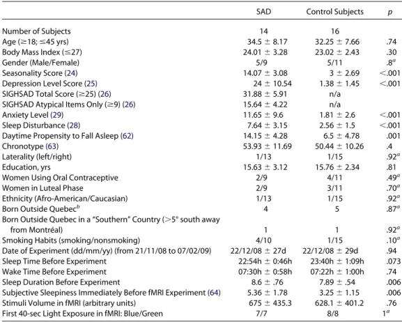
Table 1. Subjects Characteristics
SAD Control Subjects p
Number of Subjects 14 16
Age (ⱖ18; ⱕ45 yrs) 34.5⫾ 8.17 32.25⫾ 7.66 .74
Body Mass Index (ⱕ27) 24.01⫾ 3.28 23.02⫾ 2.43 .30
Gender (Male/Female) 5/9 5/11 .8a
Seasonality Score (24) 14.07⫾ 3.08 3⫾ 2.69 ⬍.001
Depression Level Score (25) 24⫾ 10.54 1.38⫾ 1.45 ⬍.001
SIGHSAD Total Score (ⱖ25) (26) 31.88⫾ 5.91 n/a
SIGHSAD Atypical Items Only (ⱖ9) (26) 15.64⫾ 4.22 n/a
Anxiety Level (29) 11.65⫾ 9.6 1.81⫾ 2.6 ⬍.001
Sleep Disturbance (28) 7.64⫾ 3.15 2.56⫾ 1.5 ⬍.001
Daytime Propensity to Fall Asleep (62) 14.15⫾ 4.28 6.5⫾ 4.78 .001
Chronotype (63) 53.93⫾ 11.69 50.44⫾ 10.26 .4
Laterality (left/right) 1/13 1/15 .92a
Education, yrs 15.63⫾ 3.12 15.76⫾ 2.34 .81
Women Using Oral Contraceptive 2/9 4/11 .49a
Women in Luteal Phase 2/9 3/11 .70a
Ethnicity (Afro-American/Caucasian) 1/13 1/15 .92a
Born Outside Quebecb 4 5 .87a
Born Outside Quebec in a “Southern” Country (⬎5° south away
from Montréal) 1 1 .92a
Smoking Habits (smoking/nonsmoking) 4/10 1/15 .10a
Date of Experiment (dd/mm/yy) (from 21/11/08 to 07/02/09) 22/12/08⫾ 27d 22/12/08⫾ 29d .94
Sleep Time Before Experiment 22:54h⫾ 0:46h 23:40h⫾ 1:09h .073
Wake Time Before Experiment 07:30h⫾ 0:58h 07:22h⫾ 1:00h .74
Sleep Duration Before Experiment 8.6⫾ .76 7.89⫾ .54 .006
Subjective Sleepiness Immediately Before fMRI Experiment (64) 5.36⫾ 1.78 3.25⫾ 1.15 .006
Stimuli Volume in fMRI (arbitrary units) 675⫾ 435.3 628.1⫾ 401.2 .76
First 40-sec Light Exposure in fMRI: Blue/Green 7/7 8/8 1a
Values are mean⫾ SD. None of the characteristics of the subjects showing a significant difference between seasonal affective disorder (SAD) and control subjects alone explained the reported differences in functional magnetic resonance imaging (fMRI) activations, as indicated by regression analyses in SPM5. Therefore the present result cannot be attributed to a single clinical symptom such as levels of depression or anxiety, seasonality, or sleep/wake distur-bances.
SIGHSAD, Structured Interview Guide for the Hamilton Depression Rating Scale—Seasonal Affective Disorder Version.
ap values computed with2test, otherwise with unpaired t test.
All showed normal scores on the 21-item Beck Anxiety Inventory (29) and Beck Depression Inventory II (25) (scores⬍11).
Participants. All participants were moderate alcohol consum-ers (⬍7 alcohol unit/week) and were not taking medication. They were asked to refrain from alcohol-containing beverages for at least 36 hours before the experiment. Smokers were included, but smok-ing was not allowed for the duration of the laboratory experimen-tations. None had worked on night shifts during the preceding year or traveled across more than one time zone during the last 2 months. All subjects had been living in the province of Quebec for at least 3 years. Women were not pregnant or breast-feeding and were more than 1 year postpartum. Absence of ophthalmic disor-der (e.g., glaucoma, color blindness) was assessed by an optome-trist (standard examination). Participants completed additional questionnaires, but the scores of these questionnaires were not used as inclusion criteria (Table 1).
At least 1 week before the experiment, participants were famil-iarized with the magnetic resonance environment during a short MRI session during which a structural image of the brain was ac-quired. Volunteers were asked to follow a regular sleep schedule based on their preferred sleep times and durations during the 5 days preceding the experimentation. Compliance was verified with sleep logs and actigraphy (Actiwatch-L; MiniMitter/Respironics, Bend, Oregon).
Experimental Protocol
Participants arrived at the laboratory 2 hours after habitual wake time and were maintained in dim light (⬍5 lux) for 1.5 hours (Figure 1A). One drop of tropicamide 1% was administered in each eye 20 min before entering the scanner to inhibit pupillary constriction. During the fMRI session (12 min), subjects performed an emotional auditory task while being exposed to alternating 40-sec periods of blue (480 nm) and green (550 nm) monochromatic lights (full width
at half maximum [FWHM]: 10 nm), separated by 15–25-sec periods of darkness (30) (order blue and green light was counter-balanced across subject within each group). The fMRI session was followed by photopic and scotopic electroretinogram (ERG) recordings charac-terizing cone and rod photoreception, respectively.
Technical Issue. In accordance with our previous studies (31,32) and work of others (e.g., [12,13]), we set the photon densities of both monochromatic lights used in fMRI at an equal level, so that comparisons between blue and green exposures could reveal non-classic modulation of brain responses. The irradiance used (1013
photon/cm2/sec) was intermediate between the two irradiances of our prior investigation of the impact of light on emotion processing (30) and had successfully been used in another study on the impact of light on auditory working memory (32). A technical problem, however, accidentally set blue and green light irradiance levels at 1.1⫻ 1013
and .9 ⫻ 1013
photons/cm2
/sec, respectively (which corresponds to 1.5 and 20 lux, respectively). This affected all data acquisitions and prevented direct comparisons between blue and green exposures but did not compromise comparisons between patients and control subjects for blue and green light separately.
fMRI Task. Acoustic stimuli consisted of three meaningless words (“goster,” “niuvenci,” “figotleich”) pronounced by profes-sional actors (half women) with two different modalities, anger and neutral prosody, as validated by extensive behavioral assessments (33) and in previous experiments (30,34,35). Note that negative and positive emotions are mediated through common (but not com-pletely identical) pathways (36), but our experience is that negative emotion elicits more robust responses, less influenced by individual valence perception (37). Stimuli were presented to the subject via headphones from an audio player. The task of the subject was to press one of two buttons on a keypad (with their right hand) upon discriminating the gender of the speaker pronouncing the pseudo-word. The goal of the study to measure brain responses to emo-tional words was hidden from the subjects. Stimuli were matched in term of duration (750 msec) and mean acoustic energy. Anger and neutral prosodies were evenly assigned to each light condition (blue, green, darkness).
fMRI Acquisitions. The fMRI data were acquired with a 3-T magnetic resonance scanner (TIM-TRIO, Siemens, Erlangen, Ger-many). Multislice T2*-weighted fMRI images were obtained with a gradient echo-planar sequence (32 axial slices; voxel size: 3.4⫻ 3.4⫻ 3 mm3
with 30% of gap; matrix size 64⫻ 64 ⫻ 32; repetition time⫽ 2180 msec; echo time ⫽ 40 msec; flip angle ⫽ 90°). Structural brain images consisted of a T1-weighted 3D magnetization prepared rapid gradient echo (MP-RAGE) sequence (repetition time⫽ 7.92 msec, echo time⫽ 2.4 msec, time of inversion ⫽ 910 msec, flip angle⫽ 15°, field of view ⫽ 256 ⫻ 224 mm2
, matrix size⫽ 256 ⫻ 224, voxel size⫽ 1 ⫻ 1 ⫻ 1 mm3
).
ERG Acquisitions. The ERG recordings were undertaken 4.5 hours after habitual wake time, after the fMRI session. One drop of tropicamide 1% was administered in each eye again 15 min before the first ERG. Recordings were obtained with DTL electrodes (Shiel-dex 33/9 Thread, Bremen, Germany) placed deep in the conjuncti-val sac, with reference electrodes placed on the canthi and ground on the forehead (38). Flash stimulations were administered with a Ganzfeld Dome (ColorDome, Diagnosys, Lowell, Massachusetts) to achieve full field retinal stimulation. Participants were first adapted to a background light (25.5 cd/m2
) for 15 min before being admin-istered a series of white light flashes of increasing intensity (range: ⫺1.12–1.375 log cd/m2
/sec; stimuli interval: 1–5 sec) to generate a photopic luminance response. Participants were then dark-adapted for 30 min (0 lux) before being presented with a series of light flashes (480 nm broadband blue light to better stimulate rods, Figure 1. Experimental design. (A) General protocol. Arrow: pupil dilator
administration. Time relative to scheduled wake time (hours). Subject per-formed an emotional task in functional magnetic resonance imaging (fMRI) (see B for details) before photopic and scotopic electroretinograms were recorded. (B) Detailed fMRI procedures. Time (seconds) relative to t0, a time point arbitrarily chosen as a green light onset of the session. The task consisted of a gender discrimination of auditory vocalizations while ex-posed to alternating blue (480 nm) and green (550 nm) monochromatic light (counterbalanced order). Light exposures lasted 40 sec and were sep-arated by 15–25-sec periods of darkness. Anger (red bars) and neutral (white bars) prosody vocalizations of the three pseudo-word type (“goster,” “niu-venci,” or “figotleich”) were pseudorandomly and evenly administered throughout each light condition across the entire session (interstimuli inter-val: 3–11 sec; mean: 4.8 sec).
which present peak sensitivity at around 505 nm) of increasing intensity (range:⫺4.25 to ⫺1.00 log cd/m2
/sec; stimuli intervals: 1.5-sec low-intensity; 5-sec high-intensity), to generate a scotopic luminance response.
Data Analysis
Behavior. Behavioral data were analyzed with Statistica 6.1 (StatSoft France, Maisons-Alfort, France). Mixed analyses of vari-ance with group as the between-subjects factor (SAD, control sub-jects) and prosody (neutral, anger) as the within-subject factor were used to compare reaction times and accuracy on the fMRI task.
fMRI. Brain functional volumes were analyzed with Statistical Parametric Mapping software (SPM5,http://www.fil.ion.ucl.ac.uk/ spm). They were realigned, coregistered, spatially normalized (Montreal Neurological Institute space; standard SPM5 parame-ters), and smoothed (FWHM: 8 mm). The analysis was conducted in two steps, accounting for individual-level fixed effects and group-level random effects, respectively. Changes in regional brain re-sponses were estimated with a general linear model in which emo-tional and neutral stimuli in each light condition; blue and green light onset and offset were modeled with stick functions (“events”) convolved with a canonical hemodynamic response function. A parametric modulation was added to each regressor to track any linear change of the amplitude of brain responses across time. Regressors derived from the realignment of functional volumes were considered as covariates of no interest. High-pass filtering was implemented in the matrix design with a cutoff period of 256 sec to remove low-frequency drifts from the time series. Serial correla-tions in the fMRI signal were estimated with an autoregressive (order 1) plus white noise model and a restricted maximum likeli-hood algorithm.
The summary statistic images resulting from the contrasts of interest were further smoothed (FWHM: 6 mm) and entered in the random effects analyses. This second level analyses consisted of two-sample t test on independent measures with unequal variance, which constituted maps of the t statistics thresholded at puncorrected⫽
.001. One-sample t tests were also computed to identify whether the observed effect was significant in each population separately. Statistical inferences were performed after correction for multiple comparisons at a threshold of pcorrected⫽ .05. Corrections for
multi-ple comparisons were computed on the entire brain volume (Fam-ily Wise Error) or on small spherical volumes around a priori loca-tions of activation (10 mm radius), which were expected in structures involved in the processing of emotional auditory stimuli (34,35), in arousal regulation (39,40), in the impact of light on non-visual brain function (30 –32,41), or in brain areas to which the melanopsin-expressing ipRGC project (42,43). Multiple regression analyses were carried out with questionnaire scores (Table 1) with standard SPM5 procedure.
ERG. One control subject did not complete the photopic and scotopic ERG assessment, because of technical problems, and pho-topic ERG data of another control subject were accidentally not recorded. Log K, which is the intensity necessary to reach half of the saturating amplitude of the ERG b-wave and constitutes a measure of retinal sensitivity (9,38), was computed for scotopic and photopic data of all the other subjects with sigmoidal curve fitting (Prism 4, GraphPad, La Jolla, California). Two-sample t tests compared sco-topic and phosco-topic LogK.
Results
Demographic Data
As expected, patients with SAD presented high SIGHSAD scores and were significantly more seasonal, anxious, and depressed than
control subjects (Table 1). SAD patients reported feeling sleepier than control subjects during the day in general and presented significantly more sleep disturbances. Sleep duration and subjec-tive sleepiness immediately before the experiment were also signif-icantly higher in SAD patients. By contrast, possible confounds such as age, body mass index, education level, and chronotype did not differ significantly between groups. Wake time before the experi-ment and the date of the experiexperi-ment were also similar in both groups.
Performance of the fMRI Task
Accuracy in the gender discrimination task was high (⬎ 90%) in both light conditions but tended to be higher for neutral than anger prosody [mean⫾ SD; blue: neutral (97.8 ⫾ 3.6%) ⬎ anger (95.4 ⫾ 5.7%), F (1,28)⫽ 3.94, p ⫽ .057; green: neutral (97.3 ⫾ 5.1%) ⬎ anger (94.1⫾ 7.1%), F(1,28) ⫽ 3.36; p ⫽ .077] (Figure 2A). In both light conditions, reaction times were significantly slower for anger than neutral prosody [mean⫾ SD; blue: neutral (1034 ⫾ 211 msec) ⬍ anger (1101⫾ 195 msec), F(1,28) ⫽ 7.22, p ⫽ .012; green: neutral (1022⫾ 210 msec) ⬍ anger (1101 ⫾ 204 msec), F(1,28) ⫽ 9.63, p ⫽ .004] (Figure 2B). Critically, accuracy and reaction times did not differ between patients and control subjects [F (1,28)⬍ 2.5, p ⬎ .12] with no group⫻ prosody interactions [F(1,28) ⬍ 2.2, p ⬎ .14].
These results indicate that the emotional content of the stimuli was equally well perceived by patients and control subjects, pre-venting behavior bias in the fMRI analyses comparing both groups. fMRI Results
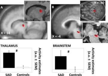
The clinical manifestation of a mood disorder (i.e., the depres-sive episode) alters normal brain function (23). Therefore, before investigating how blue and green light modulate brain responses to neutral or angry prosody stimuli, we assessed whether brain responsiveness to all stimuli differed between patients and control subjects (i.e., irrespective of the light and prosody conditions). We found that, as compared with control subjects, patients with SAD presented increased responses to all auditory stimuli in a dorso-posterior area of the thalamus compatible with the dorsal pulvinar and a dorsal area of brainstem located next to superior cerebellar peduncle and encompassing several nuclei of the ascending arous-ing system (Figure 3,Table 2). Multiple regression analyses showed Figure 2. Behavioral results of the functional magnetic resonance imaging
(fMRI) task. (A) Accuracy (mean⫾ SD); (B) reaction times (mean ⫾ SD). *Significant differences (pⱕ .05); nonsignificant difference (p ⬎ .05). ANG, anger prosody; CON, control subjects; NEU, neutral prosody; SAD, seasonal affective disorder patient.
that the group differences in thalamic and brainstem responsive-ness were not related to single characteristic of subjects that dif-fered between SAD and control participants (cf.Table 1).
Brain responses to auditory stimuli under blue or green light exposures were compared with brain responses in darkness to take into account the global difference in brain responsiveness and allow for group comparisons. Analyses revealed that, as compared
with darkness, blue light exposure increased responses to angry prosody stimuli in the posterior hypothalamus, dorsolateral to mammillary bodies, in patients with SAD (Figure 4,Table 2). In contrast, under green light exposure, responses to these emotional stimuli were decreased in a slightly more ventral hypothalamic area Figure 3. Significant differences between patients with seasonal affective
disorder (SAD) and healthy control subjects (Controls) in the brain re-sponses to all auditory stimulus types (irrespective of light and prosody condition). (A) Thalamus (dorsal and posterior); (B) brainstem (median-posterior, next to superior cerebellar peduncle). Results are overlaid over the mean structural image of all subjects. Insets: enlargements in represen-tative subjects. Graphs: activity estimates (arbitrary unit [a.u.]⫾ SEM) of the brain responses to all auditory stimulus types. #pcorrectedⱕ .05 (group
differ-ence).
Table 2. fMRI Results
Brain Areas Side X Y Z Z Score p
All Stimuli Types (irrespective of the light and prosody conditions) SAD⬎ Control Subjects
Thalamusa R 14⫺18 4 3.61 .010
Brainstema L ⫺2 ⫺28 ⫺22 3.58 .011
Control Subjects⬎ SAD No significant voxel Anger Prosody Stimuli
[Blue⬎ Dark] ⫻ [SAD ⬎ Control Subjects]
Hypothalamusbc L ⫺2 ⫺2 ⫺12 3.21 .027
[Blue⬎ Dark] ⫻ [Control Subjects ⬎ SAD] No significant voxel
[Green⬎ Dark] ⫻ [SAD ⬎ Control Subjects] No significant voxel
[Green⬎ Dark] ⫻ [Control Subjects ⬎ SAD]
Hypothalamuscd L ⫺4 ⫺2 ⫺18 3.13 .032
XYZ: relative coordinates (mm) in Montreal Neurological Institute space.
fMRI, functional magnetic resonance imaging; L, left; R, right; SAD, seasonal affective disorder.
aThe same two significant clusters of voxel are obtained in the thalamus and brainstem if the analyses only included: 1) all stimuli in darkness; 2) all stimuli
under green light exposure; 3) all stimuli under blue light exposure; 4) emotional stimuli under blue or green light exposure; 5) neutral stimuli under blue or green light exposure (i.e., these differences between groups are observed for every stimuli subgroup).
bClusters not affected by an exclusive mask (p⫽ .05) of the (Neutral ⫻ [Blue ⬎ Dark] ⫻ [SAD ⬎ Control Subjects]) contrast, indicating that the light
condition effect was specific to the emotional (angry prosody) stimuli.
cBecause of the difference in irradiance level between blue and green light (see technical issue in Methods and Materials), the contrast computing the
interaction between blue and green light conditions [(Blue⬎ Green)] ⫻ [SAD ⬎ Control Subjects] is not valid. However, if this contrast is nevertheless computed, it shows a single significant difference in the hypothalamus (⫺2 0 ⫺18 mm; Z ⫽ 3.73; psvc⫽ .006) which further strengthens the results obtained
for each light condition separately.
dClusters not affected by an exclusive mask (p⫽ .05) of the (Neutral ⫻ [Green ⬎ Dark] ⫻ [SAD ⬎ Control Subjects]) contrast, indicating that the light
condition effect was specific to the emotional (angry prosody) stimuli.
Figure 4. Significant differences between patients with SAD and Controls in
the impact of blue and green light exposure on the brain responses to auditory emotional stimuli. Results are overlaid over the mean structural image of all subjects. Inset: enlargement in a representative subject. Graphs: change in activity estimates (a.u.⫾ SEM) between the light condition (blue, green) and the darkness condition for the processing of auditory emotional stimuli. *pcorrectedⱕ .05 (in SAD patients taken in isolation); ⬃pcorrected⫽ .07
(in SAD patients taken in isolation); nonsignificant difference puncorrected⬎ .1
(in control subjects taken in isolation); #pcorrectedⱕ .05 (group difference). B,
brainstem; CP, corpus collusum; F, fornix; MB, mammillary bodies; OT, optic tract; SC, superior colliculus; T, thalamus; 3V, third ventricle; other abbrevia-tions as inFigures 1and3.
in patients with SAD. Importantly, these effects of blue and green light were not observed in control subjects and were significantly different between patients and control subjects. Again, multiple regression analyses showed that these results were not significantly related to single characteristics of subjects that differed between groups (cf.Table 1). Finally, no impact of light wavelength on the processing of neutral auditory stimuli was found in either group, demonstrating the specificity of the effects for emotional stimuli and for the patients with SAD.
ERG Results
Scotopic and photopic light sensitivity, as indicated by LogK, did not differ between patients and control subjects [mean⫾ SD; Pho-topic LogK: patients (.108⫾ .093), control subjects (.108 ⫾ .102), t (26)⫽ .008, p ⫽ .99; Scotopic LogK: patients (⫺2.72 ⫾ .12), control subjects (⫺2.75 ⫾ .11), t (27) ⫽ .66, p ⫽ .51].
Discussion
These results demonstrate that exposure to light has an acute impact on emotional brain processing in untreated symptomatic patients with SAD in fall/winter and that this impact depends on light spectral composition; blue light increased and green light decreased responses to auditory emotional stimuli in the posterior hypothalamus, as compared with healthy control subjects. This study also reveals that, in the context of our protocol, SAD patients presented increased thalamic and brainstem responsiveness to vo-cal stimuli, regardless of their emotional content and of the light condition.
Compared with control subjects, patients showed higher tha-lamic activation to auditory stimuli in the dorsal pulvinar and in regions of the brainstem compatible with the locus coeruleus and dorsal raphe nucleus (although fMRI spatial resolution does not allow identification of specific brainstem nuclei). The locus coer-uleus and dorsal raphe nucleus are implicated in reward regulation and depression (44) and constitute an important source of norepi-nephrine and serotonin, respectively. Interestingly, serotonin levels seem to be influenced by season and bright sunlight (45), and altered serotonin receptor functions have been described in SAD (2,3,46). Animal data also showed that complete light deprivation reduced noradrenergic projections from the locus coeruleus to the prefrontal cortex (47), which is essential for cognition (48). In addi-tion, metabolic and serotoninergic dysfunction in the pulvinar has been related to depression (49). Therefore, the differential respon-siveness to vocal stimuli could constitute a marker of a general increased sensitivity in SAD during the fall/winter depressive epi-sode, speculatively related to serotonin and norepinephrine func-tions.
Emotional processing was affected by blue and green light in a single area of the brain—taking into account baseline differences between groups (i.e., responses under blue or green light were compared with darkness)—pointing to light-induced variation in hypothalamic reactivity specific to SAD, at least during the fall/ winter symptomatic episode. These effects were not observed with neutral stimuli, showing their specificity for the processing of emo-tional stimuli. In other words, they were not caused by an overall change in brain reactivity throughout the 40-sec light exposure. We showed that, in healthy individuals and as compared with green light, blue light exposure increased the functional connectivity be-tween the amygdala, temporal cortex voice-sensitive area, and a hypothalamic area located in the vicinity of the present significant hypothalamic cluster (30). Dysfunction in hypothalamus-related functions is typically observed in SAD, as indicated by changes in sleep, feeding, metabolism, and motivation (2,3,5–7). One plausible
implication of our findings is that exposure to light participates in the long-term normalization of these hypothalamic functions and leads to remission. However, on the basis of our protocol, we can-not determine whether it is the case or whether these abnormal hypothalamic responses to light constitute a trait-marker trigger-ing the disorder when light availability declines or a state-marker secondary to other phenomenon.
Importantly, the data showed no performance differences be-tween groups, which ensures that our results are not due to behav-ioral differences during data acquisition (e.g., differences in task difficulty). Moreover, both populations did not differ for many other possible confounds, such as age, gender, education level, wake time, or dates of experiments (cf. Table 1). As expected, however, SAD patients differed from control subjects for several aspects typ-ically related to their pathology, such as daytime sleepiness, anxiety and depression levels, sleep duration, and seasonality. Although several of these factors are likely to have contributed to our results, none of them was identified by regression analyses as significantly contributing to the results on their own.
The spatial resolution of fMRI does not allow determination of which hypothalamic nucleus was specifically affected by light, but a number of posterior hypothalamic nuclei receive retinal projec-tions directly, or indirectly through the suprachiasmatic nucleus (17,50). Some of them, such as the hypocretin/melanin-concentrat-ing hormone postero-lateral hypothalamus, are involved in the regulation of sleep, wakefulness, motivation and metabolism. Through their numerous projections, hypocretin/melanin-concen-trating hormone neurons regulate activity in nuclei of the ascend-ing arousal system of the brainstem, includascend-ing the locus coeruleus and dorsal raphe nucleus, and in the thalamus (51). Likewise, light of various wavelengths could also affect the processing of emotional stimuli in the paraventricular nucleus of the hypothalamus, which is involved in emotional responses (52) and vegetative regula-tion (53).
Scotopic (rod-dependent) light sensitivity did not differ be-tween patients and control subjects, which contrasts with our pre-dictions on the basis of previous observations of lower rod sensitiv-ity in SAD (9,54,55). This discrepancy cannot be attributed to the sample of patients, because SIGHSAD scores, depression, and sea-sonality levels were similar to those reported in previous studies on SAD (e.g., [19,46]), including those investigating retinal sensitivity (9,54). It should be noted, however, that it is the seasonal change in rod retinal sensitivity that has been most reported to be abnormal in SAD, whereas differences between patients and control subjects were not systematically detected in fall/winter (54 –56). With regard to cone function, only one study so far reported decreased function in symptomatic patients with SAD in fall/winter (9), a result that was not observed in the current study. Only short-term light history (preceding hours) was closely controlled in the present protocol. We cannot therefore exclude that longer-term light history influ-enced our ERG results (57). However, there seems to be no indica-tion in the literature for difference in light history between SAD patients and healthy control subjects (58). In spite of this, our results suggest that abnormal rod or cone function cannot account for the altered hypothalamic responses observed in SAD under blue and green light exposures.
The irradiance level we used is compatible with the recruitment of melanopsin-expressing ipRGCs (59), and a polymorphism in the melanopsin gene has been linked to SAD (60). However, all photo-receptors are likely to have contributed (17), especially given the results obtained with green light, and further research is warranted to identify how each photoreceptor participates in the influence of light on emotional brain processing in patients and healthy
individ-uals. Nonetheless, our results support that the wavelength of light is an important factor for light therapy as well as for optimal indoor lighting, particularly for individuals more vulnerable to seasonal light variation, such as SAD patients, but also for an important part of the population, namely subsyndromal SAD sufferers (up to 18% of the North American general population), who experience inter-mediate seasonal emotional, mood, and vigilance problems that— although bothersome— do not reach clinical significance (61).
The acute impact of light on emotional brain responses might not be related to its long-term impact on mood regulation. How-ever, emotions and mood are intimately related. Mood alteration in mood disorders modify emotional brain responses, whereas emo-tional responses can greatly influence (subsequent) mood (23). Furthermore, although the impact of light on emotional processing might differ between negative and positive stimuli, common brain pathways respond to emotional stimuli, regardless of emotional valence direction (36), supporting that similar effects of light likely take place for positive emotions.
As a whole, the results provide experimental evidence for a central role of the hypothalamus in the seasonal-light-decline sen-sitivity present in SAD. Abnormal light responsiveness in the poste-rior hypothalamus constitutes a neurobiological substrate of SAD during the fall/winter depressive episode that could trigger the disorder or, conversely, lead to remission. Future studies should address these questions and compare symptomatic and asymp-tomatic states in the same individuals, in fall/winter, before and after light therapy, and spring/summer.
This study was supported by a grant from the Instituts de Recherche en Santé du Canada (IRSC-CHIR) and fellowships from the Fonds de la Recherche en Santé du Québec (FRSQ) and the Fonds Québecois de la Recherche sur la Nature et les Technologies (FQRNT). We thank N. Brault, O. Collignon, A. Cyr, M. Desrosiers, S. Frenette, C. Hurst, F. Les-age, P. Orban, N. Ouakli, J. Paquet, F. Peters, C. Phillips, and G. Poirier for their help.
The authors reported no biomedical financial interests or potential conflicts of interest.
Supplementary material cited in this article is available online.
1. Rosenthal N, Sack D, Gillin J, Lewy A, Goodwin F, Davenport Y, et al. (1984): Seasonal affective disorder: A description of the syndrome and preliminary findings with light therapy. Arch Gen Psychiatry 41:72– 80. 2. Westrin A, Lam RW (2007): Seasonal affective disorder: A clinical update.
Ann Clin Psychiatry 19:239 –246.
3. Levitan RD (2007): The chronobiology and neurobiology of winter sea-sonal affective disorder. Dialogues Clin Neurosci 9:315–324.
4. Magnusson A, Partonen T (2005): The diagnosis, symptomatology, and epidemiology of seasonal affective disorder. CNS Spectr 10:625– 634. 5. Cajochen C, Brunner DP, Krauchi K, Graw P, Wirz-Justice A (2000): EEG
and subjective sleepiness during extended wakefulness in seasonal affective disorder: Circadian and homeostatic influences. Biol Psychiatry 47:610 – 617.
6. Schwartz PJ, Rosenthal NE, Wehr TA (2001): Band-specific electroen-cephalogram and brain cooling abnormalities during NREM sleep in patients with winter depression. Biol Psychiatry 50:627– 632.
7. Pinchasov BB, Shurgaja AM, Grischin OV, Putilov AA (2000): Mood and energy regulation in seasonal and non-seasonal depression before and after midday treatment with physical exercise or bright light. Psychiatry
Res 94:29 – 42.
8. Terman M, Terman JS (2005): Light therapy for seasonal and nonsea-sonal depression: Efficacy, protocol, safety, and side effects. CNS Spectr 10:647– 663.
9. Lavoie MP, Lam RW, Bouchard G, Sasseville A, Charron MC, Gagne AM, et
al. (2009): Evidence of a biological effect of light therapy on the retina of
patients with seasonal affective disorder. Biol Psychiatry 66:253–258.
10. Duffy JF, Czeisler CA (2009): Effect of light on human circadian physiol-ogy. Sleep Med Clin 4:165–177.
11. Brainard GC, Hanifin JP (2005): Photons, clocks, and consciousness. J Biol
Rhythms 20:314 –325.
12. Lockley SW, Evans EE, Scheer FAJL, Brainard GC, Czeisler CA, Aeschbach D (2006): Short-wavelength sensitivity for the direct effects of light on alertness, vigilance, and the waking electroencephalogram in humans.
Sleep 29:161–168.
13. Cajochen C, Munch M, Kobialka S, Krauchi K, Steiner R, Oelhafen P, et al. (2005): High sensitivity of human melatonin, alertness, thermoregula-tion, and heart rate to short wavelength light. J Clin Endocrinol Metab 90:1311–1316.
14. Vandewalle G, Maquet P, Dijk DJ (2009): Light as a modulator of cogni-tive brain function. Trends Cogn Sci 13:429 – 438.
15. Provencio I, Rodriguez IR, Jiang G, Hayes WP, Moreira EF, Rollag MD (2000): A novel human opsin in the inner retina. J Neurosci 20:600 – 605. 16. Berson DM, Dunn FA, Takao M (2002): Phototransduction by retinal
ganglion cells that set the circadian clock. Science 295:1070 –1073. 17. Hatori M, Panda S (2010): The emerging roles of melanopsin in
behav-ioral adaptation to light. Trends Mol Med 16:435– 446.
18. Thorne HC, Jones KH, Peters SP, Archer SN, Dijk DJ (2009): Daily and seasonal variation in the spectral composition of light exposure in hu-mans. Chronobiol Int 26:854 – 866.
19. Glickman G, Byrne B, Pineda C, Hauck WW, Brainard GC (2006): Light therapy for seasonal affective disorder with blue narrow-band light-emitting diodes (LEDs). Biol Psychiatry 59:502–507.
20. Anderson JL, Glod CA, Dai J, Cao Y, Lockley SW (2009): Lux vs. wave-length in light treatment of seasonal affective disorder. Acta Psychiatr
Scand 120:203–212.
21. Desan PH, Weinstein AJ, Michalak EE, Tam EM, Meesters Y, Ruiter MJ, et
al. (2007): A controlled trial of the Litebook light-emitting diode (LED)
light therapy device for treatment of seasonal affective disorder (SAD).
BMC Psychiatry 7:38.
22. Strong RE, Marchant BK, Reimherr FW, Williams E, Soni P, Mestas R (2009): Narrow-band blue-light treatment of seasonal affective disorder in adults and the influence of additional nonseasonal symptoms.
De-press Anxiety 26:273–278.
23. Price JL, Drevets WC (2010): Neurocircuitry of mood disorders.
Neuro-psychopharmacology 35:192–216.
24. Rosenthal N, Bradt G, Wehr T (1984): Seasonal Pattern Assessment
Ques-tionnaire (SPAQ). Bethesda Maryland: National Institute of Mental
Health.
25. Steer RA, Ball R, Ranieri WF, Beck AT (1997): Further evidence for the construct validity of the Beck depression Inventory-II with psychiatric outpatients. Psychol Rep 80:443– 446.
26. Williams J, Link M, Rosenthal N, Amira L, Terman M (1994): Structured
Interview Guide for the Hamilton Depression Rating Scale—Seasonal Af-fective Disorder Version (SIGH-SAD) (revised edition). New York: New York
State Psychiatric Institute.
27. Hirschfeld RM, Williams JB, Spitzer RL, Calabrese JR, Flynn L, Keck PE, Jr.,
et al. (2000): Development and validation of a screening instrument for
bipolar spectrum disorder: the Mood Disorder Questionnaire. Am J
Psy-chiatry 157:1873–1875.
28. Buysse DJ, Reynolds CF 3rd, Monk TH, Berman SR, Kupfer DJ (1989): The Pittsburgh Sleep Quality Index: A new instrument for psychiatric prac-tice and research. Psychiatry Res 28:193–213.
29. Beck AT, Epstein N, Brown G, Steer RA (1988): An inventory for measur-ing clinical anxiety: Psychometric properties. J Consult Clin Psychol 56: 893– 897.
30. Vandewalle G, Schwartz S, Grandjean D, Wuillaume C, Balteau E, Deg-ueldre C, et al. (2010): Spectral quality of light modulates emotional brain responses in humans. Proc Natl Acad Sci U S A 107:19549 –19554. 31. Vandewalle G, Gais S, Schabus M, Balteau E, Carrier J, Darsaud A, et al.
(2007): Wavelength-dependent modulation of brain responses to a working memory task by daytime light exposure. Cereb Cortex 17:2788 – 2795.
32. Vandewalle G, Schmidt C, Albouy G, Sterpenich V, Darsaud A, Rauchs G,
et al. (2007): Brain responses to violet, blue, and green monochromatic
light exposures in humans: Prominent role of blue light and the brains-tem. PLoS One 2:e1247.
33. Banse R, Scherer KR (1996): Acoustic profiles in vocal emotion expres-sion. J Pers Soc Psychol 70:614 – 636.
34. Grandjean D, Sander D, Pourtois G, Schwartz S, Seghier ML, Scherer KR, et al. (2005): The voices of wrath: Brain responses to angry prosody in meaningless speech. Nat Neurosci 8:145–146.
35. Sander D, Grandjean D, Pourtois G, Schwartz S, Seghier ML, Scherer KR, et al. (2005): Emotion and attention interactions in social cognition: Brain regions involved in processing anger prosody. Neuroimage 28: 848 – 858.
36. Sergerie K, Chochol C, Armony JL (2008): The role of the amygdala in emotional processing: A quantitative meta-analysis of functional neu-roimaging studies. Neurosci Biobehav Rev 32:811– 830.
37. Sterpenich V, Albouy G, Boly M, Vandewalle G, Darsaud A, Balteau E, et al. (2007): Sleep-related hippocampo-cortical interplay during emo-tional memory recollection. PLoS Biol 5:e282.
38. Hébert M, Lachapelle P, Dumont M (1996): Reproducibility of electro-retinograms recorded with DTL electrodes. Doc Ophthalmol 91:333– 342.
39. Portas CM, Rees G, Howseman AM, Josephs O, Turner R, Frith CD (1998): A specific role for the thalamus in mediating the interaction of attention and arousal in humans. J Neurosci 18:8979 – 8989.
40. Schmidt C, Collette F, Leclercq Y, Sterpenich V, Vandewalle G, Berthomier P, et al. (2009): Homeostatic sleep pressure and responses to sustained attention in the suprachiasmatic area. Science 324:516 –519. 41. Vandewalle G, Balteau E, Phillips C, Degueldre C, Moreau V, Sterpenich
V, et al. (2006): Daytime light exposure dynamically enhances brain responses. Curr Biol 16:1616 –1621.
42. Ecker JL, Dumitrescu ON, Wong KY, Alam NM, Chen SK, LeGates T, et al. (2010): Melanopsin-expressing retinal ganglion-cell photoreceptors: Cellular diversity and role in pattern vision. Neuron 67:49 – 60. 43. Brown TM, Gias C, Hatori M, Keding SR, Semo M, Coffey PJ, et al. (2010):
Melanopsin contributions to irradiance coding in the thalamo-cortical visual system. PLoS Biol 8:e1000558.
44. Kranz GS, Kasper S, Lanzenberger R (2010): Reward and the serotonergic system. Neuroscience 166:1023–1035.
45. Lambert GW, Reid C, Kaye DM, Jennings GL, Esler MD (2002): Effect of sunlight and season on serotonin turnover in the brain. Lancet 360: 1840 –1842.
46. Willeit M, Sitte HH, Thierry N, Michalek K, Praschak-Rieder N, Zill P, et al. (2008): Enhanced serotonin transporter function during depression in seasonal affective disorder. Neuropsychopharmacology 33:1503–1513. 47. Gonzalez MM, Aston-Jones G (2008): Light deprivation damages
mono-amine neurons and produces a depressive behavioral phenotype in rats. Proc Natl Acad Sci U S A 105:4898 – 4903.
48. Koechlin E, Hyafil A (2007): Anterior prefrontal function and the limits of human decision-making. Science 318:594 –598.
49. Young KA, Holcomb LA, Bonkale WL, Hicks PB, Yazdani U, German DC (2007): 5HTTLPR polymorphism and enlargement of the pulvinar: un-locking the backdoor to the limbic system. Biol Psychiatry 61:813– 818. 50. Saper CB, Scammell TE, Lu J (2005): Hypothalamic regulation of sleep
and circadian rhythms. Nature 437:1257–1263.
51. Adamantidis A, de Lecea L (2008): Sleep and metabolism: Shared cir-cuits, new connections. Trends Endocrinol Metab 19:362–370. 52. Phelps EA, LeDoux JE (2005): Contributions of the amygdala to emotion
processing: From animal models to human behavior. Neuron 48:175– 187.
53. Kalsbeek A, Palm IF, La Fleur SE, Scheer FA, Perreau-Lenz S, Ruiter M, et al. (2006): SCN outputs and the hypothalamic balance of life. J Biol Rhythms 21:458 – 469.
54. Hebert M, Beattie CW, Tam EM, Yatham LN, Lam RW (2004): Electroreti-nography in patients with winter seasonal affective disorder. Psychiatry Res 127:27–34.
55. Hebert M, Dumont M, Lachapelle P (2002): Electrophysiological evi-dence suggesting a seasonal modulation of retinal sensitivity in subsyn-dromal winter depression. J Affect Disord 68:191–202.
56. Gagne AM, Hebert M (2011): Atypical pattern of rod electroretinogram modulation by recent light history: A possible biomarker of seasonal affective disorder. Psychiatry Res 187:370 –374.
57. Beaulieu C, Rufiange M, Dumont M, Lachapelle P (2009): Modulation of ERG retinal sensitivity parameters with light environment and photope-riod. Doc Ophthalmol 118:89 –99.
58. Dumont M, Beaulieu C (2007): Light exposure in the natural environ-ment: Relevance to mood and sleep disorders. Sleep Med 8:557–565. 59. Lall GS, Revell VL, Momiji H, Al Enezi J, Altimus CM, Guler AD, et al. (2010):
Distinct contributions of rod, cone, and melanopsin photoreceptors to encoding irradiance. Neuron 66:417– 428.
60. Roecklein KA, Rohan KJ, Duncan WC, Rollag MD, Rosenthal NE, Lipsky RH, et al. (2009): A missense variant (P10L) of the melanopsin (OPN4) gene in seasonal affective disorder. J Affect Disord 114:279 –285. 61. Kasper S, Wehr T, Bartko J, Gaist P, Rosenthal N (1989): Epidemiological
findings of seasonal changes in mood and behavior. Arch Gen Psychiatry 46:823– 833.
62. Johns MW (1991): A new method for measuring daytime sleepiness: The Epworth sleepiness scale. Sleep 14:540 –545.
63. Horne JA, Ostberg O (1976): A self-assessment questionnaire to deter-mine morningness-eveningness in human circadian rhythms. Int J Chro-nobiol 4:97–110.
64. Akerstedt T, Gillberg M (1990): Subjective and objective sleepiness in the active individual. Int J Neurosci 52:29 –37.