© Zhifei Li, 2020
Compulsive eating of binge- like eating prone rats under
conditioned fear and exploration of the neural
mechanism with c-fos expression
Thèse
Zhifei Li
Doctorat en médecine moléculaire
Philosophiæ doctor (Ph. D.)
Compulsive eating of binge- like eating prone rats under
conditioned fear and exploration of the neural
mechanism with c-fos expression
Thèse
Zhi Fei Li
Sous la direction de :
ii
Résumé
Le trouble de l'hyperphagie boulimique (THB) est un trouble de l'alimentation défini de manière autonome dans la 5e édition du Manuel diagnostique et statistique des troubles mentaux (DSM-5) en mai 2013. Le THB est caractérisé par des épisodes d'ingestion d'une quantité anormalement élevée de nourriture dans une courte période de temps sans comportements compensatoires tels que des vomissements auto-induits. La prévalence du THB a augmenté très rapidement en raison de la grande disponibilité d'aliments riches en calories et du stress croissant dans la vie moderne. Malheureusement, l'étiologie du THB est encore mal comprise et les traitements cliniques actuels du THB sont principalement limités à la thérapie cognitive comportementale, dont le pronostic est également assez limité. Afin d'étudier le THB, notre laboratoire a précédemment développé un modèle de THB chez le rat en utilisant une combinaison de stress causé par des chocs électriques aux pattes et d'un accès intermittent d'une heure à une solution de sucrose à 10%. Dans notre modèle de rat THB, les rats sujets à la frénésie alimentaire (BEP; de l'anglais binge-eating prone) ont consommé plus de sucrose que les rats résistants à la frénésie alimentaire (BER, de l'anglais binge-eating resistant) à la fois dans des conditions normales et ces rats ont augmenté davantage leur consommation après avoir vécu le stress. Nous avons également observé une alimentation compulsive dans les rats BEP avec notre test modifié de boîte claire / sombre. Une alimentation compulsive est la caractéristique la plus obstinée du THB. Mon projet de thèse se concentre sur l'observation de l'alimentation compulsive dans le modèle de THB chez le rat avec un test concurrentiel, dans lequel les comportements d'alimentation et d'immobilité ont été surveillés en présence d'un stimulus auditif conditionné de manière aversive. Les rats BEP ont montré une consommation persistante élevée de sucrose et ont montré une réponse inhibée à la peur induite en situation stressante en comparaison aux rats BER, indiquant respectivement un déficit de dévaluation de l'appétence et une réponse anxiolytique plus forte au sucrose. Après l'observation de l'alimentation compulsive dans nos rats BEP, nous avons analysé les activités cérébrales de ces rats avec l'hybridation in situ de l'ARNm c-fos. Nous avons trouvé que, dans les rats BEP, le sucrose réduisait l'activité c-fos du noyau paraventriculaire de l'hypothalamus, tout en augmentant l'activité dans la zone hypothalamique latérale face au stimulus conditionné aversif. La résistance à la dévaluation de l'appétence de la nourriture pourrait être le résultat d'un recrutement atténué de la réponse
iii
du cortex préfrontal médian et d'une réponse persistante du noyau accumbens à la consommation de sucrose. Ces résultats suggèrent que le système de récompense a pris le dessus sur les systèmes homéostatiques et répondant au stress. Étonnamment, l'apport de sucrose sous la peur conditionnée n'a pas inhibé l'activité de l'amygdale centrale, mais l'a plutôt activée à la place. Cette étude a exploré le mécanisme de l'alimentation compulsive dans un modèle de THB et a fourni certaines cibles cérébrales, telles que le noyau accumbens, pour de futures recherches thérapeutiques.
iv
Abstract
The binge eating disorder (BED) is an eating disorder that was defined in the 5th edition of Diagnostic and Statistical Manual of Mental Disorders (DSM-5) in May 2013. The BED is characterized by episodes of ingestion of abnormally large amounts of food in a short period of time without compensative behaviors such as self-induced vomiting. The prevalence of the BED is on the rise due to the availability of high-calorie food and the stressors of modern life. Unfortunately, the etiology of the BED is still poorly understood, and current clinical treatments of the BED are mostly limited to cognitive behavioral therapy, of which the prognosis is also quite limited. In order to study the BED, our lab previously developed a rat model of the BED with combination of foot-shock stress and intermittent 1 h access to a 10% sucrose solution. In our BED rat model, the binge-like eating prone rats (BEPs) consumed more sucrose than the binge-like eating resistant rats (BERs) in normal conditions and consumed more sucrose in response to stress. We also observed compulsive eating in the BEPs with our modified light/dark box test. Compulsive eating is the most obstinate feature of the BED. My PhD project focuses on the observation of compulsive eating in the BED rat model with a conflicting test, in which the feeding and freezing behaviors were monitored in the presence of an aversively conditioned auditory stimulus. The BEPs showed persistently high sucrose intake and inhibited fear response under stress when comparted with BERs, respectively indicating a deficiency in palatability devaluation and stronger anxiolytic response to sucrose. After the observation of compulsive eating in the BEPs, we further analyzed the brain activities of the BEPs and BERs by analyzing the expression of c-fos mRNA using in situ hybridization. In the BEPs, we found that sucrose reduced c-fos expression in the paraventricular nucleus of the hypothalamus (PVN) in response to an aversively conditioned stimulus (CS), but enhanced activities in the lateral hypothalamic area (LHA) in response to the CS. The resistance to devaluating the palatable food could be a result of attenuated recruitment of the medial prefrontal cortex (mPFC) and persistent nucleus accumbens (Acb) response to the sucrose intake. These findings suggest that the rewarding system overrode the homeostatic and the stress-responding systems. Surprisingly, the sucrose intake under fear conditions did not inhibit the activity of the central amygdala, but further activated it instead. Current study explored the mechanism of compulsive eating in the BED, and suggests that the mPFC and Acb should be examined for further therapeutic research.
v
Table of Contents
Résumé ...ii
Abstract ...iv
List of figures and tables ...ix
Acknowledgements ... xiii
Foreword ... xiv
Introduction ... 1
1.1 Two forms of food intake regulation ... 1
1.1.1 Homeostatic eating ... 1
1.1.2 Hedonic eating/Eating for reward ... 2
1.2 Endocrine and neuronal signals involved in food intake regulation ... 3
1.2.1 Hunger-related signals ... 3
1.2.1.1 Decreased plasma glucose ... 3
1.2.1.2 Orexin ... 4 1.2.1.3 Ghrelin ... 4 1.2.1.4 Neuropeptide Y (NPY) ... 5 1.2.2 Satiety-related signals: ... 6 1.2.2.1 Insulin ... 6 1.2.2.2 Leptin ... 7 1.2.2.3 Serotonin (5-HT) ... 8 1.2.2.4 Cholecystokinin (CCK) ... 9 1.2.2.5 Oxytocin ... 9
1.2.3 Reward-related signals in hedonic eating ... 10
1.2.3.1 Dopamine (DA) ... 10
1.2.3.2 Opioid ... 10
1.3 Brain circuits involved in food intake regulation ... 11
1.3.1 Brain regions involved in homeostatic eating ... 11
1.3.1.1 The Lateral hypothalamic area (LHA) ... 11
1.3.1.2 The Arcuate nucleus of the hypothalamus (ARC) ... 13
1.3.1.3 The Parabrachial (PBN) ... 14
1.3.2 Brain circuits involved in reward-induced feeding ... 14
1.3.2.1 The Ventral tegmental area (VTA) ... 15
vi
1.4 The binge eating disorder (BED) ... 16
1.4.1 What is BED ... 16
1.4.2 Inducers of the BED ... 18
1.4.2.1 Dieting and limited/ intermittent access to palatable food ... 18
1.4.2.2 Stress ... 18
1.4.2.3 Interaction between dieting and stress ... 19
1.4.3 Hormones and neurotransmitters involved in the development of the BED ... 20
1.4.3.1 Glucocorticoid ... 20 1.4.3.2 Ghrelin ... 21 1.4.3.3 Orexin ... 21 1.4.3.4 Dopamine (DA) ... 22 1.4.3.5 Serotonin (5-TH) ... 23 1.5 Stress response... 24
1.5.1 What is stress response ... 24
1.5.2 Fear conditioning paradigm ... 25
1.5.2.1 What is fear conditioning ... 25
1.5.2.2 Conditioned fear responses ... 26
1.5.3 Fast and slow neural responses to stress ... 27
1.5.4 Brain circuits underlying stress response ... 28
1.5.4.1 Thalamic sensory-related structures ... 28
1.5.4.2 The paraventricular nucleus of the hypothalamus (PVN) ... 29
1.5.4.3 The amygdala ... 30
1.5.4.4 The medial prefrontal cortex (mPFC) ... 31
1.5.4.5 The bed nucleus of stria terminalis (BNST) ... 33
1.5.4.6 The lateral hypothalamic area (LHA) ... 33
1.6 Compulsive eating in spite of aversive consequence ... 34
1.6.1 ‘Food addiction’—a concept under debate ... 34
1.6.2 Compulsive eating in human patient with BED... 35
1.6.3 Impaired decision-making in patients with BED ... 36
1.6.4 Brain regions involved in decision-making ... 37
1.6.4.1 The amygdala ... 37
1.6.4.2 The medial prefrontal cortex (mPFC) ... 38
vii
1.6.4.4 The ventral tegmental area (VTA) ... 40
1.6.5 Interaction between reward system and the HPA axis in the development of the BED ... 41
1.7 Sexual dimorphism in binge eating disorder, stress responses and cue-induced inhibition of feeding behaviors ... 42
1.8 Methods used in this study ... 43
1.8.1 Rate model of binge-like eating ... 43
1.8.2 Monitoring food intake in rats ... 45
1.8.2.1 Feeding behavior in rats... 45
1.8.2.2 Methods of monitoring feeding behavior in animals ... 46
1.8.3 Use of c-fos expression as an indicator of neuronal activity ... 47
1.8.3.1 What is c-fos ... 47
1.8.3.2 Mechanism of c-fos expressing and functioning ... 48
1.8.3.3 Using c-fos expression as an indicator of neuronal activation ... 50
1.9 Hypotheses and Objectives ... 51
1.9.1 Hypotheses and Objectives 1 ... 51
1.9.2 Hypotheses and Objectives 2 ... 52
Chapter 1. Compulsive eating in rat model of binge eating disorder (BED) under conditioned fear and exploration of the neural mechanism with c-fos mRNA expression ... 53
2.1 Key points summary ... 53
2.2 Résumé ... 54
2.3 Abstract ... 55
2.4 Introduction ... 56
2.5 Materials and Methods ... 57
2.5.1 Animals ... 57
2.5.2 Classification of the BEPs and BERs ... 57
2.5.3 Fear-Conditioning test... 58
2.5.3.1 Habituation and Appetitive sessions:... 59
2.5.3.2 Fear Conditioning sessions: ... 59
2.5.3.3 Test session:... 60
2.5.4 Licking microstructure analyses ... 61
2.5.5 Analysis of c-fos mRNA expression as a marker for brain activity... 61
2.5.5.1 Brain preparation ... 61
2.5.5.2 In situ hybridization for c-fos ... 61
viii
2.5.6 Statistical analyses ... 63
2.6 Results... 63
2.6.1 Classification of the BERs and BEPs ... 63
2.6.2 Sucrose intake behavior during the Appetitive and Test sessions ... 63
2.6.3 Freezing behavior during Fear Conditioning sessions and the Test session ... 67
2.6.4 The c-fos mRNA expression ... 68
2.6.4.1 Activation of the amygdala by the CS was not inhibited by sucrose intake ... 69
2.6.4.2 The LHA was activated by the CS ... 69
2.6.4.3 The activation of the PVN by the CS was inhibited by sucrose intake in the BEPs .. 73
2.6.4.4 Persistent Acb response to sucrose intake in the BEPs under conditioned fear ... 75
2.6.4.5 The BNST only responded to the CS in BEPs ... 75
2.6.4.6 The mPFC is less recruited in response to the CS in BEPs with access to sucrose ... 77
2.7 Discussion ... 78
2.8 Additional information ... 85
2.9 Reference ... 85
Discussion and perspectives ... 88
3.1 Possibility of an effect of genetic background in the development of the BED ... 88
3.2 A theory about the mechanisms of stress-induced eating ... 88
3.3 Possible pathways for devaluation of palatable food ... 89
3.4 Each animal model has strengths and weaknesses related to validity and use. ... 90
3.5 Using a single time point of c-fos mRNA ... 91
3.6 Current treatments of the BED... 91
3.7 Outlook: ... 92
Conclusion... 92
Bibliography ... 95
Annex A... 113
ix
List of figures and tables
Figure 1- 1. Homeostatic eating regulating system. ... 1
Figure 1- 2. Brain circuits involved in the regulation of food intake. ... 11
Figure 1- 3.Brain circuits integrating information about adiposity and satiety ... 12
Figure 1- 4. Brain circuits involved in the stress response. ... 29
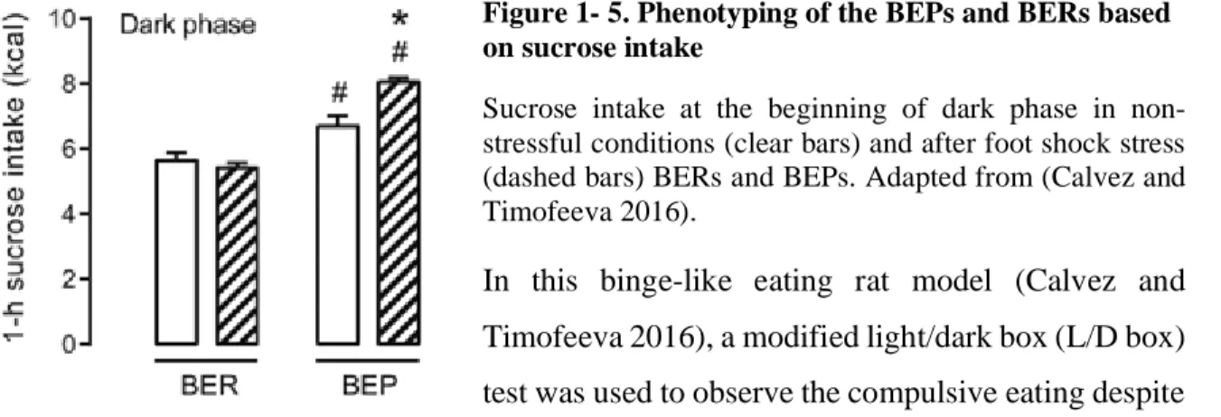
Figure 1- 5. Phenotyping of the BEPs and BERs based on sucrose intake ... 44
Figure 1- 6. Compulsive-like eating behavior in BER and BEP rats. ... 44
Figure 2- 1. Diagram of treatments and groups in the fear conditioning test. ... 60
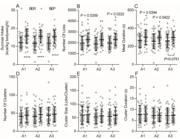
Figure 2- 2. Habitual over-eating of the BEPs during Appetitive sessions. ... 64
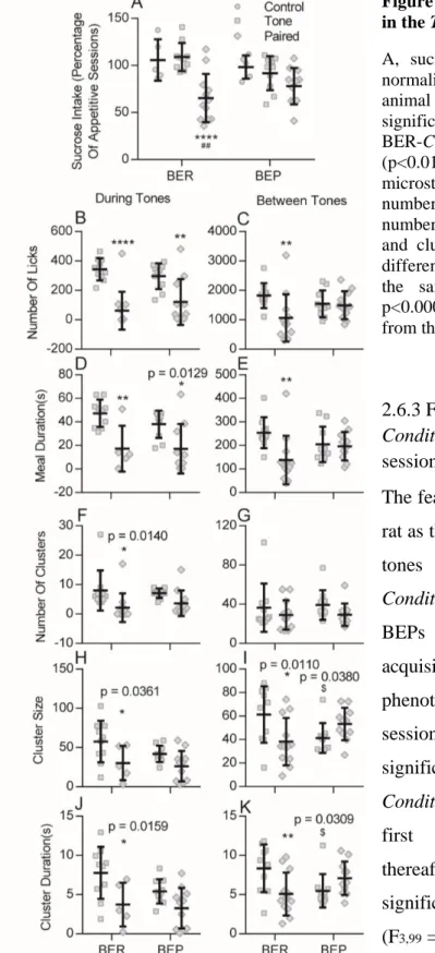
Figure 2- 3. Compulsive eating of the BEPs in the Test session. ... 67
Figure 2- 4. Conditioned fear response of the BERs and BEPs during Fear Conditioning sessions and the Test session... 68
Figure 2- 5. Activation of the amygdala by the CS was not inhibited by sucrose intake ... 70
Figure 2- 6. The LHA was activated by the CS ... 71
Figure 2- 7. The activation of the PVN by the CS was inhibited by sucrose intake in the BEPs .... 74
Figure 2- 8. Persistent Acb response to sucrose intake in the BEPs under conditioned fear ... 76
Figure 2- 9. The BNST only responded to the CS in BEPs ... 77
Figure 2- 10. The mPFC is less recruited in response to the CS in BEPs with access to sucrose .... 79
Table 2- 1. Effects of phenotype, Appetitive session and their interaction on the feeding behavior in Appetitive sessions ... 65
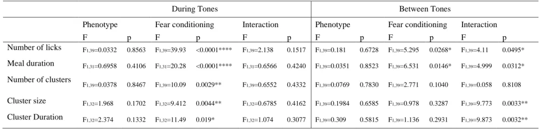
Table 2- 2. Effects of phenotype, fear conditioning and their interaction on the feeding behavior both during tones and between tones in the Test session ... 65
Table 2-3 A Effects of sucrose, fear conditioning and their Interaction on the relative c-fos mRNA expression in related brain regions of all groups ... Error! Bookmark not defined. Table 2-3 B Effects of phenotype, fear conditioning and their Interaction on the relative c-fos mRNA expression in related brain regions of all groups ... Error! Bookmark not defined. Table 2-3 C Effects of phenotype, sucrose and their Interaction on the relative c-fos mRNA expression in related brain regions of Paired groups ... Error! Bookmark not defined. Figure 3- 1. Predatory stress may induce hunger response. ... 89
Figure SD 1. Sucrose intake in non-stressful condition and after foot-shock stress of the BERs and BEPs. ... 113
Figure SD 2. First lick latency. ... 113
x
3V 3rd ventricle
5-HT 5-hydroxytryptamine, serotonin
5-HT1A 5-hydroxytryptamine receptor 1A
5-HT1B 5-hydroxytryptamine receptor 1B
5-HT2C 5-hydroxytryptamine receptor 2C
5-HT4 5-hydroxytryptamine receptor 4
5-HTIAA 5-hydroxyindoleacetic acid
ac anterior commissure
aca anterior part of the anterior commissure
Acb nucleus accumbens
AcbC nucleus accumbens core
AcbSh nucleus accumbens shell
ACh acetylcholine
ACTH adrenocorticotrophic hormone
AgRP agouti-related protein
AMPA a-amino-3hydoxy-5-meghyl-4isoxazolepropionate
AP-1 activating protein complex 1
ARC arcuate nucleus of the hypothalamus
ARCAgRP/NPY AgRP neurons in the ARC
ARCPOMC POMC neurons in the ARC
ARCOXTR Oxytocin receptor expressing neurons in the ARC
BED binge eating disorder
BEP bigne-like eatign prone
BEPs bigne-like eatign prone rats
BER binge-like eating resistant
BERs binge-like eating resistant rats BIS-11 Barratt impulsiveness scale
BMI body mass index
BNST bed nucleus of the stria terminalis
CaMK calcium/calmodulin-dependent protein kinase
Ce central amygdala
CGRP calcitonin gene-related peptide
Cl claustrum
CNS central neverous system
CREB cAMP responsive element binding protein
CRH corticotrophin releasing hormone
CS conditioned stimulus
xi
DA dopamine
DAT dopamine transporter
db diabetes
DNA deoxyribonucleic acid
f fornix
fmi forceps minor of the corpus callosum
GHS-R1A growth hormone secretagogue receptor 1A, a ghrelin receptor HPA axis hypothalamic-pituitary-adrenal axis
IEG immediate early gene
IL infralimbic part of the medial prefrontal cortex
LHA lateral hypothalamic area
LRG late response gene
L-VSCCs L type voltage-sensitive calcium channels
MAPK Ras-mitogen-associated protein kinase
MC4R melanocortin-4 receptor
MEF2 myocyte enhancer factor 2
MG medial geniculate nucleus
mPFC medial prefrontal cortex
mRNA messenger RNA
NMDA N-methyl-D-aspartate
NPY neuropeptide Y
NTS nucleus tractus solitaries
ob obese
ob-R leptin receptor
opt optic tract
PAG periaqueductal gray area
PBN parabrachial nucleus
PBNCGRP PBN CGRP neurons
PeFLH perifornical part of the LHA
PFC prefrontal cortex
PIN posterior intralaminar nuclei
PLH peduncular part of the LHA
POMC proopiomelanocortin
PrL prelimbic part of the medial prefrontal cortex
PV paralvumin
PVN paraventricular nucleus of the hypothalamus
PVNm magnocellulair part of the PVN
xii
PVNp parvocellulair part of the PVN
PVNMC4R MC4R expressing neurons in the PVN
RNA Ribonucleic acid
SERT serotonin transporter
SON supraoptic nucleus
SRF serum response factor
SST somatostatin
STD dorsal part of the BNST
STV ventral part of the BNST
TFs transcription factors
UPPS-P urgency, premeditation, perseverance, sensation seeking, and positive urgency
US unconditioned stimulus
VIP vasoactive intestinal peptide
vlPAG ventrolateral PAG
VMH ventromedial nucleus of the hypothalamus
xiii
Acknowledgements
These four years of PhD study in Laval University, and living in Quebec has been a life-changing experience for me. First of all, I really want to thank my admirable supervisor, Dr. Elena Timofeeva, for giving me the opportunity to study in her lab, for patiently directing me, for encouraging and motivating me and most importantly, for showing me what an excellent scientist and supervisor is like. She was elegant, nice, tolerant, responsible, strong-minded, hard-working etc. It is not exaggerating to use every good word on her. Even if I will not have the ability or opportunity to be a scientist like her, I will try my best to be a good person like her. I am also grateful to my current supervisor, Dr. Igor Timofeev, who took me over, and guided me during the last two years.
I would like to thank my previous and current colleagues in both labs: Dr. Juliane Calvez, Dr. Arojit Mitra, Dr. Geneviève Guèvremont, Dr. Sandrine Chometton, Christoph Lenglos, Richard Quansah Amissah, Yavar Korkian, Camila de Avila, Dr. Sylvain Chauvette, Dr. Josée Seigneur, Sergiu Ftomov, Dr. Olga Bukhtyarova, Dr. Sara Soltani, Diellor Basha, Anastasiia Ozur, Julia Potey. I really enjoyed working and talking with these wonderful people. Specially thanks to Juliane, Arojit and Geneviève for illustrating me the procedures of my protocols, also to Sandrine for revising my figures and manuscript.
I am sincerely grateful to my family, the support from my parents and my sister. Most sincerely thanks to my wife and soulmate, Zhao Xia Li, and my little son, Jason Li. You make my life so beautiful and make me know how deep my love can be.
xiv
Foreword
The manuscript presented in Chapter 1 has not been submitted yet. I am the first author of this manuscript. I was involved in the experiments presented in all figures. The experiment was designed by Dr. Elena Timofeeva, Dr. Juliane Calvez and me. The procedures were conducted by me, with assistance from Geneviève Guèvremont. The manuscript was written by me, and revised by Dr. Sandrine Chometton and Dr. Igor Timofeev. The programs used in the Med Associates system were written by me, the MATLAB programs used to analysis the licking events and optical density of the c-fos mRNA signals were adapted from Christoph Longlos and modified by me to our particular purposes.
The results were obtained using unbiased approaches. Licking events were detected using a lickometer of Med Asociates system, recorded with TDT system, and analyzed with customized MATLAB program. As for the measurements of c-fos mRNA, images were taken using an optical microscope with the StereoInvestigator software, and the optical density of the areas of interest on the images were quantified using customized MATLAB program.
1
Introduction
1.1 Two forms of food intake regulation 1.1.1 Homeostatic eating
For animals, food is the main source of all kinds of nutritions, such as proteins, carbohydrates, fat, and vitamins. The amount of energy intake is controlled via the regulation of feeding behaviors by complicated neural and endocrine systems (Figure 1-1). The cooperation of these systems ensures a stable bodyweight by making a dynamic balance between energy intake and expenditure over a long period, which is called ‘energy homeostasis’. The concept of energy homeostasis was first proposed by Kennedy in 1953 (Kennedy 1953, Morton and Schwartz 2001). Sometimes, the energy homeostasis could be disrupted, and the circuits regulating the normal food intake could be replaced by some ‘emergency feeding circuits’. When the energy intake is larger than the expenditure, the bodyweight will increase as a way to store the extra energy.
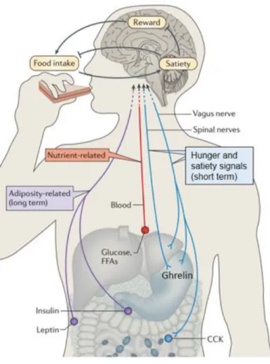
Figure 1- 1. Homeostatic eating regulating system.
The CNS integrates input from long-term energy stores (for example, leptin) and short-term meal-related signals (nutrients and gut-derived satiety signals) to regulate food intake and energy expenditure in a manner that maintains stable body fat stores over time. Positive energy balance induced by overfeeding inhibits the rewarding properties of food while enhancing meal-induced satiety, thereby reducing food intake. In response to energy deprivation, CNS adaptive responses are engaged that both increase the rewarding properties of food and reduce the response to satiety signals, collectively resulting in increased food consumption until deficient fat stores are replenished. CCK, cholecystokinin; FFAs, free fatty acids; GLP1, glucagon-like peptide. Adapted from (Morton, Meek, and Schwartz 2014).
2
Feeding behaviors are regulated by delicate feedback systems, which are modulated not only by the stored and circulating nutrients but also by various physical, psychological and environmental factors. The initiation and cessation of food intake is basically controlled by hunger and satiety signals generated in peripheral tissues such as the digestive tract (e.g. ghrelin and cholecystokinin) and adipose tissue (e.g. leptin) as well as the central nervous system (Figure 1-1). The sensation of hunger and the motivation to seek food are the central events of feeding behaviors, while the satiety signals mediate the termination of food ingestion largely through inhibiting the effects of hunger signals (Nakamura and Nakamura 2018).
The discovery and identification of appetite regulating signals started with the realization of the importance of satiety peptides (e.g. leptin), in controlling the termination of food intake. It has long been recognized that endocrine factors play critical roles in the regulation of appetite and food ingestion. Until now, more than 20 regulatory adiposity and gastrointestinal hormones have been found with a wider variety of functions related to feeding (Suzuki, Simpson et al. 2010). More metabolism and appetite regulating factors from different sources are being discovered, such as some bone-derived hormones (Mera, Ferron et al. 2018). Several hunger and satiety peptides revealed numerous new knowledge in appetite regulation and pharmacological treatment of eating disorders, such as anorexia nervosa and binge eating disorder (BED), which will be discussed in detail in the following part of the introduction. 1.1.2 Hedonic eating/Eating for reward
Hedonia is the sensation of pleasant stimuli, which is a subjective perception. The rewarding feature of the palatable food can motivate the animal to consume more food, and the hedonic response to palatable foods is very effective to trigger eating even when food ingestion is not necessary (Tepper and Yeomans 2017). This type of eating has been termed as ‘non-homeostatic eating’, because its purpose is not to restore the energy depletion. Schwartz argued that the regulation of response to the rewarding food is integral to how energy homeostasis is achieved, because the levels of hedonic response to foods are influenced by the metabolic status of the body, and accordingly lead to different levels of motivation for food consumption (Morton, Meek et al. 2014). Nevertheless, an undeniable truth is that food consumption motivated by hedonic responses to palatable foods can result in excessive
3
caloric intake, which plays an important role in the increasingly high rates of obesity in modern society (Timofeeva and Mitra 2013).
The taste of palatable food provokes its intrinsic reinforcing properties, while the interaction between taste, metabolism and reward systems makes this effect more dynamic. The taste system has different thresholds for different tastants, the taste-provoking chemical molecules. Largely, the threshold concentrations of the taste system are much higher than that of the olfactory system. The perceptual threshold is 2 mM for citric acid, 10 mM for salt (NaCl), and 20 mM for sucrose. It is not surprising that a higher sensitivity for poisonous substances is vitally important to for animals. For example, the perceptual threshold is 0.08 mM for quinine, and 0.0001 mM for the deadly substance strychnine (Hoegg and Alba 2006). Rats can identify an aversive tastant with relatively few licks, and change their licking pattern as a result (Weiss and Di Lorenzo 2012).
One possible reason for the higher threshold for nutritious components is that the body needs more nutrients, such as carbohydrates and salt, to maintain the homeostasis of energy and electrolyte. The blunted sensitivity to nutritious components can promote adequate ingestion of specific food likely by being proportional to the requirement of the body. Considering the short history of the prevalence of sweeteners, such as sugar and honey, it is reasonable to speculate that the decreasing sensitivity to sweet taste could be one of the reasons of increasing ingestion of sugar in the modern world.
1.2 Endocrine and neuronal signals involved in food intake regulation
Fat and glucose are two main form of energy in the animal body. The quantity of them ingested, stored and circulating in the body is controlled to keep the energy homeostasis in the body (Figure 1-1). To maintain the energy homeostasis the regulating systems need to detect, integrate and respond to various internal and external signals, which involves the interaction between the nervous system and a variety of long-term and short-term signals related to the metabolic status of the organism and nutrient content of individual meals. 1.2.1 Hunger-related signals
1.2.1.1 Decreased plasma glucose
As the main form of direct energy supply for the body, plasma glucose also signals to the brain about the quantity of immediately available fuel (Figure 1-1). The deficiency of
4
circulating glucose in the plasma during hypoglycemia can trigger the feeling of hunger and subsequent brain responses to motivate physical and behavioral activities, such as food seeking and eating, to restore the energy availability and storage (Morton, Meek et al. 2014). 1.2.1.2 Orexin
Orexin is a neuropeptide well known for its functions in regulating arousal, wakefulness and appetite (Davis, Choi et al. 2011). Two subtypes of the orexins, orexin A and B, derive from the same precursor, a 130 amino acid named prepro-orexin. Both orexin A and B are produced in the lateral and posterior hypothalamus and projected widely into many brain regions. The receptors for orexins belong to the G-protein coupled receptor family (Sakurai, Amemiya et al. 1998). Animal studies showed that fasting could activate the orexin neurons and that intracerebroventricular injection of orexin A or B had prominent orexigenic effects (Sakurai, Amemiya et al. 1998). More interestingly, orexin participates in the regulation of food intake not only via the metabolic pathway. The Orexin/hypocretin receptor 1 has been found to be necessary for the Palvovian appetitive conditioning and extinction, which means it is also very important for learned feeding-related behaviors (Keefer, Cole et al. 2016). Since the discovery of orexin numerous studies have suggested a dichotomy in orexin functions, in regulation of both arousal and feeding (Harris and Aston-Jones 2006). Harris and Aston-Jones hypothesized that orexin neurons in different sub-regions of the hypothalamus have different functions. They proposed that orexin neurons in the lateral hypothalamus are involved in the process of reward information related to food intake and drug abuse, whereas those in the dorsomedial and perifornical hypothalamus mediate arousal and stress response (Harris and Aston-Jones 2006). Furthermore, Saper proposed a role of orexin in coordinating the sleep/awake cycle with the need of eating. ‘After all, animals can only eat when they are awake’. He came up with a flip-flop model, in which the orexin plays a critical role in switching between sleep and wakefulness. After integrating the information about the energy storage, carried by circulating signals such as leptin and ghrelin, the dorsomedial hypothalamus (DMH) relays to the lateral hypothalamic area (LHA), and modulates the arousal states according to the need of food ingestion (Saper 2006).
1.2.1.3 Ghrelin
Synthesized and released by specialized gastrointestinal endocrine cells (Figure 1-1), ghrelin (the “hunger hormone” ) is a 28-amino acid acylated peptide functioning as a gut-brain
5
hunger hormone that typically stimulates food ingestion (Wren, Seal et al. 2001). Ghrelin levels in the blood fluctuate with a circadian rhythm in harmony with eating schedules, peaking before and descending rapidly after meals (Cummings, Purnell et al. 2001). Studies in animals have demonstrated that administration of ghrelin is sufficient to increase food intake, as well as to enhance appetite for palatable foods such as fat and sucrose (Schellekens, Finger et al. 2012).
The facilitating effect of ghrelin on food intake may be via increasing the motivation for food, because it can enhance dopamine transmission in the VTA (Abizaid, Liu et al. 2006, Jerlhag, Egecioglu et al. 2007). Indeed, the ventral tegmental area (VTA), a central component of reward system, has been found with high levels of ghrelin receptors (50-60%) (Abizaid, Liu et al. 2006, Zigman, Jones et al. 2006). Administration of ghrelin into the VTA leads to increased DA turnover in the nucleus accumbens (Acb) (Abizaid, Liu et al. 2006). Consistently, suppression of drug induced reward related activities (Jerlhag, Egecioglu et al. 2010) and reduced alcohol consumption was observed after the blockage of ghrelin signaling by an antagonist (Landgren, Simms et al. 2012). Interestingly, ghrelin levels are positively correlated with the neuronal activities induced by the view of palatable food, indicating its participation in cue-induced motivation for food (Kroemer, Krebs et al. 2013), and ghrelin antagonism with a ghrelin receptor antagonist GHRP-6 [D-Lys3] can reduce the appetitive response to food-associated cues (Dailey, Moran et al. 2016).
Additionally, the dysfunction of the ghrelin receptors has been found to induce a decrease in reward seeking activities. For example, mice with abnormal ghrelin receptor (GHS-R1A) gene, and rats treated with a GHS-R1A antagonist both have lower intake of palatable food in a free choice model (Egecioglu, Jerlhag et al. 2010).
1.2.1.4 Neuropeptide Y (NPY)
NPY is a 36 amino acid peptide functioning via specific receptors, Y1-Y6 (Krysiak, Obuchowicz et al. 1999). NPY has a major role in appetite regulation as an orexigenic neural transmitter (Gehlert 1999), and intraventricular administration of NPY was found to significantly enhance feeding and drinking in rats during light phase when they usually ingest small amounts of food (Levine and Morley 1984). A recent study found that the small-molecule NPY receptor antagonist can suppress biting and blood-feeding of mosquitos on
6
live hosts (Duvall, Ramos-Espiritu et al. 2019), suggesting that the appetite-regulating effect is a highly conserved function of NPY in the processes of evolution.
Interestingly, NPY positive neurons in the arcuate nucleus (ARC) are activated by orexin-A administration, and NPY antagonism can partially inhibit the orexin-induced feeding behavior. These findings indicate that the NPY system is possibly one of the downstream pathways in the feeding-enhancing effects of orexin-A (Yamanaka, Kunii et al. 2000). 1.2.2 Satiety-related signals:
The sensation of satiety is composed of the generation of satiety signals in response to food ingestion, as well as the suppression of hunger signals. The transmission of satiety signals, such as gastrointestinal hormones (e.g. cholecystokinin), includes a pathway from the periphery to the brain via the vagus nerve and/or the circulation of blood (Figure 1-1). 1.2.2.1 Insulin
Insulin is synthesized and released by the β-cells of the pancreas in response to increased blood glucose (Figure 1-1). Peripherally, insulin decreases the blood sugar concentration by promoting energy storage and glucose metabolism in insulin sensitive tissues, such as liver, muscle and adipose tissue. It can pass through the blood brain barrier and induce satiety via the insulin receptors in the brain (Baskin, Lattemann et al. 1999), and studies in rats found that intracerebroventricular admission of insulin could reduce the sucrose intake (Sipols, Bayer et al. 2002). Moreover, the metabolic and satiety signaling functions of insulin are possibly mediated by different receptor systems. For example, in mice the global deletion of nucleobindin-2 protein caused insulin resistance in obesity, without affecting satiety (Ravussin, Youm et al. 2018).
In this way, insulin is involved in two main aspects of energy homeostasis. Insulin in the blood is increased following a meal to promote the storage of excess glucose in the form of glycogen thus making a balance between the circulating and stored energy. Dynamic level of insulin in the blood can also work as a signal of the glucose accessibility, and regulate the feeding behaviors by provoking satiety (Baskin, Lattemann et al. 1999). Studies have demonstrated that fasting leads to a larger meal size, partially due to greatly reduced insulin penetrating into the brain (Strubbe, Porte Jr et al. 1988).
7 1.2.2.2 Leptin
Before the finding of leptin, there had already been a hypothesis about the existence of a feedback loop keeping a dynamic homeostasis of energy intake and expenditure, and in this modulating system, the hypothalamus plays as the monitor of energy storage by sensing a product of fat tissue circuiting in the plasma. This hypothesis is also called the lipostasis theory (Mayer 1955).
Zhang et al found that the obesity of ob/ob (obese) mice resulted from a mutation of the ob gene. After locating and sequencing this ob gene with exhausting work of positional cloning, they revealed that it coded for a hormone synthesized and secreted by the white adipose tissue. They latter dubbed the product of ob gene as ‘leptin’, derived from ‘leptos’ (Greek, ‘thin’), and predicted it as the signaling molecule in the lepostasis theory.
Leptin is a 16 kDa peptide, synthesized by in fat tissue (Figure 1-1), and emitted into the blood in proportion to the quantity of adipose storage. The concentration of leptin in the blood directly represents energy storage. It was found that food deprivation significantly lowered the leptin level, which could be reversed by refeeding the animals. Leptin has a strong appetite-stimulating effect via its receptor (ob-R), which was first isolated by Tartaglia (Tartaglia, Dembski et al. 1995). In this way, the CNS centers can monitor the energy storage by detecting the level of the circulating leptin and regulating the energy intake and expenditure accordingly to keep an energy homeostasis in the body. Shortly after the finding of leptin, it was confirmed that the abnormal feeding behavior and bodyweight of the ob/ob mice could be reversed by administration of exogenous leptin (Campfield, Smith et al. 1995, Halaas, Gajiwala et al. 1995).
Leptin inhibits food consumption by enhancing the response to other satiety signals (Morton, Blevins et al. 2005) and inhibiting the reward value of foods (Davis, Choi et al. 2010). The incentive value of foods can be assessed with behaviors such as conditioned place preference (CPP) and the breaking points of lever-pressing for food rewards. For example, ob/ob mice show higher CPP for a high fat diet, which can be decreased by leptin treatment independent of obesity (Shimizu, Son et al. 2017). Thus, leptin has been proposed to suppress the hedonic response to palatable food. Interestingly, the inhibition of reward system by leptin may have more functions other than inhibiting the food consumption. For example, leptin antagonism
8
enhances cocaine-induced CPP, accompanied by upregulated level of dopamine in the Acb (Shen, Jiang et al. 2016). Considering the crucial roles of leptin in regulating the energy homeostasis, it is not surprising that a malfunction of the leptin/leptin receptor system will result in many health problems such as obesity and diabetes. Obese individuals have high level of leptin, but there is no corresponding inhibition of food intake or reduction in bodyweight, possibly due to a deficit of leptin receptors instead of leptin itself. Approximately one year after the identification of leptin, a mutation in the long form of the leptin receptor was proved to be underlying the phenotype of db/db (diabetes) mouse (Chen, Charlat et al. 1996).
1.2.2.3 Serotonin (5-HT)
Studies using brain microdialysis found increased release of 5-HT in the hypothalamus of rats during feeding and pre-ingestive events (Schwartz, Hernandez et al. 1990). Furthermore, it was also found that the hypothalamic release of 5-HT after ingestion of a meal may be underlying the generation of satiety signals to terminate the meal (Haleem 1993).
The caudal 5-HT receptor-expressing neurons in the nucleus tractus solitarius (NTS) send glutamatergic projections to the parabrachial nucleus (PBN). Inhibition of these 5-HT receptors can reverse the feeding inhibiting effects induced by ablation of agouti-related protein (AgRP) neurons in the arcuate nucleus (Wu, Clark et al. 2012). Pharmacological agents which can increase the release of 5-HT (e.g. d-fenfluramine) (Gibson, Kennedy et al. 1993) or selectively inhibit 5-HT reuptake (e.g. fluoxetine) (Tao, Fray et al. 2002) have also a function of suppressing feeding in humans and experimental animals (Heisler, Kanarek et al. 1999, Halford, Harrold et al. 2007). Some researchers have proposed that postsynaptic 5-HT1B and 5-HT2C receptors in the hypothalamus may be underlying the feeding inhibiting effect of these 5-HT related agents (Kennett and Curzon 1988).
Besides the hypothalamus, 5-HT signals in the Acb have also been found involved in the regulation of food intake. The Acb has long been recognized to play critical roles in the perception of food-related rewards and modulation of motivation to eat (Georgescu, Sears et al. 2005). The 5-HT4 receptors are richly expressed in the Acb, and inhibition of these receptors in mice can reduce feeding behavior in fed mice, although not in food-deprived
9
ones (Jean, Conductier et al. 2007), suggesting a role of hypothalamus 5-HT4 receptors in inhibiting the motivation for food in sated animals.
Taken together, the 5-HT/ 5-HT receptors system in the hypothalamus controls food intake by enhancing the satiety signals, while Acb 5-HT modulates the motivation for food possibly via interaction with the dopamine system.
1.2.2.4 Cholecystokinin (CCK)
CCK is synthesized and secreted by enteroendocrine cells in the duodenum (Figure 1-1 and Figure 1-3) and causes the release of digestive enzymes and bile from the pancreas and gallbladder, respectively. It acts as a hunger suppressant and is the first gut peptide that is proposed to act as a satiety signal. As a peptide hormone, CCK mediates satiety by acting on the CCK receptors distributed widely throughout the central nervous system.
A study in rats found that 48h food deprivation attenuated theCCK-induced satiety, which could be prevented by leptin replacement (McMinn, Sindelar et al. 2000). This indicates that the fasting-induced leptin deficiency impairs the efficiency of CCK signaling for satiety, and that leptin regulates food intake particularly through sensitivity modulation to satiety signals such as CCK.
1.2.2.5 Oxytocin
Oxytocin is mainly produced in the hypothalamus and released via the posterior pituitary. A single dose of intranasal oxytocin is enough to significantly decrease food intake in
humans. The effects of oxytocin are more prominent in obese individuals (Lawson 2017). Oxytocin seems to enhance the hindbrain response to gut-derived satiety signals such as CCK, leading to the consumption of smaller meals. In support of this concept, leptin-responsive oxytocin neurons in the paraventricular nucleus of the hypothalamus (PVN) project to the NTS and leptin-induced anorexia requires oxytocin signaling (Blevins, Schwartz et al. 2004). It has been recently found that oxytocin expressing glutamatergic neurons (ARCOXTR) in the arcuate nucleus of the hypothalamus (ARC) plays a very important
role in facilitating the acute food intake inhibiting effect of leptin (Fenselau, Campbell et al. 2017), which will be discussed later in details. Another possibility is that oxytocin can decrease the motivation for eating by inhibiting the hedonic responses to palatable foods. For example, studies using fMRI found that oxytocin administration in men significantly
10
decreased functional connectivity between the VTA with other food intake-related brain regions, such as the insula, oral somatosensory cortex and primary visual cortex in response to viewing pictures of palatable foods (Kerem, Hadjikhani et al. 2019).
1.2.3 Reward-related signals in hedonic eating 1.2.3.1 Dopamine (DA)
The dopamine system is the most thoroughly investigated component of the reward system (Volkow, Wang et al. 2011), with primary importance for incentive motivation. The dopamine system involved in food intake regulation is mainly composed of a mesoaccumbal projection from the VTA to the Acb, as well as projections from the VTA to the prefrontal cortex, amygdala and hypothalamus (Egecioglu, Skibicka et al. 2011). A study in human found a positive correlation between DA release in the dorsal striatum and the self-reported level of pleasure induced by ingestion of palatable food (Small, Jones-Gotman et al. 2003). The dopaminergic projections from the VTA to the Acb is activated upon first exposure to new or unexpected access to palatable foods (Norgren, Hajnal et al. 2006). However, after repeated exposure to the same palatable food, the DA response habituates to the stimulus of the food itself, and gradually shifts to the stimuli (e.g. smell) associated with the food, or cues predicting the availability (Epstein, Temple et al. 2009, Schultz 2010). Thus, the DA release in the Acb also signals a ‘reward prediction error’ in the absence of expected food reward or receiving unexpected food reward (Nieh, Matthews et al. 2015).
Interestingly, a recent study found that dopamine also participates the timing of feeding behavior. In this study, D1 dopamine receptor (Drd1) signaling in response to high fat diet was found to perturb circadian feeding rhythms, and Drd1-knockout mice are resistant to this effect. Drd1 rescue within the suprachiasmatic nucleus, the central circadian clock, completely abolished this perturbing influence of high fat diet on the circadian feeding rhythms (Grippo, Tang et al. 2020).
1.2.3.2 Opioid
The endogenous opioid system is another major component of the reward system in the brain, and involved in both hedonic perception and motivational responses to pleasant/rewarding stimuli, including consumption of palatable food (Berridge, Ho et al. 2010, Nummenmaa and Tuominen 2018). Administration of μ-opioids into the mesolimbic system, such as the VTA
11
is hedonic, and rats can learn to ‘work’ (lever-pressing) for intracranial self-administration of opioid (Bozarth and Wise 1981). Ingestion of palatable food can stimulate the release of opioid in the Acb, and administration of opioid agonist and antagonist into the Acb can respectively increase and decrease the consumption of palatable food (Woolley, Lee et al. 2006). Moreover, genetic ablation of μ-opioids receptors has recently been found sufficient to reduce palatable food intake in binge-like eating mice (Awad, Roeckel et al. 2019). 1.3 Brain circuits involved in food intake regulation
1.3.1 Brain regions involved in homeostatic eating 1.3.1.1 The Lateral hypothalamic area (LHA)
The hypothalamus accounts for only ~3% of the brain volume, but it directly controls many critical homeostatic functions. The hypothalamus is divided into many regions such as the supraoptic nucleus (SON), paraventricular nucleus of the hypothalamus (PVN), arcuate nucleus (ARC) and ventromedial nucleus (VMH), based on their functions, gene expression patterns, or classical anatomical boundaries. Nevertheless, the lateral hypothalamic area composes a large portion of the hypothalamus and consists of a large area of neurons and fibers, with much less clear anatomical definition.
The LHA is located anterior to the VTA, and posterior to the preoptic area. It integrates information from cortical and subcortical regions, such as the amygdala and basal forebrain networks, and consequently mediates some specific behaviors via projecting to downstream circuits involved in reward (e.g. the VTA, in Figure 1-2) and feeding regulation (e.g. brain stem motor pattern generator in Figure 1-2 and the NTS in Figure 1-3) (Rossi and Stuber 2018).
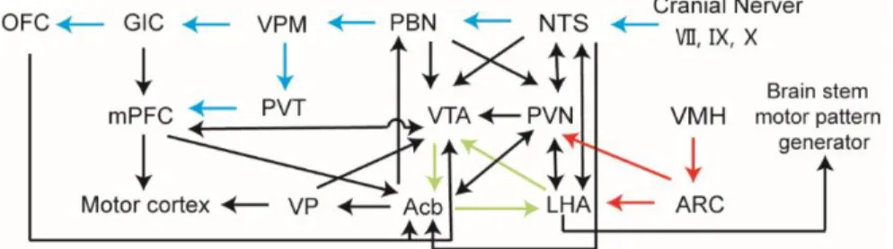
Figure 1- 2. Brain circuits involved in the regulation of food intake.
This diagram of brain connections includes the neural pathways underlying the taste information procession (pathways with blue arrows), homeostatic eating regulation (pathways with red arrows), and reward value perception (pathways with green arrows). Acb, nucleus accumbens. ARC, arcuate
12
nucleus of the hypothalamus. GIC, gustatory insular cortex. LHA, lateral hypothalamic area. NTS, nucleus tractus solitarius. OFC, orbitofrontal cortex. PBN, parabrachial nucleus. PFC, prefrontal cortex. PVN, paraventricular nucleus of the hypothalamus. PVT, paraventricular nucleus of thalamus. VMH, ventromedial hypothalamus. VPM, ventral posteromedial nucleus. VP, ventral pallidum.
The LHA is a highly heterogeneous brain area, with the glutamatergic and GABAergic neurons in the LHA playing opposing roles in modulating the homeostatic and hedonic feeding behaviors (Stuber and Wise 2016, Qualls-Creekmore and Münzberg 2018). The appetite-promoting effect of the LHA is mediated by excitation of its GABAergic neurons, which inhibit the GABAergic interneurons in the VTA. The final effect is the disinhibition of the dopaminergic neurons in the VTA, and augmentation of the hedonic response to food intake (Nieh, Vander Weele et al. 2016, Stuber and Wise 2016). The activation of glutamatergic neurons in the LHA can inhibit feeding by projecting to the lateral habenula (LHb). For example, mice with genetic ablation of glutamatergic neurons in the LHA showed higher daily caloric intake and larger weight gain when they had access to a high-fat diet compared with the wild-type controls. Moreover, optogenetic stimulation of the glutamatergic LH--LHb projections can acutely inhibit food consumption in mice, and optogenetic inhibition of these projections produced the opposite effect (Stamatakis, Van Swieten et al. 2016).
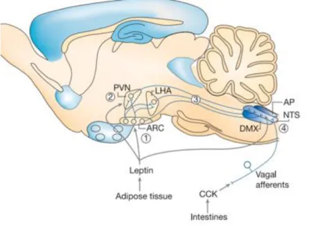
Figure 1- 3. Brain circuits integrating information about adiposity and satiety
Afferent input in proportion to body fat mass (for example, leptin) enhances the response to satiety signals such as CCK that are generated in response to food consumption and lead to meal termination. Whereas the hypothalamus is a key target for leptin action, the satiety effect of CCK involves activation of vagal afferent fibres that terminate in the NTS. Integration of these inputs can involve the actions of leptin in the ARC (1) or other hypothalamic areas (2) involving neurons that project to the NTS (3) and influence the response to CCK (4). In addition, leptin can act directly in the NTS. AP, area postrema; DMX, dorsal motor nucleus of the vagus nerve; LHA, lateral hypothalamic area; NTS, nucleus of the solitary tract; PVN, paraventricular nucleus. Adapted from (Morton, Cummings et al. 2006).
13
1.3.1.2 The Arcuate nucleus of the hypothalamus (ARC)
The ARC of the hypothalamus is a small sub-region of the mediobasal hypothalamus, adjacent to the median eminence and third ventricle. The ARC is the main site where the leptin signals enter the brain (Figure 1-3). Briefly, leptin inhibits food intake via inhibiting the AgRP/NPY (agouti-related peptide/neuropeptide Y) neurons (ARCAgRP/NPY neurons) and
activating the POMC (proopiomelanocortin) neurons (ARCPOMC) in the ARC. Moreover,
these ARCAgRP/NPY neurons send GABAergic projections to ARCPOMC neurons (Newton,
Hess et al. 2013).
ARCAgRP/NPY neurons are GABAergic and co-express two inhibitory neuropeptides, NPY and
AgRP. The intrinsic firing rate of ARCAgRP/NPY neurons increases after food deprivation, but
this modulation depends on the presence of leptin (Takahashi and Cone 2005). Optogenetic activation of ARCAgRP/NPY neurons was sufficient to stimulate voracious food consumption
in sated mice without training, by decreasing the latency to take the food and increasing the duration of meal (Atasoy, Betley et al. 2012). This effect is very rapid, usually within minutes, and disappears immediately after the end of the stimulation. The intensity of this regulation depends on the frequency and duration of the photo stimulation (Aponte, Atasoy et al. 2011). Ghrelin can stimulate eating by activating ARCAgRP/NPY neurons. Ghrelin receptor (GSH)
mRNA is expressed in 94% NPY expressing ARC neurons (Tannenbaum, Lapointe et al. 1998). ARCPOMC neurons are recognized as the counterpoint to ARCAgRP/NPY neurons
(Palmiter 2017). However, unlike ARCAgRP/NPY neurons, activation of ARCPOMC neurons is
extremely slow to take effect to inhibit food consumption (many hours). Long-term (24h) excitation of ARCPOMC neurons in this region was sufficient to inhibit the food intake and
decrease the bodyweight. In complementary to the ARCPOMC neurons, oxytocin receptor
expressing glutamatergic neurons in the ARC (ACROXTR) rapidly promote satiety when
chemo- or optogenetically activated (Fenselau, Campbell et al. 2017, Palmiter 2017). The same study found that these oxytocin responding neurons project to melanocortin-4 receptor expressing satiety neurons (PVNMC4R) in the paraventricular hypothalamus (PVN), which
also converge the projections from GABAergic ARCAgRP/NPY neurons. Interestingly,
transmission across these glutamatergic ARCOXTR--PVNMC4R projections is potentiated by
14
ARCAgRP/NPY neurons promote food intake mainly through projections to the LHA and PVN,
and optogenetic stimulation of the ARC--LHA and ARC--PVN projections (Figure 1-2, Figure 1-3) can induce comparable feeding behaviors as to activation of ARCAgRP/NPY neurons
themselves (Betley, Cao et al. 2013). ARCPOMC neurons also densely innervate the LHA and
PVN, as well as other feeding related regions such as the VMH and dorsomedial hypothalamus (DMH) (Waterson and Horvath 2015).
1.3.1.3 The Parabrachial (PBN)
Campos et al. found that parabrachial CGRP (calcitonin gene-related peptide) neurons (PBNCGRP neurons) relay satiety information from the NTS, and initiate meal termination by
projecting to the central amygdala (Ce). Inactivation of PBNCGRP neurons in mice leads to
impaired ability to terminate meals. It was also found that PBNCGRP neurons receive
inhibiting projections from the orexigenic ARCAgRP/NPY neurons. Simultaneous inactivating
PBNCGRP neurons and activating ARCAgRP/NPY neurons leads to voracious feeding (Campos,
Bowen et al. 2016).
Since the feeding behaviors are influenced by a large number of factors, including the physical conditions of the organism itself, environmental conditions and stimuli, the body needs a complicated system composed of a large number of signals to present these factors and maintain proper feeding behaviors. Based on aforementioned signals and brain regions, we can see that the energy homeostasis is achieved via a system where the circulating signals transfer the information about circulating and stored energy to the brain, and after integrating all related information the brain makes corrective adjustment to food intake.
1.3.2 Brain circuits involved in reward-induced feeding
Feeding behavior is also modulated by the rewarding properties of foods, other than homeostatic requirements. As most of subjective perceptions, it is susceptible to the experience, physical status and environmental situations etc. This hedonic regulation of food intake has been found to be largely modulated by the mesolimbic reward system and influenced by hypothalamic inputs. Taste information is perceived by oral taste receptors, projected in turn to the NTS by sensory fibers and subsequently transmitted to multiple structures in the hindbrain (e.g. the PBN), midbrain (e.g. the VTA), and forebrain (e.g. the Acb), thalamus and cerebral cortex (e.g. the mPFC and OFC), which assigns the rewarding
15
value to the taste via integrating and discriminating different tastes and textures (Morton, Cummings et al. 2006) (Figure 1-2). The evaluation of reward is mediated by the release of dopamine from the VTA to the Acb, striatum and some other brain regions. This dopamine signal is not directly responsible for the hedonic perception itself (‘liking’), but potently drives the motivation to obtain a rewarding stimulus (‘wanting’). Instead, the opioid signaling in the Acb and other adjacent forebrain regions, partially activated by dopamine release, is underlying the hedonic experience (Morton, Cummings et al. 2006).
1.3.2.1 The Ventral tegmental area (VTA)
The post-ingestive rewarding effect of nutritious food also enhances the craving for energy-dense food. Neurons in the LHA, which co-express orexin and glutamate, send excitatory projections to the VTA dopaminergic neurons. The activities of these LHA neurons can be influenced by peripheral signals of energy state, such as blood glucose (Sheng, Santiago et al. 2014). Moreover, the LHA also send GABAergic projections into the VTA, and activation of these projections significantly increases food intake (Nieh, Matthews et al. 2015). Dopaminergic neurons in the VTA synthesize dopamine (DA) and sent it to the Acb to facilitate its functions (Ikemoto 2007). Moreover, the projections directly or indirectly from the frontal lobe (e.g. the mPFC in Figure 1-2) and the amygdala to the VTA bias the responses towards reward and this effect is modulated by the DA signaling (Gottfried, O'Doherty et al. 2003, Stefani and Moghaddam 2006).
1.3.2.2 The Nucleus accumbens (Acb)
The name of the nucleus accumbens septi was derived from latin meaning ‘nucleus adjacent to the septum’. The Acb is made up by two components, the core (AcbC) and the shell (AcbSh). It is located between the caudate and putamen, without a specific demarcation from either of them. While the caudate nucleus and putamen comprise the dorsal striatum, the Acb and the olfactory tubercle comprise the ventral striatum. The striatum is part of the basal ganglia, and the majority of striatal neurons are medium spiny neurons (MSNs) (Yager, Garcia et al. 2015). Roitman et al. found a very precise and temporally discrete striatal dopamine release in trained animals during sucrose consumption (Roitman, Stuber et al. 2004). Carlezon proposed that the effect of reward has predominant association with a decreased activity of neurons in the Acb, such as the medium spiny neurons (Carlezon Jr and Thomas 2009).
16
There is a paucity of evidence suggesting that rostral part of the Acb is responsible for the ‘liking’ of rewards, while the caudal part works as a hedonic cold spot. Moreover, as the environment shifts between safe and stressful, some changes may occur on the functional map of the Acb. Reynolds and colleagues found that a stressful environments recruited ~90% of the Acb shell in the caudal, whereas the fear-generating zones shrink in the home environment, while the appetitive-generating zones expended caudally to fill ~90% of the Acb shell (Reynolds and Berridge 2008).
In the Acb, multiple separate, but interacting neurochemical systems are involved in the modulation of discrete aspects of appetitive motivation. For example, the GABAergic projections from the Acb shell to the hypothalamus has a direct influence on the feeding motor patterns. The dopamine signal in the Acb is involved in the control of general motoric and arousal process underlying pleasant response and response selection. The hedonic impact of palatable food is mediated by the enkephalinergic neurons that distributed widely in the Acb (Kelley, Baldo et al. 2005).
The special connections of the Acb with other brain regions involved in the regulation of food intake and energy homeostasis makes it a good candidate of integrating point of feeding related information. For example, the Acb receives taste information from the brain stem and gastrointestinal sensory information about the ingested food via direct connection with the NTS. Moreover, the Acb also has a bidirectional connections with the lateral hypothalamus, which plays essential roles in maintaining energy homeostasis (Erlanson-Albertsson 2005). 1.4 The binge eating disorder (BED)
1.4.1 What is BED
Under normal conditions, the homeostasis is capable of maintaining intake and expenditure of energy in a dynamic balance over time. However, when the homeostatic regulation of feeding behaviors is disrupted, various eating disorders may consequently occur. Eating disorders are manifested as a disturbance of eating habits or weight-control behaviors. Nevertheless, not all abnormal eating behaviors can be attributed to eating disorders. A basic criterion for the diagnosis of eating disorders is that the behavioral disturbance or associated core eating features should stem directly from significantly impaired physical health or
17
psychosocial functioning, rather than a result of general medical disorders or psychiatric condition.
Before the fifth edition of Diagnostic and Statistical Manual of Mental Disorders (DSM-5), eating disorders were classified into three diagnostic categories: Anorexia nervosa, bulimia nervosa, and atypical eating disorders not otherwise specified, the last of which included the binge eating disorder. In May 2013, the binge eating disorder was defined as an autonomous eating disorder in the DSM-5.
Binge eating disorder is characterized by discrete episodes of consuming more food than would normally be consumed under similar conditions during a similar period of time (Bogduk 2013). Binge-eating episodes are associated with three or more of the following: 1) eating until feeling uncomfortably full;
2) eating large amounts of food when not physically hungry; 3) eating much more rapidly than normal;
4) eating alone because one is embarrassed by how much she/he is eating; 5) feeling disgusted, depressed, or guilty after overeating;
6) marked distress or anxiety regarding binge eating.
People with binge eating will eat until uncomfortably full. Not surprisingly, binge eating is often associated with overweight and obesity. About 35% of those who frequently binge eats have the problems of overweight or obesity. Furthermore, binging subjects have higher risk of weight regain after treatment than non-binging ones (Stunkard and Wadden 1992, Swanson, Crow et al. 2011).
Severe eating compulsiveness often results into low mood, negative feelings and feeling of out of control, which in turn lead to more serious psychopathological impairment if not well coped with (Carrard, Crépin et al. 2012). Personality disorders and mood disorders, can be frequently seen among BED patients, and in some cases with substance abuse. All these comorbidities eventually lead to more complicated psychopathological problems and worse prognosis (Carrard, Crépin et al. 2012). Because of the close relation to the BED, these comorbidities were proposed as markers of severity of the BED, instead of simply as
18
associated conditions. All of these hazards caused by the BED can lead to a tremendous impact on our society and economy, because of reduced life quality, work efficiency, and increased health service cost.
1.4.2 Inducers of the BED
1.4.2.1 Dieting and limited/ intermittent access to palatable food
The attractiveness of food is induced by taste or nutritional value, such as carbohydrate and fat, but the hedonic value of palatable food is also influenced by the nutritional state of the body. Long-term dietary restriction, as well as intermittent feeding, have been found to augment the rewarding value of food and various drugs of abuse (De Vaca and Carr 1998, Carr 2002). In other words, the reward system can be sensitized by food restriction, which in turn increases the craving for food, as well as for drugs and alcohol (Söderpalm and Hansen 1999). As a result, it is not surprising that food restriction can provoke binge eating in both human and animals (Hagan, Chandler et al. 2003).
Sugar dependence has been triggered in rodents with intermittent access to concentrated sugar solutions (Colantuoni, Rada et al. 2002). Binge-like eating animal models developed with intermittent feeding protocol are often used to support the concept of food addiction, and to explore the mechanism underlying it.
1.4.2.2 Stress
An even more common inducer of eating disorders in humans is stress (Corning and Viducich 2019). Stress is a state in which the organism is threatened and the homeostatic balance is disrupted. Under some emergent or stressful situations, the stress-responding circuits override the homeostasis system, and induce some abnormal feeding behavior. The abnormal eating of palatable food induced by stress is also known as ‘non-homeostatic eating’ or ‘emotional eating’.
Most studies in animals demonstrate that stress inhibits feeding, unless animals are given the access to palatable food when they are stressed, which is consistent with reports about feeding responses to stressful situations in human (Pecoraro, Reyes et al. 2004, Tomiyama, Dallman et al. 2011). For example, people tend to eat palatable, high-calorie foods during stress, while maintain or decrease intake of lower-calorie foods, such as vegetables (Zellner, Loaiza et al. 2006). Individuals with stress-induced overeating problems just over-eat palatable food
19
which they usually tried to avoid for weight-control or health purposes (Zellner, Loaiza et al. 2006).
Psychogenic stressors, such as social defeat and loss of family members, can induce eating disorders in humans. (Over-) eating induced by emotional stressors is also called ‘emotional eating’. A study in male Japanese workers found that obesity and abnormal eating behaviors are associated with psychological stress response of ‘tension/anxiety’ induced by high job demand and/or low job latitudes (Nishitani and Sakakibara 2006). In students, a correlation has been found between academic stress (e.g. exam or high workload) and the intake of high-calorie foods (Michaud, Kahn et al. 1990) or consumption of less healthy diets (Weidner, Kohlmann et al. 1996).
1.4.2.3 Interaction between dieting and stress
Human studies also showed that the direction (increase or decrease) of change in food intake induced by stress is also modulated by dietary restraint. Healthy individuals with history of dieting are more likely to increase their food intake in response to stress (Zellner, Loaiza et al. 2006). The interaction between stress and feeding is complicated. Numerous evidences have demonstrated that stress may enhance eating, leading to obesity in chronic situations (Levine and Morley 1981, Tamashiro, Hegeman et al. 2007). Several studies also found that this feeding response to stress could be significantly modulated by dieting. Individuals classified as restrained eaters respond to stress with enhanced eating, instead of inhibited eating in control/non-restrained eaters. For example, restrained eaters ate more than the controls following an interpersonal stressor , in an intensity dependent way, which means that the greater the restraint, the more restrained eaters ate (Tanofsky‐Kraff, Wilfley et al. 2000).
Studies with animal models showed that food restriction itself only moderately increased the food consumption. However, when food restriction is followed by foot shock stress, and tested with palatable foods, this effect was tremendously enhanced, and animals showed binge-like eating behaviors. An example in human is that restrained eaters ate more than unrestrained after a stressful reaction-time task, whereas the result was opposite when test was done in a relaxation condition (Lattimore and Caswell 2004).
20
Besides physical stressor, other stressful situations are created by manipulating various environmental factors, such as maternal separation-- a very widely used model of early life stress. Maternal separated rodents display anxiety-like behaviors and depression in adulthood, along with disturbed serotonin levels (Lee, Kim et al. 2007), but without obvious sign of overeating or overweight. Nevertheless, when the maternal separation induced stress is followed by repeated fasting/refeeding protocol during adolescent time, animals start to exhibit binge-like food intake.
1.4.3 Hormones and neurotransmitters involved in the development of the BED 1.4.3.1 Glucocorticoid
It has long been known that HPA axis governs the endocrine response to stress. As a main product of activation of the HPA axis by stress, glucocorticoid is found to be involved in appetite regulation (Wolkowitz, Epel et al. 2001) and energy homeostasis by increasing circulating nutrient via lipolysis and gluconeogenesis (Epel, Lapidus et al. 2001). Acute stress typically induces an increase of cortisol, which in turn influences the releasing and functioning of neurotransmitters and hormones involved in appetite regulation, energy metabolism and feeding behaviors (Piazza, Rougé-Pont et al. 1996, Adam and Epel 2007). It is true that acute increase of glucocorticoid plays a protective role in coping with stress, but repeatedly or persistently elevated levels of glucocorticoid may lead to eating disorders including the BED.
A study in humans explored the relationship between laboratory stress-induced cortisol changes and food intake. This study found that in healthy female participants, after a cognitive stress task, individuals with higher levels of cortisol consumed significantly larger amount of food compared to individuals with lower cortisol levels (Epel, Lapidus et al. 2001). In a study examining the BED individuals, a positive correlation was found between binge eating severity and baseline salivary cortisol concentration (Coutinho, Moreira et al. 2007). In healthy female individuals without any eating disorder, stress-induced cortisol is positively correlated with food consumption (Epel, Lapidus et al. 2001). Studies with BED animal models showed that binge-like eating rats having significantly elevated corticosterone levels compared with non-binging rats (Artiga, Viana et al. 2007).