Using bioinformatic analyses to understand Prostate
Cancer Cell Biology
Mémoire
Raghavendra Tejo Karthik Poluri
Maîtrise en médecine moléculaire - avec mémoire
Maître ès sciences (M. Sc.)
Résumé
Le cancer de la prostate (CaP) affecte 1 homme sur 7 au cours de sa vie. C’est le cancer numéro un diagnostiqué chez l'homme. Il s'agit du quatrième cancer le plus fréquent au Canada. Le CaP est une maladie hormonodépendante diagnostiquée chez l'homme. Les androgènes jouent un rôle vital dans la progression de la maladie. La première ligne de traitement, suivant une intervention chirurgicale ou un traitement de radiothérapie, est la thérapie de déprivation aux androgènes. Malgré une réponse initiale positive à l'inhibition des androgènes, la progression de la maladie vers un cancer de la prostate résistant à la castration (CRPC) est presque inévitable. Aux différentes étapes du CaP, le récepteur des androgènes joue un rôle majeur. Ainsi, cette thèse décrit les méthodes développées et utilisées pour mieux comprendre la biologie du CaP et le rôle joué par les androgènes dans cette maladie. Le travail démontré dans cette thèse se compose principalement d'analyses bioinformatiques effectuées sur des ensembles de données accessibles au public et d'un « pipeline » construit pour analyser des données RNA-Seq. Un pipeline RNA-Seq a été développé pour comprendre l'impact des androgènes et des gènes régulés lors du traitement aux androgènes dans les modèles de cellules de CaP. Ce pipeline bioinformatique se compose de divers outils qui ont été décrits ci-dessous dans le chapitre 1. L'objectif principal de ce projet était de développer un pipeline pour analyser les données RNA-Seq qui aide à comprendre et à définir les voies et les gènes métaboliques qui sont régulés par les androgènes, et qui jouent un rôle important dans la progression du CaP. Le flux de travail expérimental consistait en deux lignées cellulaires positives aux récepteurs aux androgènes LNCaP et LAPC4. Toutes les données utilisées dans ce projet ont été rendues publiques pour que la communauté de recherche puisse effectuer diverses autres études et analyses comparatives pour comprendre les fonctions des androgènes dans un sens beaucoup plus profond afin de développer de nouvelles thérapies pour traiter le CaP.
Dans un autre projet décrit au chapitre 2, des analyses bioinformatiques ont été réalisées sur des données accessibles au public pour comprendre la fréquence de la perte et de l'altération génomique du gène PTEN localisé à 10q23. Ces analyses ont mis en évidence la fréquence d'altération génomique de PTEN qui est beaucoup plus élevée dans le CRPC que dans le CaP localisé. Ces analyses ont également aidé à identifier d'autres gènes altérés dans le CaP. Ces gènes n’ont pas été beaucoup étudiés dans la littérature, mais il semble que certains d’entre eux possèdent des caractéristiques de suppresseurs de tumeurs. Ces résultats pourraient être un bon début pour des analyses plus approfondies concernant la perte de gènes.
La compréhension des fonctions de AR et de la suppression de PTEN aidera à développer de nouvelles stratégies et approches pour diagnostiquer et traiter le CaP. L'intégration des analyses bioinformatiques à la recherche clinique ouvre une nouvelle perspective dans le domaine de la recherche du CaP.
Abstract
Prostate Cancer (PCa) affects 1 in 7 men in their lifetime and is the number one diagnosed cancer in men. It is the 4th most common cancer in Canada. PCa is a hormone-dependent disease diagnosed in men. Androgens play a vital role in the disease progression. The standard of care to treat PCa, following surgery or radiation therapy, is the androgen deprivation therapy (ADT). In spite of initial positive response to androgen inhibition, the progression of the disease to castration-resistant prostate cancer (CRPC) is almost inevitable. Across the various stages of PCa, the androgen receptor (AR) plays a major role. This thesis portrays the methods developed and used to understand PCa biology. The work demonstrated in this thesis majorly consists of bioinformatic analyses performed on publicly available data sets and a pipeline built to analyse RNA-Seq data. An RNA-Seq pipeline has been developed to understand the impact of androgens and the genes regulated upon androgen treatment in PCa cell models. This bioinformatic pipeline consists of various tools which have been described below in chapter 1. The major goal of this project was to develop a pipeline to analyse the RNA-Seq data which helps to understand and define the metabolic pathways and genes regulated by androgens which play an important role in PCa disease progression. The experimental workflow consisted of two androgen receptor positive cell lines LNCaP and LAPC4. All the data used in this project has been made publicly available for the research community to perform various other comparative studies and analyses to understand the functions of androgens in a much deeper sense to develop novel therapies to treat PCa.
In another project described in chapter 2, bioinformatic analyses have been performed on publicly available data to understand the loss and genomic alteration frequency of the gene PTEN occurring at 10q23. These analyses highlighted that the genomic alteration frequency of PTEN is much higher in CRPC than in localised PCa, and also helped in identifying other genes which are lost along with PTEN. The lost genes have not been studied much in literature, but few studies demonstrated that they might possess tumor suppressor characteristics. These results might be a good start for further deeper analyses regarding the lost of genes.
Understanding the functions of AR and the deletion of PTEN will help for the development of novel strategies and approaches to diagnose and treat PCa. Integration of bioinformatic analyses with clinical research open up a new perspective in the PCa research domain.
Table des matières
Résumé ... ii
Abstract ... iii
Table des matières ... iv
Liste des figures ... vi
Liste des tableaux ... vii
Liste des abréviations, sigles, acronymes ... viii
Remerciements ... xii
Avant-propos ... xvii
Introduction ... 1
1. Cancer Genomics ... 1
1.1 Role and Impact ... 1
1.2 Computational considerations in Cancer genome analysis ... 2
1.3 Transcriptomics ... 3
2. What is a Prostate ? ... 5
2.1 The Etiology of Prostate Cancer ... 8
3. Androgen Receptors ... 9
3.1 What are Androgens and Androgen receptors? ... 9
3.2 Androgen Receptor Structure ... 10
3.3 Androgen Deprivation Therapy ... 11
3.4 Progression of Prostate Cancer to Castration-Resistant disease ... 12
4. PTEN: Tumor Suppressor ... 13
5. Hypotheses, Objectives and Methodologies ... 15
Chapitre 1. RNA Sequencing Data of Human Prostate Cancer Cells Treated With Androgens ... 16
Résumé ... 16
Abstract ... 17
Value of the Data ... 18
Data ... 18
Experimental Design, Materials, and Methods ... 18
Cells ... 18
Sequencing ... 18
RNA-seq analysis ... 19
Differential gene expression and GSEA analysis ... 19
Acknowledgments ... 19
Figure Legends ... 20
Figures ... 21
Tables ... 21
Chapitre 2. Genomic Deletion at 10q23 in Prostate Cancer : More than PTEN Loss ? ... 22
Résumé ... 22
Abstract ... 23
Introduction ... 24
PTEN is more frequently altered through CNA rather than through a specific gene mutation in PCa ... 25
CNA at 10q23 leads to loss not only of PTEN but also of several additional genes ... 25
Future direction in PCa genomic alteration studies ... 26
Concluding remarks ... 28 References ... 29 Figure legends ... 31 Figures ... 32 Conclusion ... 35 Bibliographie ... 40
Liste des figures
Figure 1: RNA-Seq data uses short reads of mRNA…………...5
Figure 2: TCA Cycle…………...…………...7
Figure 3: Zonal anatomy of Prostate Gland...…………...8
Figure 4: Schematic chart depicting the synthesizing of DHT...…………...11
Figure 5: The three functional domains of AR...…………...12
Figure 6: The major biological function of PTEN...…………...15
Liste des tableaux
Liste des abréviations, sigles, acronymes
ADT Androgen Deprivation Therapy
AR Androgen Receptor
CNA Copy Number Alterations CGP Cancer Genome Project
CRPC Castration-Resistant Prostate Cancer DHT Dihydrotestosterone
ER Estrogen Receptors
FDR False Discovery Rate GEO Gene Expression Omnibus GnRH Gonadotropin-releasing hormone GR Glucocorticoid Receptors GSEA Gene Set Enrichment Analyses
ISUP International Society of Urologic Pathology LBD Ligand Binding Domain
LH Luteinizing Hormone
MR Mineralocorticoid Receptors NES Normalised Enrichment Score NGS Next generation sequencing
NTD N Terminal Domain
PCa Prostate Cancer
PHTS PTEN hamartoma tumor syndrome PR Progesterone Receptors
PSA Prostate Specific Antigen QC Quality Control
RP Radical Prostatectomy TCA Tricarboxylic Acid
TCGA The Cancer Genome Atlas TPM Transcripts Per Million TSG Tumor Suppressor Genes
Dedicated to my granny Mrs.Vijaya Lakshmi
Sathyavolu
गतस्य
शोचनं
नािस्त
Gone is gone. You shouldn’t worry about
Remerciements
I don’t understand the concept of acknowledgements and thanking every person for inspiration and non-existence assistance or some casual reference. Going with the flow and formality, I would like to “THANK” people who have provided me with impeccable “existing and real” support during my stay in #LABEAW housed at CHU-Quebec.
First things first, a super big Thank you (genuinely) to “The Boss” Dr. Étienne Audet-Walsh for accepting me as your first grad student. I’m pretty sure I will never be erased out of your memory but Mr. Kevin Gonthier can be erased, removed and deleted (as he is the second grad student :P). I might have let you down and I might have given you nightmares on various occasions, but all this was on purpose so that you can never forget me J. Leaving the fun aside, thank you for letting me grow and for guiding me onto the right path. Prostate cancer wouldn’t have never become a huge part of my career if it wasn’t for you. I still remember (I’m sure even you do too) my first lab meeting where I presented my slides with yellow borders and grey background consisting of all computational information and no biology. My career graph after joining your lab is almost similar (let’s say identical) to the prostate cancer disease progression graph. After the initial identification and deployment in your lab, the first standard of care is poster and oral presentations followed by ChIP protocols and TC protocols. In spite of initial positive response to TC protocols, the progression of fear in the hood of TC was inevitable. Across all these stages, EAW played a major role in progression of my career. In more than 80% of the cases (instances), EAW is always reactivated when my brain starts working on PCa. We (#LABEAW) all by now know that AR (EAW) indirectly controls various pathways in disease progression through mTOR and SREBF1. Here, mTOR and SREBF1 are Mr. Poluri and Mr. Gonthier. Terrific and troubleshooting Thursday lab meetings were always exhausting (at least when I was presenting) but I learnt the art of destroying every slide (inclusive of title slide). Tuesday individual meetings at the temple of enlightenment (EAW’s cave) consisted of flashes of pure awesomeness and epiphany jiffs. Drive to Château Bromont in 2018, Bombay Sapphire at Estérel in 2019, playing cards for one whole day at airports (YQB, YYZ) along with Dr.JLP will be the ultimatum. I believe I have been absolutely lucky and blessed to have you as my PI/Boss for my master’s. It is sad that I’m leaving the team as things didn’t go as well as we planned, but still I’m taking tonnes of positives on my way out. I also thank you for putting out a super huge Avengers’ team around me who became my tight knit circle much more than just lab mates or colleagues. Thank you to the members of #LABEAW (especially KG and CL) for supporting me emotionally and mentally during KitKat, Oreo, Hazelnut Hot Choc and Butter phases.
*theme music plays* Bad boys … bad boys … Whatcha gonna do …. Whatcha gonna do ... When they come for you!!!
Mr. Gonthier, I have no words to describe the moments we had and the time we spent together. You have told my mother that you are my Canadian twin and you stayed on what you have said. I believe we have equally discussed bullcrap and PCa over the past 2 years (I think the lab will not agree to this but it’s a fact). One day we might find a cure to PCa but nothing can cure your absence around me when I move to Kingston. The experiment performed with you at 6:00 AM on a beautiful Thursday changed me into a better student/researcher. CRCQ and CCRC 2019 are the elite highlights for various reasons, but Luigi’s mansion and sharing the same blanket with you will be on the zenith of my moments. The 4/5 hours preparation for our first journal club during the C’mas season of 2018 was as good as Oreo shake and Domino’s pizza. C’mas 2018 reminds me of fun filled dinner party at EAW’s domicile and the drive back in your beautiful blue “FROZEN” car. I have honed my skills in bullcrapping from you. One example of this is the spontaneously knit beautiful story of elephants and tigers. Thank you for introducing me to the rousse and noire boréale. Along with you these two are the best things that have happened to me. The conversation with you after orally consuming the amalgamation of the left-over liquids at La Cage/3 Brasseurs felt like a budding Buddha preaching under a bodhi tree. Every cup of mushroom soup at the cafeteria with you was a pure nirvana. You have promised me to teach WB but never did. Will never ever forgive you for this. Thank you for letting me be me. Thank you for not making me wear any masks or put on any fronts. Thank you for being part in my happiest moments, and for genuinely feeling the same; for listening to my weird exotic stories and radiating compassion and empathy from wherever you are. Thank you for being the only person I ever want to confide in.
You will always be my BLUE DRAGON. « I love you ».
Ms. Lafront, I’m privileged to share my workstation next to yours. I have learnt so much just by staring at your color annotated lab experiment book and watching you work. It was a pure bliss. The wonderful first lab meeting of yours was legendary. The presentation along with you for the scientific communication course was a heroic saga. I know I have irritated and disturbed you on various ocassions by bullcrapping but thank you for taking them on your chin and being a sport. You have destroyed me and Kevin on every opportunity given to you. Class given by you on economy of Zimbabwe at CRCQ 2019 was historic. Thank you for being the Wizard. And super huge shout out for critical reading of this thesis (with out which this would have been a pure mess with spelling and grammatical errors :P)
Cindy, you have been an absolute blessing and it was wonderful working with you. You have always been super positive and had the patience to hear out my frustrations. Stealing cookies with Mr.KG from your office felt like a heist. Thank you for the unlimited cookies, snacks and chocolates, thank you for helping me out on various experiments and thank you for sharing your office space for couple of months.
I don’t even know the names of the summer interns (being mean here :P). Thank you, Mr. Luke the Nuke, for all the donkey jokes and elephant stories. You are the only person who could understand the bioinfo errors and frustrations. Working with you for last minute analyses for EAW’s grant was epic … Felt like sitting on a grenade without pin. Loved working for the article you have authored (sarcasm). Thanks mate for everything. Thank you Lilliane for teaching me how to be social, thank you Jolyane for showing 8 full qPCR plates can be done in one single day. Thank you Virginie for showing me how to be productive, punctual and focussed.
Thank you, Mariam Dessay, for being the shoulder I can lean on whenever I needed one. Thank you for keeping my desi taste buds active. Thank you for torturing me to death to attend the 6:00 AM swimming sessions even during the chilly winters. In spite of so many ups and downs in the past year and a half, your love, affection, generosity and concern towards me remained the same. You are an unbelievable pocket-sized beast.
Thank you Mr. Lucien Tchara for your help and inputs on graphical aspects of this thesis. Thank you for all the evenings at the bar and for insights on life along with science. A huge shout out my project collaborators Dr. Eric Alain P and Dr. Charles Joly-Beauparlant.
Special ‘Thank you’ to Baba, Amma, Chellai and Ammama (Thanking you people doesn’t make sense … Your acts have been selfless so that I could take a step forward). This would have never been possible without you (Technically I would have never been possible without you). Thank you for being super supportive, encouraging and trusting my choices and career decisions. Thank you for giving me the strength and belief to reach for the stars and chase my dreams.
Thank you Raghu annayagaru and Leela vadhina for your continous mentoring and advices regarding my career decisions. It has been a great privilege to grow under your shadow, guidance, love and care and hope that continues for many more years to come. Thank you Subramanyam annayagaru and Bujji vadhina for being with me and helping me out during the COVID-19 pandemic. Thank you to Manas, Tejas and Krish for being available at any given time to talk and discuss anything and everything.
Thank you Chintu anna for showing me the art of tackling obstacles and hurdles in life with ease. Thank you for each and every effort you have put in to inspire the me. The fancy and edificational nights at Plaza Dorval, MTL with you and Anjali Vadhin will always remain special of mutliple reasons. Thank you for always encouraging me and being on my side whenever needed.
All the cronies (Abhi, Anky, Chottu, Gok, Puppy, Satam, SK) and extended associates (B, Sa, Sh, T) of the #bulbuls group played a huge part in this journey of mine. The journey with you guys over the past decade has been nothing less to a roller coaster ride. Most of the ‘firsts’ I have experienced involved you guys. You guys
have been pizza sauce to my pizza base (I mean pizza without sauce is horrible … At least super bad). The gaming sessions (from Assassins Creed, NFS Most Wanted, FIFA to LUDO King and COD Mobile) we had/ we are having/we will be having are best I had/I’m having/I will be having. Thank you for being with me during my hardest times. As our family is growing bigger year by year, I believe and hope the fun and bonding will be doubled and the journey in the next coming decades will be a terrific ride.
"Not all races are won on the track." - Darius, NFS Carbon, 2006
Thank you to my dearest Gauri Matta. I don’t say it too often but thank you. Thank you for being that person that I know I can rely on even if I feel like the world has turned against me. Most of all, thank you for showing me that love still exists and for always sticking around when things get hard. Thank you for being the light of my life and for showing me that I am yours too. Thank you for supporting all my stupid ideas and my ambitious choices. Thank you for introducing me to Grey’s Anatomy, due to which this thesis writing has been put on hold :P for straight couple of days.
"If there's a crisis, you don't freeze, you move forward. You get the rest of us to move forward because you've
seen worse, you've survived worse, and you know we'll survive too. You say you're all dark and twisty. That's not a flaw; that's a strength. It makes you who you are. I love you and I want to spend the rest of my life with you." - Derek Shepherd, season 5, episode 19.
Thank you, Patrick Russo and Mario Russo at Patrizio & co., and at Birra & Basta. It has been absolute fun and pleasure to work along with you guys and grow into an average « Pizzaolo ». Thank you for being patient and teaching me the art of cooking Italian dishes. #ciaobella
Merci à tous J <3 #desperatefrenchattempt
Finally, I would like to leave the remaining space (including the next page with a star) in the memory of people who lost their lives in the process of fighting with/against COVID-19.
Avant-propos
This thesis titled “Using bioinformatic analyses to understand the Prostate Cancer Cell Biology” is written and adapted to article insertion format and is presented to the Graduate Faculty of Université Laval for a M.Sc. Degree in Molecular Medicine. This section explains and defines contribution of other authors in my articles. I have also contributed as a co-author in other projects from the lab which are not discussed in this thesis.
“Genomic Deletion at 10q23 in Prostate Cancer: More Than PTEN Loss?” is the title of the first article I contributed and authored. This article was published in Frontiers of Oncology journal on June 29th, 2018. The impact factor of this journal is 4.137. This study was conceptualized and established by Dr. Étienne Audet-Walsh. The analyses were performed by Dr. Étienne Audet-Audet-Walsh. For my part, my contribution was involved in the interpretation of the results obtained from the analyses, and also involved in manuscript writing along with Dr. Étienne Audet-Walsh. All the data to perform the analyses has been obtained from The Cancer Genome Atlas (TCGA) portal. I estimate the percentage of my contribution to this article would be around 20%.
The article titled “RNA Sequencing Data of Human Prostate Cancer Cells Treated With Androgens” was published in the Data in Brief journal in August 2019 with an impact factor of 0.970. This study was designed and conceptualized by Dr. Étienne Audet-Walsh and me. I have contributed to this article by performing the wet lab experiments and bioinformatic analyses. I have performed wet lab experiments like cell treatments and RNA extraction. I have also majorly participated in the writing of the manuscript and graphical representations. Dr. Étienne Audet-Walsh also contributed by fine tuning the manuscript and the graphical representation. The RNA-Seq bioinformatic was established in collaboration with Dr. Charles Joly-Beauparlant and Dr. Arnaud Droit. They have contributed in building and debugging the pipeline. I believe my contribution in this article would be around 75%.
Introduction
1. Cancer Genomics
1.1 Role and Impact
In the recent decade, the rapid surge in awareness that somatic mutations and other aberrations drive human malignancies has led us within reach of personalized cancer medicine. The cumulative phenotypic consequence of somatically acquried genetic, genomic and epigenetic alterations in cancer cells are influenced by both heterotypic interactions with a unique tissue microenvironment and the host germline. The greatest advantage that cancer genomics holds is the potential to inform the prognosis and guide evidence-based management of early stage diseases, which comprise an increasing proportion of cancer diagnoses. Cancer genome drives the transition from a morphology-based to a genetics-based taxonomy of cancer, which means point of care decisions will become increasingly customized to the unique genomic and proteomic features of a patient’s tumor. These molecular features may be assessed in tumor biopsies, in circulating tumor cells or in body fluids using companion diagnostics that can predict the likelihood of clinical benefit for all treatment options1.
The information which is being gained from studying cancer genome has changed the dimensions of cancer research. Defining the biological relevance of genomic data is a key step in realizing the full clinical potential of cancer genetics and genomics.
A point mutation in HRAS gene was the first cancer related mutation discovered nearly thirty years ago, which causes a glycine to valine mutation at codon 122. The subsequent identification of similar mutations in the family members KRAS3–5 and NRAS6 ushered in a new field of cancer research activity3. The high cost and time taking efforts like using a PCR-based exon-by-exon direct sequencing approach concentrated in a highly draggable class of genes. Two cancer genes V600 and BRAF, which are now the subject of intense therapeutic development, were identified during the early phase of the work4. The extension of these PCR-based resequencing efforts to larger and larger segments of the coding genome has substantially increased the number of newly identified cancer genes and broadened the range of biology that has been shown to be subverted by mutations in human cancer.
The sudden hike in clinical opportunities has been because of the analysis of ever-increasing genomic datasets. The identification of mutations in the JAK2 kinase in myelo-proliferative neoplasms has resulted in new drug development opportunities for diseases for which there are currently no approved therapies5. Targeted PCR-based sequencing of candidate genes has yielded an expanding list of genomic discoveries that are being translated into personalized cancer management. Some notable ones include kinase activating mutations in the oncogene KIT. Integrative cancer genome characterization can generate the genomic and statistical evidence
for new candidate cancer targets for therapeutic and diagnostic development. Converting these into therapeutic agents and biomarkers will require deep biological insights to inform drug discovery and to define a clinical path for development.
1.1.1 Accession of Cancer Genomics Data
Most of the large-scale cancer genomics data has been made publicly available but accessing and using that data remains a major obstacle. Depending on the degree of computational modification and integration applied to the data, it is divided into four categories: (1) raw, (2) processed or normalized, (3) interpreted, and (4) summarized categories. These are also even known or referred as levels I to IV data. Projects like The Cancer Genome Atlas (TCGA), Cancer Genome Project (CGP) at Wellcome Trust Sanger Institute are huge large-scale projects, whose genomics data is generally available for the public or can be accessed upon request6. The data available does not have to be a high-quality data. Various quality control analyses have to be performed to check for experimental noise, analytical accuracy and batch effects, as these have an impact on validation steps. Multiple factors have to be considered before performing or progressing ahead with publicly accessed data.
1.2 Computational considerations in Cancer genome analysis
For the analysis of cancer genome data, several general considerations are looked upon apart from the challenges thrown by the process. These include Quality Control (QC) of data, the accurate estimation of the signal and noise in large data sets, reproducible approaches to complex genomic analysis and achieving sufficient power in the face of multiple hypotheses testing. The quality of the genomic data should first be verified whether it is sufficient for the analysis and experimental purposes. The QC can be performed using multiple computational QC tools, but the most commonly used is the FastQC7. This tool generally checks for Per Base Sequence Quality, Adaptor Content, Kmer Content and other parameters. The sample mismatching is also one of the biggest problems in the generation of the data. One should ensure that the data obtained or downloaded is from the intended sample. To address the aforementioned challenge, TCGA road map developed by Robbins et al. can be implemented, which provides a data model and system for capturing, indexing and annotating metadata for a subset of TCGA data8. One of the key objectives of the TCGA roadmap project was to develop a programmatic interface that enables the end-user to navigate and discover subsets of files based on file-level provenance annotations and rich metadata. This enables, for example, discovery of the exact set of files that led to a particular research result, even if each patient sample, experiment, and platform have several raw data files9.
There are several limitations to using next-generation sequencing. Next-generation sequencing provides information on a number of molecular aberrations. For many of the identified abnormalities, the clinical significance is currently unknown. Building of validated bioinformatic pipelines is another major step in the
analyses. The pipelines must be updated and maintained in order to be working on par with latest and validated tools as well as data. The performance of the pipelines should be very much optimized without compromising quality and results, in order to give more room for the people with low configuration units to implement the pipelines in their workflow.
Reproducibility is another key element which should be considered in the genome analysis. For example, if 25 genes are differentially expressed between two tumor subtypes, one would expect to obtain the approximate result even when the process is done for the second time using the exact parameters, tools used, and process involved.
Building of validated bioinformatic pipelines is another major step in the analyses. The pipelines must be updated and maintained in order to be working on par with latest and validated tools as well as data. The performance of the pipelines should be very much optimized without compromising quality and results, in order to give more room for the people with low configuration units to implement the pipelines in their workflow.
In the next coming years, the considerations or the challenges mentioned above will be drastically reduced as the current era is largely progressing forward. Further developments in “omic” analysis of data must aim to ease interoperability of multiple data sets and to develop a framework that can help in seamless analysis of data.
1.3 Transcriptomics
The health science research sector is currently undergoing a huge transformation in order to integrate latest technologies and produce new data to unravel the mystery of cancer. The importance of identification of proper predictive biomarker has been show by the success and failures of integration of transcriptomics in the new age of oncology care and research10, 11. Concentrations of RNA molecules in biological samples and mRNAs for protein coding genes can be understood and analysed precisely by transcriptomics12. Transcriptomics is the go-to approach in this era go-to generate go-top quality high throughput gene expression data13. Transcriptomics can be defined as the study of all RNA molecules consisting in a cell. This approach focuses more messenger (m)RNA molecules only, which provides with expression levels or state of the genes. Transcriptomics uses RNA sequencing (RNA-Seq) data, which uses high-throughput sequencing to capture all sequences to determine the quantity of a transcript14. Many labs across the globe are working towards personalized medication through transcriptomics, which has increased the deposition of transcriptomic data on public repositories like Gene Expression Omnibus (GEO)15 or TCGA, Array-Express16. The use of transcriptomics for functional interrogation of gene expression has been justified by drawing a correlation between ribosomal profiling and transcriptomic data for mRNA molecules17-19.
Over the period, the potential to understand gene expression between genotypes, drug treatments or tissues has been transformed by evaluating the content and abundance of RNA. This technique helps to improve the depth of investigation to the entire transcriptome of known and novel RNAs20. This is because RNA-Seq is not only limited to genomic sequences but can also identify transcripts. Low background signal and RNA-Seq data can be more quantitative than the data obtained from microarrays21. Pathway analyses, differentially expressed genes, identification of novel transcripts and splicing events can be accessed by various bioinformatic tools like Gene Set Enrichment Analysis, DESeq2, g:Profiler and Whippet.
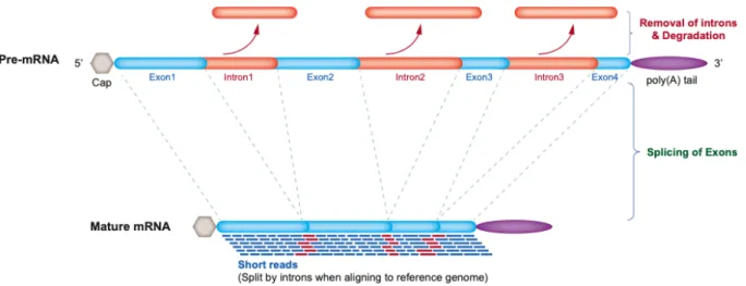
RNA-Seq is a powerful tool which can help the researchers to better understand genes. For example, a gene with an unknown function expressed in various tissues can be emphasized through transcriptome. Novel alternative splicing events/regions can be discovered (Fig.1). These events cannot be identified by DNA sequencing20.This approach will enable the identification of the genes that may be susceptible to prostate cancer (PCa)22. On the basis of RNA-seq data, the transcriptome profiles of primary PCa are identified, including gene fusions, long non-coding RNAs, alternative splicing and somatic mutations23, 24.
.
Figure 1: RNA-Seq data uses short reads of mRNA which is free of intronic non-coding DNA. The reads must be aligned to the reference genome. Aligning the reads to the reference genome will help for the identification
of novel gene expressions and splicing events/regions.
Next generation sequencing (NGS) played an important role in the recent cancer biological insights and breakthroughs; however, the main challenge in translating the large amounts of data into information which can be interpretable and accessible for cancer care still lies ahead. A fruitful relationship has to be established between bioinformaticians, statistical geneticists and molecular biologists to increase the translating rate of
omics data into clinical practice. The access to the databases and the availability of proper workflows and pipelines for NGS will greatly increase omics-based cancer diagnosis and personalized treatment strategies20, 21.
The induction of omics approaches in clinical practice will allow analysis of changes in patients at a global level by improving diagnosis and choice of therapeutic plans which are so far based on a few markers only. This approach also helps in developing models that can accurately predict patient survival using prognostic and predictive biomarkers in the era of precision oncology. In the near future, omics technologies will lead to a significantly improved biomarkers identification, compared to for example the top genes obtained by conventional differential gene expression analysis. It will also help in guided biomarker identification, which will play an important role in early tumor prognostic so that treatments can start earlier. Moreover, the identification of novel targets will decrease reliance on morbid therapies, thus improving the quality of life for PCa patients. A revolutional step forward in the field of precision medicine could derive from a pan-cancer analysis of multiple omics profiles on a genome-wide scale, in order to understand the shared patterns across multiple cancer types and identify shared actionable targets at a multilayered level. It therefore appears evident that the integration of omics data represents a powerful tool to allow clinical translation of this integrated dissection of cancer biology.
2. What is a Prostate ?
Behind the man’s penis is the localization of a walnut-sized gland called prostate. In the male reproductive system, the prostate gland is the largest accessory gland. It is important for the proper functioning of the male reproductive system. The vital function of the prostate is the production of thin, slightly alkaline fluid that forms a portion of the seminal fluid which is responsible for the protection and transportation of the sperms. The collagen forms an outer layer of the prostate capsule and the inner layer consists of a smooth muscle25.
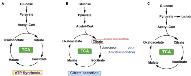
Another important function of prostate gland is the accumulation and secretion of high levels of citrate. Healthy prostate epithelial cells exhibit a highly specialized behaviour regarding their metabolic pathways. Typically, cells rely on citrate oxidation as a key step in the Krebs cycle for the progression of aerobic respiration26, 27. Benign prostatic cells accumulate zinc in high concentrations, and this excess zinc inhibits the oxidation and metabolism of citrate within the citric acid cycle, resulting in the production of citrate (Fig.2). The citrate is subsequently secreted as a component of semen.
Figure 2: TCA cycle is a central route for oxidative phosphorylation in cells, and fulfills their bioenergetic, biosynthetic, and redox balance requirements. (A) TCA cycle in the classic cells. (B) TCA cycle in normal prostate cells. These healthy cells exhibit a highly specialized behavior with respect to their metabolic pathways. Prostate cells, especially epithelial cells in the peripheral zone of the prostate, are programmed to produce and not oxidize citrate.(C) TCA cycle in PCa cells. PCa cells lose their ability to accumulate zinc, thus
leading to a continuation of the TCA cycle.
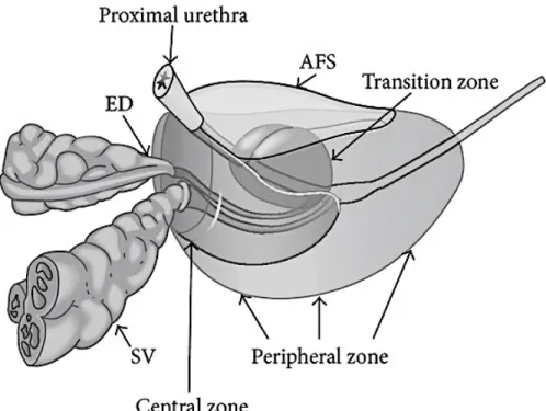
The prostate gland is located posterior to the lower portion of the symphysis pubis, anterior to the rectum, and inferior to the urinary bladder in the sub peritoneal compartment between the pelvic diaphragm and the peritoneal cavity. The prostate gland surrounds the proximal urethra as it exits from the bladder28.
The prostate consists of 4 regions, the Central Zone, Transition Zone, Peripheral Zone, and Anterior fibromuscular stroma, and also abides an apex, base and anterior, posterior, and inferior-lateral surfaces (Fig.3)28. One third of the lower prostate is taken by the apex, the middle one third of the prostate is midprostate which comprises of the verumontanum in the midprostatic urethra, and the upper one third of the prostate is formed by the base situated right below the urinary bladder28.
Figure 3: Zonal anatomy of the prostate gland. Ejaculatory Ducts (ED), Seminal Vesicles (SV), Anterior Fibromuscular Stroma (AFS). 28
Out of all the 4 zones, peripheral zone is the largest of all the zones, comprising around 70% of the glandular tissue. It surrounds the distal urethra and it extends from the base to the apex along the posterior surface. Approximately around 70% of all PCa arise from peripheral zone, which is primarily derived from the urogenital sinus29. By contrast, a very low incidence of prostate cancer is found in the central zone which is derived from the Wolffian duct30. The transition zone shares a similar embryologic origin as the peripheral zone; however, the percentage of prostate cancer arising from the transition zone is lower, in the order of 25%28.
The location of the central zone is at the base of the prostate between peripheral and transition zones, and is responsible for approximately 25% of the glandular tissue28. It is in the form of a cone and narrows down to an apex at verumontanum, and adjoining are the ejaculatory ducts. The transition zone comprises of two small lobules of glandular tissue that surrounds the proximal prostatic urethra just superior to the verumontanum and this zone makes up 5% of glandular tissue. This is the portion of the glandular tissue that enlarges due to benign prostate hyperplasia28. Finally, the anterior fibromuscular stroma forms the convexity of the anterior external surface and is devoid of glandular tissue; it is instead composed of fibrous and smooth muscular elements28.
2.1 The Etiology of Prostate Cancer
PCa is the most commonly diagnosed cancer in men in Canada31. PCa is often diagnosed in North American men when compared to rest of the world. The majority of PCa cases are diagnosed in men aged 50 and above31. A tumor is formed when the healthy epithelial cells of prostate change and grow out of control. The malignancy occurs by a major alternation in the mechanism of epithelial cells32. The tumor formed can be either cancerous or benign. The major difference between the former two is cancerous tumor can grow and spread to other parts of the body whereas benign cancer can grow but will not spread to other parts of the body. At the time of initial diagnosis, more than 90% of patients have PCa confined in the organ or only locally advanced33. PCa can be a slow-progression disease, such that many men die with PCa rather than from PCa. Many men have tumors that grow very slowly, whereas others develop a very aggressive disease, which metastasizes rapidly spreading elsewhere in the body.
PCa clinical follow-up has been revolutionized by the discovery of prostate-specific antigen (PSA), and to this day it is the only widely used biomarker for prognosis and follow-up of the disease. However, the survival benefit of PSA screening for PCa diagnostic is still being studied34, 35. This human protease is a protein encoded by the gene KLK3, which is located on chromosome 19q13.4 and is a member of the tissue kallikrein family of proteases36. PSA is responsible for dissecting semenogelin I and II in the seminal coagulum; it is produced in prostate ductal and acinar epithelium and is secreted into the lumen where the dissecting happens37, 38. Research studies conducted during the early 1990s suggested the use of PSA for detecting the presence of PCa. Serum PSA was clearly a much more sensitive screening tool, but it lacked specificity when compared digital rectal examination39, 40. On the other hand, when compared to prostatic acid phosphatase, serum PSA was demonstrated to be a more sensitive marker for PCa detection. The introduction of these tools for screening of PCa led to the increase of organ-confined PCa cases38.
Another method to evaluate the prognosis of men with prostate cancer is the Gleason grading system with prostate biopsy samples41. The classical Gleason system defines five histological growth patterns (grades). It scales from favourable prognosis which is Gleason 1 to poor prognosis which is Gleason 5. The scoring has been significantly modified after two major consensus meetings conducted by the International Society of Urologic Pathology (ISUP) in 2005 and 2014, respectively
.
A new simplified grading system is integrated into a strategy of prostate cancer staging that predicts prognosis and helps to guide treatment42. By using these known baseline clinical parameters, we can predict the prognosis and efficacy of novel treatments for prostate cancer. However, they are not perfect for selecting the best treatment sequence. Achieving precision medicine will require more precise tissue- or liquid-based biomarkers with prognostic and predictive value beyond these clinical parameters.The treatment approaches for localized PCa involve radical prostatectomy (RP) and radiotherapy. These approaches are considered to be curative approaches for localized PCa43, 44. Observation studies have been conducted to understand the outcomes of these above-mentioned approaches, with surgery being beneficial especially in young men with intermediate and high risk of localized PCa44. The majority of patients with localized PCa will be cured after local therapy with a five-year survival near 100%; but once the tumor progresses by developing distant metastasis, the disease often become incurable45. Various comparative studies have been done by teams across the globe to understand the outcomes of radiotherapy and surgery, but no clear and strong evidence has been found46.
3. Androgen Receptors
3.1 What are Androgens and Androgen receptors?
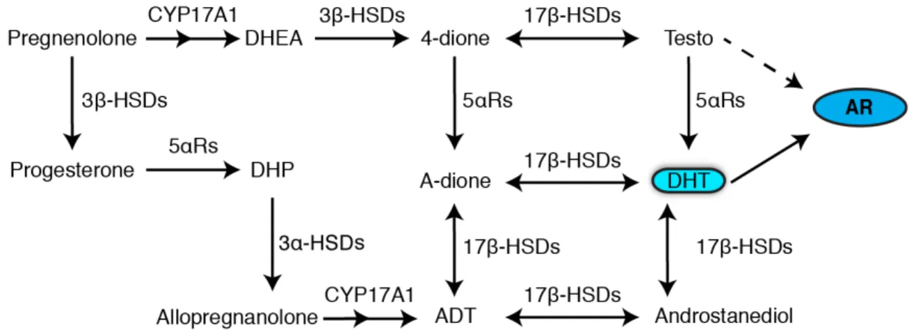
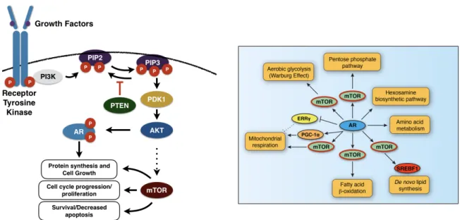
Testosterone and Dihydrotestosterone (DHT) together are known as androgens. Androgens are the male sex hormones which play a vital role in the development of the male reproductive system. Testosterone can be converted into DHT by the enzyme 5α-reductase, which is the most potent form of androgens, and to estradiol by the enzyme aromatase (Fig.4). Actions of DHT and testosterone are mediated through the androgen receptor AR, a ligand-dependent transcription factor. The androgen receptor is part of the hormonal nuclear receptor family which consists of estrogen receptors (ER), glucocorticoid receptors (GR), progesterone receptors (PR) and mineralocorticoid receptors (MR)47. The localization of the gene AR is on the X chromosome and is also expressed in a variety of tissues. Androgens are studied and subjected to various biological functions in bone, muscle, prostate, and immune and neural systems. The initiation of male sexual development is done when AR binds to its native ligands testosterone and DHT. The AR, when bound by a ligand, complexes with DNA at androgen response elements in the promoter region of target genes. The transcriptional regulatory effects of the AR include pathways involved in cell growth and proliferation, cell cycle progression, protein synthesis, and cell death.
Figure 4: Schematic chart depicting the synthesizing of DHT, the most potent form of androgen. 48
Prostate cancer is driven by the androgen receptor signalling27, 49. AR signalling axis is considered to be an important driver of prostate carcinogenesis and subsequent phases of the disease because of the vital and critical role of AR in the normal prostate. Androgen receptor indirectly/directly promotes tumor growth and disease progression through reprogramming specific cellular metabolic pathways such as aerobic glycolysis, mitochondrial respiration, fatty acid ß-oxidation, and de novo lipid synthesis.For that reason, androgen inhibition or deprivation therapy is the standard of care to treat PCa following disease recurrence after surgery or radiotherapy. In most of the cases, in spite of initial positive responses to the AR inhibition/deprivation therapy, the progression of the disease to CRPC is almost inevitable, which generally occurs within the period of 18-24 months50. This is the most lethal form of PCa51, 52.
Recent research work has shown that CRPC remains dependant on AR signalling pathways, and the most important therapeutic target has been the AR signalling pathway even in low androgen environment. The incremented expression, gene duplication and gene copy number of AR is believed to be the cause of dependence on AR in CRPC. Increased levels of AR gene copy number and mRNA amplification has been recorded in patients with CRPC53. The promotion of resistance to variety of AR targeting agents54 and conversion of AR antagonists to agonists have been linked to the elevated levels of AR.
3.2 Androgen Receptor Structure
The localization of the AR gene is on the X chromosome at the locus Xq11-Xq12 47, 55, 56. The protein coding region comprises of 8 exons, and the introns varies in sizes from 0.7 to 2.6kb. A 110kDa protein consisting of 919 amino acids is encoded by AR57, 58. AR is the only receptor in the nuclear receptor family which consists of 3 functions domains, namely (i) the N-Terminal Domain (NTD), (ii) the DNA binding domain (DBD) and (iii) the C-Terminal ligand Binding Domain (LBD), which is connected to the DBD by a flexible hinge region (Fig.5)47. For
the receptor to function, all the three domains are important. Currently, there is no complete structural information of the full-length AR receptor, but both DBD and LBD have been studied separately57, revealing critical details of the ligand dependant androgen receptor mechanism action of this receptor.
Figure 5: The three functional domains of AR: (i) the N-Terminal Domain (NTD), (ii) the DNA binding domain (DBD) and (iii) the C-Terminal ligand Binding Domain (LBD). (H – Hinge Region, AF-1 – transcriptional activating function 1, AF-2 – Transcriptional activating function 2, NLS – Nuclear localisation Signal, NES –
Nuclear Export Signal)47.
3.3 Androgen Deprivation Therapy
It was demonstrated by Huggins and Hodges that PCa is dependent on AR activation for its growth and survival59. The main therapy to treat PCa is Androgen Deprivation Therapy (ADT) or Androgen Inhibition Therapy60. ADT is the process of testosterone deprivation produced by testicles, which is done either by chemical castration or by surgery (orchidectomy).
By using ADT, AR ligand availability and subsequent AR mediated proliferative effects on prostate have been decreased by the reduction of circulating androgen levels and the rates of testicular androgen synthesis since androgens play a major role in PCa disease progression. The common approaches of ADT include orchiectomy or chemical castration through the chronic administration of gonadotropin-releasing hormone (GnRH) agonists and analogs of the LHRH61; other medical approaches include estrogen therapy, which also results in impaired androgen production and castration levels of circulating testosterone62. The use of GnRH agonists is the preferred means of achieving anorchid testosterone levels in clinical practice. Cyclic GnRH stimulation of the anterior pituitary stimulates the release of luteinizing hormone (LH), which in turn stimulates testicular androgen synthesis in the normal host. Depot GnRH agonists, such as leuprolide, goserelin, buserelin and others, induce an initial transient increase in LH release, followed by tachyphylaxis resulting from nonphysiologic and nonpulsatile stimulation of GnRH receptors. The potential therapeutic benefit of inhibiting both AR ligand production and binding led to the development of AR antagonists, such as bicalutamide, enzalutamide, flutamide and nilutamide, but this remains controversial62.
3.4 Progression of Prostate Cancer to Castration-Resistant disease
PCa cannot be completely cured with ADT; indeed, after the median of 1-2 years of ADT, the clinical progression occurs63. This stage is termed as CRPC. AR signaling is maintained by various mechanisms like mutations or truncations, amplifications and extragonadal androgen production even in the drastically low circulating levels of serum testosterone64. Randomized controlled clinical trials (RCT) are the clinical proof that showed CRPC are androgen dependent and also showed the survival benefits with more innovative ways of androgen inhibition or blockade. Enzalutamide, an androgen inhibitor and a second-generation AR antagonist with higher affinity for AR than first-generation antagonists, blocks and degrades AR, resulting in survival benefits in CRPC stage65. The reactivation of AR in CRPC stage should be studied in much deeper sense to understand the functions of AR in order to develop novel therapies.
If castrate levels of serum testosterone are less than 20ng/dL during ADT, along with satisfying at least one of the below mentioned three criteria, then PCa is considered as being at the castration-resistant stage. The three criteria are: (1) a rise of prostate-specific antigen (PSA) serum levels (biochemical progression), (2) development of symptoms in the presence of pre-existing cancer (clinical progression), or (3) detection of new metastatic lesions on imaging (radiographic progression)66, 67. CRPC was also termed as “androgen-independent” or “hormone-refractory” PCa68, but as the research led to new discoveries, especially the discovery of reactivation of AR in CRPC, it is now referred as “castration-resistant”69.
The concept of disease progression to CRPC is still unclear and is being studied to understand and discover novel therapies. Few researchers pointed out that the continued AR signaling despite of ADT might be the central point for the disease progression to CRPC70, 71. In recent times, majority of the research is done to understand how AR signaling is reactivated in CRPC and to understand the functions of AR in CRPC69.
The mammalian target of rapamycin (mTOR), which is a serine/threonine kinase that plays central roles in various biological processes, is a major component of the protein complexes, mTOR complex 1 (mTORC1) and mTOR complex 2 (mTORC2)72. mTOR phosphorylates distinct sets of substrates in response to growth factors (GFs), stress, nutrient availability, and other stimuli73, 74. Nuclear mTOR has been shown as an important transcriptional effector of PCa cellular metabolism and a key integrator of the androgen signaling pathway. It has also been shown that activation of mTOR-dependent metabolic gene networks is essential for the androgen-mediated metabolic reprogramming of cancer cells75.
In metastatic CRPC, mTOR is often hyperactivated through one of the most frequent genomic alternations which is loss of PTEN76, 77.
4. PTEN: Tumor Suppressor
The impact on Tumor Suppressor Genes (TSG) from the alterations that lead to the development of cancer are higher than compared to oncogenes in the cancer genomes. The major set of cancer driving genes consisted of TSGs which was justified by the results from exome sequencing analyses performed on various human cancers78, 79. In the recent times, the analyses on data obtained from The Cancer Genome Atlas (TCGA) supported the initial findings, with the majority of copy number alterations (CNA) in this genome analyses comprising deletions of putative TSGs. According to the findings provided by Zack et al., approximately 60% of peak regions of copy number alteration in cancer are deletions, and the majority of genes within these peaks are either known TSGs or appear to be novel TSGs80.
A novel gene PTEN was identified on the chromosome 10q23 in the year 1997 as a tumor suppressor redefining the regulation of cell behaviour81, 82. PTEN is abbreviated for phosphate and tensin homolog. PTEN is also known to be playing vital roles in the processes of apoptosis, embryonic development and cell migration83, 84. The mutation or deletion of this gene on chromosome 10 has serious impacts on the progression of human brain, breast and prostate cancer.
The dephosphorylation of phosphatidylinositol 3,trisphosphate (PtdIns(3,4,5)P3) to phosphatidylinositol 4,5-biphosphate (PtdIns(4,5)P2) is the major biological function of PTEN. This biological process inhibits the phosphatidylinositide 3-kinase (PI3K) signaling pathway (Fig.6)85. The PI3K signaling pathway is known to play major roles in cell metabolism, survival, proliferation, apoptosis, growth and migration. The suppression of PI3K pathway allows PTEN to bring its tumor suppressive functions to play86. PTEN loss also hyperactivates mTOR and its upstream regulator (PI3K/Akt) (Fig.4), and has been observed in metastatic castration-resistant prostate cancer (CRPC)87, 88. Activated AR enhances and reprograms mTOR chromatin-binding profiles and that nuclear mTOR activity is essential for androgen-mediated transcriptional reprogramming of metabolism in PCa cells75(Fig.7).
Figure 6: The major biological function of PTEN: the inhibition of PI3K pathway, which plays an important role in cell metabolism, survival, proliferation, apoptosis, growth and migration.85
Figure 7: mTOR pathway (left). mTOR directly/indirectly controls various cellular metabolic pathways linked with disease progression (right).
From the database of catalogue of somatic mutations in cancer (COSMIC) by sanger institute, 1478 unique mutations of PTEN have been recorded. The major enzymatic activity of PTEN can be impaired by these unique mutations89. The most typical pathology induced by PTEN mutation is PTEN hamartoma tumor syndrome (PHTS). PHTS confers an increased risk for specific malignancies, mostly breast, thyroid, renal and endometrial cancers90.
Various studies have demonstrated the tumor suppressor characteristics of PTEN gene86, 91. In the recent times, numerous studies have shown that PTEN regulates many proteins involved in immune cell development and immune signaling pathways, as well as other immunological activities89.
5. Hypotheses, Objectives and Methodologies
AR is known as the master regulator of cellular energy metabolism as it controls metabolic pathways like Glycolysis, lipid metabolism and protein synthesis along with Electron Transport Chain and TCA cycle in the mitochondria. AR also indirectly controls various pathways related to disease progression, majorly through mTOR. For example, AR controls pathways like Fatty acidβoxidation and mitochondrial respiration through mTOR. Here mTOR pathway is an important intracellular signaling pathway in regulating cell cycle. There it is directly related to proliferation. Phosphorylation and activation of AKT can have many downstream effects out of which one is the activation of mTOR (Fig.7). This pathway is antagonized by various factors along with PTEN, which will be further detailed in this thesis92.
The major hypotheses for my master’s degree are: (i) AR controls mRNA expression of various genes associated with various metabolic pathways, and (ii) the loss of PTEN is hyperactivating the mTOR pathway and is much more than just a PTEN loss at 10q23. The objectives developed and designed to understand and assess the above mentioned hypothesis are: (i) to develop and establish a RNA-Seq bioinformatic pipeline which identifies differentially expressed genes and pathways regulated upon treatment with androgens (R1881) from the sequencing data, then define metabolic genes regulated by androgen receptor, and (ii) to perform bioinformatic analyses on the publicly available data to study PTEN in details. The results of the above-mentioned work are described in chapters 1 and 2 respectively.
For the wet lab experiments, two AR positive cell lines LNCaP and LAPC4 have been selected, maintained and treated with androgens. RNA extraction was performed, and samples were sent for sequencing. The sequencing of these samples was performed by CHU-Québec sequencing platform.
The knowledge of methodologies obtained and used in this work are majorly bioinformatic tools required to analyze the data. The bioinformatic pipeline for RNA-Seq analyses has been built from scratch in collaboration with Dr. Arnaud Droit team. The pipeline consists of tools for quality control, pseudo alignment and quantification, and is used to identify differentially expressed genes and pathways regulated. The majority of the analyses were performed on compute canada servers. Statistical analyses were perfomed on local system using R-Studio.
Chapitre 1. RNA Sequencing Data of Human
Prostate Cancer Cells Treated With Androgens
Résumé
Le cancer de la prostate (CaP) est le cancer le plus fréquent chez les hommes en Amérique du Nord et les cellules de CaP dépendent du récepteur aux androgènes (AR) pour leur croissance et leur survie. Pour comprendre l'effet du AR dans les cellules cancéreuses, nous avons traité des cellules LNCaP et LAPC4, deux lignées cellulaires humaines immortalisées de CaP in vitro, avec l'androgène synthétique R1881, puis nous avons effectué des analyses par RNA-Seq. Les données de séquençage de haute qualité obtenues ont été analysées en utilisant notre pipeline bioinformatique qui comprend le FastQC pour le contrôle de la qualité,
Trimmomatic pour l’épuration des données et Kallisto pour le pseudo-alignement sur le transcriptome. Les gènes
présentant un différentiel d’expression ont été identifiés en utilisant DESeq2 suivant un ajustement pour le False Discovery Rate (FDR) (valeurs q FDR < 0,05) et une normalisation de l'expression logarithmique relative (RLE). Une analyse de l'enrichissement des ensembles de gènes (GSEA) a également été effectuée pour identifier les voies biologiques significativement modulées par les androgènes. Les analyses GSEA ont permis d'identifier la voie de signalisation des androgènes, ainsi que plusieurs sentiers métaboliques, comme étant significativement enrichis après une stimulation aux androgènes. Ces analyses ont mis en évidence les sentiers métaboliques les plus significativement régulées à la hausse suite à l'activation du AR. Les données brutes et traitées de RNA-Seq ont été déposées et rendues disponibles publiquement sur le Gene Expression Omnibus (GEO ; GSE128749). Ces données ont été intégrées dans un récent article décrivant les fonctions du AR en tant que régulateur clé du métabolisme des cellules CaP. Pour plus de détails sur l'interprétation de ces résultats, veuillez-vous référer à l’article "Functional genomics studies reveal the androgen receptor as a master regulator
of cellular energy metabolism in prostate cancer" par Gonthier K et al. (doi : 10.1016/j.jsbmb.2019.04.016).
Abstract
Prostate cancer (PCa) is the most frequent cancer in North American men and PCa cells rely on the androgen receptor (AR) for growth and survival. To understand the effect of AR in cancer cells, we have treated LNCaP and LAPC4 cells, two immortalized human PCa cells in vitro, with the synthetic androgen R1881 and then performed RNA-seq analyses. High quality sequencing data have been analyzed using our bioinformatic pipeline which consists of FastQC for quality controls, Trimmomatic for trimming, and Kallisto for pseudoalignment to the transcriptome. Differentially expressed genes were identified using DESeq2 after adjustment for false-discovery rate (FDR q values < 0.05) and Relative Log Expression (RLE) normalization. Gene Set Enrichment Analysis (GSEA) was also performed to identify biological pathways significantly modulated by androgens. GSEA analyses identified the androgen signaling pathway, as well as several metabolic pathways, as significantly enriched following androgen stimulation. These analyses highlight the most significant metabolic pathways up-regulated following AR activation. Raw and processed RNA-seq data were deposited and made publicly available on the Gene Expression Omnibus (GEO; GSE128749). These data have been incorporated in a recent article describing the functions of AR as a master regulator of PCa cell metabolism. For more details about interpretation of these results, please refer to "Functional genomics studies reveal the androgen receptor as a master regulator of cellular energy metabolism in prostate cancer" by Gonthier et al. (doi: 10.1016/j.jsbmb.2019.04.016).
This article is published in Data in Brief 2019; 25:104372
Keywords: Castration-resistance; Fatty acid metabolism; Glycolysis; Hormone receptor; Metabolic reprogramming; Metabolism; Mitochondria; Nuclear receptor; Steroid.
Value of the Data
• Bioinformatic analyses of differentially expressed genes and biological pathways regulated by androgens can be studied for a better understanding of the effect of AR in PCa.
• Validation in two distinct PCa cell lines allow for the identification of more reproducible results.
• These data highlight a new function of AR in PCa as a master regulator of cellular energy metabolism.
• These data may allow the discovery of new therapies targeting the unique PCa cell metabolic program.
Data
The raw data (.fastq files) generated from Illumina sequencing were deposted on the Gene Expression Omnibus (GEO) with the reference number GSE128749. The comma separated value files (.csv) which have been produced after the quantification and pseudoalignment with the transcriptome hg38 using Kallisto were also uploaded on GEO. These files contain the raw counts, the transcripts per million (TPM) values, and the fragments per kilobase million (FPKM) values for every sample. Differentially expressed genes on normalized data were identified using a FDR q value < 0.05.
Experimental Design, Materials, and Methods
Cells
LNCaP and LAPC4, two androgen receptor (AR) positive human PCa cell lines, were initially obtained from the ATCC and re-authentified in 2016 93. After resuscitation, the cells were not kept in culture for more than 3 months. Cells were grown in RPMI 1640 supplemented with 10% fetal bovine serum (FBS), streptomycin, penicillin, and sodium pyruvate in 37°C incubators with 5% CO2. Before androgen stimulation, cells were trypsinized and seeded at a 70% confluence in RPMI-1640 media with no phenol-red and supplemental with 5% charcoal-stripped serum (CSS), streptomycin, penicillin, and sodium pyruvate, as described previously 75. After hormonal deprivation (48h), media was changed and fresh media containing 10nM of the synthetic androgen R1881 or vehicle (EtOH 96%). 24h later, cells were harvested for RNA purification using the RNA purification kit RNeasy plus mini kit from QIAGEN.
Sequencing
Excellent RNA integrity was confirmed using a TapeStation 2200 (Agilent); all samples had an RNA integrity number equivalent (RINe) > 8.5. mRNA enrichment and library preparation were performed using the NEBNext Ultra II Directional RNA library prep kit following the manufacturer’s protocol. RNA was then sent to the Genomic
Centre of the Centre de recherche du CHU de Québec - Université Laval for sequencing using a HiSeq 2500 (125bp paired-end sequencing).
RNA-seq analysis
After sequencing, raw data were obtained in the fastq format. FastQC7 was used for validating the quality of the data. Trimming of the adaptor content and over-represented sequences was performed using Trimmomatic94. Also note that trimming was performed with the minimal length (MINLEN) set at 36. Quality check using FastQC was performed again on the trimmed sequences (Table 1). For the pseudoalignment of the trimmed sequences to the hg38 transcriptome, the Kallisto tool was used95. Final normalization was performed using the Relative Log Expression (RLE) method96. We have used the R-package called Tximport to convert the transcript quantifications to gene quantifications 97.
Differential gene expression and GSEA analysis
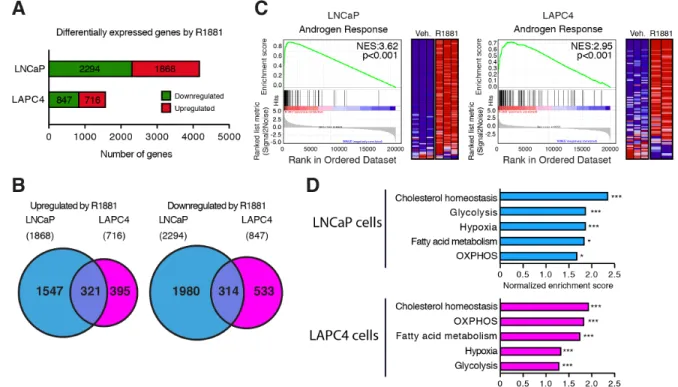
To study genes regulated by AR in PCa cells, differential expressed genes were identified using a FDR q value < 0.05 with DESeq298. Overall, 1868 and 716 genes were up-regulated in LNCaP and LAPC4 cells and 2294 and 847 genes were significantly down-regulated in LNCaP and LAPC4 cells, respectively (Fig. 1A). Of these, 321 common genes were up-regulated while 314 common genes were down-regulated in both cell lines (Fig. 1B). GSEA analyses 99 were also performed using TPM values to identify the most significantly up-regulated pathways following activation of AR in these human PCa cells. In both cell lines, the androgen signaling pathway was highly enriched following R1881 treatment (Fig. 1C). In addition, several metabolic pathways were also enriched in both LNCaP and LAPC4 cells following AR activation (Fig. 1D).
Acknowledgments
This work was supported by funding to EAW from the Cancer Research Society and the Canadian Institutes for Health Research (CIHR; 23123) and the CIHR (PJT159530).
References
1. Audet-Walsh, E., et al., Androgen-Dependent Repression of ERRgamma Reprograms Metabolism in Prostate Cancer. Cancer Res, 2017. 77(2): p. 378-389.10.1158/0008-5472.CAN-16-1204
2. Audet-Walsh, E., et al., Nuclear mTOR acts as a transcriptional integrator of the androgen signaling pathway in prostate cancer. Genes Dev, 2017. 31(12): p. 1228-1242.10.1101/gad.299958.117 3. Andrews, S. FastQC A Quality Control tool for High Throughput Sequence Data. 2010; Available
from: https://www.bioinformatics.babraham.ac.uk/projects/fastqc/.
4. Bolger, A.M., M. Lohse, and B. Usadel, Trimmomatic: a flexible trimmer for Illumina sequence data. Bioinformatics, 2014. 30(15): p. 2114-20.10.1093/bioinformatics/btu170
5. Bray, N.L., et al., Near-optimal probabilistic RNA-seq quantification. Nat Biotechnol, 2016. 34(5): p. 525-7.10.1038/nbt.3519
6. Anders, S., et al., Count-based differential expression analysis of RNA sequencing data using R and Bioconductor. Nat Protoc, 2013. 8(9): p. 1765-86.10.1038/nprot.2013.099
7. Soneson, C., M.I. Love, and M.D. Robinson, Differential analyses for RNA-seq: transcript-level estimates improve gene-level inferences. F1000Res, 2015. 4: p.
1521.10.12688/f1000research.7563.2
8. Love, M.I., W. Huber, and S. Anders, Moderated estimation of fold change and dispersion for RNA-seq data with DESeq2. Genome Biol, 2014. 15(12): p. 550.10.1186/s13059-014-0550-8
9. Mootha, V.K., et al., PGC-1alpha-responsive genes involved in oxidative phosphorylation are coordinately downregulated in human diabetes. Nat Genet, 2003. 34(3): p. 267-73.10.1038/ng1180
Figure Legends
Figure 1: Transcriptomic analyses of the androgen signaling pathway functions in human prostate cancer cells. A) Number of genes significantly up- or down-regulated following treatment with R1881 in LNCaP and LAPC4 cells. A FDR q value < 0.05 was used to identify differentially expressed genes. B) Venn diagrams showing the overlap between genes up-regulated (left) and down-regulated (right) by R1881 in LNCaP and LAPC4 cells. C) Gene set enrichment analysis (GSEA) plots for the “Hallmarks - Androgen Response” signature in LNCaP and LAPC4 cells. NES: normalized enrichment score. D) GSEA signatures enrichment scores for significantly enriched metabolic pathways in LNCaP and LAPC4 cells following 24h treated with R1881. OXPHOS: oxidative phosphorylation (mitochondrial respiration) * p < 0.05; ** p < 0.01; *** p < 0.001.
Figures
Tables
Table 1: Number of reads for raw and trimmed sequences of PCa cells treated with androgens.