OATAO is an open access repository that collects the work of Toulouse researchers and makes it freely available over the web where possible
Any correspondence concerning this service should be sent
to the repository administrator: [email protected]
This is an author’s version published in: http://oatao.univ-toulouse.fr/22751
To cite this version:
Kheir, Joyce and Kallassy, Mireille and Salameh, Dominique and Lteif, Roger and Brandam, Cédric and Strehaiano, Pierre Brettanomyces yeasts isolated from lebanese wines showing difference in their molecular pattern. (2014) European Scientific Journal, 10 (9). ISSN 1857-7881
BRETTANOMYCES YEASTS ISOLATED FROM
LEBANESE WINES SHOWING DIFFERENCE IN
THEIR MOLECULAR PATTERN
Joyce Kheir, PhD
Mireille Kallassy, Prof.
Dominique Salameh, PhD
Roger Lteif, Prof.
Université Saint-Joseph, Faculté des Sciences. Mar Roukoz, Mkallès, Beirut, Lebanon
Cedric Brandam, PhD
Pierre Strehaiano, Prof.
Université de Toulouse, INPT, UPS, Laboratoire de Génie Chimique, Toulouse, France
Abstract
The action of Brettanomyces yeast is a leading cause for organoleptic alterations of wine. To evaluate the presence of this yeast in Lebanon, 100 red wine samples selected from 14 wineries located in different regions of the Lebanese territory, were analyzed. Only 3 samples gave positive results. A first PCR showed that all isolates belonged to the species B. bruxellensis. Strains profiles were compared to 2 other French isolates. The PCR-RFLP was then used with 3 endonucleases for an inter-species characterization. The obtained results revealed a degree of polymorphism between strains isolated from different geographical origins. A comparison of the sequence of ITS regions, including the 5.8S rDNA and partial sequences of 18S and 28S rDNA illustrate potential similarities or differences between strains belonging to the same group.
Keywords: Brettanomyces, red wine, PCR, sequencing Introduction
Brettanomyces/Dekkera is a wine spoilage microorganism that causes economic loss in wineries all over the world. These yeasts are able to increase volatile acidity in wine because of the production of acetic acid which constitutes more than 90% of wine’s volatile acidity (Van Der Walt & Van Kerken, 1958). Brettanomyces/Dekkera as well can synthesize
2-acetyltetrahydropyridine and 2-ethyltetrahydropyridine molecules associated to mousiness off-flavor (Heresztyn, 1986). Finally, this contaminant yeast is responsible of the production of phenolic off-flavors (Chatonnet et al., 1992; 1995), especially volatile phenols which represent a large family of aromatic compounds, mainly 4-vinylphenols and 4-ethylphenols. The presence of these compounds is considered to be a direct indicator of Brettanomyces bruxellensis activity in red wine (Oelofse et al., 2009).
In Lebanon, the winemaking sector has been witnessing a significant growth for the last 10 years. In the 1960s, Lebanon had only 3 wine producers, but their number increased to 5 in 1998 and to more than 30 active ones nowadays. All major wineries have their vineyards in the southern Bekaa Valley that the country benefits from 300 days of sunshine per year. Concerning the grape varieties, Lebanese winemakers have favored French ones, particularly Cabernet Sauvignon, Merlot, Syrah and Rhone varietals, such as Carignan, Cinsault and Grenache. However, Lebanon has a rich heritage of indigenous grapes, as the blend of Obaideh and Merwah used for white wine.
Brettanomyces/Dekkera spp. are detected in all winemaking processes, although their population’s concentration is generally minor in the presence of other rapidly fermenting yeasts. Their occurrence is largely described in winery equipment, more commonly in vats, pumps, transfer lines, air, and on materials that are difficult to clean or sterilize. As Brettanomyces are known to be slow growing yeasts, their development require nutritionally favorable conditions (Fugelsang et al., 1993). Their proliferation is usually observed during stuck or slow fermentations, most commonly in malolactic fermentation due to their ability to survive at high ethanol content (14-14.5%) and at low levels of residual sugars, in the presence of free sulfur dioxide (Dias et al., 2003; Renouf et al., 2006a; Wedral et al., 2010). Detection and isolation have been described in stainless steel or concrete tanks and in finished bottled wine, but ageing in oak barrels can be beneficial since the transfer of small amounts of oxygen is allowed by the porous microstructure of wood and the presence of cellobiose that can serve as a sugar resource (Oelofse et al., 2008).
Brettanomyces in wine can be detected, whether directly, by selective culturing medium, or indirectly, by metabolites analysis. Isolation media often employ two antimicrobial agents (Rodrigues et al., 2001), including ethanol and cycloheximide that inhibits Saccharomyces, genus, which is subject to proteosynthesis pathway inhibition (Barnett et al., 1990), and a wide range of bacteria and other indigenous yeasts that can interfere. p-Coumaric acid is added as a precursor of 4-ethylphenols production while sugar can be used to decrease acetic acid levels that can interrupt olfactory detection of 4-ethylphenols by trained investigators (Couto et al., 2005).
Plating on selective growth media is unable to recover Brettanomyces from a viable but not culturable (VBNC) state where yeasts are metabolically active, but unable to undergo cellular division for growth (Oliver, 1993; Oelofse et al., 2008). This dormant state can remain for long periods in bottled wine and volatile compounds can be present even when yeast cells are undetectable. Direct methods, after culturing, also include polymerase chain reaction (PCR) techniques or fluorescence in situ hybridization, using peptide nucleic probes which have been recently developed (Wedral et al., 2010). Cocolin et al. 2004 put in place a specific PCR targeting the D1-D2 loop of the 26S rRNA, followed by the digestion of the PCR products (PCR-RFLP) to differentiate between B. bruxellensis and B. anomalus, using the DdeI restriction enzyme. Several molecular methods have been applied to characterize the biodiversity of isolates from different geographical origins (de Barros Lopes et al., 1998; Agnolucci et al., 2009). Some of these methods are a polymerase chain reaction denaturing gradient gel electrophoresis fingerprinting (PCR-DGGE) (Renouf et al., 2006b); restriction enzyme analysis of mitochondrial DNA (mtDNA) and RADP-PCR with OPA-primers (Martorell et al., 2006); restriction enzyme analysis and pulsed field gel electrophoresis (REA-PFGE) (Miot-Sertier & Lonvaud-Funel, 2007).
Indirect methods of detection rely on the physiological, sensory and chemical analysis using trained tasters or gas chromatography/mass spectrometry for the detection of produced volatile phenols compounds.
The presence of Brettanomyces in Lebanese wine and wineries has never been seriously assessed. Yet, the problem has affected many wineries in the whole world. A comprehensive study is therefore necessary to evaluate the presence of this type of yeasts and the level of contamination of wine. Isolated strains were then compared to others found in French wineries by the PCR-RFLP technique, using the restriction patterns generated from the region spanning between the internal transcribed spacer (ITS1 and ITS2) and the 5.8S rRNA gene (White et al., 1990; Guillamón et al., 1998).
Materials and methods Microorganisms and media Lebanese wine samples
Samples of this study were chosen to have representative data covering the Lebanese wineries. Thus, 100 red wine samples were chosen from 14 different wineries located in different regions with various vintages (varying from 1998 to 2011) and different grape varieties. The number of bottled wine was lower compared to other samples; as bottled wine could have been sterile-filtered, thereby removing all microbes prior to bottling. Thus, only 38 bottles (38%) found in the market were selected for this study
while 62 other samples (62%) were directly collected from different Lebanese wineries. Indeed, 25 samples were taken from Inox tanks, 25 others from wood barrels, 10 from plastic tanks and 2 from concrete tanks.
French strains
Two French strains were isolated and detected in a same winery situated in the southern region of France, using the detection procedure described below.
Yeast strains isolation
Three different selective media for screening and isolation were used to detect Brettanomyces yeasts. The isolation medium with chloramphenicol and cycloheximide (IACC) was formulated by (Medawar, 2003). This medium prevents any development of lactic, acetic and other bacteria or other Saccharomyces species. It has no fungistatic action which could hide Brettanomyces development. IACC composition was: 10 g l-1 glucose, 3 g l-1 yeast extract, 3 g l-1 peptone, 15 g l-1 agar, 0.3 g l-1 chloramphenicol and 0.2 g l-1 cycloheximide. Samples tested with IACC were incubated for 8 days at 30˚C.
While the IACC medium is not able to detect low concentrations of yeasts, an enrichment medium in liquid phase (EMPL) is required to recover or activate stressed yeasts (Medawar, 2003). EMPL consists of 15 g l-1 glucose, 4 g l-1 yeast extract, 4 g l-1 peptone, 0.4 g l-1 chloramphenicol and 0.2 g l-1 cycloheximide. Samples were added (v/v) to EMPL and then incubated for 8 days at 30˚C with continuous shaking.
A solid medium (SM) (10 g l-1 glucose, 5 g l-1 yeast extract, 20 g l-1 agar, 0.1 g l-1 chloramphenicol and 0.1 g l-1 cycloheximide, 0.05 g l-1 p -coumaric acid and 3.5 g l-1 calcium carbonate) was used to test the acidity character of all formed colonies on IACC and EMPL media (Barbin, 2006). Colony aspects of all positive samples were observed by microscopy examination to check that microorganisms exhibited Brettanomyces morphology features.
Molecular and sequencing analysis
Yeast culture conditions and genomic DNA extraction
Cells originating from a colony grown from a single cell of each of the isolated Brettanomyces/Dekkera strains were inoculated in 100 ml of a liquid culture medium consisting of 20 g l-1 glucose, 1 g l-1 yeast extract, 2 g l-1 (NH4)2SO4, 0.3 g l-1 citric acid, 3 g l-1 malic acid, 2 g l-1 tartaric acid, 0.4 g l-1 MgSO4,7H2O and 5 g l-1 KH2PO4 (pH 3.5) for at least 48 h at 30˚C with continuous shaking.
Cells from exponential growing cultures were harvested by centrifugation at 13 000 rpm and resulting cell pellets were re-suspended with 0.2 ml lysis buffer [2% Triton X-100, 1% SDS, 100 mM NaCl, 10 mM Tris-HCl (pH 8.0), 1 mM EDTA (pH 8.0)]. Cells were then thermally disrupted using two cycles of temperature comprising 2 minutes in a -80˚C freezer, followed by 2 minutes in a +95˚C water bath. 0.2 ml of chloroform was added to yeast lysates and the mixture was centrifuged at 13 000 rpm in order to remove all protein debris. The aqueous layer was allowed to precipitate at room temperature with 0.4 ml of ice-cold 100% ethanol. The DNA collected by centrifugation was washed with 0.5 ml of ice-cold 70% ethanol and DNA pellets were dried at 60˚C for 15 minutes. DNA pellets were finally suspended in TE buffer [10 mM Tris-HCl, 1 mM EDTA (pH 8.0)]. To improve DNA quality, the subsequent step of RNAase treatment was performed according to Hoffman & Winston, 1987 or phenol:chloroform (1v:1v) treatment to remove remaining protein debris.
PCR amplification for Brettanomyces/Dekkera bruxellensis
Specific primers DBRUXF
5’-GGATGGGTGCACCTGGTTTACAC-3’ and DBRUXR
5’-GAAGGGCCACATTCACGAACCCCG-3’ were used for the amplification of the 26S ribosomal DNA (rDNA) gene, producing a 79 pb fragment specific to D. bruxellensis (Phister & Mills, 2003). The PCR was performed with 25 ng of DNA matrix in a 25 µL master mix volume containing: 1X Reaction buffer, 2 mM MgCl2, 0.2 mM dNTP, 0.6 µM of each primer and 2U of DNA Taq polymerase (Fermentas). PCR reactions were run on a BioRad (IQTM5) thermocycler programmed as follow: an initial 3 min denaturing step at 98˚C followed by 35 cycles of denaturation at 95˚C for 30 s, annealing at 55˚C for 45 s and extension at 72˚C for 30 s. 5 µL of each PCR product was analyzed by electrophoresis in a 2.5% agarose gel stained with 0.5 mg ml-1 ethidium bromide and photographed under UV light. A 100 base-pair DNA ladder marker (Fermentas) served as the size standard.
PCR-RFLP analysis
After a positive amplification with the primers specific to D. bruxellensis, a specific PCR-RFLP pattern for each of the isolated strain was performed. Primer pairs ITS1 5’TCCGTAGGTGAACCTGCGG-3’ and ITS4 5’-TCCTCCGCTTATTGATATGC-3’ targeting the region ITS1-ITS2 and the 5.8S rRNA gene and thermal cycling parameters for PCR reactions were chosen according to Guillamón et al. 1998.
PCR products were purified with the QiaQuick PCR Purification Kit (Qiagen, Chatsworth, CA) and three restriction endonucleases HinfI, HaeIII, and CfoI (Roche Diagnostics) were used in order to screen the isolates
belonging to B. bruxellensis species. The digestion of PCR products was performed according to the supplier’s instruction and restriction fragments were separated on a 3% agarose gel using 1X TAE buffer [40 mM Tris acetate, 1 mM EDTA, pH 8.0] and stained with ethidium bromide. Gels were examined under UV light.
Sequencing analysis
The sequencing of PCR products, amplified with ITS1/ITS4 primers, was undertaken by the “Unité de génétique médicale, Faculté de médicine, Université Saint-Joseph, Beyrouth, Liban” using the BigDye Terminator v3.1 Cycle Sequencing Kit (Applied Biosystems, Foster City, CA, USA). ChromasPro 1.34 was used for sequence analysis.
After sequencing, the assembled contigs were blasted using BLASTN. The sequence alignment of the PCR fragments was done using ChromasPro while the analysis of nucleotide polymorphism based on the sequence similarity was performed with Clustal W (version 1.83).
Nucleotide sequence accession numbers
The 5.8S rRNA gene sequences, including partial sequences from ITS1 and ITS2, which were determined from the various Brettanomyces yeast strains isolated in our study, have been deposited in the National Center for Biotechnology Information (NCBI) GenBank data library under the accession numbers GenBank ID: JQ327829, JQ327830, JQ327831, JQ327832 and JQ327833 for respectively F1, F2, L1, L2 and L3 strains.
Results
Detection of Brettanomyces species in wine samples
A screening procedure of several steps was made for Brettanomyces/Dekkera spp. isolation from post-alcoholic fermentation of red wine. Samples were aseptically taken from the tanks and barrels of different wineries located in different Lebanese territory regions, while bottles were selected according to vintages and grape varieties. All samples were analyzed in the same conditions starting with microbiological detection on selective liquid medium EMPL. The IAAC medium was used in parallel to EMPL in order to assess the contamination level depending on the number of colonies formed without any enrichment phase. Color and odor changes and colony forming units (CFU) occurring in the medium were verified over time. If CFU appeared, preliminary identification of yeast cells was checked by microscope observation since Brettanomyces presented a specific morphology, but confirmatory tests were necessary to verify the results. Among 14 wineries chosen for this study, only 2 of them revealed the presence of Brettanomyces/Dekkera in their wine.
IACC recovered two isolates (L1 and L2) from two different plastic tanks (3 and 5) belonging to the same winery while EMPL assured the detection of a third one (L3) after an enrichment phase of 8 days in a liquid media, followed by agar plating. Six others strains were detected on EMPL and IACC media, but gave negative results when testing the acidity character. The characterization of these strains was not of interest to this study. To conclude this part, the IACC medium detected eight isolates (8%), while the second confirmed the presence of a ninth one (9%). The latest step corresponding to a confirmatory test ensured the presence of 3 isolates (3%) belonging to Brettanomyces species among the 100 tested samples.
Strain identification
Molecular identification of Brettanomyces bruxellensis species
The three Lebanese isolates and the two French ones were subjected to a molecular analysis. When using DBRUXF-DBRUXR primer pair, only D. bruxellensis and its synonyms can give PCR products of 79 pb. The obtained result (Fig. 1) showed that the 3 Lebanese Brettanomyces isolates and the 2 French ones produced a specific 79 pb PCR amplicon, confirming the specific profile of B. bruxellensis.
Different RFLP profiles of Brettanomyces bruxellensis strains
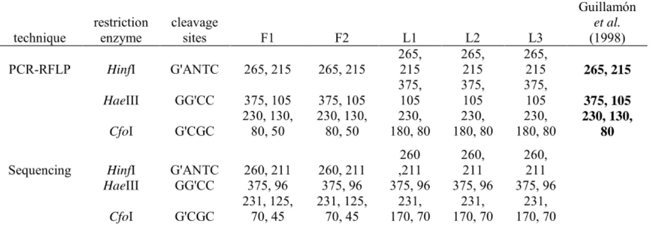
In order to study whether the B. bruxellensis strains were genetically identical, we performed a PCR-RFLP technique using the couple of primers ITS1/ITS4 targeting the region covering the internal transcribed spacer ITS1 and ITS2, including the 5.8S rDNA. Our results revealed the specific 500 pb fragments for the 5 strains of Brettanomyces tested in this study. The generated PCR products were digested with the three endonucleases HinfI, HaeIII and CfoI in order to compare the corresponding profiles (Fig. 2). Out of the 3 tested enzymes, only CfoI endonuclease provided distinctive restriction profiles and was able to differentiate between Brettanomyces strains belonging to the 2 different countries (France and Lebanon). This polymorphism was shown by the molecular weights of the restriction fragments generated as French isolates gave 4 bands of 230-130-80-50 pb while all Lebanese ones gave 3 bands of 230-180-80 pb. Restriction fragments obtained by HinfI and HaeIII had the same bands for all isolated strains. Molecular weights of the generated fragments were estimated at 265 and 215 pb when the endonuclease was HinfI and 375 and 105 pb when the endonuclease was HaeIII.
Multiple alignment and Blast result of PCR amplified products with ITS1 and ITS4 primers
The purified DNA of the 5 isolates was sequenced and aligned. As illustrated in the RFLP results, all strains present one restriction site belonging to each of the endonucleases HinfI and HaeIII, leading to two bands on the gel. French strains showed 3 restriction sites where CfoI can cleave while Lebanese ones showed only two. The sequencing results confirmed the polymorphism detected at the CfoI restriction site. Thus, the digestion by CfoI allows the differentiation between B. bruxellensis subspecies isolated from different geographical origins. Table 1 summarizes results obtained by PCR-RFLP and sequencing analysis. Cleavage sites were clearly observed in the five aligned sequences (Fig. 3) and fragments length appeared to be approximately the same in both analyses. Polymorphism was also detected when some nucleotide positions were not identical between strains. Therefore, a multiple alignment for the sequence fragment was performed, in order to show if a polymorphism can be detected. The Blast analysis of the Lebanese sequences revealed different sequences compared to the registered strains in the GenBank.
Discussion
Many studies have described detection and discrimination of Brettanomyces/Dekkera species, using various PCR techniques in different wine and wineries all over the world. The principal aim of this study is to describe the presence of Brettanomyces/Dekkera yeasts for the first time in Lebanese wine and wineries and to genetically compare these strains with others that are isolated from the French wine.
In the Lebanese collected wine, we have found that only 2 out of 62 (3.2%) showed growth of Brettanomyces when plating on the IACC medium without any enrichment phase (L1 and L2) and one after enrichment (L3). The winery, from which we isolated both L1 and L2, uses plastic tanks for conservation. In the past years, the employment of plastic tanks has increased in the wine industry, especially for small and medium wine cellars, which employ tanks from 2 to 22 hL and after solving all problems arising from the transfer of plastic odors through the employment of suitable materials. These porous tanks are permeable to oxygen due to the polymers from which they are manufactured and may have a controlled permeability, which makes their use more attractive to cellars. They are used for conservation and for aging wine by the addition of wood pieces that can produce wine with characteristics similar to those aged in barrels. In addition, these tanks allow the winemaker to control the amount of wine oxygenation (Del Álamo et al., 2010). The previously described
characteristics may favor the presence of Brettanomyces in the plastic tanks of this winery.
Millet & Lonvaud-Funel, 2000 reported that microorganisms which are able to survive in wine after wine-making for long periods and after the sulfating process may have a size reduction and form micro colonies that are not visible within the usual incubation time as shown in our results with the L3 strain. This result shows that the population that appeared, after an enrichment phase of 8 days, was either present in low cell concentrations in the bottled sample or it could be an example of a dormant state for 12 years. The isolate remained metabolically active, and a simple traditional agar medium was not able to reach detectable population levels. It cannot describe a VBNC state as this latest refers, by definition, to those cells that are not culturable, even on non-selective media no matter the length of incubation time. This fact should be taken into consideration by winemakers, since a sample may be “Brett negative” when using the traditional plate for detection while it is positive when analyzed after an enrichment phase of several days. The L3 strain was the sole one isolated from bottled wine among the 38 samples reviewed in this study.
Under aerobic or semi-aerobic conditions, Brettanomyces is capable of synthesizing significant quantities of acetic acid from glucose or ethanol, thus verifying the "acidifying" properties of this yeast (Ciani & Ferraro, 1997). This acidification is highlighted by the appearance of a translucent circle around positive colonies (L1, L2 and L3). The circle results from the dissociation of calcium carbonate according to the following reaction: CaCO3 + 2H+Ca2+ + CO2 + H2O; Ca2+ being soluble, it gives a clear gel. Six other samples revealed colonies on both IACC and EMPL, but did not exhibit any trace of translucent circle These strains could be one of the following, Kloeckera apiculata, Hanseniaspora uvarum, Pichia guilliermondi, Schizosaccharomyces pombe or Candida parapsilosis as they all show resistance between 10 and 100 mg l-1 of cycloheximide (Benito et al., 2006) as well as D. anomala and D. bruxellensis.
For all isolates, detection started with microscopic observation. As morphology is not a criterion in itself for confirming whether our isolates belong to the Brettanomyces genus or not, the samples were subjected to genetic tests.
With DBRUXF-DBRUXR primer pair, our results were similar to those of Phister & Mills, 2003 as we obtained the same band at 79 pb for all strains tested confirming that all strains isolated from wine belong to the species B. bruxellensis as was illustrated by Mitrakul et al. 1999. This latest primer pair was tested against 36 other wine-related yeasts with three bacteria and did not produce any amplicons.
The region spanning between the internal transcribed spacer ITS1 and ITS2, including the 5.8S rDNA, was amplified from the confirmed isolates, using the described primers ITS1 and ITS4. All French and Lebanese isolates produced a band of 500 pb, knowing that sizes of the generated fragments were between 450 and 550 pb for all Brettanomyces yeasts and between 600 and 900 pb for other grape and wine-specific yeasts (Egli et al., 1999). In order to study the genetic variety of the Lebanese isolates, we performed the PCR-RFLP technique and the profile of PCR digested fragments was compared with both endonucleases HinfI (265 and 215 pb) and HaeIII (365 and 105 pb). The digestion profile was identical to that obtained by Guillamón et al. 1998 and by Puig et al. 2011 whereas CfoI revealed another restriction band profile and was able to differentiate between Lebanese and French strains, thus confirming the discrepancy at subspecies level. Sizes of restriction fragments obtained by CfoI by Guillamón et al. 1998 were 230, 130, 80 pb for B. bruxellensis with a total size of 440 pb while our results gave 4 bands of 230, 130, 80, 50 pb for French strains and 230, 180, 80 pb for Lebanese ones, making a total size of 490 pb for all our isolates. This last discrimination allowed the distribution of our isolates into 2 groups of subspecies.
To complete the results obtained by PCR-RFLP, the alignment of partial sequences confirmed the presence of all restriction sites in the region ITS1-ITS4. Table 1 compares the sizes of different fragments obtained by both techniques. With some acceptable differences, values appear similar for each endonuclease. So far, there are poor genetic characterization data of D. bruxellensis. Woolfit et al. 2007 carried out a genome survey sequencing to obtain 0.4 x coverage of the genome. The studied sequences thus contain numerous uncharacterized enzymes, which may be involved in the contribution of the “Brett” character in wine and may facilitate the analyses of population structure and variation in D. bruxellensis. Hellborg & Piškur, 2009 analyzed the genome structures of 30 isolates of D. bruxellensis originating from different geographical locations around the world. These isolates revealed different numbers and sizes of chromosomes with a variation in the number of copies of several analyzed genes and their sequences.
Acknowledgment
The research work was supported by the “Conseil de la Recherche de l’Université Saint-Joseph” of Beirut.
References:
Agnolucci, M., Vigentini, I., Capurso, G., Merico, A., Tirelli, A., Compagno, C., Foschino, R. & Nuti, M. (2009). Genetic diversity and physiological traits of Brettanomyces bruxellensis strains isolated from Tuscan Sangiovese wines. International Journal of Food Microbiology 130, 238-244.
Barbin, P. (2006). Maîtrise de la contamination des vins rouges par Brettanomyces au cours des procédés de vinification (PhD Thesis). In INP (Institut National Polytechnique). Toulouse, France.
Barnett, J. A., Payne, R. W. & Yarrow, D. (1990). Yeasts: Characteristics and Identification, Cambridge University Press edn.
Benito, S., Palomero, F., Morata, A., Calderón, F. & Suárez-Lepe, J. A. (2006). Detección de Brettanomyces/Dekkera en vinos tintos mediante el uso de medios selectivo-diferenciales. Tecnología del vino 32, 27–31.
Chatonnet, P., Dubourdieu, D., Boidron, J. N. & Pons, M. (1992). The origin of ethylphenols in wines. Journal of the Science of Food and Agriculture 60, 165-178.
Chatonnet, P., Dubourdieu, D. & Boidron, J. N. (1995). The Influence of Brettanomyces/Dekkera sp. Yeasts and Lactic Acid Bacteria on the Ethylphenol Content of Red Wines. Amercican Journal of Enology and Viticulture 46, 463-468.
Ciani, M. & Ferraro, L. (1997). Role of oxygen on acetic acid production by Brettanomyces/Dekkera in winemaking. Journal of the Science of Food and Agriculture 75, 489-495.
Cocolin, L., Rantsiou, K., Iacumin, L., Zironi, R. & Comi, G. (2004). Molecular Detection and Identification of Brettanomyces/Dekkera bruxellensis and Brettanomyces/Dekkera anomalus in Spoiled Wines. Applied and Environmental Microbiology 70, 1347-1355.
Couto, J. A., Barbosa, A. & Hogg, T. (2005). A simple cultural method for the presumptive detection of the yeasts Brettanomyces/Dekkera in wines. Letters in Applied Microbiology 41, 505-510.
de Barros Lopes, M., Soden, A., Martens, A. L., Henschke, P. A. & Langridge, P. (1998). Differentiation and species identification of yeasts using PCR. International Journal of Systematic Bacteriology 48, 279-286. Del Álamo, M., Nevares, I., Cárcel, L., Crespo, R. & Gonzalez-Muñoz, C. (2010).Wine ageing in controlled permeability HDPE tanks. In Food innova: International conference on food innovation. Universidad Politecnica De Valencia.
Dias, L., Pereira-da-Silva, S., Tavares, M., Malfeito-Ferreira, M. & Loureiro, V. (2003). Factors affecting the production of 4-ethylphenol by the yeast Dekkera bruxellensis in enological conditions. Food Microbiology 20, 377-384.
Egli, C. M., Mitrakul, C. & Henick-Klong, T. (1999).Molecular identification methods of Brettanomyces yeasts. In Proceedings of the 12th International Enology Symposium, pp. 201-217. Edited by E. Lemperle. Montréal, Canada.
Fugelsang, K. C., Osborn, M. M. & Muller, C. J. (1993). Brettanomyces and Dekkera. Implications in winemaking. In Beer and wine production: analysis, characterization and technological advances pp. 110-131. Edited by B. H. Gump. Washington DC: American Chemical Society.
Guillamón, J. M., Sabaté, J., Barrio, E., Cano, J. & Querol, A. (1998). Rapid identification of wine yeast species based on RFLP analysis of the ribosomal internal transcribed spacer (ITS) region. Archives of Microbiology 169, 387-392.
Hellborg, L. & Piškur, J. (2009). Complex Nature of the Genome in a Wine Spoilage Yeast, Dekkera bruxellensis. Eukaryotic Cell 8, 1739-1749.
Heresztyn, T. (1986). Formation of Substituted Tetrahydropyridines by Species of Brettanomyces and Lactobacillus Isolated from Mousy Wines. American Journal of Enology and Viticulture 37, 127-132.
Hoffman, C. S. & Winston, F. (1987). A ten-minute DNA preparation from yeast efficiently releases autonomous plasmids for transformation of Escherichia coli. Gene 57, 267-272.
Martorell, P., Barata, A., Malfeito-Ferreira, M., Fernandez-Espinar, M. T., Loureiro, V. & Querol, A. (2006). Molecular typing of the yeast species Dekkera bruxellensis and Pichia guilliermondii recovered from wine related sources. International Journal of Food Microbiology 106, 79-84.
Medawar, W. (2003). Etude physiologique et cinétique des levures du genre Brettanomyces dans un contexte œnologique (PhD Thesis). In INP (Institut National Polytechnique), Toulouse, France & Saint-Jospeh University, Beirut, Lebanon.
Millet, V. & Lonvaud-Funel, A. (2000). The viable but non-culturable state of wine micro-organisms during storage. Letters in Applied Microbiology 30, 136-141.
Miot-Sertier, C. & Lonvaud-Funel, A. (2007). Development of a molecular method for the typing of Brettanomyces bruxellensis (Dekkera bruxellensis) at the strain level. Journal of Applied Microbiology 102, 555-562.
Mitrakul, C. M., Henick-Kling, T. & Egli, C. M. (1999). Discrimination of Brettanomyces/Dekkera yeast isolates from wine by using various DNA fingerprinting method. Food Microbiology 16, 3-14.
Oelofse, A., Pretorius I.S. & du Toit, M. (2008). Significance of Brettanomyces and Dekkera during Winemaking: A Synoptic Review. South African Journal of Enology and Viticulture 29, 128-144.
Oelofse, A., Lonvaud-Funel, A. & du Toit, M. (2009). Molecular identification of Brettanomyces bruxellensis strains isolated from red wines and volatile phenol production. Food Microbiology 26, 377-385.
Oliver, J. D. (1993). Formation of viable but nonculturable cells. In Starvation in Bacteria, pp. 239-271. Edited by S. Kjelleberg. New York: Plenum Press.
Phister, T. G. & Mills, D. A. (2003). Real-Time PCR Assay for Detection and Enumeration of Dekkera bruxellensis in Wine. Applied and Environmental Microbiology 69, 7430-7434.
Puig, A., Bertran, E., Franquet, R., García, J. & Mínguez, S. (2011). Brettanomyces bruxellensis prevalence in wines produced and marketed in Spain. Annals of Microbiology 61, 145-151.
Renouf, V., Falcou, M., Miot-Sertier, C., Perello, M. C., De Revel, G. & Lonvaud-Funel, A. (2006a). Interactions between Brettanomyces bruxellensis and other yeast species during the initial stages of winemaking. Journal of Applied Microbiology 100, 1208-1219.
Renouf, V., Miot-Sertier, C., Strehaiano, P. & Lonvaud-Funel, A. (2006b). The wine microbial consortium: a real terroir characteristic. Journal International des Sciences de la Vigne et du Vin 40, 209-216.
Rodrigues, N., Gonçalves, G., Pereira-da-Silva, S., Malfeito-Ferreira, M. & Loureiro, V. (2001). Development and use of a new medium to detect yeasts of the genera Dekkera/Brettanomyces. Journal of Applied Microbiology 90, 588-599.
Van Der Walt, J. & Van Kerken, A. (1958). The wine yeasts of the cape. Antonie van Leeuwenhoek 24, 239-252.
Wedral, D., Shewfelt, R. & Frank, J. (2010). The challenge of Brettanomyces in wine. LWT - Food Science and Technology 43, 1474-1479.
White, T. J., Bruns, T., Lee, S. & Taylor, J. (1990). Amplification and direct sequencing of fungi ribosomal RNA genes for phylogenetics. In PCR protocols A guide to methods and applications, pp. 315-322. Edited by G. D. Innis MA, Sninsky JJ, White TJ. San Diego: Academic Press.
Woolfit, M., Rozpędowska, E., Piškur, J. & Wolfe, K. H. (2007). Genome Survey Sequencing of the Wine Spoilage Yeast Dekkera (Brettanomyces) bruxellensis. Eukaryotic Cell 6, 721-733.
Table 1. Comparison between the sizes of the restriction fragments (pb) obtained by
PCR-RFLP and by sequencing.
Isolates
technique restriction enzyme cleavage sites F1 F2 L1 L2 L3
Guillamón et al. (1998) PCR-RFLP HinfI G'ANTC 265, 215 265, 215 265, 215 265, 215 265, 215 265, 215 HaeIII GG'CC 375, 105 375, 105 375, 105 375, 105 375, 105 375, 105 CfoI G'CGC 230, 130, 80, 50 230, 130, 80, 50 180, 80 230, 180, 80 230, 180, 80 230, 230, 130, 80
Sequencing HinfI G'ANTC 260, 211 260, 211
260 ,211 260, 211 260, 211 HaeIII GG'CC 375, 96 375, 96 375, 96 375, 96 375, 96 CfoI G'CGC 231, 125, 70, 45 231, 125, 70, 45 170, 70 231, 170, 70 231, 170, 70 231, Figure 1. Identification of Brettanomyces bruxellensis strains by the PCR amplification of
DBRUXF and DBRUXR elements. Lanes (1,2): French strains F1, F2; Lanes (3,4,5): Lebanese strains L1, L2, L3; Lane (C-): negative control reaction where no DNA was added; Lane (M): 100-pb ladder molecular weight marker.
Figure 2. Restriction fragments of the PCR products using ITS1 and ITS4 primers obtained
with 3 restriction endonucleases. Products were separated by electrophoresis on 3% agarose gel. Digests of respectively (a) HaeIII, (b) HinfI and (c) CfoI. Lanes (1,2,3): Lebanese strains L1, L2, L3; Lanes (4,5): French strains F1, F2; Lane (M): 100-pb ladder molecular weight marker. M 1 2 3 4 5 C -100 - pb M 1 2 3 4 5 M 1 2 3 4 5 1 2 3 4 5 M 100 400 -pb 100 300 -pb - 100 - 300 pb (a) (b) (c)
Figure 3. Sequence alignment (by Clustal W version 1.81) of ITS1-ITS4 amplified fragments from the
five Brettanomyces bruxellensis strains used in this work. Conserved nucleotides were shown by asterisks while differences were indicated by blanks. The 5.8S rDNA stretches from position 105 to 262, 18S rDNA ends at position 11 and 28S rDNA starts at position 395 and ends at 431 before the primer ITS4 which stretches from position 432 to 451. The region ITS1 is defined between positions 12 and 104 while ITS2 is between 263 and 394.
[1-60] L1_ AAGGATCATTACAGGATGCTGGGCATAAGCCCGTGCAGACACGTGGATAAGCAACGATAA L2_ AAGGATCATTACAGGATGCTGGGCATAAGCCCGTGCAGACACGTGGATAAGCAACGATAA L3_ -AGGATCATTACAGGATGCTGGGCATAAGCCCGTGCAGACACGTGGATAAGCAAGGATAA F1_ AAGGATCATTACAGGATGCTGGGCGCAAGCCCGTGCAGACACGTGGATAAGTAAGGATAA F2_ -AGGATCATTACAGGATGCTGGGCGCAAGCCCGTGCAGACACGTGGATAAGTAAGGATAA *********************** ************************* ** ***** [61-120] L1_ AAAATACATTAAATTTATTTAGTTT-AGTCAAGAAAGAATTTTAAAACTTTCAACAATGG L2_ AAAATACATTAAATTTATTTAGTTT-AGTCAAGAAAGAATTTTAAAACTTTCAACAATGG L3_ AAA-TACATTAAATTTATTTAGTTT-AGTCAAGAAAGAATTTTAAAACTTTCAACAATGG F1_ AAA-TACATTAAATTTATTTAGTTTTAGTCAAGAAAGAATTTTAAAACTTTCAACAATGG F2_ AAA-TACATTAAATTTATTTAGTTTTAGTCAAGAAAGAATTTTAAAACTTTCAACAATGG *** ********************* ********************************** [121-180] L1_ ATCTCTTGGTTCTCGCGTCGATGAAGAGCGCAGCGAATTGCGATACTTAATGTGAATTGC L2_ ATCTCTTGGTTCTCGCGTCGATGAAGAGCGCAGCGAATTGCGATACTTAATGTGAATTGC L3_ ATCTCTTGGTTCTCGCGTCGATGAAGAGCGCAGCGAATTGCGATACTTAATGTGAATTGC F1_ ATCTCTTGGTTCTCGCGTCGATGAAGAGCGCAGCGAATTGCGATACTTAATGTGAATTGC F2_ ATCTCTTGGTTCTCGCGTCGATGAAGAGCGCAGCGAATTGCGATACTTAATGTGAATTGC ************************************************************ [181-240] L1_ AGATTTTCGTGAATCATCGAGTTCTTGAACGCACATTGCGCCCTCTGGTATTCCGGAGGG L2_ AGATTTTCGTGAATCATCGAGTTCTTGAACGCACATTGCGCCCTCTGGTATTCCGGAGGG L3_ AGATTTTCGTGAATCATCGAGTTCTTGAACGCACATTGCGCCCTCTGGTATTCCGGAGGG F1_ AGATTTTCGTGAATCATCGAGTTCTTGAACGCACATTGCGCCCTCTGGTATTCCGGAGGG F2_ AGATTTTCGTGAATCATCGAGTTCTTGAACGCACATTGCGCCCTCTGGTATTCCGGAGGG ************************************************************ [241-300] L1_ CATGCCTGTTTGAGCGTCATTTCCTTCTCACTATTTAGTGGTTATGAGATTACACGAGGG L2_ CATGCCTGTTTGAGCGTCATTTCCTTCTCACTATTTAGTGGTTATGAGATTACACGAGGG L3_ CATGCCTGTTTGAGCGTCATTTCCTTCTCACTATTTAGTGGTTATGAGATTACACGAGGG F1_ CATGCCTGTTTGAGCGTCATTTCCTTCTCACTATTTAGTGGTTATGAGATTACACGAGGG F2_ CATGCCTGTTTGAGCGTCATTTCCTTCTCACTATTTAGTGGTTATGAGATTACACGAGGG ************************************************************ [301-360] L1_ TGTTTTCTTCAAAGGAAAGAGGGGAGAGTGAGGGGATAATGATTTAAGGTTTCGGCCGTT L2_ TGTTTTCTTCAAAGGAAAGAGGGGAGAGTGAGGGGATAATGATTTAAGGTTTCGGCCGTT L3_ TGTTTTCTTCAAAGGGAAGAGGGGA--GTGAGGGGATAATGATTTAAGGTTTCGGCCGTT F1_ TGTTTTCTTCAAAGGAAAGAGGGGAGAGTGAGGGGATAATGATTTAAGGTTTCGGCCGTT F2_ TGTTTTCTTCAAAGGAAAGAGGGGAGAGTGAGGGGATAATGATTTAAGGTTTCGGCCGTT *************** ********* ********************************* [361-420] L1_ CATTATTTTTTTCTTCTGCCCCAATTATCAAGTTTGACCTCAAATCAGGTAGGAGGACCC L2_ CATTATTTTTTTCTTCTGCCCCAATTATCAAGTTTGACCTCAAATCAGGTAGGAGGACCC L3_ CATTATTTTTT-CTTCTCCCCCAGTTATCAAGTTTGACCTCAAATCAGGTAGGAGGACCC F1_ CATTATTTTTT-CTTCTCCCCCAGTTATCAAGTTTGACCTCAAATCAGGTAGGAGGACCC F2_ CATTATTTTTT-CTTCTCCCCCAGTTATCAAGTTTGACCTCAAATCAGGTAGGAGGACCC *********** ***** ***** ************************************ [421-451] L1_ GCTGAACTTAAGCATATCAATAAGCGGAGGA L2_ GCTGAACTTAAGCATATCAATAAGCGGAGGA L3_ GCTGAACTTAAGCATATCAATAAGCGGAGGA F1_ GCTGAACTTAAGCATATCAATAAGCGGAGGA F2_ GCTGAACTTAAGCATATCAATAAGCGGAGGA *******************************