OATAO is an open access repository that collects the work of Toulouse
researchers and makes it freely available over the web where possible
Any correspondence concerning this service should be sent
to the repository administrator:
[email protected]
This is an author’s version published in:
http://oatao.univ-toulouse.fr/23138
To cite this version:
Brandicourt, Pierre and Delanoé, Franck and Roux,
Franck-Emmanuel and Jalbert, Florian and Brauge, David and
Lauwers, Frédéric Reconstruction of cranial vault defect with
polyetheretherketone implants.
Official URL:
https://doi.org/10.1016/j.wneu.2017.04.049
Reconstruction of Cranial Vault Defect with Polyetheretherketone Implants
Pierre Brandicourt1, Franck Delanoe´2, Franck-Emmanuel Roux1, Florian Jalbert2, David Brauge1, Fre´de´ric Lauwers2
-OBJECT:Reconstruction of a cranial vault defect is a frequent challenge in neurosurgery. Polyetheretherketone (PEEK) is used in many types of prostheses and has been employed for 10 years in our institution (University Hospital of Toulouse, France). The objectives of this study are to describe the benefits and drawbacks of reconstructing the cranial vault defect with a PEEK prosthesis.
-METHODS:Clinical data of the 37 patients who received a reconstruction with a custom-made PEEK prosthesis from 2007 2015 were retrospectively analysed. Operative tech-nique, postoperative complications, and patient’s satis-faction with the aesthetic result—on a scale ranging from 1 (very dissatisfied) to 5 (very satisfied)—were studied.
-RESULTS:Average follow-up was 4.3 years (from 2 months 9 years). The placement of the prosthesis was performed 195 days on average (from 0 1051 days, stan-dard deviation 258 days) after the initial bone flap removal. One infection (2.7%), which required the removal of the prosthesis, was described. Six patients (16%) were reop-erated by the maxillofacial surgery team to treat a lack of temporal projection related to muscle atrophy, using a fat cell autograft taken from the abdominal region. Overall, 30 patients (81%) answered the question about their aesthetic satisfaction, with good results on the satisfaction scale (average 4.5; from 3 5).
-CONCLUSION:The use of a PEEK prosthesis in cranial vault defect reconstruction is a reliable technique with a high patient satisfaction rate and few complications. Cor-rections of the temporal muscle atrophy by fat grafting may be performed in addition, without increasing the rate of complications.
INTRODUCTION
D
ecompressive craniectomies used in intractable elevated intracranial pressure raise the issue of cranial bone reconstruction. Many authors have reported elevated rates of complications with autologous bone reconstructions, such as aseptic bone resorptions, skin or boneflap infections, or brain abscesses.1Many different methods of cranial bone reconstructionhave been described in the past, such as metal prostheses (gold, silver, aluminium, titanium) and allografts from cadavers or autografts using a rib or scapula.2 Currently, a choice of synthetic materials is available for the procedure: methyl methacrylate, hydroxyapatite, ceramic, or acrylic resin.3 Polyetheretherketone (PEEK), manufactured for the first time in 1978 and marketed since 1981, is a linear, polyaromatic, semi-crystalline polymer that has many advantages.4 It is a tough, rigid, biocompatible material that is used in the fabrication of many prostheses in orthodontics or orthopedic surgery.5,6 How-ever, few studies of its use are available in the neurosurgical literature.7 The objectives of this study were to evaluate the complications and aesthetic result concerning patients in whom the cranial vault was rebuilt with a PEEK prosthesis because of a defect. The patients were all treated in a single institution.
MATERIAL AND METHODS
We retrospectively studied all patients who received a PEEK custom-made implant as part of the reconstruction of a cranial vault defect at the University Hospital of Toulouse between 2007 and 2015. Thirty-seven patients were analyzed: 15 women (40%) and 22 men (60%), with an average age of 40 years (from 12e80 years old; standard deviation [SD] 15). Because of the various indications of PEEK implants, 3 different groups were formed according to their surgical history (Figure 1). Thefirst group of patients underwent a craniotomy or decompressive craniectomy with an autologous cranioplasty, followed by removal of the autologousflap for osteitis or aseptic lysis (Table 1). The second group comprised patients who had never had autologous
Key words -Cranioplasty -Polyetheretherketone -Prosthesis
-Reconstruction of the cranial vault Abbreviations and Acronyms CT: Computed tomography PEEK: Polyetheretherketone SD: Standard deviation
From the Departments of 1
Neurosurgery and 2
Maxillofacial Surgery, Hospital Center University of Toulouse, Paul Sabatier University, Toulouse, France
To whom correspondence should be addressed: Pierre Brandicourt, M.D. [E mail: [email protected]]
37 patients Craniectomy or Craniotomy
23 patients Craniectomy 8 patients Osteo-meningioma 6 patients
\
Ablation of bone flap 23 patients
No cranioplosty One-step reconstruction
PEEK implants 37 Patients
Figure 1. The 3 different groups of the series. acx:ording to their surgical history.
cranioplasty (i.e., they had decompressive craniectomy without
autologous cranioplasty because of flap infection or a complex
cranial fracture)
(Table 2).The last group was composed of
Table 1. Characteristics of Group of 23 Patients with Decompressive Craniotomy and Autologous Cranioplasty Patients Total Women Men 23 patients 9 14 Average age at time of decompressive craniectomy/craniotomy 28.1 years Median Glasgow score after decompressive 14 (7-15) craniectomy/craniotomy
Cause of decompressive craniectomy: Traumatic brain injury
lntracranial hemorrhage
Various (stroke. raised intracranial pressure. spontaneous infection)
Meningioma
Average age during the autologous cranioplasty
Average time between decompressive craniectomy/craniotomy and autologous cranioplasty
Average time between autologous cranioplasty and removal of bone flap Cause Aseptic lysis Osteitis 12 6 3 2 28.4 years 101.6 days 7751 days 12 11
patients who had reœived 1-step surgery for ablation and recon
struction of an osteomeningioma
(Table 3).Each patient was
presented to the pharmacovigilance committee, which validated
the cost for the prosthesis and gave its agreement for surgery.
For this study, operative data (length of stay, operative rime)
and postoperative complications (infèction, hematoma, hydro
œphalus) were reviewed. To evaluate the aesthetic results, each
Table 2. Characteristics of Group of 8 Patients Who Had Craniectomy without Autologous Cranioplasty
Patients Total Women Men
Average age at time of decompressive craniectomy Median of Glasgow scores after the decompressive craniectomy
Indication of craniectomy Traumatic brain injury lntracranial hematoma Infection
Meningioma
Aeason for no autologous cranioplasty:
Oecompressive craniectomy carried out abroad (not possible to transfer the bone flap)
Oepressed bone fractures. multiple fractures of the flap Other 8 patients 3 5 42.15 years 14 (13-15) 4 2 2 2 4
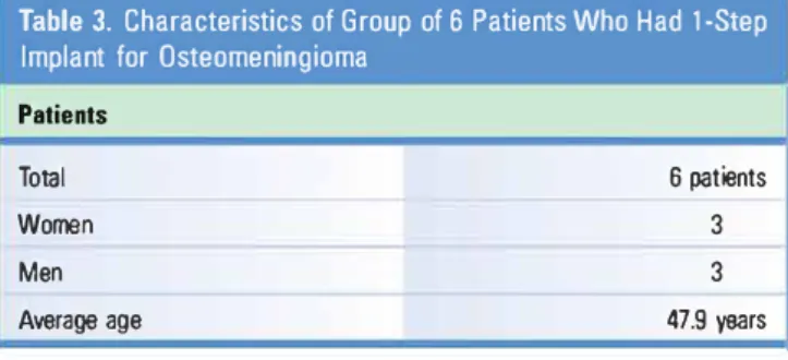
Table 3. Characteristics of Group of 6 Patients Who Had 1-Step Implant for Osteomeningioma
Patients
Total
Women
Men
Average age
6 patients
3
3
47.9 years
patient was contacted by phone and questioned on the aesthetic
(self-assessment) result of the prosthesis. A scale similar to that
employed by Rosenthal et al
7and ranging from 1 (very dissatisfied)
to 5 (very satisfied) was used
(Table 4).Implant costs (including
taxes) were calculated and depended on the number of PEEK
blocks (prosthesis volume) needed to make the prostheses.
TECHNICAL SPECIFICITY: MANUFACTURE OF THE PROSTHESIS
After the decision to implant a PEEK prosthesis with the patient's
agreement, a 3-dimensional (_31)) high-resolution computed to
mography (CT) scan with millimeter slices was made so that the
ideal implant conformation could be calculated according to the
defèct to be filled. This
cr
scan was sent to the manufacturer, and
the suggested area of resection (if there was a lysis component or
persistent bone fragment) and implant design were sent to the
surgeons in a portable document format file. The project was
validated (or modified then validated) before fàbrication of the
implant started. Depending on the size of the defèct, the implant
could be made of 1-3 pieces. A total of 24 implants were provided
by the Synthes Holding AG, Soluthurn, Switzerland/West Chester,
Pennsylvania (2007-2015) and 13 by the Stryker Corporation,
Kalamazoo, Michigan (2013-2016).
TECHNICAL SPECIFICITY: RECONSTRUCTION IN 1 STEP
This technique was used to design implants in preoperative
planning before excision of large osteomeningiomas. It allowed
the prosthesis to be ready for use before the intervention, thus
avoiding further surgery for cranial vault reconstruction. From a
3D high-resolution
cr
scan, the lesion was bounded using the
open-source software Osirix to create a _31) volume with the
planned bone resection. The PEEK prosthesis was then designed
on the basis of this tracking. An STL file (widely used for rapid
Table 4. Scale of Satisfaction with Aesthetic Result
Satisfaction
Very dissatisfied
Little satisfied
2
Neutral
3
Satisfied
4
Very satisfied
5prototyping, _31) printing and computer-aided manufacturing)
containing the limits predefined for the excision was then pro
vided by the manufacturer to be integrated in the neuronavigation
software (Brainlab Kolibri, Munich, Germany). In the operating
room, a monoblock excision, guided by the neuronavigation
software and predefined limits, was conducted by a double
neurosurgical and maxillofàcial team. The prosthesis was then
fixed immediately.
TECHNICAL SPECIFICITY: LIPOSTRUCTURE TECHNIQUE
During follow-up, based on a request from the patient, seconda
rycorrections could be made on the temporal muscle projection,
which was ofi:en atrophied afi:er several cranial surgeries and
frontotemporoparietal approaches. In such cases, lipostructure
can be used to correct a Jack of temporal projection related to
muscle atrophy and improve the aesthetic results for long-term
reconstruction, using �1 injections of autologous fat
Described in 1995 by Sydney R. Coleman, lipostructure is a fàt
cell autografi: technique in which fat cells are taken from the
abdominal region most of the time.
8Afi:er being purified by
centrifugation, the cells are fèd back into a _31) lattice and
provide good results in sofi: tissue reconstruction.
RESULTS
The mean follow-up was 43 years (from 2.7 months to 9.1 years,
SD 2.7 years). The average age at the rime of reconstruction was
35.7 years (48-77, SD 16.2 years). Implantation of the bone flap
was ofi:en performed more than 6 months after craniectomy. The
average delay was 195 days (from 0-1051, SD 258.4). The average
operating rime was 2 hours and 19 minutes (from
Ihour and
8 minutes to 4 hours and 57 minutes, SD
Ihour). The average
hospitalization rime was 6.8 days (from 3-18 days, SD 2.8 days).
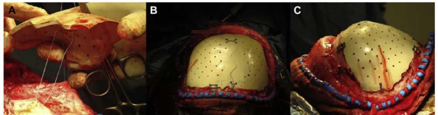
Figure 2illustrates the intraoperative implantation of the
prosthesis. Depending on the defèct size, 1-3 blocks of PEEK
were used to make the implant, with an average cost per
implant of 78g6 euros (from 3200-17107 euros, SD 2840 euros).
Postoperative Complications of Prosthetic Cranioplasty
Three subcutaneous hematomas occurred in the immediate
postoperative period, which did not require any surgery. An
infection of the PEEK prosthesis was observed in
Ipatient whose
autologous cranioplasty flap had already been infècted (Pseudo
monas
aeruginosa infèction of the operating site). A PEEK prosthesis
was implemented 91 days after ablation of this flap, and antibiotic
treatment was given. Unfortunately, he had to be reoperated
3 months later for a new empyema (P. aeruginosa), with removal of
the prosthesis.
Technical and Aesthetic Results
A correction (drilling) of the prosthesis during the operation was
used in only 2 cases (s.4
°/o) in order to improve its implementa
tion. In 6 patients (16%), a defèct in the projection of the temporal
muscle was noticed postoperatively, leading to
11lipofilling pro
cedures in these patients (1-3 procedures/patient). The lipofilling
was ofi:en performed a long rime after the insertion of the pros
thesis (on average 800 days; SD 440 days). No complications were
reported.
Thirty patients responded to the survey on their satisfaction with the aesthetic result of the reconstruction. Five could not be reached (by phone or letter). At the last follow-up, 2 patients had died from complications of their initial brain injury. As shown in Figure 3, a majority of patients (19) were very satisfied (an example of the aesthetic result is shown inFigure 4).Table 5details all the complications and aesthetic results for the whole cohort and 3 groups of this study.
DISCUSSION
This large cohort of patients who received cranial vault recon-struction with a PEEK prosthesis illustrates the multiple in-dications of this material for cranial vault reconstruction. The PEEK material can be used with very few complications after aseptic or septic bone resorption, post-traumatic decompressive craniectomy, or for osteomeningioma-scheduled bone sacrifice. The excellent level of aesthetic satisfaction of patients and the perfect intraoperative adaptation of the prosthesis attest to the
interest and usefulness of prostheses that are custom-made from a high-resolution scan of the patient, making it possible to obtain personalized reconstruction with few complications. Our results are in line with those of Rosenthal et al,7 who published a neurosurgical series of PEEK reconstructions from 3 different centers, showing a significant infection rate of 7.6% and secondary ablation of the prosthesis in 9.1% of patients but otherwise high levels of satisfaction.
PEEK material is increasingly used in neurosurgery or other surgical specialties.9-13 Its compatibility with biologic and
radiologic techniques (absence of artifacts in CT and MRI) make it a first-choice material. The biomechanical characteristics of PEEK prostheses are also interesting. PEEK is a comfortable material that is less dense and lighter than other implants. The Young’s modulus, or elastic modulus, of PEEK and bone are similar. It does not conduct heat as a metallic implant would.14 PEEK implants are frequently used for trauma and orthopedic and spinal defects.6 Jonkergouw et al15 found a much higher rate of infection (13%) for 40 patients in 2 different centers with PEEK prostheses and 12.5% of secondary removal of prostheses due to infection. Hanasono16 reported a series of 6 patients with 0% of infections. In a comparative study between PEEK and titanium cranioplasty, Thien et al17did not show significant differences in terms of complications between the 2 materials but detected a trend toward fewer complications with the PEEK.
However, other techniques and materials are available for cranial vault reconstruction.2,3 Hydroxyapatite, a mineral
component of bone, has been described as safe and effective18 but with low mechanical resistance and thus a risk of fracture of the cranial implant.19-21 Huang et al22 reported a series of 22 patients rebuilt with implants in solid polymethylmethacrylate. They reported a good satisfaction result in 20 patients and no infection but secondary removal of the implant to correct a cosmetic defect in 2 of them. In another series described by Lee et al,23 17 patients received a Figure 2. Intraoperative implantation of the prosthesis. Bifrontal cranial
vault reconstruction of a 23 year old patient, injured in a traffic accident, with serious head injury requiring a bifrontal decompressive flap. An autologous cranioplasty was performed 66 days after the decompressive craniectomy but became infected 3 weeks later. A methicillin sensitive Staphylococcus aureus infection of the surgical site was diagnosed and
necessitated the removal of the bony component. After antibiotic treatment, a bifrontal polyetheretherketone prosthesis was implanted, 153 days after removal of the component. (A) Exposure of cranial defect and suspension of the dura. (B) Fixing of the implant with screw and plates. (C) Photo of the perfect implantation of the tailored prosthesis.
Figure 3. Bar graph showing the results for patients’ satisfaction. Among 30 patients who responded to the questionnaire, 19 were very satisfied, 8 satisfied, and 3 neutral.
prosthesis in polymethylmethacrylate with an infection rate of 5. 8%. Kumar et al24described a series of 5 patients with a large
defect to fill (longer axis of >15 cm). They used a porous polyethylene prosthesis and reported no complications. Williams et al,25 in a series of 151 titanium cranioplasties, showed that the main complication was an infection of the material in 4% of their patients. This statement matched results for the series of Klinger et al,26 who showed an infection rate of 5.8% in 120 patients receiving acrylic cranioplasty for cranial vault reconstruction. The infection rate of 2.8% in our series is not greater than the latest available data from the literature.
To the best of our knowledge, there are no recommendations in the literature on the necessary waiting period between the removal of an infected bone flap and the placing of a cranial prosthesis. For Cheng et al27 and Matsuno et al,28 multiple
operations and short delays are risk factors of infection in cranioplasties. To reduce the risk of such infection, a longer period should be proposed to these patients. However, the timing of cranioplasty remains open to discussion. In a meta-analysis of 18 articles, Yadla et al29 showed that early
reintervention (<3 months) was not associated with a higher risk of infection. Nevertheless, the best timing regarding the infection complication rate remains debated.30 Further studies Figure 4. Aesthetic result of a frontotemporoparietal
prosthesis in a 40 year old man, injured in a traffic accident, with complex cranial fracture and subdural hematoma. Decompressive craniectomy with evacuation of hematoma was performed, but the bone
flap could not be kept because of multiple fragments. Top: Before the reconstruction. His neurologic outcome was good, and a polyetheretherketone (PEEK) implant was placed 191 days later. Bottom: 2 months after PEEK reconstruction.
Table 5. Results and Complications After Implantation of Prosthesis
Decompressive Craniotomy Decompressive Craniect1111y
with Autologous without Previous Autologous 1 -Step Prosthesis
Whole Cohort Cranioplasty Group Cranioplasty Group Group
Number of patients
36
23
8
6
Median Glasgow score after reconstruction
15 (7-15)
15 (7-15)
15 (14-15)
15 (15-15)
Length of hospitalization in days
6.8 (3-18, SD 2.8)
7 (4-18, SD 3.3)
5.4 (3-8, SD 1.9)
7.8 (6-10, SD 1.5)
Su bcuta neous hematoma
3
2
0
lntracranial fluid collection
0
0
0
0
Infection
Su perf.:ial
0
0
0
0
Deep
0
0
Lipostructure
Number of patients
5
3
2
Number of procedures
10
5
2
3
Average satisfaction score
4.5 (3-5)
4.6 (3-5)
4.4 (3-5)
4.4 (4-5)
Follow up (years)
4.3 (0.2-9.1, SD 2.7)
5.1 (01-9.1, SD 2.8)
2.5 (0.4-6.9, SD 2.3)
3.9 (1.6-6.5, SD 1.8)
are needed to better understand the risks associated with the
timing of the surgery.
using fàt taken from the abdominal area, with a transplant
survival rate of 90%.
8 CONCLUSIONA temporal projection defect is a common problem in
neurosurgery after a pterional or frontotemporoparietal flap and
various corrections have already been proposed.
31-33In our se
ries, 16% of patients were corrected by autografi: of fat tissue by
lipostructure.
34These patients reported a high level of
satisfaction. This original method, coupled with a prosthetic
cranioplasty, constitutes a simple and effective solution. No
infections or other complications were reported in these
patients. It allows the temporal projection to be increased
This study reports the largest series of PEEK prosthesis cranial vault
reconstructions from a single institution. With a very low rate of
complications, in terms of infèction and secondary implant removal,
the use of PEEK should form part of the annamentarium available to
the surgeon in the reconstruction of cranial vault defècts. In addition,
coupled with autologous fàt injection to correct a temporal projection
defèct, it offers excellent rates of patient satisfàction.
REFERENCES
1. Wachter D, Reineke K, Behm T, Rohde V. Cra nioplasty after decompressioe hemicraniectomy. underestimated surgery-associated complications? Clin Nturol Neurosur9. 2013;II5:n93-n97. 2. Harris DA, Fong A}, Buchanan EP, Manson L,
Khechoyan D, Lam S. Histoiy of �nthetic mate rials in alloplastic cranioplasty. Nturosur9 Foo,s. 2014;36:Elo.
3. Shah AM, Jung H, Skiiboll S. Materials used in cranioplasty: a history and analysis. Neurosur9 FOOIS. 2014;36:E19.
4. Abdullah MR, Goharian A, Abdul Kadir MR, Wahit MU. Biomechanical and bioactivity con ceptS of polyetheretherketone composites for use in orthopedic implants a review: biomechanical and bioactivity concepts of PEEK.
J
Biomed MaterRts A. 2015;103:3689-3702.
5. Schwitalla A, Müller WO. PEEK dental implants: a
review of the literature.
J
Oral Implantol. 2013;39:74:t-749-6. Kurtz SM, Devine )N. PEEK biomaterials in uauma, olthopedic, and spinal implants. Bio mamls. 2007;28:4845-486!).
7. Rosenthal G, Ng I, Moscovici S, Lee KK, Lay T, Martin C, et al. Pol)'etheretherketone implants for the repair of large cranial defects: a ;tcenter experience. Nturosur9ery. 2014;75:52;t-529.
8. Coleman SR. Structural fàt grafting: more than a permanent filler. Piast Rtœnstr Sur9. 2oo6;n8(suppl 3):1o8S-120S.
9. Goodson ML, Farr D, Keith D, Banks R). Use of two-piece pol)'etheretherketone (PEEK) implants in orbitozygomatic reconstruction. Br
J
Oral Max illojiu Sur9. 2on;50:268-26g.10. Scolozzi P, Martinez A, Jaques B. Complex orbito fronto-temporal reconstruction using
computer-designed PEEK implant
J
Craniefat Sur9. 2007;18: 224-228.11. Najeeb S, Zafar MS, Khuishid Z, Siddiqui F. Applications of pol)'etheretherketone (PEEK) in oral implantology and prosthodontics. J Prostlto<lont Rts. 2016;60:12-19.
n. Jalbert F, Lauwers F. Custom-made implants for craniofàcial reconstruction. Rtu Stomafl>I Citer Max illo-Facial, Chir Oralt. 2013;m1:211-2r8.
13. Jalbert F, Boetta S, Nadon F, Lauwers F, Schmidt E, Lapez R. One-step primary recon struction for complex crnniofàcial resection with PEEK custom-made implants. J Cranio-Maxillofat Sur9. 2014;42:141-148.
14. Lethaus B, Safi Y, ter Laak-Poort M, Kloss Brandstàtter A, Banki F, Robbenmenke C, et al. Cranioplasty with customized titanium and PEEK implants in a mechanical stress mode!.
15. Jonkergouw J, van de Vijfeijken SE, Nout E, Theys T, Van de Casteele E, Folkersma H, et al. Outcome in patient-specific PEEK cranioplasty: a two-center cohort study of 40 implants. J Cranio-Maxillofac Surg. 2016;44:1266-1272.
16. Hanasono MM, Goel N, DeMonte F. Calvarial reconstruction with polyetheretherketone im-plants. Ann Plast Surg. 2009;62:653-655. 17. Thien A, King NK, Ang BT, Wang E, Ng I.
Comparison of polyetheretherketone and titanium cranioplasty after decompressive craniectomy. World Neurosurg. 2015;83:176-180.
18. Stefini R, Esposito G, Zanotti B, Iaccarino C,
Fontanella MM, Servadei F. Use of “custom made” porous hydroxyapatite implants for cranioplasty: postoperative analysis of com-plications in 1549 patients. Surg Neurol Int. 2013;4:12.
19. Adetchessi AT, Pech-Gourg G, Metellus P, Fuentes S. [Fracture of macroporous hydroxy-apatite prosthesis]. Neurochirurgie. 2012;58: 382-385.
20. Staffa G, Nataloni A, Compagnone C, Servadei F. Custom made cranioplasty prostheses in porous hydroxy-apatite using 3D design techniques: 7 years experience in 25 patients. Acta Neurochir (Wien). 2007;149:161-170.
21. Hardy H, Tollard E, Derrey S, Delcampe P, Peron JM, Freger P, et al. [Clinical and ossification outcome of custom-made hydroxyapatite proth-esis for large skull defect]. Neurochirurgie. 2012;58: 25-29.
22. Huang GJ, Zhong S, Susarla SM, Swanson EW, Huang J, Gordon CR. Craniofacial reconstruction
with poly(methyl methacrylate) customized cra-nial implants. J Craniofac Surg. 2015;26:64-70. 23. Lee SC, Wu CT, Lee ST, Chen PJ. Cranioplasty
using polymethyl methacrylate prostheses. J Clin Neurosci. 2009;16:56-63.
24. Kumar NG, Sreenivas M, Gowda S. Cranioplasty of large cranial defects with porous polyethylene implants. J Craniofac Surg. 2016;27:e333-e335. 25. Williams LR, Fan KF, Bentley RP. Custom-made
titanium cranioplasty: early and late complications of 151 cranioplasties and review of the literature. Int J Oral Maxillofac Surg. 2015;44:599-608. 26. Klinger DR, Madden C, Beshay J, White J,
Gambrell K, Rickert K. Autologous and acrylic cranioplasty: a review of 10 years and 258 cases. World Neurosurg. 2014;82:e525-e530.
27. Cheng YK, Weng HH, Yang JT, Lee MH, Wang TC, Chang CN. Factors affecting graft infection after cranioplasty. J Clin Neurosci. 2008;15: 1115-1119.
28. Matsuno A, Tanaka H, Iwamuro H, Takanashi S, Miyawaki S, Nakashima M, et al. Analyses of the factors influencing bone graft infection after delayed cranioplasty. Acta Neurochir (Wien). 2006; 148:535-540.
29. Yadla S, Campbell PG, Chitale R, Maltenfort MG, Jabbour P, Sharan AD. Effect of early surgery, material, and method of flap preservation on cranioplasty infections: a systematic review. Neurosurgery. 2011;68:1124-1129.
30. Corallo F, De Cola MC, Lo Buono V, Marra A, De Luca R, Trinchera A, et al. Early vs late
cranio-plasty: what is better? Int J Neurosci. 2017;127: 688-693.
31. Choudhry OJ, Christiano LD, Arnaout O, Adel JG, Liu JK. Reconstruction of pterional defects after frontotemporal and orbitozygomatic craniotomy using Medpor Titan implant: cosmetic results in 98 patients. Clin Neurol Neurosurg. 2013;115: 1716-1720.
32. Badie B. Cosmetic reconstruction of temporal defect following peritonal craniotomy. Surg Neurol. 1996;45:383-384.
33. Kubo S, Takimoto H, Kato A, Yoshimine T. Endoscopic cranioplasty with calcium phosphate cement for pterional bone defect after fronto-temporal craniotomy: technical note. Neurosurgery. 2002;51:1094-1096.
34. Coleman SR. Facial recontouring with lip-ostructure. Clin Plast Surg. 1997;24:347-367.