Université de Montréal
EVALUATION OF OXYTOCIN PHARMACOKINETIC /
PHARMACODYNAMIC PROFILE AND ESTABLISHMENT OF ITS
CARDIOMYOGENIC POTENTIAL IN SWINE.
par
NORMA THELMA YBARRA NAVARRO
Département de biomédecine vétérinaire
Faculté de médecine vétérinaire
Thèse présentée à la Faculté de médecine vétérinaire
en vue de l’obtention du grade de
Philosophiae doctor (Ph.D.)
en sciences vétérinaires
option pharmacologie
Août, 2010
Université de Montréal
Faculté de médecine vétérinaire
Cette thèse intitulée
EVALUATION OF OXYTOCIN PHARMACOKINETIC /
PHARMACODYNAMIC PROFILE AND ESTABLISHMENT OF ITS
CARDIOMYOGENIC POTENTIAL IN SWINE.
présentée par
Norma Thelma Ybarra Navarro
a été évaluée par un jury composé des personnes suivantes
Bruce D. Murphy, président-rapporteur
Éric Troncy, directeur de recherche
Jérôme R.E. del Castillo, codirecteur
Jean-François Tanguay, membre du jury
Joanne Paquin, examinateur externe
Christopher A. Price, représentant du doyen
RÉSUMÉ
La thérapie cellulaire est une avenue pleine de promesses pour la régénération myocardique, par le remplacement du tissu nécrosé, ou en prévenant l'apoptose du myocarde survivant, ou encore par l'amélioration de la néovascularisation. Les cellules souches de la moelle osseuse (CSMO) expriment des marqueurs cardiaques in vitro quand elles sont exposées à des inducteurs. Pour cette raison, elles ont été utilisées dans la thérapie cellulaire de l'infarctus au myocarde dans des études pre-cliniques et cliniques. Récemment, il a été soulevé de possibles effets bénéfiques de l'ocytocine (OT) lors d’infarctus. Ainsi, l’OT est un inducteur de différenciation cardiaque des cellules souches embryonnaires, et cette différenciation est véhiculée par la voie de signalisation du monoxyde d’azote (NO)-guanylyl cyclase soluble. Toutefois, des données pharmacocinétiques de l’OT lui attribue un profil non linéaire et celui-ci pourrait expliquer les effets pharmacodynamiques controversés, rapportés dans la lttérature.
Les objectifs de ce programme doctoral étaient les suivants :
1) Caractériser le profil pharmacocinétique de différents schémas posologiques d'OT chez le porc, en développant une modélisation pharmacocinétique / pharmacodynamique plus adaptée à intégrer les effets biologiques (rénaux, cardiovasculaires) observés.
2) Isoler, différencier et trouver le temps optimal d’induction de la différenciation pour les CSMO porcines (CSMOp), sur la base de l'expression des facteurs de transcription et des protéines structurales cardiaques retrouvées aux différents passages.
3) Induire et quantifier la différenciation cardiaque par l’OT sur les CSMOp. 4) Vérifier le rôle du NO dans cette différenciation cardiaque sur les CSMOp.
Nous avons constaté que le profil pharmacocinétique de l’OT est mieux expliqué par le modèle connu comme target-mediated drug disposition (TMDD), parce que la durée du séjour de l’OT dans l’organisme dépend de sa capacité de liaison à son récepteur, ainsi que de son élimination (métabolisme).
D'ailleurs, nous avons constaté que la différenciation cardiomyogénique des CSMOp médiée par l’OT devrait être induite pendant les premiers passages, parce que le nombre de passages modifie le profile phénotypique des CSMOp, ainsi que leur potentiel de différenciation. Nous avons observé que l’OT est un inducteur de la différenciation cardiomyogénique des CSMOp, parce que les cellules induites par l’OT expriment des marqueurs cardiaques, et l'expression de protéines cardiaques spécifiques a été plus abondante dans les cellules traitées à l’OT en comparaison aux cellules traitées avec la 5-azacytidine, qui a été largement utilisée comme inducteur de différenciation cardiaque des cellules souches adultes. Aussi, l’OT a causé la prolifération des CMSOp. Finalement, nous avons observé que l'inhibition de la voie de signalisation du NO affecte de manière significative l'expression des protéines cardiaques spécifiques.
En conclusion, ces études précisent un potentiel certain de l’OT dans le cadre de la thérapie cellulaire cardiomyogénique à base de cellules souches adultes, mais soulignent que son utilisation requerra de la prudence et un approfondissement des connaissances.
Mots-clés: Ocytocine, cellules souches adultes, différenciation cardiomyogénique, monoxyde d’azote, porc, pharmacokinetic, pharmacodynamic
ABSTRACT
Cell therapy has been suggested as a promising treatment for myocardial regeneration through cardiomyocyte replacement or by preventing apoptosis of surviving myocardium and/or improving neovascularisation. Bone marrow stem cells (BMSCs) express cardiac markers in vitro upon stimulation with different inducers. The BMSCs have been used as cell therapy after myocardial infarction (MI) in pre-clinical and clinical studies. Recent reports have uncovered the potential beneficial effects of oxytocin (OT) after MI. Particularly, OT is an inducer of cardiomyogenic differentiation of embryonic stem cells and this differentiation is mediated by the nitric oxide (NO)-soluble guanylyl cyclase pathway. However, some studies have shown that OT exhibits nonlinear pharmacokinetics and that this could explain the previously described controversial hemodynamic alterations.
Therefore the objectives of the present work were to:
1) Characterize the pharmacokinetic profile of different dosing regimens of OT in swine, by using a more suitable pharmacokinetic / pharmacodynamic modelization that could explain the time-course of cardiovascular and renal effects observed following OT administration.
2) To isolate, differentiate and find the optimum time of porcine BMSC (pBMSC) differentiation based on the expression of cardiac related transcription factors and structural proteins expressed at different passages.
4) To document the role of the NO pathway in the OT-mediated cardiomyogenic differentiation of pBMSCs.
We found that OT pharmacokinetics are better explained by target-mediated drug disposition (TMDD) kinetics, because the time-course of plasma OT concentration depends on the binding capacity to its receptor, as well as OT elimination (metabolism).
Also, we found that OT-mediated cardiomyogenic differentiation of pBMSCs should be induced during the first passages, because passaging affects the phenotypic profile of pBMSCs, as well as the differentiation potential of pBMSCs.
We observed that OT induces cardiomyogenic differentiation of pBMSCs, because OT-induced cells expressed cardiac markers, and the expression of cardiac specific proteins was more abundant in OT-treated cells vs. 5-azacytidine-treated cells, which has been used widely as a cardiomyogenic differentiation inducer of adult stem cells. Moreover, OT improved proliferation of pBMSCs. Finally, we observed that the inhibition of the NO pathway significantly affects the expression of cardiac specific proteins.
To conclude, these studies demonstrate some interesting potential in cardiomyogenic differentiation of adult stem cells for OT, but its precise role in cell therapy will need prudence and further investigations.
Keywords: Oxytocin, adult stem cells, cardiomyogenic differentiation, nitric oxide, swine, pharmacokinetics, pharmacodynamics.
TABLE OF CONTENTS
RÉSUMÉ ... III
ABSTRACT ... V
LIST OF TABLES ... XII
LIST OF FIGURES ... XIII
LIST OF ABBREVIATIONS ... XVI DEDICATORY...XXV
ACKNOWLEDGMENTS...XXVI
CHAPTER 1. INTRODUCTION ... 1
CHAPTER 2. LITERATURE REVIEW ... 5
2.1 Stem cells ... 6
2.1.1 Definition and properties ... 6
2.1.2 Classification ... 6
2.1.3 Embryonic stem cells (ESCs) ... 6
2.1.4 Adult stem cells ... 8
2.1.5 Cardiomyogenic potential of embryonic stem cells ... 9
2.1.6 Adult stem cells with cardiomyogenic potential ... 14
2.1.6.1 Resident cardiac stem cells (CSCs) ... 15
2.1.6.2 The c-Kit positive cells ... 15
2.1.6.3 The Sca-1 positive cells ... 16
2.1.6.4 Side population cells ... 17
2.1.6.5 Non-resident cardiomyogenic stem cells ... 18
2.1.6.7 Mesenchymal stem cells ... 20
2.2. Oxytocin (OT) ... 23
2.2.1 Definition and structure ... 23
2.2.2 Oxytocin synthesis... 24
2.2.3 Other sites of OT synthesis ... 24
2.2.3.1 Heart ... 25
2.2.3.2 Vasculature ... 26
2.2.3.3 Ovary and uterus ... 26
2.2.4 Regulation of oxytocin production and secretion ... 27
2.2.5 Central regulation of oxytocin production and secretion ... 28
2.2.6 Other factors controlling oxytocin secretion ... 30
2.2.7 Oxytocin receptor: Classification and activation ... 32
2.2.8 Oxytocin receptor ... 33
2.2.8.1 Brain ... 33
2.2.8.2 Kidney ... 34
2.2.8.3 Heart and cardiovascular system ... 35
2.2.9 Oxytocin effects extern to the reproductive system ... 36
2.2.10 Oxytocin renal effects ... 36
2.2.11 Oxytocin cardiovascular effects ... 38
2.2.12 Oxytocin cardio-protective effects ... 41
2.2.13 Oxytocin in cardiomyogenesis ... 43
2.2.14 Oxytocin in cardiomyogenic differentiation of stem cells ... 43
2.2.15 Oxytocin pharmacokinetics and therapeutic use ... 44
2.2.15.1 Oxytocin elimination ... 47
2.2.15.2 Evidences of non-linear oxytocin pharmacokinetics ... 50
2.3 Nitric oxide ... 53
2.3.1 Definition and synthesis ... 53
2.3.2 Nitric oxide cardio-protective effects ... 55
2.3.3 Nitric oxide in cardiomyogenesis ... 57
CHAPTER 3. FIRST ARTICLE ... 62
3.1 ABSTRACT ... 64
3.2 INTRODUCTION ... 65
3.3 MATERIALS AND METHODS ... 67
3.4 RESULTS ... 71
3.5 DISCUSSION ... 74
3.7 REFERENCES ... 79
3.8 TABLES AND FIGURES ... 82
CHAPTER 4. SECOND ARTICLE ... 96
4.1 ABSTRACT ... 98
4.2 INTRODUCTION ... 99
4.3 MATERIALS AND METHODS ... 101
4.3.1 Isolation and culture of pBMSCs ... 101
4.3.2 Characterization ... 102
4.3.3 Cardiomyogenic differentiation ... 104
4.3.4 Immunocytochemistry ... 105
4.3.5 Western Blot analysis ... 106
4.3.6 Transmission electron microscopy ... 107
4.4.1 Isolation and characterization of pBMSCs ... 108
4.4.2 Cardiomyogenic differentiation induction ... 109
4.5 DISCUSSION ... 110
4.7 REFERENCES ... 117
4.8 TABLES AND FIGURES ... 124
CHAPTER 5. THIRD ARTICLE ... 131
5.1 ABSTRACT ... 133
5.2 INTRODUCTION ... 134
5.3 MATERIAL AND METHODS ... 136
5.3.1 Isolation and culture of pBMSCs ... 136
5.3.2 Cell treatment ... 137
5.3.3 Effect of the treatments on OT-mediated cardiomyogenic differentiation ... 138
5.3.4 Statistical analyses ... 141
5.4 RESULTS ... 142
5.4.1 Expression of NOS in pBMSCs cardiomyogenic differentiation ... 143
5.4.2 Effect of the inhibitors on OT-mediated cardiomyogenic differentiation of pBMSCs ... 143
5.4.3 Effect of treatments on cell count ... 144
5.5 DISCUSSION ... 145
5.7 REFERENCES ... 151
5.8 TABLES AND FIGURES ... 156
CHAPTER 6. GENERAL DISCUSSION ... 165
6.1 Oxytocin pharmacokinetics and pharmacodynamics ... 166
6.2 Cell therapy in cardiovascular disease: why promote adult stem cells and particularly bone marrow derived stem cells? ... 170
6.3 How to optimize BMSCs for cell therapy ... 173
6.4 Potential of oxytocin as a therapy for myocardial infarction: effects on embryonic and adult stem cells, on isolated ex vivo organs, and preclinical models in rats and pigs. ... 175
6.5 Mechanisms of action of OT-induced cardiomyocyte differentiation (role of NO, future implications, propositions of future tests) ... 177
6.6 Potential roles of oxytocin in the future therapies: with BMSCs, hypotheses of research, as well as limitations ... 180
CHAPTER 7. GENERAL CONCLUSIONS ... 186
7.1 Oxytocin pharmacokinetics and pharmacodynamics ... 187
7. 2 Isolation and characterization of pBMSCs ... 187
7.3 Cardiomyogenic differentiation induction of pBMSCs ... 187
7.4 Involvement of the NO-sGC pathway in the OT mediated cardiomyogenic differentiation of pBMSCs ... 188 CHAPTER 8. REFERENCES ... I
LIST OF TABLES
CHAPTER 2. LITERATURE REVIEW
TABLE 1. ENZYMES PARTICIPATING IN OXYTOCIN METABOLISM ... 48
CHAPTER 3. FIRST ARTICLE
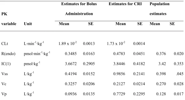
TABLE 1. TIME-POINTS OF SERIAL BLOOD SAMPLING FOR DETERMINING PLASMA OXYTOCIN CONCENTRATIONS ... 82
TABLE 2. AVERAGE MOMENT PHARMACOKINETIC PARAMETERS AND VARIABLES AS A FUNCTION OF OXYTOCIN ADMINISTERED DOSE ... 83
TABLE 3. SUMMARY OF MEAN OVERALL CHANGES IN CARDIOVASCULAR PARAMETERS AFTER BOLUS OR CONSTANT RATE INFUSION OF OXYTOCIN IN PIGS. ... 84
CHAPTER 4. SECOND ARTICLE
TABLE 1. PCR PRIMERS AND REACTION CONDITIONS. ... 124
CHAPTER 5. THIRD ARTICLE
LIST OF FIGURES
CHAPTER 2. LITERATURE REVIEW
FIGURE 1. EMBRYONIC STEM CELLS DERIVED FROM EMBRYOS AT MORULAE AND BLASTOCYST STAGE ... 7
FIGURE 2. ADULT STEM CELLS DERIVED FROM ADULT TISSUES ... 8
CHAPTER 3. FIRST ARTICLE
FIGURE 1. PLASMA OXYTOCIN CONCENTRATIONS ASSOCIATED WITH INTRAVENOUS ADMINISTRATION OF EXOGENOUS OT IN PIGS. ... 85
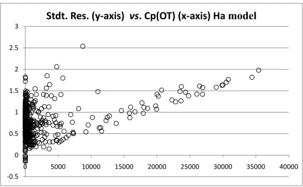
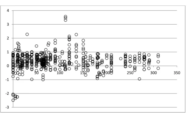
FIGURE 2. COMPARISON OF NULL-HYPOTHESIS AND ALTERNATIVE-HYPOTHESIS MODELS ADEQUACY. ... 88
FIGURE 3. OXYTOCIN PHARMACOKINETIC / PHARMACODYNAMIC (CARDIOVASCULAR) CORRELATION. ... 93
CHAPTER 4. SECOND ARTICLE
FIGURE 1. FLUORESCENT-ACTIVATED CELL SORTING ANALYSIS OF PORCINE BONE MARROW STEM CELLS (PBMSCS) SURFACE MARKERS. ... 125
FIGURE 2. SEMI-QUANTITATIVE RT-PCR ANALYSIS OF GENE EXPRESSION PROFILE IN PORCINE BONE MARROW STEM CELLS (PBMSCS). ... 126
FIGURE 3. SEMI-QUANTITATIVE RT-PCR ANALYSIS OF GENE EXPRESSION PROFILE AFTER DIFFERENTIATION INDUCTION. ... 127
FIGURE 4. CARDIAC PROTEIN EXPRESSION ON PBMSCS. ... 128
FIGURE 5. WESTERN BLOT ANALYSIS OF CARDIAC TROPONIN I ... 129
FIGURE 6. TRANSMISSION ELECTRON MICROSCOPE IMAGES OF PBMSCS ... 130
CHAPTER 5. THIRD ARTICLE
FIGURE 1. EFFECTS OF OXYTOCIN (OT) ON ENDOTHELIAL (ENOS) AND INDUCIBLE (INOS) NITRIC OXIDE SYNTHASE TRANSCRIPTS AND PROTEINS EXPRESSION OF BOTH ENZYMES. ... 158
FIGURE 2. REPRESENTATIVE IMAGE OF SEMI-QUANTITATIVE RT-PCR ANALYSIS OF CARDIAC MARKERS GENE EXPRESSION 10 DAYS AFTER INITIAL TREATMENT ... 159
FIGURE 3. EXPRESSION OF CARDIAC TROPONIN-T PROTEIN ON TREATED CELLS IN THE PRESENCE AND ABSENCE OF NITRIC OXIDE PATHWAY INHIBITORS. ... 160
FIGURE 4. EXPRESSION OF CARDIAC MYOSIN HEAVY CHAIN PROTEIN ON TREATED CELLS IN THE PRESENCE AND ABSENCE OF NITRIC OXIDE PATHWAY INHIBITORS. ... 162
FIGURE 5. SEMI-QUANTITATIVE WESTERN BLOT ANALYSIS OF PROTEIN EXPRESSION OF THE CARDIAC SPECIFIC MARKER CARDIAC TROPONIN I, EFFECTS OF INHIBITORS OF NITRIC OXIDE PATHWAY ... 164
LIST OF ABBREVIATIONS
5-Aza 5-azacytidine µg Micrograms
µmol/min Micromolar per minute
µU Microunits 3-D Three-dimensional
ABC ATP-binding cassette transporters
Abcg2 ATP-binding cassette, sub-family G member 2 ANOVA Analysis of variance
ANP Atrial natriuretic peptide
ATP Adenosine triphosphate
ATPase ATP hydrolytic enzyme
AVP Arginine vasopressin
B220 Surface marker of human hematopoietic stem cells, equivalent to murine CD45
BM Bone marrow
BM MNC Mononuclear population cells of the bone marrow Bmax Maximum receptor density
BMP-2 Bone morphogenic protein-2
Bmpr1a Bone morphogenetic protein receptor, type IA BMSCs Bone marrow stem cells
BNP Brain natriuretic peptide
bp Base pairs
Ca Blood concentration (arterial)
Ca2+ Calcium
CAD Coronary artery disease
cAMP Cyclic adenosine monophosphate CD4 Cluster of differentiation 4 CD8 Cluster of differentiation 8 CCD camera Charge-coupled device CD20 Cluster of differentiation 20 CD31 Cluster of differentiation 31 CD34 Cluster of differentiation 34 CD45 Cluster of differentiation 45
CD45RO Cluster of differentiation 45 RO isoform CD90 Cluster of differentiation 90
cDNA Complementary DNA
cGMP Cyclic guanosine monophosphate
c-Kit Also known as CD117 cytokine receptor Cl Clearance
Cl- Chloride ion
CLd Distribution clearance
CLt Elimination clearance
cm2 Squared centimetre
cMHC Cardiac myosin heavy chain CNS Central nervous system
CO Cardiac output
COS-1 Cell line obtained by immortalizing a CV-1 cell line derived from kidney cells of the African green monkey. CRI Constant rate infusion
CSCs Cardiac stem cells
CSF Cerebrospinal fluid
CSMO Cellules souches de la moelle osseuse CSP Cardiomyocyte structural proteins cTNI3 Cardiac troponin I-3
cTnT or TNNT2 Cardiac troponin T-2
ctrl Control
Cu Urine concentration
Cv Blood concentration (venous)
CVP Central venous pressure Cys-1 Amino acid cysteine, position 1
DAG Diacylglycerol DEA/NO 2-(N,N-diethylamino)-diazenolate-2-oxide DMEM-LG Dulbecco's Modified Eagle Medium-low glucose
DMSO Dimethyl sulfoxide
DNA Deoxyribonucleic acid
EBs, EB Embryoid bodies, embryoid body
ECG Electrocardiogram tracings
ECMs Embryonic cardiomyocytes
EDTA Ethylenediaminetetraacetic acid
eNOS Endothelial nitric oxide synthase
Erk1/2 Extracellular signal-regulated kinases 1 and 2 ERPF Effective renal plasma flow
ES Embryonic stem
ESCs Embryonic stem cells
F Forward
FBS Fetal bovine serum
FiO2 Inspired fraction of oxygen
FITC Fluorescein isothiocyanate isomer 1 Flk-1 Kinase insert domain receptor Flt-1 Fms-related tyrosine kinase 1
fmol Fentomoles
GATA-4 GATA binding protein 4, family of zinc-finger transcription factors
GFP Green fluorescent protein GPCR G protein-coupled receptors Gr-1 Surface marker myeloid cells
h Hour
H+ Hydrogen cation
HR Heart rate
HSC Hematopoietic stem cell
HUVEC Human umbilical vein endothelial cells
I/R Ischemia/reperfusion ICC Immunocytochemistry IgG Immunoglobulin IL-1β Interleukin one beta
IL-6 Interleukin 6 iNOS Inducible nitric oxide synthase
IP3 Inositol 1,4,5-trisphosphate
K+ Potassium
KATP channels ATP-sensitive potassium channels
Kd Constant of dissociation
kg Kilograms
L/min Liter per minute
Lin− Lineage negative
L-NAME Nω-Nitro-L-arginine methyl ester hydrochloride
M Molar Mac-1 Macrophage-1 antigen
MAP Mean arterial pressure
MAP kinase Mitogen-activated protein kinase
MDRs Multidrug resistances
Mef2 Myocyte enhancer factor MEF-2C Myocyte enhancer factor-2C
Mg2+ Magnesium
MHC Myosin heavy chain
MI Myocardial infarction
min Minutes mL Millilitres mL/beat Milliliter per beat
mL/min Milliliter per minute
MLC-2v Myosin light chain-ventricular mmHg Millimeter of mercury
MNCs Mononuclear cells
mRNA Messenger ribonucleic acid MSCs Mesenchymal stem cells
mU Milliunits
Na+ Sodium ion
ng Nanograms ng/kg or ng kg-1 Nanograms per kilogram
ng/kg/h Nanograms per kilogram per hour ng/mL or ng mL-1 Nanograms per millilitre
Nkx2.5 NK2 transcription factor related, locus 5 nmol Nanomoles nNOS Neuronal nitric oxide synthase
NO Nitric oxide
NOS Nitric oxide synthase
O2 Oxygen
ºC Degrees Celsius
ODQ 1H-[1,2,4]Oxadiazolo[4,3-a]quinoxalin-1-one OT Oxytocin
OTase Oxytocinase
OTR Oxytocin receptor
P Probability
P1 Passage one
PAP Pulmonary artery pressure pBMSCs Porcine bone marrow stem cells PBS Phosphate buffered saline
PCR Polymerase chain reaction
PCV Packed cell volume
PCWP Pulmonary capillary wedge pressure
PD Pharmacodynamics PDE Phosphodiesterase pg/mL or pg mL-1 Picograms per millilitre
pH Potential for hydrogen ion concentration
PIP2 Phosphatidylinositol 4,5-bisphosphate
PK Pharmacokinetics pKa Potential of acid dissociation constant
PKC Protein kinase C
PKG Protein kinase G
PLB Phospholamban
PLC Phospholipase C
pM Picomolar pBMSC Porcine bone marrow stem cells
PVDF Polyvinylidene difluoride
PVN Paraventriculear nucleus
R Reverse
RNA Ribonucleic acid
RPE R-Phycoerythrin
RPFc Corrected renal plasma flow
RT-PCR Reverse transcriptase polymerase chain reaction SAS Statistical analysis software
Sca-1 Stem cell antigen-1
SD Standard deviation
SDS-PAGE Sodium dodecyl sulfate polyacrylamide gel electrophoresis
SE Standard error
SEM Standard error of the mean
SERCA-2 Sarco/Endoplasmic Reticulum Ca2+-ATPase type 2
sGC Soluble guanylyl cyclise
SNAP S-nitroso-N-acetyl-d,l-penicillamine
SON Supraoptic nucleus
SP Side population
SpO2 Pulse oximetry
SV Stroke volume
SVR Systemic vascular resistance TBS Tris buffered saline
TBX5 T-box 5
Tef-1 Transcriptional enhancer factor-1 TER119 Marker of erythroid lineage Thy1 or CD90 Thymocyte differentiation antigen
TMDD Target-mediated drug disposition
Tyr-2 Aminoacid tyrosine, position 2 U Units
UF Urine flow
uNOS Universal nitric oxide synthase (antibody detecting three NOS isoforms)
USMC Human uterine smooth muscle cells UV Ultraviolet V Volts
V1a-type Arginine vasopressin receptor type 1 (vascular) V2 Arginine vasopressin receptor (renal)
V3 (also known as V1b) Arginine vasopressin receptor (pituitary) Vc Central volume of distribution
VEGF Vascular endothelial growth factor vs. Versus Vss Steady-state volume of distribution
Wnt coined as a combination of Wg (wingless) and Int
α1 Alpha one
α-MHC Alpha myosin heavy chain
β1 Beta one
β-MHC Beta myosin heavy chain
ACKNOWLEDGMENTS
First, I would like to thank Dr. Éric Troncy for being my director during my MSc and PhD programs. I want to thank you for your support and guidance during the last almost 6 years, thanks for giving me the opportunity to work with you. Thanks for your trust and support during this time; I really appreciate your kindness and patience. Thanks for all the good advices and for also being a friend. Thanks for playing an important role in my professional and personal development.
I would also like to thank Dr Jerome del Castillo, for being my co-director and for his enthusiasm for explaining pharmacokinetics and statistics, aunque ambos sabemos no compartimos la misma pasión. Thanks for sharing your knowledge with your students, also thanks for your patience and for always having a good disposition to offer your help, also I enjoyed the good jokes in Spanish. Muchas gracias.
I want to thank Valerie Morin, who studied her master degree under the supervision of Dr Troncy, thanks a lot for being my first friend in Quebec, thanks for all your support and friendship especially in those moments of confusion, and for all the fun times we spent together (sometimes dangerous), I will never forget the times when we worked until very late during the night and we were so exhausted, but you still had energy to make jokes and make me laugh.
I want to thank Dr Simon Authier for all his invaluable help and support during all the experiments performed during my master’s degree program, your enthusiasm is always inspirational. I wish I could have your energy!
I want to thank my laboratory collegues, Dominique Gauvin for her help and support during these years, and for sharing her knowledge in laboratory techniques and protocols.
Thanks to Colombe Otis for her help in data analysis and protocols preparations.
Thanks to Pascale Rialland for all the nice times and her friendship also thanks for the good jokes and the time we laughed together.
I want to especially thank Dr Bruce Murphy for very precious help and support during my PhD program, thanks a lot for always have been willing to share your knowledge and to always have open doors of your lab to other students that sometimes are not from the CRRA. Thanks a lot for always having the will of help the others; I also appreciate your jokes.
I want to thank Mira Dobias, for her invaluable help and for always been willing to share her experiences and knowledge, also thanks a lot for always saying hello with a smile in your face, and always make me feel welcome to your lab.
I want to thank Dr. Lawrence Smith and his research associates, Patrick Vincent, France, Jacinthe, for being so open and willing to share knowledge, without mentioning your ever present smiles and kindness. Thanks Patrick for your enthusiasm in sharing your knowledge and for always being willing to help.
Mercie beaucoup Mme St Germain pour toutes les fois que vous m’avais auxilié avec toute sorte des questions et aussi des problèmes, merci beaucoup pout tout votre aide depuis le debut. Thanks a lot to all the staff working in the secretariat. Thanks to Mme Rodier for her help and advice.
In fact, thanks to all the CRRA staff and students!
I also want to thank my dear friends at the CRRA: Adriana Verduzco, Joelle Demarais, Simon Demers not only for your precious friendship but also for sharing with me your knowledge and experience in the lab.
Also thanks a lot to all my friends in the faculty for sharing your time with me, thanks for the fun times you made me have during my post-graduate studies: Adriana, Joelle, Simon, Ricardito, Aron, Lan, Virginie, Pavine, Johanna.
But of course, I want to thank the most important person in my life, my mother for always being there for me. Simplemente a ti te debo todo lo que soy, no tengo palabras para expresarte mi gratitud y admiración. I want to thank my whole family, my brothers, my uncles, thanks for your love and support. Gracias a mis hermanos. Especialmente gracias tío Miguel, por siempre estar con nosotros apoyándonos, gracias porque siempre podemos contar contigo y por darnos tu cariño incondicional. A mi madrina, porque a pesar de la distancia siempre está al pendiente de mi, gracias por tus palabras de aliento.
A mis mejores amigas Karla, Mickey y Lucia, estaremos lejos físicamente pero siempre están ahí para apoyarme y quererme, gracias por siempre escucharme y hacerme sentir mejor cuando más las necesito. Gracias a todos mis amigos.
Cardiovascular diseases remain the leading cause of mortality and morbidity worldwide. Despite substantial improvements in acute management, survivors of myocardial infarction often progress to heart failure (Siu, Moore et al. 2007). Heart failure is a condition that can result from any structural or functional cardiac disorder. It is defined as the inability of the left ventricle to fill with and/or eject sufficient blood to meet the metabolic requirements of the body. It is undoubtedly an increasing common condition (Harris 1994).
Despite major advances in treatment, the prognosis after a diagnosis of heart failure is poor and comparable to that of several forms of cancer. Even though, the rate of fatalities per case associated with heart failure has declined, the crude number of deaths attributed to the condition has increased, primarily because of increasing prevalence of the condition. Several epidemiological investigations have identified the main risk factors for heart failure, which are: increasing age, hypertension, coronary artery disease (CAD), diabetes, obesity, valvular heart disease, and the metabolic syndrome (Kenchaiah, Narula et al. 2004).
The relative contribution of risk factors to the occurrence of heart failure in the community may be changing over time. The aging of the population, the better treatment of CAD, and the improved survival and ‘‘salvage’’ of patients with myocardial infarction (MI) with subsequent progression to pump failure are believed to be some factors contributing to the growing burden of heart failure. Heart failure is also predicted to increase in line with increased atrial fibrillation and diabetes among the elderly population. Nowadays, hypertension and CAD are the major modifiable risk factors for heart failure. Globally, CAD and arterial hypertension alone or in combination account for more than 90% of cases of heart failure (Lloyd-Jones, Evans et al. 2002; Ezekowitz and Kaul 2010).
Although human cardiomyocytes are reported to proliferate and contribute to the increase in muscle mass of the myocardium after myocardial infarction (Beltrami, Urbanek et al. 2001), their capacity for regeneration, mitigation of the adverse effects of ventricular remodelling,
and contribution to cardiac function is limited. Among other factors, cardiac wall thinning and cardiac remodelling lead to a diminished capacity of the heart to pump blood effectively leading to eventual heart failure and ultimately death of the patient. Heart transplantation is currently the last resort for end stage heart failure, but is hampered by rejection and a severe shortage of donor organs. The main purpose of cell-based therapies as applied for the treatment of myocardial infarction is to prevent heart failure by either rescuing the host myocardium or regenerating cardiac cells (Christoforou and Gearhart 2007; Tao and Li 2007; Dimmeler and Zeiher 2009).
In the past, a variety of cell types have been used experimentally, obtaining promising results, mainly by improving neovascularization, reducing infarct size, limiting wall thinning, and providing proangiogenic and antiapoptotic factors promoting tissue repair in a paracrine manner. By all these mechanisms cell therapy may curb the degeneration to heart failure (Tao and Li 2007).
The cell types used previously include skeletal myoblasts, as a non-cardiac contracting cell, and foetal cardiomyocytes, but these approaches are also limited by cell availability or side effects, mainly due to a non-cardiac identity, as observed in the case of skeletal myoblasts. There is a special interest in the use of stem cells in cell-based therapies, because of their capacity of cell renewal, and differentiation potential. In vitro studies examined the ability of stem cells to differentiate into cardiomyocytes and then proceeded to investigate the functional characteristics of these cells. In vivo studies examined the capacity of stem cells to graft into the host myocardium and then assayed the functional recovery of the diseased heart.
During the latest years, pre-clinical and clinical studies exploiting stem cells from different sources for transplantation in animal models and patients have reported favourable outcomes (Christoforou and Gearhart 2007; Tao and Li 2007; Dimmeler and Zeiher 2009).
Unfortunately, full recovery has not been achieved yet, due to many variables that need to be considered. For instance, the optimal cell type, which in turn should generate contractile cells that integrate both functionally and structurally into the surrounding viable myocardium. These cells have to beat in a synchronized manner to avoid alterations in the electrical conduction and syncytial contraction of the heart, these cells should also be able to survive in the hostile environment into which they will be grafted (Tao and Li 2007; Dimmeler and Zeiher 2009).
2.1 Stem cells
2.1.1 Definition and properties
Stem cells are unspecialized cells, with the long-term capacity of symmetrical self-renewal. Differentiation is a process that involves undifferentiated or unspecialized cells progressing into specialized cells with restricted developmental potential. They can differentiate into specialized cells, including cardiomyocytes (Blau, Brazelton et al. 2001; Bishop, Buttery et al. 2002). The differentiation capacity of these specialized cells in vivo to form mature cell types ultimately depends on the state of commitment of the cell and both intrinsic factors and the extra-cellular environment (niche) (Perino, Yamanaka et al. 2008).
The genetic and cellular mechanisms that initiate stem cell differentiation are poorly understood. Transplanted stem cells also undergo a “homing” process in which they are attracted to the site of injury (Hardy 1995). The exact homing mechanism and organ-specific differentiation signals for stem cells are not clearly understood but may be related to microenvironmental factors that are favourable to stem-cell growth and function, integrin and other adhesion molecules, homing receptors, ischemia, and increased expression of different paracrine factors and cytokines (Lee, Wolf et al. 2000; Caplice and Gersh 2003).
2.1.2 Classification
Stem cells have been classified based on their source of isolation, and by their differentiation potential into embryonic stem cells, adult stem cells and induced pluripotent stem cells.
2.1.3 Embryonic stem cells (ESCs)
Embryonic stem cells can be derived from the embryo at morulae stage, based on their differentiation potential, they are known as totipotent cells because they can give rise to
extraembryonic and embryonic tissues. Stem cells derived from the inner cell mass of the embryo at blastocyst stage are known as pluripotent cells. These pluripotent cells can differentiate into the three primary germ layers, endoderm, mesoderm, and ectoderm, meaning they have the potential to differentiate into all tissue specific cells of an embryo, and if the embryo is implanted in utero following tetraploid aggregation, an entire ES cell-derived embryo, excluding some extra-embryonic tissues, can be formed (Nagy, Gocza et al. 1990).
Figure 1. Embryonic stem cells derived from embryos at morulae and blastocyst stage
Source: http://www.stemcellresearchfoundation.org
Embryonic stem cells can also be used for the generation of chimeric animals, in which the ES cell genotype can be passed through the germline (Illmensee and Mintz 1976). Another pluripotent cell type includes germ cells, progenitor cells of the germline, these cells are found in a specific part of the foetus called the gonadal ridge, germ cells can also give rise to cells from the three germ layers (Moreno-Ortiz, Esteban-Perez et al. 2009).
2.1.4 Adult stem cells
Adult stem cells possess a more restricted developmental potential, giving rise to a subset of cells belonging to the same germ layer, they are generally considered to be multipotent to unipotent. They typically produce only cells of a closely related family (cells from the tissue or organ in which they reside), providing “new” cells in order to replenish damaged
specialized cells in the adult (Wobus and Boheler 2005; Perino, Yamanaka et al. 2008).
Figure 2. Adult stem cells derived from adult tissues
Source: National institutes of health. www.nih.gov
Both embryonic and adult stem cells are capable of cell division in the undifferentiated state (self-renewal). Embryonic stem cells readily form tumours when implanted outside the blastocyst, adult stem cells (e.g., hematopoietic, mesenchymal, neuronal) may or may not form tumours, but both ES and adult stem cells are different to cancer stem cells (Perino, Yamanaka et al. 2008).
Noteworthy, cancer stem cells are oncogenic, have lost the ability to prevent uncontrolled proliferation or differentiation, and are therapeutically unviable, properties that make them different from normal stem cells (Maenhaut, Dumont et al. 2010).
2.1.5 Cardiomyogenic potential of embryonic stem cells
Embryonic stem cells are conducive to cell implantation therapy, mainly because they are pluripotent. These unique pluripotent cell lines can be propagated in the undifferentiated state in culture and coaxed to differentiate into cell derivatives of the three germ layers, including cardiomyocytes (Capi and Gepstein 2006; Tao and Li 2007).
Doetschman and colleagues (Doetschman, Eistetter et al. 1985) made the initial observation that when ESCs are grown in suspension culture and under conditions favourable for differentiation (without feeder layer and cytokines that maintain an undifferentiated state, i.e. leukaemia inhibitory factor –LIF–), ESCs aggregated to form spherical structures called embryoid bodies (EBs). Stochastic differentiation within EBs resulted in the juxtaposition of different developmental fields, thereby mimicking induction cues that occurred during normal embryogenesis. As a consequence, cell lineages of endoderm, ectoderm, and mesoderm origin were observed to appear in differentiating EBs. Prominent cardiomyogenesis occurred during EB differentiation, as evidenced by the presence of well-formed myofibers, as well as spontaneous contractile activity. Thus, differentiating ESCs might provide a surrogate source of donor cardiomyocytes for therapeutic cell transplantation (Rubart and Field 2006; Perino, Yamanaka et al. 2008).
Studies with ESCs derived cardiomyocytes revealed that cardiomyogenic differentiation in EBs closely paralleled that observed for early stages of heart development in vitro (Doevendans, Kubalak et al. 2000), and that cardiomyocytes with the typical characteristics of the primitive heart tube and early chamber myocardium could be easily identified (Fijnvandraat, van Ginneken et al. 2003; Fijnvandraat, van Ginneken et al. 2003). The
temporal phenotypic changes in myofiber structure in ES-derived cardiomyocytes closely paralleled those observed in cardiomyocytes in vivo (Guan, Rohwedel et al. 1999). Similarly, ES-derived cardiomyocytes exhibited a temporal pattern of cell cycle withdrawal and multinucleation similar to that observed in vivo (Klug, Soonpaa et al. 1995). Electrophysiological studies revealed that cells with characteristics of atrial, ventricular, and sinus-nodal foetal cardiomyocytes were present following terminal differentiation of ESCs (Maltsev, Rohwedel et al. 1993), a result that was confirmed via molecular analyses (Miller-Hance and Chien 1993). The developmental changes in the electrophysiological properties of ESCs derived cardiomyocytes from initial cardiomyoblast commitment (Kolossov, Fleischmann et al. 1998), through formation of three-dimensional, spontaneously contracting structures (Maltsev, Rohwedel et al. 1993; Banach, Halbach et al. 2003) have been well characterized.
The reproducible differentiation observed in cultured EBs has been exploited not only to identify factors that could be able to enhance cardiomyogenic induction, but also it has been useful to identify transcription factors or signalling pathways important for cardiomyogenic differentiation. Examples of these transcription factors are: Nkx2.5, GATA-4 and Mef2, which also have been used as markers of differentiation. Some studies have implicated the signalling pathways of bone morphogenic proteins and Wnt family members in cardiomyogenic differentiation of ESCs (Winnier, Blessing et al. 1995; Czyz and Wobus 2001; Behfar, Zingman et al. 2002; Kawai, Takahashi et al. 2004; Terami, Hidaka et al. 2004). In addition, signalling through the fibroblast growth factor (Dell'Era, Ronca et al. 2003; Kawai, Takahashi et al. 2004), insulin growth factor-II (Morali, Jouneau et al. 2000), Cripto (Parisi, D'Andrea et al. 2003), cardiotrophin (Sauer, Neukirchen et al. 2004), transforming growth factor beta (Behfar, Zingman et al. 2002), and dynorphin B (Ventura, Zinellu et al. 2003) pathways enhanced cardiomyogenesis during EB differentiation. Cardiomyogenic differentiation has being promoted also by indirect mechanisms, such as
LIF-regulated cardiomyogenesis in ESCs cultures via its effect on parietal endoderm differentiation (Bader, Al-Dubai et al. 2000). Thus, altering the relative content of other cell lineages subsequently provided cardiomyogenic inducing factors, that enhances the yield of cardiomyocytes.
In addition to these defined factors, co-culture experiments revealed that precardiac endoderm contained factors capable of enhancing ESC cardiomyogenic differentiation (Rudy-Reil and Lough 2004). Other inducers used in enhancing ESC cardiomyogenic differentiation are some organic compounds such as dimethysulfoxide (DMSO) (Rudnicki, Jackowski et al. 1990), a novel butyric and retinoic acid linked ester of hyaluronan (Ventura, Maioli et al. 2004), and inorganic compounds including lithium (Schmidt, Guan et al. 2001), reactive oxygen species (Sauer, Neukirchen et al. 2004), and nitric oxide (NO) and its signalling components which apparently play a pivotal role during cardiomyogenesis (Bloch, Fleischmann et al. 1999), and enhance the differentiation of ESCs into cardiomyocytes (Kanno, Kim et al. 2004; Mujoo, Krumenacker et al. 2006). Also the neurohypophyseal hormones arginine vasopressin and oxytocin (OT) enhanced the ESC cardiomyogenic differentiation (Paquin, Danalache et al. 2002; Gassanov, Jankowski et al. 2007).
Gene transfer experiments identified additional pathways that enhanced cardiomyogenic differentiation in ESCs. For example, overexpression of GATA-4 (Arceci, King et al. 1993; Grepin, Nemer et al. 1997), alpha-1,3-Fucosyltransferase (Sudou, Muramatsu et al. 1997), and TBX5 (Fijnvandraat, Lekanne Deprez et al. 2003) resulted in increased yields of cardiomyocytes in differentiating EB cultures.
In summary, the reports described above identified many factors that could increase cardiomyocyte content in differentiating ESCs cultures. The information obtained from such studies can in turn be exploited to enhance the production of ESCs derived cardiomyocytes for clinical applications, or to discover signalling pathways that could be used to improve the differentiation of adult stem cells.
In spite of all the interesting qualities of ESCs, there are some ethical concerns about their use in cell therapy. The use of human ESCs in research requires the generation of human ESC lines. The lines currently used were mostly produced from fertilized oocytes that had undergone about 7-8 divisions. The major objection to the use of ESCs is that their generation is purported to involve an “act of killing”. The resulting controversy has delayed or stopped human ESCs research in some countries, and could affect the extent to which human ESCs derived therapies are developed and used (Robertson 2001).
Many obstacles remain to be overcome before ESCs can be expected to undergo evaluation in clinical trials. Further, the multiple regulatory issues that surround the use of ESCs and their derivatives, including the difficulties this potentially raises with respect to federal funding, continue to heavily influence the research that is conducted with these cells. Despite this, steady progress continues to be made toward understanding the mechanisms/pathways that control ESCs. A particularly important issue is the potential of ESCs to induce teratoma formation when transplanted. This may be dependent on many factors, including the experimental model and the exact nature of the ESCs population that is administered (Gonzales and Pedrazzini 2009). Currently, the consensus view is that the administration of ESCs that are at least partially committed to a certain fate may considerably reduce the risk of teratoma formation (Kovacic, Harvey et al. 2007).
Given the possible risk of graft rejection, the ethical and other concerns surrounding the use of human ESCs, a recent possible alternative for the possible graft rejection, could be the ESCs like induced pluripotent stem (iPS) cells initially derived from murine fibroblasts. The iPS cells have been created by the retroviral introduction of Oct3/4, Sox2, c-Myc, and Klf4, these factors have been related to pluripotency in ESCs (Kovacic, Harvey et al. 2007). The iPS could be considered a substitute in the evolution of the stem cell field and for cell-based therapies. Unfortunately, the high risk of teratoma formation remains. Furthermore, various safety concerns exist that must be resolved before iPS cell therapy becomes a reality.
Potential risks are related to the delivery of the endogenous factors, alterations in target cells, the cellular effects of the expression and reactivation of the factors that induce pluripotency, and safety issues related to the incorrect characterization and incomplete differentiation of the reprogrammed cells (Jalving and Schepers 2009).
An interesting alternative to the controversial destruction of a human embryo and the possible teratoma formation, is the use of stem cells obtained from adult tissues (Dickens and Cook 2007).
Cell therapy with autologous stem cells from adults, i.e. bone marrow cells is completely justified ethically, except for the small numbers of patients with direct or indirect bone marrow disease (e.g. myeloma, leukaemic infiltration) in whom there would be lesions of mononuclear cells (Strauer, Brehm et al. 2008).
The use of human autologous adult (bone marrow) stem cells is clinically justified and ethically unquestionable because no side-effects have been reported, especially with regard to teratocarcinoma. Moreover, in contrast to differentiation of embryonic stem cells immunosuppressive therapy is unnecessary. Thus, the therapeutical advantage clearly prevails, and clinical use has already been realized, at least in cell therapy for MI, obtaining promising results and no arrhythmogenic potential has been reported (Strauer, Brehm et al. 2008).
Further and intensified research using autologous human bone marrow stem cells is needed and is essential in order to promote stem cell therapy for the numerous cardiac diseases. Clinical use of autologous bone marrow mononuclear cells has no ethical problems. Therapy is performed with usual cardiac catheterization techniques (Strauer, Brehm et al. 2008).
2.1.6 Adult stem cells with cardiomyogenic potential
Whereas ESCs clearly havethe capacity to differentiate into a variety of cell types andgive rise to all tissues, adult stem cells are morespecified (more lineage committed) and the use of adult stemcells for cell therapy appears to be rather limited.
The cardiomyogenic potential of adult stem cells is a continuous source of controversy, due to the diversity of experimental protocols, cell sources, the limited in vitro functional tests, and the difficulties in the follow up of transplanted cells in vivo. The follow up of the in vivo differentiation of adult stem cells represents a special challenge because of the presence of two types of artefacts, the transfer of cell labels used to track implanted cells to neighbouring cells (Burns, Ortiz-Gonzalez et al. 2006), and heterotypic cell fusion, which is a process whereby donor and host cells merge, resulting in a fused cell with the genetic information of both cells, including any genetic marker (Nygren, Jovinge et al. 2004; Reinecke, Minami et al. 2008). Both artefacts have been observed in vitro and in vivo, and complicate the demonstration of adult stem cell in vivo differentiation potential (Reinecke, Minami et al. 2008).
Demonstration of in vitro differentiation potential of adult stem cells also represents some challenges, for instance the demonstration of an unambiguous cardiac phenotype. The most common approach is to show the expression of one or more cardiac specific markers by immunocytochemistry (ICC), reverse transcriptase-polymerase chain reaction (RT-PCR) (Antonitsis, Ioannidou-Papagiannaki et al. 2007), and western blot (Genovese, Spadaccio et al. 2009). In reality, there are very few specific cardiac markers, and so this approach requires the judicious selection of multiple markers and appropriate controls. Ideally, such phenotyping by cardiac markers is accompanied by functional assays (Rubart and Field 2006), such as action potential recordings or fluorescent calcium (Ca2+) imaging (Yoon, Choi et al. 2008).
Even with all the difficulties listed above there are several reports showing the existence of adult stem cells with cardiomyogenic potential which reside mainly in the heart itself or in the bone marrow.
2.1.6.1 Resident cardiac stem cells (CSCs)
Several independent laboratories have reported the existence of CSCs, based on the presence of the receptor tyrosine kinase c-Kit (Beltrami, Barlucchi et al. 2003), stem cell antigen-1 (Sca-1) (Oh, Bradfute et al. 2003), or the presence of side population (SP) cells that express multidrug resistance transporter genes and exclude Hoechst dye (Liang, Tan et al. 2010). Proponents of the stem cell theory of cardiac self-renewal postulate that these cells, isolated from cardiac tissues, easily differentiate into cardiomyocytes. Some reports have described these observations as experimental artefacts or cell fusion. Perhaps more importantly, opponents have suggested that some of the putative stem cell derivatives may arise from de-differentiated adult cardiac myocytes, trans-determination or even trans-differentiation events and therefore these cells are not authentic or resident stem cells. The origin of cardiomyogenic stem cells is also open to debate, partly because hematopoietic stem cell markers such as c-Kit and Sca-1 are most prominent outside the heart, suggesting that they may originate elsewhere (Braun and Martire 2007). Although SP cells have been associated at least partially with hematopoietic stem cell (HSC) populations, others have shown that the HSCs were equally distributed in the non-SP population, suggesting that precaution should be taken with respect to the claim that SP cells represent a stem cell population (Goodell, Brose et al. 1996; Morita, Ema et al. 2006).
2.1.6.2 The c-Kit positive cells
Beltrami et al. first reported the discovery of a c-Kit+ population of resident stem cells that could be isolated from adult rat heart and expanded in vitro under limiting dilution conditions
(Beltrami, Urbanek et al. 2001). These relatively small cells were calculated to be present at only about 1 per 100 cardiomyocytes and were lineage negative (Lin−) for blood lineage
surface markers [cluster differentiation (CD) CD34, CD45, CD20, CD45RO and CD8]. The c-Kit+ cells were also very heterogeneous, with less than 10% of the cells expressing Nkx2.5,
GATA-4 and Mef2 cardiac transcription factors, and less than 0.5% of the cells expressing sarcomeric proteins. Under appropriate conditions, these cells could differentiate into cardiomyocytes (Beltrami, Urbanek et al. 2001). Based on the proof of cell division, using Ki-67 protein as a recognized marker strictly associated to cell proliferation, the authors also indicated that repair occurred independently of cell fusion. However, most of the derived cardiomyocytes appeared immature with either limited sarcomeric structures or the presence of stress fibers that are typical of fibroblasts or myofibroblasts (Urbanek, Quaini et al. 2003).
2.1.6.3 The Sca-1 positive cells
Sca-1 is one of the most recognized HSCs markers in mice, and an anti-Sca-1 antibody is routinely used to identify and isolate murine HSC from bone marrow. Oh et al. identified a small number of Sca-1+ cardiac cells that overlapped with a SP of cells from heart. The
cardiac Sca-1+ cells lacked blood lineage surface markers (CD4, CD8, B220, Gr-1, Mac-1,
and TER119), c-Kit, Flt-1, Flk-1, vascular endothelial-cadherin, von Willebrand factor, and HSC markers CD45 and CD34. Moreover, these cells expressed GATA-4, Mef2 transcription factors and Tef-1, but not Nkx2.5 or genes that encoded cardiac sarcomeric proteins. The Sca-1+ stem/progenitor cells did not spontaneously differentiate into cardiomyocytes, but
following induction with 5-azacytidine, a DNA demethylating agent that causes pronounced epigenetic modifications, genes for Nkx2.5, α-myosin heavy chain (MHC), β-MHC, and the type 1A receptor for bone morphogenetic proteins (Bmpr1a) were induced, and contraction was observed. The cells were however mononucleated and fibroblast-like in structure (Oh, Bradfute et al. 2003).
On the other hand, Matsuura et al. reported that OT, but not 5-azacytidine, induces Sca-1+
cells from the adult murine heart to differentiate into functional, spontaneously beating, immature cardiomyocytes with Ca2+ transients typical to those found in heart cells (Matsuura,
Nagai et al. 2004). The 5-azacytidine-treated cells however developed a fibroblast-like morphology and never spontaneously contracted. In this study, both treatments up-regulated cardiac transcription factors Nkx2.5, GATA-4, and MEF-2C and structural proteins for α- and β-MHC, myosin light chain (MLC)-2a, MLC-2v, and cardiac α-actin. The OT-treated cells that formed cardiomyocytes were few in number, but these cells had positive inotropic responses to isoproterenol via β1-adrenergic receptor signalling (Matsuura, Nagai et al. 2004).
Given the apparently small number of cardiomyocytes generated in vitro from this induction, it seems that cardiomyogenesis is not a default pathway for these cells, and the potential to differentiate into true cardiac progenitors and cardiomyocytes requires further investigation (Matsuura, Nagai et al. 2004).
2.1.6.4 Side population cells
The ATP-binding cassette (ABC) transporter family contains 50 members that use the hydrolysis of ATP to pump toxins from cells. The ABC transporters encoding multidrug-resistance genes (MDRs) also efflux the DNA binding dye Hoechst 33342, which allows for the easy identification and sorting of ABC transporter-positive and transporter-negative cells by flow cytometry. In most cases, cells positively expressing ABC transporters comprise a very small percentage of freshly isolated cells, which appear as a “side population” (SP) of cells relative to the majority of non-SP cells as observed using flow cytometry. The SP cells and especially the Abcg2-dependent SP cell populations, have been associated with stem/progenitor cells and with long-term self-renewal (Goodell, Brose et al. 1996). The ATP-binding cassette, sub-family G, member 2, also known as Abcg2 gene is robustly expressed in the developing heart (Martin, Meeson et al. 2004). At later developmental stages and in the
adult heart, this transporter could also be used to identify a rare cell population, this SP phenotype comprising around 1% of all cells in the mouse heart and these cells did not express hematopoietic surface markers that was not enriched for the hematopoietic markers CD34, c-Kit, Sca-1, Flk-2, and Thy1 (Hierlihy, Seale et al. 2002). Further experiments, in which cardiac SP cells from adult green fluorescence protein (GFP) transgenic mice were isolated and co-cultured with cardiac main population cells from wild-type mice, showed that after 14 days, an unspecified fraction of the GFP positive SP-derived cells immunostained for the striated muscle marker α-actinin. The purified myocardial SP cells from GFP transgenic mice were capable of forming cardiomyocytes, but similarly to the c-Kit+ cells previously
described, only did so under co-culture conditions with primary cardiomyocytes.
2.1.6.5 Non-resident cardiomyogenic stem cells
Many non-resident stem cell populations with cardiomyogenic potential have been described. Many clinical trials have shown the safety and therapeutic potential of bone marrow mononuclear cells after myocardial infarction (Strauer, Brehm et al. 2002; Lipinski, Biondi-Zoccai et al. 2007; Arminan, Gandia et al. 2008). However, this population is very heterogeneous and is not enriched with stem or progenitor cells, therefore recent clinical trials using cellular cardiomyoplasty have focused on the potential of specific subpopulations, such as mesenchymal stem cells (MSCs), or HSCs.
2.1.6.6 Hematopoietic stem cells
Therapeutic applications of HSCs in patients with infarcted myocardium have resulted in very different results. Probably, this was due to differences in purification protocols, influencing the outcome of the therapies. Transplanted cells may have contained different contents of stem cells or maybe the selected cells were a heterogeneous population containing other cells with regenerative potential (paracrine effects). The latter is quite possible because
different CD surface antigens were used to purify the definitive stem cell populations. The HSCs are the most studied and well characterized population of stem cells. These cells display a unique set of markers (Murine: c-Kit+, Sca-1+, Lin−; Human: CD34+, Thy-1+, Lin−).
Cells expressing these markers correspond to a resident stem cell population suitable for long-term replacement therapy. Some reports have shown that HSCs have the ability to promote myocardial regeneration; Orlic et al. mobilized HSCs from bone marrow to show that the “trans-differentiated cells” could repair the infarcted heart (Orlic, Kajstura et al. 2001; Perino, Yamanaka et al. 2008). In a complementary study, they isolated HSCs from mice that constitutively expressed the GFP and used these cells to look for cardiac integration and repair (Orlic, Kajstura et al. 2001). After two weeks of myocardial infarction induction, the mice transplanted with the GFP-labelled HSCs presented improved function of the myocardium, and the newly formed myocardium contained GFP-labelled cells that occupied up to 68% of the infarcted portion of the ventricle. These labelled cells also expressed sarcomeric actins and myosins, troponin and several cardiac-associated transcription factors; however, the sarcomeric structures appeared disorganized. These results were further supported with a report by Rota et al. (2007) who showed that bone marrow cells engraft, survive, and grow within the spared myocardium after infarction (Rota, Kajstura et al. 2007). More specifically, they reported that locally delivered bone marrow cells generate new myocardium composed of integrated cardiomyocytes and coronary vessels, and that the cardiomyogenesis was independent of cell fusion, but dependent on close contact with resident cardiomyocytes (Rota, Kajstura et al. 2007).
On the other hand, several reports have contradicted these findings. Murry et al. reported that the HSCs did not express cardiac specific genes, and that they could not detect any increase in cardiomyocytes in a murine model of MI region of the heart (Murry, Soonpaa et al. 2004). Also, Kawada et al. showed that mobilized murine HSCs expressing GFP do not differentiate into cardiomyocytes after myocardial infarction (Kawada, Fujita et al. 2004).
2.1.6.7 Mesenchymal stem cells
Mesenchymal stem cells possess multipotent capabilities; they can proliferate in vitro and in vivo. These cells are able to induce angiogenesis, and to differentiate into cell types belonging to the mesodermal lineage, such as osteogenic, chondrogenic, adipogenic and myogenic cells. The MSCs have been first isolated from bone marrow (Pittenger, Mackay et al. 1999), which is the main source of MSCs. But they have also been isolated from other sources, like dental pulp (Pierdomenico, Bonsi et al. 2005), adipose tissue (Izadpanah, Trygg et al. 2006), umbilical cord blood (Wang, Seshareddy et al. 2009), chorionic villi of the placenta (Igura, Zhang et al. 2004), amniotic fluid (Tsai, Lee et al. 2004), and others.
Although, MSCs obtained from different sources share the expression of certain genes, differences in self-renewal, proliferation, and differentiation have been reported to exist among these various sources of MSCs (Arminan, Gandia et al. 2008).
Animal studies and early clinical studies suggest that therapeutically delivered MSCs can improve heart function after acute MI. More specifically, MSCs seemed to improve contractility, wall thickness and decrease necrosis (Nagaya, Fujii et al. 2004). It has been proposed that the observed therapeutic benefits are probably mediated through the release of a variety of signalling molecules, which may be anti-inflammatory, anti-apoptotic and angiogenic.
The paracrine factors secreted by bone marrow mononuclear cells and endothelial progenitor cells are well characterized (de Macedo Braga, Lacchini et al. 2008). More recently, the gene expression profiles of cultured MSCs were determined by microarray. In particular, transcripts of IL-6, leukemia inhibitory factor (LIF) and vascular endothelial growth factor (VEGF) family members were found to be expressed in human MSCs. Also expressed are several mRNAs for matrix-mediating factors such as matrix metalloproteinase (MMP)-2, inhibitors such as tissue inhibitor of metalloproteinase-1, 2, and matricellular proteins
(thrombospondin-1 and tenascin C) were also highly expressed in human MSCs. The results of the microarray analysis demonstrated that cultured human MSCs expressed mRNAs for a variety of secreted factors that may be cardioprotective and reparative (Iso, Spees et al. 2007). The anti-apoptotic role and broader spectrum of all the paracrine molecules produced by MSCs suggest that the release of these mediators could augment angiogenesis, reduce apoptosis and promote the recruitment of circulating progenitor stem cells into the injured tissue, leading to the favourable remodelling of the damaged heart (Kinnaird, Stabile et al. 2004).
Studies by Tang et al. have reported that the expression of basic fibroblast growth factor (FGF), vascular endothelial growth factor (VEGF), and stromal cell-derived factor (SCDF-1) is increased after MSCs in a rat model of MI. The FGF, is a mostly mitogenic protein, is produced by MSCs and stimulates sustained quiescence and proliferation in uncommitted and committed MSCs (Benavente, Sierralta et al. 2003), and it has been suggested that SCDF-1 may have tissue protective and regenerative roles in solid tissues, this by activating the cell-survival pathway mediated by protein kinase B (Tang, Zhao et al. 2005; Saxena, Fish et al. 2008).
These data suggest that paracrine effects are responsible for the cardioprotection seen with MSC therapy cells in the bone marrow.
Some authors claim that there is only limited data suggesting cardiomyogenic differentiation of MSCs (Li, Guo et al. 2009). But apparently, MSCs have a wider differentiation potential than what it has been previously thought. Some reports have shown that MSCs can differentiate into vascular endothelial cells, and smooth muscle cells (Song, Lee et al. 2007), but more importantly, it has been shown that MSCs are able to differentiate into cardiomyocytes in vivo and in vitro. For instance, in vitro studies have shown that MSCs express some cardiac transcription factors and structural proteins when exposed to 5-azacytidine, a DNA demethylating agent, which induces pronounced epigenetic changes. The
reputed formation of cardiomyocytes after 5-azacytidine exposure was based on the spontaneous contraction of cells in culture, action potentials similar to those found in foetal cardiomyocytes, the expression of sarcomeric proteins, and the expression of atrial natriuretic peptide (ANP) and brain natriuretic peptide (BNP). These peptides are expressed in cardiac muscle; but the sarcomeric proteins used as markers, skeletal α-actin and β-MHC, are associated with both cardiac and skeletal muscle cells. In addition, published images clearly showed multinucleated cells, which are most typical of skeletal muscle (Makino, Fukuda et al. 1999). These results suggest that the MSCs exposed to 5-azacytidine were not authentic cardiomyocytes, but probably a mixed population of muscle cells, due to the hypomethylation of regulatory genes caused by 5-azacytidine treatment. Therefore, it is very possible that 5-azacytidine differentiation induction is not cardiac specific (Bittira, Kuang et al. 2002).
Differentiation of MSCs has also been achieved by co-culture with cardiomyocytes (Rangappa, Entwistle et al. 2003); these cells after co-culture with cardiomyocytes were positive for β-myosin heavy chain, cardiac troponin T, cardiac troponin I, cardiac α-actin, and desmin. They also exhibit Ca2+ transients, and several types of action potentials (Li, Yu et al.
2008). Furthermore, it has been reported that even without stimulation, MSCs express certain cardiac markers, such as connexin-43, α-actinin, and GATA-4, but fail to express cardiac specific structural proteins (Bayes-Genis, Roura et al. 2005). The ability of MSCs to form cardiomyocytes is still in doubt and in almost all the cases in which cardiomyogenic differentiation has been reported, the cells had to go through a cultivation period that may have altered some basic properties. In vivo differentiation has also been reported, there are some studies stating that MSCs can differentiate into cardiomyocytes when injected into healthy murine heart (Toma, Pittenger et al. 2002). The MSCs however remain attractive as a vehicle for cell transplantation or for tissue engineering, because they can be obtained in relatively large numbers and are easily expanded in culture, in addition they can be obtained
from the bone marrow of the patient, eliminating the problems related with graft rejection. Therefore, MSCs remain a promising source for cell therapy in cardiac diseases.
There are many factors influencing the capabilities of MSC differentiation, such as: culture medium, presence and concentration of undetermined growing factors in the foetal bovine serum (FBS), number of passages, as well as differentiation inducer(s) added to the medium. In fact, some reports have shown that MSCs have certain predisposition to differentiate into cardiomyocytes (Bayes-Genis, Roura et al. 2005) or have passage-restricted differentiation potential into cardiomyocyte-like cells depending on the time they have been in culture, with a very possible variation among species (Zhang, Li et al. 2005). Phenotype and gene expression are affected during culture (Vacanti, Kong et al. 2005). For these reasons, it is possible that full and effective cardiomyogenic differentiation of MSCs has not been achieved yet.
2.2. Oxytocin (OT)
2.2.1 Definition and structure
The peptide hormone OT has many functions in the human body, including facilitating milk expulsion during lactation (Nishimori, Young et al. 1996), and mediating maternal behaviour (Young, Wang et al. 1998). However, OT is best known as a powerful uterotonic agent that stimulates uterine contractions during labour, for which it is frequently used to augment labor and to prevent postpartum haemorrhage (Wise and Clark 2008). Oxytocin was the first hormone to have its structure elucidated and the first to be chemically synthesized in a biologically active form (Du Vigneaud, Ressler et al. 1953). Oxytocin is a cyclic nonapeptide (single linear chain of nine amino acids), derived from large precursor proteins called pro-peptides, which in turn are derived from even larger molecules known as pre-propeptides. The molecular formula of OT is C43H66N12012S2 and it has a molecular weight of 1,007 Da. It
effect. There is a close homology between OT and vasopressin, the difference consisting only in two amino acids. The similarity in chemical structure explains several common properties which can become evident when OT is administered in high doses (Lippert, Mueck et al. 2003).
2.2.2 Oxytocin synthesis
The synthesis of OT occurs in the neural cell bodies of the supraopticus nucleus (SON) and paraventricularis nucleus (PVN) of the hypothalamus. From this nuclei, it is transported into the neurohypophysis in the form of a precursor molecule, oxytocin-neurophysin, this complex passes down the axons of hypothalamic nuclei to be stored as a dimeric in dilated nerve terminals in the posterior lobe of the pituitary gland (neurohypophysis), from which it is released into the blood stream in a pulsatile form (al-Eknah and Homeida 1991; Lippert, Mueck et al. 2003).
The inactive precursor protein is progressively hydrolyzed into smaller fragments (one of which is neurophysin I) via a series of enzymes. The last hydrolysis which releases the active oxytocin nonapeptide is catalyzed by peptidylglycine alpha-amidating monooxygenase (Sheldrick and Flint 1989).
The release of the neurosecretory product from the terminals occurs mainly as a consequence of invasion of the terminal membranes by action potentials. The electrical activity of the neuron is thus the immediate determinant of secretory output (Bicknell 1985).
2.2.3 Other sites of OT synthesis
Recent studies have shown that OT is a hormone that can be synthesized at many sites and several physiological activities have been attributed to this peptide.