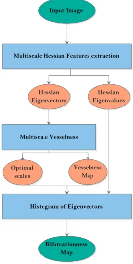
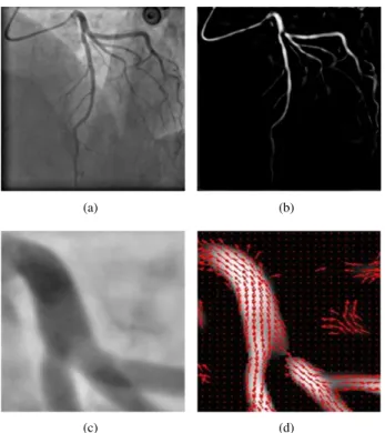
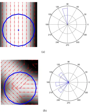
Automatic Bifurcation Detection in Coronary X-Ray Angiographies
Texte intégral
Figure
Documents relatifs
En résumé, les fibres modifient le mécanisme d’endommagement sous sollicitation statique, la première fissuration est retardée, de plus les fibres jouent un rôle de
Par la suite une réflexion spéculaire a été observée sur Kraken Mare (le plus grand lac de méthane, situé au pôle Nord) avec VIMS, confirmant la présence d’une surface liquide
L’archive ouverte pluridisciplinaire HAL, est destinée au dépôt et à la diffusion de documents scientifiques de niveau recherche, publiés ou non, émanant des
One important measurement for photon counting pixel detectors is the efficiency between the pixels.. If the threshold is too high, we lose photons because the charges are shared
The main specification for HEP is a fast sparse readout while for x-rays it is the photon counting at high rate and large dynamic range.. The conclusion is that the electronic
Long and Rinard (2016a) have performed an analysis of the search space of their two systems SPR and Prophet on the benchmark of C bugs. They show that not all repair operators are
If the subspace method reaches the exact solution and the assumption is still violated, we avoid such assumption by minimizing the cubic model using the ` 2 -norm until a
