A De e p N a s o p h a r y n g e a l S w a b V e r s u s N o n e n d o s c o p i c
B r o n c h o a l v e o l a r L a v a g e f o r Is o l a t i o n o f B a c t e r i a l P a t h o g e n s f r o m
P r e w e a n e d C a l v e s W i t h R e s p i r a t o r y D i s e a s e
L. Van Driessche, B.R. Valgaeren, L. Gille, F. Boyen, R. Ducatelle, F. Haesebrouck, P. Deprez, and
B. Pardon
Background: Nonendoscopic bronchoalveolar lavage (BAL) is a practical alternative for a deep nasopharyngeal swab (DNS) to sample the airways of a large number of calves in a short period of time. The extent of commensal overgrowth and agreement of BAL with DNS culture results in preweaned calves are unknown.
Objectives: To compare commensal overgrowth and bacterial culture results between DNS and BAL samples. Animals: A total of 183 preweaned calves (144 with bovine respiratory disease and 39 healthy animals).
Methods: Cross-sectional study. Deep nasopharyngeal swab and BAL samples were taken from each calf and cultured to detect Pasteurellaceae and Mycoplasma bovis. Agreement and associations between culture results of DNS and BAL samples were determined by kappa statistics and logistic regression.
Results: Bronchoalveolar lavage samples were less often polymicrobial, more frequently negative and yielded more pure cultures compared to DNS, leading to a clinically interpretable culture result in 79.2% of the cases compared to only in 31.2% of the DNS samples. Isolation rates were lower in healthy animals, but not different between DNS and BAL samples. Only Histophilus somni was more likely to be isolated from BAL samples. In clinical cases, a polymicrobial DNS culture
result did not increase the probability of a polymicrobial BAL result by≥30%, nor did it influence the probability of a
nega-tive culture. A significant herd effect was noted for all observed relationships.
Conclusions and Clinical Relevance: Nonendoscopic BAL samples are far less overgrown by bacteria compared to DNS samples under the conditions of this study, facilitating clinical interpretation and resulting in a higher return on investment in bacteriologic culturing.
Key words: Bacteria; Bovine respiratory disease; Sampling; Comparison.
B
ovine respiratory disease (BRD) has major
eco-nomic impact in cattle production systems
world-wide.
1It is the main indication for antimicrobial use in
calves and therefore receives considerable attention in
countries in which veterinary use of antimicrobials is in
question.
2To rationalize antimicrobial use, veterinary
formularies have been established in several European
countries such as Belgium, the Netherlands, and
Den-mark. These formularies recommend sampling of the
respiratory tract, bacterial isolation, and susceptibility
testing before certain antimicrobial classes, critical for
human medicine, can be used.
3Recently, a change in
Belgian law has been made, requiring an antibiogram
before fluoroquinolones or cephalosporins can be used.
4However, to date, there is no consensus on how the
respiratory tract should be sampled to isolate causative
pathogens.
In practice, deep nasopharyngeal swabs (DNS),
5–7transtracheal aspiration (TTA),
8and bronchoalveolar
lavage (BAL)
9,10have been used for sampling the
respi-ratory tract. Deep nasopharyngeal swab is the easiest,
fastest, and cheapest technique and therefore most
suit-able for sampling large numbers of animals.
6One major
disadvantage is that DNS does not sample the site of
interest (pneumonic lung). Previous work in a single
feedlot showed moderate agreement between DNS and
BAL culture results in calves for Pasteurellaceae
(Pas-teurella multocida, Mannheimia haemolytica sensu lato,
and Histophilus somni) and mycoplasmata.
11Transtra-cheal aspiration samples the bronchial bifurcation, but
has the disadvantage of being more time-consuming,
expensive, and invasive, while at the same time holding
a certain risk (e.g., hemorrhage, emphysema, infection)
for the animal.
12Agreement between DNS and TTA
culture results was reported in fattening bulls to be
moderate for M. haemolytica s.l.
8A BAL often is
per-formed with an endoscope, which requires costly
equip-ment and carries high risk of contamination when
sampling multiple animals successively.
11Alternatively,
BAL can be performed with a reusable sterilized BAL
catheter without endoscopic guidance.
13This makes it
From the Department of Large Animal Internal Medicine,
Faculty of Veterinary Medicine, (Van Driessche, Valgaeren, Gille,
Deprez, Pardon); and the Department of Pathology, Bacteriology and Avian Diseases, Faculty of Veterinary Medicine, Ghent University, Merelbeke, Belgium (Boyen, Ducatelle, Haesebrouck).
This study was presented at the World Buiatrics Congress 2016 in Dublin.
Corresponding author: L. Van Driessche, Salisburylaan 133, 9820 Merelbeke, Belgium; e-mail: Laura.Vandriessche@UGent.be.
Submitted October 4, 2016; Revised December 23, 2016; Accepted January 9, 2017.
Copyright © 2017 The Authors. Journal of Veterinary Internal
Medicine published by Wiley Periodicals, Inc. on behalf of the Ameri-can College of Veterinary Internal Medicine.
This is an open access article under the terms of the Creative Commons Attribution-NonCommercial License, which permits use, distribution and reproduction in any medium, provided the original work is properly cited and is not used for commercial purposes.
DOI: 10.1111/jvim.14668
Abbreviations:
BAL bronchoalveolar lavage
BRD bovine respiratory disease
DNS deep nasopharyngeal swab
TTA transtracheal aspiration
easier for large numbers of animals to be sampled at
the lung level in a short time frame and with a low cost
per calf. However, an important point of criticism is the
nasal passage of the BAL catheter, which may inoculate
the BAL sample with either respiratory pathogens of
the nasal cavity or commensal microflora.
12Despite the
high prevalence of BRD in preweaned calves,
7,14infor-mation on the performance of nonendoscopic BAL and
the agreement of DNS and BAL culture results in
weaned calves currently is not available. Results in
pre-weaned calves might substantially differ from those in
feedlot cattle, because preweaned calves are more likely
to suffer from their first BRD episode, whereas the
older feedlot cattle might relapse, and residual
patho-genic flora in the lung might differ from the dominant
nasopharyngeal flora.
Therefore, the objectives of our study were (1) to
determine the outcome of bacterial culture results,
isola-tion rates, and agreement for samples taken with DNS
and nonendoscopic BAL with respect to Pasteurellaceae
and Mycoplasma bovis. infections in preweaned calves;
(2) to determine the polymicrobial nature of DNS and
BAL samples; and (3) to determine whether a
polymi-crobial DNS culture result, caused by the
nasopharyn-geal flora or unhygienic sampling, influences BAL
culture results.
Materials and Methods
All sampling techniques and the study protocol were revised by the local ethical committee and permitted under experimental license number EC2014-164.
Sample Size Calculation, Study Design, and Animals
Sample size was calculated to detect a 30% difference in culture results (i.e., prevalence of pure cultures) between DNS and BAL samples in calves with BRD (cases) and controls with 95% confi-dence and 80% power. Required sample size for a 2-sided test was
37 observations per group.a The sample size for the cases was
increased 3.5 times to increase the probability that all major BRD pathogens would be present in the data set.
A cross-sectional study was performed on 14 commercial herds (4 veal, 10 beef) between September 2014 and May 2015. The study was divided into 2 parts. In 11 herds, animals with clinical BRD (cases) were sampled, and in 3 (2 veal and 1 beef) herds, only healthy animals were sampled (controls).
Veal calves were group-housed (4–8) on a slatted floor and fed
milk replacer, concentrates, and roughage according to European
legislation (EC2008-119). Beef calves also were group-housed (8–
12 calves per group) on straw and received milk replacer, concen-trates, and roughage. The herds with clinical BRD were reported by local veterinarians and subsequently visited by the research staff. Calves to be sampled (cases) were selected based on
previ-ously described inclusion criteria.15 Briefly, the following clinical
signs were scored on a 4-point scale (score 0–3): lethargy (from
standing to recumbency and position of the ears), cough (from
absent to spontaneous), rectal temperature (from <39°C to
>39.5°C), and nasal discharge (from absent to bilateral purulent).
An animal with a score≥5 was considered a case, independent on
how many clinical signs were abnormal. Additionally, thoracic ultrasound examination was performed with a 7.5-MHz linear
pro-beb as previously described.16 The definition for a case was the
presence of a consolidated zone in the lung of ≥1 cm3. In the
affected herds, all animals that met the inclusion criterion were sampled. To avoid subclinical infection or inflammation (bronchi-tis-pneumonia) because of exposure to BRD risk factors, controls were selected from herds that had not experienced a BRD out-break in the last month. Controls had to have a normal clinical investigation (0 on the 4-point scale) and absence of any ultra-sonographic abnormalities. Animals that were vaccinated against BRD or treated with antimicrobials 14 days before sampling were excluded from the study.
Sampling
From each calf, an unguarded DNS and then a BAL sample
were taken as previously described.13Before inserting a DNS, the
animal was restrained while standing and the nostrils were
disin-fected with 90% alcohol. A 16-cm sterile transport swabc was
used. The swab was sufficiently long to cover the distance from the nostril to the medial canthus of the eye, hereby sampling nasopharyngeal tissue. The swab was introduced medioventrally in the nasal cavity until the nasopharyngeal tissue was reached. After rotating several times, the swab was taken out and placed in Amies transport medium without charcoal formulas.
Bronchoalveolar lavage fluid was collected by a reusable
home-made polytetrafluorethylene catheterdadjusted with a 12-G
cathe-ter stylet.13 The procedure was performed in standing animals
without sedation as previously described.13 Briefly, after rinsing
the nostril with 90% alcohol, the catheter was inserted medioven-trally in the nasal cavity, passed through larynx and trachea, and gently advanced into the bronchi until the wedge position was reached. Next, 20 mL of sterile 0.9% NaCl was injected into the
lungs and immediately aspirated (recovery of 30–50% of the
fluid).13If no fluid was recovered, a second 20 mL injection was
attempted. Sample validity was checked by inspecting for the pres-ence of the characteristic foam layer, indicating contact with sur-factant. Samples were transported at ambient temperature and cultured within 12 hours after sampling. For each calf, a new ster-ilized catheter was used. Sampling was performed by different
vet-erinarians (3–5 different samplers per herd, 17 different samplers in
total).
Bacteriology
Deep nasopharyngeal swab and BAL samples (0.2 mL) were
inoculated on Columbia blood agare enriched with 5% sheep
blood and on pleuropneumonia-like organism (PPLO) agar (10.6 g
D-glucose and 40 g PPLOfin 800 mL of distilled water [pH= 7.8–
7.9]) for isolation of Pasteurellaceae and M. bovis, respectively. Blood agars were incubated overnight and PPLO agars for 5 days,
both at 35°C and 5% CO2. Bacteria were selected based on
pheno-typic characteristics and subsequently further identified by
bio-chemical tests according to as previously described.17Identification
of M. bovis was made by culturing on PPLO agar enriched with polysorbate 80. Mycoplasma bovis colonies showed the typical “fried-egg” morphology on microscopic examination. If no growth was observed after this period, incubation was continued for 48 h for Pasteurellaceae and 7 days for M. bovis. All bacteriological analyses were performed at the department of bacteriology at the Faculty of Veterinary medicine, Gent University, Belgium.
Data Management and Statistical Analysis
Culture results were interpreted as follows: A negative culture result was defined as the absence of growth of the target bacteria
or the presence of<2 colonies of contaminants after 48 h of
incu-bation for Pasteurellaceae. A polymicrobial result was defined as
morphologies on the agar of which no target bacteria could be subjected to subculture for further identification. A pure culture result was defined as the presence of 1 bacterial species on the agar
(>2 colonies). The presence of several (<5) bacterial species on the
agar with dominant growth of 1 species was defined as a dominant culture. Isolation rates of the studied bacteria were calculated by dividing the sum of pure and dominant cultures (i.e., positive cul-tures) by the total number of samples. All results, except for polymicrobial results, were considered clinically interpretable.
The experimental unit was the individual calf. To compare iso-lation rates between DNS and BAL samples, a multivariable linear mixed model was constructed (PROC GLIMMIX) with the respec-tive bacteriological result (e.g., P. multocida or pure culture) as the outcome variable and swab/BAL as a binary variable factor. A binomial distribution and logit link function with Wald’s statistics for type 3 contrasts was used. Herd was added as a random factor to account for clustering. No agreement was investigated among the different veterinarians involved.
Agreement between DNS and BAL for the isolation of P. mul-tocida, M. haemolytica s.l., H. somni, and M. bovis was determined
by means of the Kappa statistic.18 Strength of agreement for the
Kappa coefficient was interpreted as previously described19
(≤0 = poor; 0.10–0.20 = slight; 0.21–0.40 = fair; 0.41–0.60 =
mod-erate; 0.61–0.80 = substantial; and 0.81–1.0 = almost perfect).
The association between isolation of a bacterial species from the DNS sample and its isolation from the BAL sample was deter-mined by means of a multivariable linear mixed model (PROC GLIMMIX). Eight different models were constructed, separate for cases and controls, with the respective pure culture (M. haemolyt-ica s.l., P. multocida, M. bovis, and all pure cultures), a polymicro-bial culture, dominant culture, or negative result as the outcome variables. A binomial distribution and logit link function with Wald’s statistics for type 3 contrasts was used. Herd was added as a random factor to account for clustering.
To determine the effect of a polymicrobial DNS culture result on the probability of a pure culture in the BAL sample in calves with BRD, 5 different general linear mixed models were con-structed with M. haemolytica s.l., P. multocida, M. bovis, and a negative culture result as outcome variables. The same procedure as described above was followed. Model validity was evaluated by
the Hosmer–Lemeshow goodness-of-fit test for logistic models.
Significance was set at P< .05. All analyses were performed in
SAS 9.4.g
Results
Details on herd types, number of animals sampled,
and sampling results at herd level are provided in
Table 1. Mannheimia haemolytica s.l., P. multocida, and
H. somni
were found in 27.3% (3 of 11), 63.6% (7 of
11), and 18.2% (2 of 11) of the BRD outbreak herds,
respectively. Mycoplasma bovis was only found in both
veal farms with BRD outbreaks (18.2%; 2 of 11). Very
few targeted respiratory pathogens (n
= 7) could be
retrieved from the 3 control herds. In 2 herds (herds 13
and 14), 2 P. multocida isolates were retrieved, whereas
in herd 14, 2 H. somni isolates also were retrieved. In
herd 13, M. bovis was isolated from a single calf. In
herd 12, no respiratory pathogens could be isolated
(Table 1).
Isolation
rates
of
the
targeted
pathogens
(M. haemolytica s.l., P. multocida, H. somni, and M.
bo-vis) were higher in cases compared to controls both in
DNS (43.7% [63 of 144] versus 5.1% [2 of 39]; P
< .01)
and in BAL (53.5% [77 of 144] versus 17.9% [7 of 39];
P
< .01; Table 2). With DNS and BAL, both in cases
as controls, P. multocida (n
= 67) was isolated most
fre-quently, followed by M. bovis (n
= 39), M. haemolytica
s.l.
(n
= 30), and H. somni (n = 13). In case calves, the
isolation rates were not significantly different between
DNS and BAL for all studied bacteria, except for
H. somni
which was less frequently isolated from DNS
(P
< .01; Table 2). Mixed infections (i.e., isolation of
≥ 2 respiratory target bacteria from the same DNS or
BAL sample) were only seen in cases from the veal
farms (Table 3). In cases, agreement between DNS and
BAL culture results was moderate for all bacteria
(
j = 0.41–0.60), with the exception of H. somni, for
which it was slight (j = 0.16; Table 4). A positive DNS
culture result in cases significantly increased the odds of
a positive BAL for M. haemolytica s.l., P. multocida,
and M. bovis (Table 4). This relationship was
signifi-cantly affected by the herd effect (P
< .001).
Table 1.
Overview of isolated pathogens and polymicrobial culture results in the 11 case and 3 control herds.
Herd Case/ Control Type Age (Weeks) Calves (n)
Number (Percentage) of Positive Cultures For
% DNS Polymicrobial % BAL Polymicrobial Mannheimia haemolytica s.l. Pasteurella multocida Histophilus somni Mycoplasma bovis 1 Case Beef 5 3 0 (0) 0 (0) 0 (0) 0 (0) 3 (100) 0 (0) 2 Case Beef 17 7 0 (0) 1 (14.3) 0 (0) 0 (0) 6 (85.7) 0 (0) 3 Case Beef 9 10 0 (0) 2 (20) 0 (0) 0 (0) 8 (80) 1 (10) 4 Case Beef 8 10 0 (0) 1 (10) 0 (0) 0 (0) 8 (80) 1 (10) 5 Case Beef 8 10 3 (30) 0 (0) 0 (0) 0 (0) 5 (50) 2 (20) 6 Case Beef 8 10 0 (0) 0 (0) 9 (90) 0 (0) 9 (90) 0 (0) 7 Case Beef 10 15 0 (0) 3 (20) 0 (0) 0 (0) 13 (86.7) 6 (40) 8 Case Beef 8–12 5 0 (0) 1 (20) 0 (0) 0 (0) 3 (60) 2 (40) 9 Case Beef 9–13 7 0 (0) 0 (0) 0 (0) 0 (0) 7 (100) 5 (71.4) 10 Case Veal 6 35 13 (37.1) 25 (71.4) 1 (2.9) 22 (62.9) 10 (28.6) 3 (8.6) 11 Case Veal 7 32 2 (6.3) 7 (21.9) 0 (0) 4 (12.5) 27 (84.4) 10 (31.3) 12 Control Veal 3–8 9 0 (0) 0 (0) 0 (0) 0 (0) 5 (55.5) 2 (22.2) 13 Control Veal 2 18 0 (0) 3 (16.7) 0 (0) 1 (5.5) 15 (82.3) 7 (38.9) 14 Control Veal 8–28 12 0 (0) 2 (16.7) 2 (16.7) 0 (0) 12 (100) 4 (33.3)
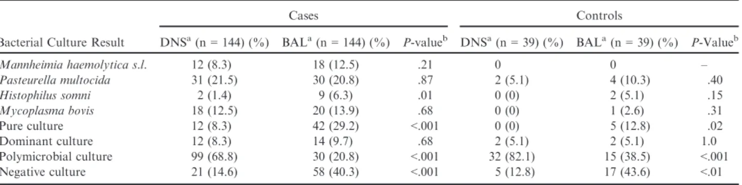
The majority of DNS cultures were polymicrobial
(68.8% [99 of 144] in cases, 82.1% [32 of 39] in
con-trols), meaning that no Pasteurellaceae or M. bovis
could be phenotypically identified from the plate.
Com-pared to DNS, BAL samples were significantly less
polymicrobial (P
< .001 for cases and controls), more
often negative (P
< .001 for cases, P < .01 for
con-trols), and more often returned pure cultures of
Pas-teurellaceae
or M. bovis (P
< .001 for cases, P < .02
for controls; Table 2). In summary, BAL samples
returned an interpretable result (either negative, pure,
or dominant culture result) in 79.2% of the cases and in
61.5% of the controls, compared to 31.2% and 17.9%
for DNS in cases and controls, respectively (P
< .01 for
both comparisons; Table 2). The polymicrobial nature
of a sample result was strongly affected by the herd
effect (P
< .001). A polymicrobial DNS and BAL
cul-ture result in at least 1 animal was present in almost all
herds (11 of 14 herds, the other 3 herds had no
polymi-crobial BAL culture result), but there was very large
variation in the percentage of polymicrobial results
among the herds sampled (Table 1). In the cases, a
polymicrobial DNS culture result did not increase the
probability of a polymicrobial BAL result by
≥30%
(P
= .09), nor did it influence the probability of a
negative culture (P = .52). However, the probability of
retrieving M. haemolytica s.l. and P. multocida from the
BAL sample still decreased when the DNS was
polymi-crobial. In contrast, there was no effect of a
polymicro-bial DNS result on the probability of isolation of
M. bovis
from the BAL sample (Table 5).
Discussion
To determine how the respiratory tract should be
sampled to isolate the causative pathogens, a
cross-sectional study was performed to compare bacterial
cul-ture results and commensal overgrowth between DNS
and BAL samples. Sampling procedures returning high
isolation rates of the major respiratory pathogens and
with a straightforward interpretation of the culture
results have the highest return on investment and are
therefore most suitable for practice.
In our study, all isolates were identified by
biochemi-cal tests and morphology instead of by polymerase
chain reaction (PCR). This approach might limit the
results with respect to bacterial species identification.
Biochemical identification was selected because it is the
routine identification method used in private
laborato-ries in Belgium and neighboring countlaborato-ries, for reasons
of speed and cost of analysis. The objective of our
study was to gain insights into the sampling and culture
methods currently used in the field. Also, no selective
media to increase Pasteurellaceae isolation rates were
used, because doing so currently is not the standard
procedure used in private laboratories. Selective media
would likely decrease contamination, whereas 1 of the
main objectives was to study differences in
contamina-tion between DNS and BAL. A final limitacontamina-tion of this
study was that, for practical reasons, the returned
lavage fluid volume was not determined. Quantification
of the target bacteria was not an objective of the study,
but differences in the returned volume might potentially
have influenced culture results.
One of the main findings in the study on preweaned
calves is that isolation rates of respiratory bacterial
pathogens in both DNS and BAL samples were lower
in
controls
compared
to
cases.
The
most
likely
Table 2.
Differences in isolation rates of bacterial respiratory pathogens and negative, pure culture, or
polymicro-bial culture results between DNS and BAL samples in 183 preweaned calves.
Bacterial Culture Result
Cases Controls
DNSa(n= 144) (%) BALa(n= 144) (%) P-valueb DNSa(n= 39) (%) BALa(n= 39) (%) P-Valueb
Mannheimia haemolytica s.l. 12 (8.3) 18 (12.5) .21 0 0 – Pasteurella multocida 31 (21.5) 30 (20.8) .87 2 (5.1) 4 (10.3) .40 Histophilus somni 2 (1.4) 9 (6.3) .01 0 (0) 2 (5.1) .15 Mycoplasma bovis 18 (12.5) 20 (13.9) .68 0 (0) 1 (2.6) .31 Pure culture 12 (8.3) 42 (29.2) <.001 0 (0) 5 (12.8) .02 Dominant culture 12 (8.3) 14 (9.7) .68 2 (5.1) 2 (5.1) 1.0 Polymicrobial culture 99 (68.8) 30 (20.8) <.001 32 (82.1) 15 (38.5) <.001 Negative culture 21 (14.6) 58 (40.3) <.001 5 (12.8) 17 (43.6) <.01
DNS, deep nasopharyngeal swab; BAL, bronchoalveolar lavage.
aResults are shown as numbers with percentages between brackets.
bP-value referring to the difference between DNS and BAL.
Table 3.
Mixed infections as diagnosed by bacterial
culture on deep nasopharyngeal swabs or
bronchoalveo-lar lavage samples in 144 preweaned calves with
respira-tory disease.
DNS BAL Pasteurella multocida+ Mannheimia haemolytica s.l.+ Mycoplasma bovis 6.7% (1/15) 25.0% (4/16) P. multocida+ M. haemolytica s.l. 20.0% (3/15) 18.8% (3/16) P. multocida+ M. bovis 66.6% (10/15) 37.4% (6/16) M. haemolytica s.l. + M. bovis 0.0% (0/15) 18.8% (3/16) M. haemolytica s.l.+ Histophilus somni 6.7% (1/15) 0.0% (0/25)Table
4.
Associations
and
agreement
between
DNS
and
BAL
culture
results
in
183
preweaned
calves.
Spec ies Case s Controls Perc entage (Numb er) of Positive B A L C ultures Asso ciation of DNS with BAL Agreem ent Perc entage of Positive BALs (Nu mber) Asso ciation of DN S w ith BAL Agreem ent DNS Culture Res ult a Negative Positive O R 95% CI P -Value Kapp a 95% CI DNS Cultu re Res ult a Negative Positive O R 95% CI P -Value Kapp a 95% CI Mannh eimia haemoly tica s.l. 7.6% (10/ 132) 66.7% (8/12) 18.9 3.3 –111.1 < .01 0.52 0.36 –0.69 0% (0/ 39) 0% (0/ 0) ND Pa steurella multoc ida 10.5% (12/ 114) 63.3% (19/30) 13.3 3.5 –50.0 < .001 0.48 0.25 –0.71 8.1% (3/ 37) 50.0% (1/ 2) 11.4 0.5 –250 .12 0.28 0.03 –0.54 Histo philu s somn i 5.6% (8/ 142) 50% (1/2) ND 0.16 0– 0.46 0% (0/ 0) 5.1% (2/ 39) ND Myco plasm a bovis 4.8% (6/ 124) 60.0% (12/20) 8.9 2.0 –38.5 < .01 0.58 0.38 –0.78 2.6% (1/ 39) 0% (0/ 0) ND Pure cult ure 24.2% (32/ 132) 83.3% (10/12) 7.8 1.4 –45.5 .02 0.28 0.12 –0.43 12.8% (5/ 39) 0% (0/ 0) ND Dominant culture 9.8% (13/ 132) 8.3% (1/12) ND ND 5.4% (2/ 37) 0% (0/ 2) ND Polym icrobial culture 8.9% (4/ 45) 26.3% (26/99) 2.9 0.8 –10.2 .09 0.12 0.03 –0.21 14.3% (1/ 7) 43.8% (14/32) 0.18 0.46 –47.6 .15 0.15 0– 0.33 Ne gative cu lture 39.0% (48/ 123) 47.6% (10/21) 3.5 1.0 –11.6 .05 0.05 0– 0.18 38.2% (13/ 34) 80.0% (4/ 5) 6.5 0.60 –71.4 .12 0.21 0.08 –0.34 DN S, de ep nasop haryng eal sw ab; BA L, bron choa lveolar lavag e; ND , n o stati stical ana lysis possib le, be cause of a too sma ll numbe r o f obser vations in o ne of the gro ups; OR, odds ratio; CI, co nfidence inte rval. Stre ngth of agreemen t for the Kapp a coefficie nt was inte rpreted accor ding to Land is and Koch : ≤ 0 = poor ; 0.10 –0.20 = slight; 0.21 –0.40 = fair; 0.41 –0.60 = mode rate; 0.61 –0.80 = subst antial and 0.81 –1.0 = almost pe rfect. aDNS culture result refe rs to isola tion of the sam e bac teria as in the BA L. He rd effec t was sign ifican t for all st udied ou tcomes, except P. multocida.explanation is that the control group consisted of
ani-mals originating from other farms than the case farms,
whereas in previous work, “apparently healthy”
in-contact animals were used as controls.
11These
appar-ently healthy animals are likely exposed to the same risk
factors as the cases and might be subclinically infected.
Therefore, in our study, controls were deliberately
cho-sen from farms without recent BRD exposure, and
ultrasound examination was used as an additional tool
to aid in selecting truly healthy animals. A disadvantage
of this approach is the environmental differences (e.g.,
bedding, herd size, air quality) that exist among herds.
To definitively determine whether isolation rates differ
between diseased and truly healthy animals in 1 herd, a
longitudinal study design would be needed.
Agreement between DNS and BAL samples was
moderate for M. haemolytica s.l., P. multocida, and
M. bovis,
similar
to
what
was
observed
for
M. haemolytica s.l.
in fattening bulls.
11Agreement was
much lower for H. somni, which can be explained by
the fact that H. somni is easily overgrown by other
bac-teria.
20Given their polymicrobial nature, DNS samples
are likely to be falsely negative for H. somni, when no
selective media are used. Current understanding of the
pathogenesis of bacterial pneumonia in calves suggests
overgrowth of Pasteurellaceae in the nasopharynx and
tonsils with subsequent colonization of the trachea and
lungs.
21Even when applying a transtracheal sampling
procedure, in diseased animals, one is probably as likely
to isolate bacteria that have descended from the
nasopharynx as those originating from the lung.
Possi-ble reasons why DNS and BAL samples do not agree
are false-negative results caused by polymicrobial
over-growth (sampling technique or presence of resident
flora), sampling of a nonaffected lung lobe with the
nonendoscopic BAL technique, or the absence of deep
bronchitis or alveolitis in case calves. The latter reason
was excluded as much as possible by the use of
ultra-sound examination in this study. Previous work showed
that this nonendoscopic BAL approach samples a
ran-dom lung lobe in nonsedated animals, and not
necessar-ily the most frequently affected cranial lobes.
13This
might in part explain why some cultures of cases were
negative. However, we doubt this is true, and our
hypothesis is that passage through trachea and deep
bronchi transfers bacteria deeper into the lung.
Interestingly, in the same animal, the DNS could be
polymicrobial, whereas the BAL yielded a pure culture,
dominant culture, or even a completely negative result.
Additionally, the polymicrobial nature of the DNS did
not affect the presence of a negative or pure culture
result in the BAL. Also, H. somni could be isolated in
pure culture from the lungs of diseased calves, whereas
it was overgrown or absent on the nasopharyngeal
cul-ture. These observations strongly suggest that, under
the conditions of our study, nasopharyngeal
contamina-tion of a BAL sample is less common than previously
assumed. To what extent a possible cleansing effect of
the DNS contributes to a pure culture result in the
BAL is unclear. On the other hand, a DNS
polymicro-bial result did decrease the probability of isolating
M. haemolytica s.l.
or P. multocida from the BAL,
whereas this effect was not observed for M. bovis for
which selective media were used. Again, this
observa-tion could be explained by BAL placement in a healthy
lung lobe in a case calf or because respiratory bacteria
are not necessarily involved in every case. Several
viruses (e.g., bovine respiratory syncytial virus, bovine
coronavirus) are capable of inducing pneumonia and
marked disease without bacterial superinfection.
Unfor-tunately, in our study, viral analysis in each case was
not possible for financial reasons. However, in our
opinion, the polymicrobial nature of DNA and BAL is
strongly influenced by the sampling (technique and
Table 5.
Results of univariable logistic regression models on the effect of a polymicrobial DNS on recovery of
res-piratory bacteria from BAL samples in 183 preweaned calves.
Species
Percentage (Number) of Positive BAL Cultures OR 95% CI P-Value Polymicrobial DNS No Yes Cases (n= 144) M. haemolytica s.l. 31.1% (14/45) 4.0% (4/99) 0.23 0.08–0.64 <.01 Pasteurella multocida 44.4% (20/45) 11.1% (11/99) 0.20 0.05–0.83 .03 Histophilus somni 2.2% (1/45) 88.9% (8/99) ND Mycoplasma bovis 24.4% (11/45) 7.1% (7/99) 1.34 0.33–5.62 .67 Negative culture 22.4% (13/45) 45.5% (45/99) 1.36 0.53–3.5 .52 Controls (n= 39) M. haemolytica s.l. 0% (0/7) 0% (0/32) ND P. multocida 14.3% (1/7) 9.4% (3/32) 0.62 0.05–7.69 .70 H. somni 0% (0/7) 6.3% (2/32) ND M. bovis 0% (0/7) 3.1% (1/32) ND Negative culture 71.4% (5/7) 37.5% (12/32) 0.24 0.04–1.53 .12
DNS, deep nasopharyngeal swab; BAL, bronchoalveolar lavage; ND, no statistical analysis possible, because of a too small number of observations in one of the groups; OR, odds ratio; CI, confidence interval.
hygiene), given that such a strong herd effect on the
sampling results was observed. To overcome the issue
of possible nasopharyngeal overgrowth in DNS and
BAL samples due to nasopharyngeal passage, both the
use of selective media for isolation of Pasteurellaceae
(e.g., addition of bacitracin
5,7) and a more quantitative
approach to BAL results
22might be suitable. Our study
focused on culture results obtained when applying DNS
and BAL as in practice. To definitively determine the
extent and diagnostic importance of possible
over-growth as a consequence of nasopharyngeal passage,
experimental work with intensive strain typing and
necropsy to confirm the infective status of the lung will
be needed.
As mentioned above, a significant herd effect was
noted on many of the outcomes studied. Deep
nasopha-ryngeal swab and BAL were performed only after
clean-ing the outer nares and without a protective sleeve as
used in previous studies.
6,11This could have increased the
risk of contamination by bacteria residing in the nostril.
In Belgium, DNS for practical reasons is routinely
per-formed without a protective sleeve, again increasing
external validity in this study. Multiple samplers
partici-pated in the study, and although all of them received at
least 1 training session from the same trainer before the
start of the study, variation in the extent of experience in
taking DNS or BAL samples and in the hygienic
proce-dures accompanying these techniques might have
influ-enced the results. Deep nasopharyngeal swab samples
might be polymicrobial due to the presence of a highly
variable nasopharyngeal microflora
23or due to
environ-mental contamination (e.g., touching the muzzle or other
objects during sampling). Other reasons might be
envi-ronmental or aerosolized dust, endotoxin, bedding
condi-tions, and issues with stable ventilation. Likely, the risk
of catheter contamination increases when repeated
attempts to enter the trachea are needed or when the
esophagus is accidently entered. Adequate training is
likely the only solution, other than considering other
pro-cedures such as protective sleeves, agar plugs, or
visual-ization of the larynx through a low-cost laryngoscope.
In conclusion, a nonendoscopic BAL results in less
contaminated (and therefore more easily interpretable
samples) compared to DNS under the conditions of this
study. It returns an interpretable result in 79.2% of the
cases, compared to 31.2% in DNS, and has better
isola-tion rates for H. somni, offering a better return on
investment for bacteriological sampling. It can be
per-formed rapidly in a representative number of animals at
low cost and likely has less impact on animal welfare
then more invasive techniques.
Footnotes
aWinepiscope 2.0, University of Zaragoza, Spain
b
Tringa Linear Vet, Esaote, the Netherlands c
TransystemTM, Copan, Brescia, Italy
d
1.5 m length; inner and outer diameter, 2 and 4 mm, respec-tively, VWR, Belgium, Leuven
eOxo€ıd, Hampshire, UK
fDifco, BD Diagnostic Systems, Sparks, MD
gSAS Institute, Cary, NC
Acknowledgments
We thank Karlijn Janssens and Pieter De Wolf for
sampling some of the calves. All practitioners are
acknowledged for reporting suitable outbreaks for this
study.
Conflict of Interest Declaration:
Authors declare no
conflict of interest.
Off-label Antimicrobial Declaration:
Authors declare
no off-label use of antimicrobials.
References
1. Snowder GD, Van Vleck LD, Cundiff LV, et al. Bovine res-piratory disease in feedlot cattle: Environmental, genetic, and
eco-nomic factors. J Anim Sci 2006;84:1999–2008.
2. Pardon B, Catry B, Dewulf J, et al. Prospective study on quantitative and qualitative antimicrobial and anti-inflammatory
drug use in white veal calves. J Antimicrob Chemother
2012;67:1027–1038.
3. Mevius DJ, Koene MGJ, Wit B, et al. Monitoring of Antimicrobial Resistance and Antibiotic Usage in Animals in the Netherlands. Lelystad, the Netherlands; 2012. Available at: http:// www.wageningenur.nl/nl/Publicatie-details.htm?publicationId= publication-way-343330383332. Accessed February 2, 2016.
4. Filip RD, De Block M, Borsus W. Koninklijk besluit betref-fende de voorwaarden voor het gebruik van geneesmiddelen door de dierenartsen en door de verantwoordelijken van de dieren. 2016, Available at: http://www.afsca.be/dierlijkeproductie/dieren/ diergeneesmiddelen/_documents/2016_07_21_KB21juli2016_AR21 juillet2016_BS_MB.pdf. Accessed August 10, 2016.
5. Catry B, Haesebrouck F, Vliegher SD, et al. Variability in acquired resistance of Pasteurella and Mannheimia isolates from the nasopharynx of calves, with particular reference to different
herd types. Microb Drug Resist 2005;11:387–394.
6. Godinho KS, Sarasola P, Renoult E, et al. Use of deep nasopharyngeal swabs as a predictive diagnostic method for
natu-ral respiratory infections in calves. Vet Rec 2007;160:22–25.
7. Pardon B, De Bleecker K, Dewulf J, et al. Prevalence of res-piratory pathogens in diseased, non-vaccinated, routinely medi-cated veal calves. Vet Rec 2011;169:278.
8. Timsit E, Christensen H, Bareille N, et al. Transmission dynamics of Mannheimia haemolytica in newly-received beef bulls
at fattening operations. Vet Microbiol 2013;161:295–304.
9. Pringle JK, Viel L, Shewen PE, et al. Bronchoalveolar lavage of cranial and caudal lung regions in selected normal calves: Cellu-lar, microbiological, immunoglobulin, serological and histological
variables. Can J Vet Res 1988;52:239–248.
10. Thomas A, Dizier I, Trolin A, et al. Comparison of sam-pling procedures for isolating pulmonary mycoplasmas in cattle.
Vet Res Commun 2002;26:333–339.
11. Allen JW, Viel L, Bateman KG, et al. The microbial flora of the respiratory tract in feedlot calves: Associations between nasopharyngeal and bronchoalveolar lavage cultures. Can J Vet
Res 1991;55:341–346.
12. Rohn M, Heckert HP, Hofmann W. Vergleichende auswer-tung der bakteriologischen untersuchungsbefunde von nasen-und
trachealtupfern sowie trachealsp€ulproben. Prakt Tierarzt
13. Van Driessche L, Valgaeren B, Schutter P, et al. Effect of sedation on the intrapulmonary position of a bronchoalveolar lavage catheter in calves. Vet Rec 2016;179:18; doi:10.1136.
14. Assie S, Seegers H, Beaudeau F. Incidence of respiratory disorders during housing in non-weaned Charolais calves in cow-calf farms of Pays de la Loire (Western France). Prev Vet Med
2004;63:271–282.
15. Pardon B, Alliet J, Boone R. Prediction of respiratory dis-ease and diarrhea in veal calves based on immunoglobulin levels and the serostatus for respiratory pathogens measured at arrival.
Prev Vet Med 2015;120:169–176.
16. Buczinski S, Forte G, Belanger A. Short communication: Ultrasonographic assessment of the thorax as a fast technique to assess pulmonary lesions in dairy calves with bovine respiratory
disease. J Dairy Sci 2013;96:4523–4528.
17. Quinn PJ, Carter ME, Markey B, et al. The Mycoplasmas (Class: Mollicutes). In: Quinn PJ, ed. Clinical Veterinary
Microbi-ology. London: Mosby International Limited; 1994:320–326.
18. Cohen JA. A coefficient of agreement for nominal scales.
Educ Psychol Measur 1960;20:37–46.
19. Landis JR, Koch GG. An application of hierarchical kappa-type statistics in the assessment of majority agreement
among multiple observers. Biometrics 1977;33:363–374.
20. Quinn PF, Carter ME, Markey B, et al. Haemophilus spe-cies. In: Quinn PJ, ed. Clinical Veterinary Microbiology. London:
Mosby International Limited; 1994:273–278.
21. Grey CL, Thomas RG. Pasteurella haemolytica in the
tra-cheal air of calves. Can J Comp Med 1971;35:121–128.
22. Rennard SI, Basses G, Lecossier D, et al. Estimation of volume of epithelial lining fluid recovered by lavage using urea as
marker of dilution. J Appl Physiol 1986;60:532–538.
23. Allen JW, Viel L, Bateman KG, et al. Changes in the bacterial flora of the upper and lower respiratory tracts and
bronchoalveolar lavage differential cell counts in feedlot
calves treated for respiratory diseases. Can J Vet Res 1992;56: