Full Terms & Conditions of access and use can be found at
http://www.tandfonline.com/action/journalInformation?journalCode=yacb20
Download by: [University of Liege] Date: 18 December 2017, At: 10:05
Acta Clinica Belgica
International Journal of Clinical and Laboratory Medicine
ISSN: 1784-3286 (Print) 2295-3337 (Online) Journal homepage: http://www.tandfonline.com/loi/yacb20
Clinical experience in idiopathic pulmonary
fibrosis: a retrospective study
Julien Guiot, Bernard Duysinx, Laurence Seidel, Monique Henket, Fanny
Gester, Olivier Bonhomme, Jean-Louis Corhay & Renaud Louis
To cite this article: Julien Guiot, Bernard Duysinx, Laurence Seidel, Monique Henket, Fanny Gester, Olivier Bonhomme, Jean-Louis Corhay & Renaud Louis (2017): Clinical experience in idiopathic pulmonary fibrosis: a retrospective study, Acta Clinica Belgica, DOI: 10.1080/17843286.2017.1399228
To link to this article: https://doi.org/10.1080/17843286.2017.1399228
Published online: 10 Nov 2017.
Submit your article to this journal
Article views: 31
View related articles
KEYWORDS
idiopathic pulmonary fibrosis; interstitial lung diseases; prognosis
© Acta clinica Belgica 2017
CONTACT Julien guiot J.guiot@chu.ulg.ac.be ORIGINAL PAPER
Clinical experience in idiopathic pulmonary fibrosis: a retrospective study
Julien Guiota, Bernard Duysinxa, Laurence Seidelb, Monique Henketa, Fanny Gestera, Olivier Bonhommea,Jean-Louis Corhaya and Renaud Louisa
aPneumology Department, cHU liège, Domaine universitaire du Sart-tilman, liège, Belgium; bDepartment of Medico-economy and
Biostatistics, Domaine universitaire du Sart-tilman, liège, Belgium
ABSTRACT
Introduction: Idiopathic pulmonary fibrosis (IPF) is a rare lung disease with an increased
incidence since the last few years. Here, we report our eight-year clinical experience in CHU of Liège, Belgium.
Methods: We have studied retrospectively patients recruited from our ambulatory care
polyclinic at CHU of Liège from 1 January 2009 to 1 January 2017. We have excluded all patients treated with a specific anti-fibrotic therapy due to incomplete follow-up. The diagnosis of IPF was made according to the ATS/ERS international recommendations (2015).
Results: Out of the 114 patients initially selected, 82 cases were found to be suitable for the
analysis. The average age was 71.1 ± 9.35 years with a male predominance. The median survival was 43.7 months (23.6–71.7) with a majority (45%) of patients in the group II of the GAP index. The median rate of annual decline in diffusion capacity of CO (DLCO) was 11%, whereas the sub analysis for group III (according to GAP index) showed a decrease annual rate of 30%.
Conclusion: Our results are in keeping with the literature. One of our major finding is that
patients in GAP III exhibit an annual rate of mortality of 42% and a median annual decline in DLCO of 30%. This observation highlights the fact that this specific subgroup of patients presents a high risk of morbi-mortality.
Introduction
Idiopathic pulmonary fibrosis (IPF) is a rare lung disease of unknown origin which leads rapidly to death [1,2]. Epidemiological studies suggest that the incidence of IPF has been increasing steadily over the last two to three decades [3]. Its incidence is evaluated to 2–29/100.000 people [4–6]. However, recent study from USA has shown that incidence rate may be as high as 76.4/100.000 in subjects above 75 years (based on a broad IPF defini-tion) [7].The sex ratio is in favor of male with an increase prevalence of smokers (or ex-smokers with a smoking history of more than 20 pack years). There are also famil-ial forms, which are associated with mutations thought to be autosomic dominant with an incomplete pene-trance (mainly TERT/TERC and SFTPC) [8].
The disease is generating a progressive fibrosing pro-cess exclusively limited to the lungs, which is very spe-cific particularity in comparison with other interstitial lung diseases. The symptomatic presentation is variable as well as the clinical course of the disease, which is rang-ing from a slow progressive evolution to a rapid decline or with an acute exacerbation pattern. Classically, the symptoms increase until pulmonary insufficiency, which is the main cause of death. Acute exacerbation in IPF is a
rare complication secondary to one hyper inflammatory state associated with a highly increased mortality rate.
Although the etiology and the pathophysiology of IPF are still incompletely understood, two antifibrotic drugs, pirfenidone and nintedanib, have recently proven to be effective in slowing down disease progression, and, are now approved as treatments [9,10]. Clinical man-agement of IPF remains difficult due to a lack of accu-rate indicators of disease progression, and an absence of simple short-term measures of therapeutic response
[11–13]. Recently, new data studying the pooled
analy-sis of the previous studies with pirfenidone identified a benefit on mortality in IPF patients [14].
The aim of our work was to analyze our eight-year clinical experience in IPF in the CHU of Liège (Belgium) and to confirm whether our clinical data are confident with the literature.
Methods
We retrospectively studied patients recruited from our ambulatory care policlinic at CHU from 1 January 2009 to 1 January 2017. We excluded all patients treated with specific anti-fibrotic therapy. The diagnosis of IPF was made according to the international recommendations
2 J. GUIOT ET AL.
of the ATS/ERS [1,15] using the respiratory function tests, high-resolution computed tomography scan (probable UIP pattern), bronchoalveolar lavage (when available), as well as the clinical history of the patient. We excluded all other causes of interstitial lung disease (such as asbestosis, hypersensitivity pneumonitis, pneu-monia associated with connective tissue disease or toxic pneumonitis). We combined the different results for the diagnosis. All cases were discussed in a multidisciplinary group about interstitial lung diseases composed of a pul-monologist, a specialist in pulmonary rehabilitation, a rheumatologist, a radiologist, a pathologist and a spe-cialist in occupational medicine.
Pulmonary function tests
We performed pulmonary function tests (PFT) in our routine respiratory laboratory of CHU of Liège. All spirometric tests performed for this study were meas-ured using the pneumotachograph JaegerMasterlab system (Erich Jaeger GmbH, Wuzburg, Germany). The forced expiratory volume in one second (FEV1) and forced vital capacity (FVC) were measured in accordance with the recommendations of the European Respiratory Society (ERS). The results were expressed in milliliter and percentage of predicted values. The Tiffeneau index or FEV1/FVC was expressed in percent. The total lung capacity (TLC) was measured by body plethysmogra-phy according to ERS recommendations (Erich Jaeger GmbH, Wuzburg, Germany). The diffusion capacity of CO (DLCO) and the report DLCO/VA were meas-ured by the single-breath carbon monoxide gas trans-fer method and expressed as percentage of predicted values (Sensor Medics 2400 He/CO Analyzer System, Bilthoven, the Netherlands).
GAP score
One easy, simple, and useful index that has been pro-posed for predicting IPF prognosis is the GAP index risk prediction model, which includes four factors: gender (G), age (A), and two lung physiologic (P) variables (FVC (% pred) and DLCO (% pred)) (Table 1) [16]. Patients were stratified based on GAP score: GAP I are patients with 0–3 points, GAP II are patients with 4 and 5 points,
and GAP III patients with 6–8 points. The patients with-out complete PFT available or withwith-out sufficient quality of collaboration for analysis were only listed in the all cohort and not included in the GAP sub-analysis. Statistical analysis
Results are expressed as frequency tables for qualitative variables and as mean and standard deviation (SD) or as median and quartiles (Q1–Q3) for continuous variables. Comparisons between groups were done by chi-square test for qualitative variables and by Kruskal–Wallis test for continuous variables. Overall, survival was repre-sented by a Kaplan–Meier curve. Survival between groups was compared by Cox regression model (HR and 95% confidence interval). To calculate the annual decrease of a parameter, a linear regression of this parameter on time since first EFR was applied for each patient. The annual decrease is calculated as the slope corrected by the number of days between the first and the last EFR and converted in percent per year. The annual decrease is assessed by the Wilcoxon sign-rank test. Comparison of annual decrease between groups was done by Kruskal–Wallis test. To compare groups two by two, a Bonferroni correction was applied. Results were significant at the 5% significance level (p < 0.05). All sta-tistical analyses were carried out by SAS version 9.4 (SAS Institute, Cary, NC, USA) and figures by R version 3.2.2.
Results
We identify 114 untreated patients suffering from IPF and excluded 32 of them because of incomplete data-set. Subject demographic and functional
characteristics
Subjects’ characteristics are listed in Table 2. The time of follow-up range from 1 to 92 months. The mean age of IPF patients was 71.1 ± 9.35 years with a male domi-nance (69.5%). Spirometric values were lower than pre-dicted values with a restrictive pattern and a reduced DLCO as expected. The GAP I group present higher values for TLC, FVC, FEV1, and DLCO than GAP II and III, whereas the GAP II group exhibit higher values for FVC, FEV1, and DLCO than GAP III. GAP III group had a significantly lower DLCO/VA than groups I and II (p < 0.0013).
Decline of PFTs
We also studied the decline of PFTs separating patients in three groups according to their GAP score (Table 3). We’ve identified that the median annual rate of decline (in absolute values) for CPT, FVC, FEV1, and DLCO was 1.90, 2.64, 3.75, and 10.80%, respectively. Interestingly, in the GAP III group sub analysis, we identified that there
Table 1. gAP index score for idiopathic pulmonary fibrosis.
notes: adapted from ley et al. [16]. gAP i: 0–3 points, gAP ii: 4 and 5 points, gAP iii: 6–8 points. FVc: forced vital capacity; DlcO: diffusion lung capacity of cO.
G (Gender) A (Age)
Female Male ≤60 61–65 ≥65
0 1 0 1 2
P (Physiology)
FVc post-BD (% predicted) DlcO (% predicted) >75% 50–75% <50% >55% 36–55% ≤35% cannot
perform
0 1 2 0 1 2 3
was no real change in the pulmonary volumes. Conversely to that observation, there was an annual rate of DLCO decline around 30% underlying the rapid degradation of the gas exchanges in patients with the more severe condition. On the other hand, the GAP I group present an annual decline of the FVC around 5% of the initial absolute value ranging from −4 to 7% with an annual DLCO reduction around 13% (Figure 1). Globally there are no differences between the three groups except for GAP III which present a significantly increased decline in DLCO (p = 0.01) compared to GAP I and II.
Survival according to the GAP index
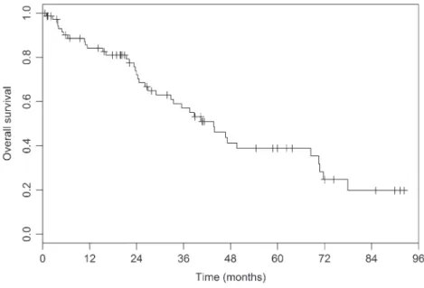
Concerning the survival data, it is widely known that median survival rate in IPF without specific therapy is around 3–5 years. In our experience, the median overall survival is 43.7 months (23.6–71.7). In our cohort of patients, based on the GAP index, we identified 34% of patients in group I; 45% in group II and 21% in group III.
The overall mortality is represented in Figure 2, while the Figure 3 is representing the mortality accord-ing to the GAP index classification. Based on the GAP index, we found a one-year mortality of 4, 14, and 42% in stages I, II, and III, respectively, whereas the three-year mortality was 17, 39, and 86%, respectively (Table 4). Cox model identified a highly significant dif-ference in mortality by separating patients with GAP score (p = 0.0002), where the GAP III group exhibit a very high mortality rate with an HR of 7.70 (2.88–20.6;
p < 0.0001) and 2.68 (1.31–5.46; p = 0.0067) in
com-parison with GAP I and III, respectively. Patients in GAP I exhibit a lower mortality than GAP II and III (p = 0.0007).
Hospitalizations
The hospitalization rate is presented in Table 5. The median hospitalization rate is four during the follow-up, whereas the median number of admission is bellow one for intensive care unit (ICU) and of two for emergency room (ER) admissions.
Table 2. Subjects characteristics.
notes: tlc: total lung capacity; FVc: forced vital capacity; FeV1: forced expired volume in one second; DlcO: diffusion lung capacity for cO; DlcO/VA: DlcO/ alveolar ventilation.
Bonferroni correction for group comparison. the differences are noticed with the same letter.
aWe had only 77 patients with full PFts.
Totala Gap I Gap II Gap III
p-value
Median (Q1–Q3) Median (Q1–Q3) Median (Q1–Q3) Median (Q1–Q3)
n = 82 n = 26 (34%) n = 35 (45%) n = 16 (21%)
gender (M/F), n 55/27 14/12 27/8 13/3 0.080
Smokers (never/ex/current), % 26.7/52/21.3 20.8/50.0/29.2 23.5/47.1/29.4 30.8/61.5/7.7 0.61
Age (years) mean ± SD 71.1 ± 9.35 66.7 ± 11.80a 71.80 ± 6.72 76.7 ± 6.20 a 0.0019
Follow-up (months) 24.6 (10.9–43.9) 27.8 (18.9–62.3) 33.4 (10.9–46.7) 13.3 (3.15–25.2) 0.044
Range 0–92 0–92 0.6–91.3 0.99–74
Dead (no/yes), % 48.8/51.1 76.9/23.1a,b 42.9/57.1a 18.8/81.2b 0.0007
Median survival (months) 43.7 (23.6–71.7) >92 (70.5 to >92) 43.7 (24.6–70.8) 21.4 (4.9–26.1) <0.0001
tlc %pred 77.0 (61.0–92.0) 92.0 (81.0–101)a,b 74.0 (61.0–85.0)a 59.5 (49.5–71.5)b <0.0001
FVc %pred 78.0 (63.0–94.0) 94.5 (88.0–106)a,b 76.0 (63.0–90.0)a,c 62.5 (43.5–68.5)b,c <0.0001
FeV1 %pred 77.0 (64.0–92.0) 89.5 (79.0–101)a,b 76.0 (60.0–88.0)a,c 61.0 (52.0–73.0)b,c <0.0001
DlcO %pred 45.5 (34.0–60.0) 62.0 (46.0–69.0)a,b 43.0 (36.0–53.0)a,c 26.0 (25.0–28.0)b,c <0.0001
DlcO/VA % 72.5 (59.5–89.0) 80.0 (65.0–94.0)b 71.0 (59.0–90.0)c 56.0 (45.0–63.0)b,c 0.0013
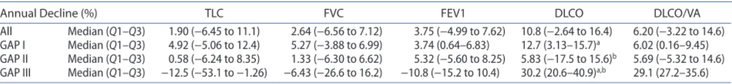
Table 3. Annual decline of the main physiological pulmonary parameters in our cohort of iPF patients.
note: significant reduction in DlcO between gAP iii vs gAP i (p = 0.0012) and gAP iii vs gAP ii (p = 0.01).
asignificant reduction in DlcO between gAP iii vs gAP i (p = 0.0012) bsignificant reduction in DlcO between gAP iii vs gAP ii (p = 0.01).
Annual Decline (%) TLC FVC FEV1 DLCO DLCO/VA
All Median (Q1–Q3) 1.90 (−6.45 to 11.1) 2.64 (−6.56 to 7.12) 3.75 (−4.99 to 7.62) 10.8 (−2.64 to 16.4) 6.20 (−3.22 to 14.6) gAP i Median (Q1–Q3) 4.92 (−5.06 to 12.4) 5.27 (−3.88 to 6.99) 3.74 (0.64–6.83) 12.7 (3.13–15.7)a 6.02 (0.16–9.45)
gAP ii Median (Q1–Q3) 0.58 (−6.24 to 8.35) 1.33 (−6.30 to 6.62) 5.32 (−5.60 to 8.25) 5.83 (−17.5 to 15.6)b 5.69 (−5.32 to 14.6)
gAP iii Median (Q1–Q3) −12.5 (−53.1 to −1.26) −6.43 (−26.6 to 16.2) −10.8 (−15.2 to 10.4) 30.2 (20.6–40.9)a,b 29.1 (27.2–35.6)
Figure 1. Annual decrease of DlcO (in absolute value) in the overall population of iPF and according to the gAP i, ii, and iii subgroups.
notes: Bonferroni correction comparing gAP i vs. ii vs. iii showing a significant difference between group i vs. iii (*p < 0.017) and ii vs. iii
(**p < 0.001).
4 J. GUIOT ET AL.
often encounter approximately 30% never-smoking IPF patients [17]. In our study, we confirm the male predom-inance with a mean age of 71 years, and 27% of the all population had never smoked. As expected, following the definition of the GAP index, we identify a altered FVC and DLCO increasingly from GAP I to GAP III. Interestingly, there was no difference between GAP II and III patients for TLC.
Concerning the survival data, it is widely known that median survival rate in IPF without a specific therapy is around 3–5 years. In our experience, the median overall survival is 43.7 months (23.6–71.7). We also have sepa-rated the patients according to the GAP index [16]. Our data are again consistent with the literature exhibiting a one-year mortality of 4, 14, and 42% in stages I, II, and
Discussion
Here, we report our eight-year clinical experience of IPF in the university hospital of Liege showing that our data are in keeping with the literature.
The epidemiological characteristics are similar than those seen in the literature [1–7], which are classically elderly males with a history of smoking. However, we
Figure 2. Overall survival of the cohort of iPF. note: Kaplan–Meier curve.
Figure 3. Survival of iPF cohort according to the gAP index.
notes: Survival after a specific repartition in three groups according to gAP index. We identified a one-year mortality of 4, 14, and 42% in stages i, ii, and iii, respectively, whereas the three-year mortality was 17, 39, and 86%, respectively.
Table 4. Mortality at 1 and 3 years in our cohort according to the gAP index group vs. what is given in the literature [16]. % of patients 1-y mortality (vs. literature) 3-y mortality (vs. literature)
gAP i 34% 4% (vs. 6%) 17% (vs. 16%)
gAP ii 45% 14% (vs. 16%) 39% (vs. 42%)
gAP iii 21% 42% (vs. 39%) 86% (vs. 77%)
multidisciplinary classification of the idiopathic interstitial pneumonias. Am J Respir Crit Care Med. 2013;188(6):733–748.
[6] Raghu G. Idiopathic pulmonary fibrosis: guidelines for diagnosis and clinical management have advanced from consensus-based in 2000 to evidence-based in 2011. Eur Respir J. 2011;37(4):743–746.
[7] Raghu G, Chen S-Y, Hou Q, et al. Incidence and prevalence of idiopathic pulmonary fibrosis in US adults 18–64 years old. Eur Respir J. 2016;48(1):179– 186.
[8] Gilani SR, Vuga LJ, Lindell KO, et al. CD28 down-regulation on circulating CD4 T-cells is associated with poor prognoses of patients with idiopathic pulmonary fibrosis. PLoS ONE. 2010;5(1):e8959.
[9] King TE, Bradford WZ, Castro-Bernardini S, et al. A phase 3 trial of pirfenidone in patients with idiopathic pulmonary fibrosis. N Engl J Med. 2014;370(22):2083– 2092.
[10] Richeldi L, du Bois RM, Raghu G, et al. Efficacy and safety of nintedanib in idiopathic pulmonary fibrosis. N Engl J Med. 2014;370(22):2071–2082.
[11] Maher TM. profileing idiopathic pulmonary fibrosis: rethinking biomarker discovery. Eur Respir Rev. 2013;22(128):148–152.
[12] Guiot J, Bondue B, Henket M, et al. Raised serum levels of IGFBP-1 and IGFBP-2 in idiopathic pulmonary fibrosis. BMC Pulm Med. 2016;16(1):86.
[13] Guiot J, Henket M, Corhay JL, et al. Sputum biomarkers in IPF: evidence for raised gene expression and protein level of IGFBP-2, IL-8 and MMP-7. In: Morty RE, editor. PLoS ONE. Vol. 12. San Francisco (CA): Public Library of Science; 2017. p. e0171344.
[14] Noble PW, Albera C, Bradford WZ, et al. Pirfenidone for idiopathic pulmonary fibrosis: analysis of pooled data from three multinational phase 3 trials. Eur Respir J. 2015;47(1):ERJ-00026.
[15] Raghu G, Rochwerg B, Zhang Y, et al. An official ATS/ ERS/JRS/ALAT clinical practice guideline: treatment of idiopathic pulmonary fibrosis. an update of the 2011 clinical practice guideline. Am J Respir Crit Care Med. 2015;192(2):e3–e19.
[16] Ley B, Ryerson CJ, Vittinghoff E, et al. A multidimensional index and staging system for idiopathic pulmonary fibrosis. Ann Intern Med. 2012;156(10):684–691. [17] Kishaba T, Nagano H, Nei Y, et al. Clinical characteristics
of idiopathic pulmonary fibrosis patients according to their smoking status. J Thorac Dis. 2016;8(6):1112– 1120.
[18] Guiot J, Moermans C, Henket M, et al. Blood biomarkers in idiopathic pulmonary fibrosis. Lung. 2017;195(3):273–280.
III, respectively, whereas the initial publication report a 6, 16, and 39% mortality for each group, respectively [16]. According to what was described in previous stud-ies, there was a highly significant difference in mortality by separating patients with GAP score where the GAP III group exhibit a very high mortality rate in comparison with GAP I and III.
One of our major finding is that patients in GAP III present an annual rate of mortality of 42% and a median decline in DLCO of 30% of the initial absolute value enhancing the fact that these patients are at high risk. Moreover, the reduction in DLCO is higher in that group than in patient in GAP II and III . Therefore, we truly consider that this is our responsibility to accelerate the access to the anti-fibrotic therapies since pirfenidone has been shown to reduce the overall mortality in IPF patients.
In this context, biomarkers are highly needed in IPF as tools for differential diagnostic, predictor of the progression of the disease and treatment response [18]. Specifically in IPF, an early diagnostic is important to reduce as much as possible the disease progression. Ideally, biomarkers should easily be sampled (in oppo-sition with BAL and surgical lung biopsy) and analyzed for a wide-spread utility in clinical practice.
Disclosure statement
No potential conflict of interest was reported by the authors.
References
[1] Raghu G, Collard HR, Egan JJ, et al. An official ATS/ ERS/JRS/ALAT statement: idiopathic pulmonary fibrosis: evidence-based guidelines for diagnosis and management. Am J Respir Crit Care Med. 2011;183(6):788–824.
[2] Guiot J, Corhay JL, Louis R. Idiopathic pulmonary fibrosis. Rev Med Liege. 2014;69(11):605–610.
[3] Raghu G, Weycker D, Edelsberg J, et al. Incidence and prevalence of idiopathic pulmonary fibrosis. Am J Respir Crit Care Med. 2006;174(7):810–816.
[4] Kaunisto J, Salomaa E-R, Hodgson U, et al. Idiopathic pulmonary fibrosis – a systematic review on methodology for the collection of epidemiological data. BMC Pulm Med. 2013;13(1):53.
[5] Travis WD, Costabel U, Hansell DM, et al. An Official American Thoracic Society/European Respiratory Society statement: update of the international
Table 5. Hospitalizations in the iPF cohort.
notes: icU: intensive care unit; eR: emergency room.
Variable N Mean SD SE Min Q1 Median Q3 Max
Hospitalization 103 5.17 5.85 0.58 0.0 2.0 4.00 7.0 41.0
icU admission 89 0.67 1.30 0.14 0.0 0.0 0.00 1.0 9.0
eR admission 89 2.25 2.63 0.28 0.0 1.0 2.00 3.0 16.0
total stay (days) 89 47.66 59.26 6.28 1.0 16.0 28.00 62.0 396.0