HAL Id: hal-02463050
https://hal.archives-ouvertes.fr/hal-02463050
Submitted on 31 Jan 2020HAL is a multi-disciplinary open access archive for the deposit and dissemination of sci-entific research documents, whether they are pub-lished or not. The documents may come from teaching and research institutions in France or abroad, or from public or private research centers.
L’archive ouverte pluridisciplinaire HAL, est destinée au dépôt et à la diffusion de documents scientifiques de niveau recherche, publiés ou non, émanant des établissements d’enseignement et de recherche français ou étrangers, des laboratoires publics ou privés.
Sex effects on structural maturation of the limbic system
and outcomes on emotional regulation during
adolescence
Pauline Bezivin Frere, Nora Vetter, Eric Artiges, Irina Filippi, Ruben
Miranda, Hélène Vulser, Marie-Laure Paillère-Martinot, Veronika Ziesch,
Patricia Conrod, Anna Cattrell, et al.
To cite this version:
Pauline Bezivin Frere, Nora Vetter, Eric Artiges, Irina Filippi, Ruben Miranda, et al.. Sex effects on structural maturation of the limbic system and outcomes on emotional regulation during adolescence. NeuroImage, Elsevier, 2020. �hal-02463050�
Sex effects on structural maturation of the limbic system and outcomes on emotional regulation during adolescence.
Pauline Bezivin Frere1, Nora C. Vetter2, Eric Artiges1, 3, Irina Filippi1, Rubén Miranda1,14, Hélène Vulser1, Marie-Laure Paillère-Martinot1, Veronika Ziesch2, Patricia Conrod4,5, Anna Cattrell4, Henrik Walter6, Jurgen Gallinat7, Uli Bromberg7, Sarah Rodehacke2, Eva Menningen2, Vincent Frouin8, Dimitri Papadopoulos-Orfanos8, Argyris Stringaris9, Jani Penttilä10, Betteke van Noort6, Yvonne Grimmer11, Gunter Schumann4, Michael N. Smolka2, Jean-Luc Martinot1,13, Hervé Lemaître1 , for the Imagen consortium12
1
Inserm, UMR 1000, Research unit NeuroImaging and Psychiatry, Paris Sud University-Paris Saclay University, Paris Descartes University, Digiteo Labs, Bâtiment 660, Gif-sur-Yvette, France
2
Department of Psychiatry and Neuroimaging Center, Technische Universität Dresden, Germany
3
Groupe Hospitalier Nord Essonne, Psychiatry Department 91G16, Orsay, France
4Institute of Psychiatry, Psychology & Neuroscience, King’s College London, United Kingdom 5
Department of Psychiatry, Université de Montreal, CHU Ste Justine Hospital, 175 Chemin de la Côte-Sainte-Catherine, Montréal, QC H3T 1C4, Canada
6
Department of Psychiatry and Psychotherapy, Campus Charité Mitte, Charité, Universitätsmedizin Berlin, Berlin, Germany
7
University Medical Centre Hamburg-Eppendorf, Hamburg, Germany
8
Neurospin, Commissariat à l'Energie Atomique, CEA-Saclay Center, Gif-sur-Yvette, Paris, France
9
10
Department of Social and Health Care, Psychosocial Services Adolescent Outpatient Clinic Kauppakatu 14, Lahti, Finland
11
Department of Child and Adolescent Psychiatry and Psychotherapy, Central Institute of Mental Health, Medical Faculty Mannheim, Heidelberg University, Mannheim, Germany
12
www.imagen-europe.com
13
Centre de Neuro-Imagerie de Recherche (Cenir), Institut du Cerveau et de la Moëlle épinière (ICM), Pitié-Salpêtrière Hospital, Paris, France
14
APHP, Department of Psychiatry and Addictology, Paul Brousse Hospital, Villejuif, France
Corresponding author: Dr. Hervé Lemaître, Digiteo Labs, Bâtiment 660, Rue Noetzlin, 91190 Gif-sur-Yvette, France; e-mail: herve.lemaitre@u-psud.fr
Abstract
Though adolescence is a time of emerging sex differences in emotions, sex-related differences in
the anatomy of the maturing brain has been under-explored over this period. The aim of this
study was to investigate whether puberty and sexual differentiation in brain maturation could
explain emotional differences between girls and boys during adolescence. We adapted a
dedicated longitudinal pipeline to process structural and diffusion images from 335 typically
developing adolescents between 14 and 16 years. We used voxel-based and Regions of Interest
approaches to explore sex and puberty effects on brain and behavioral changes during
adolescence. Sexual differences in brain maturation were characterized by amygdala and
hippocampal volume increase in boys and decrease in girls. These changes were mediating the
sexual differences in positive emotional regulation as illustrated by positive attributes increase in
boys and decrease in girls. Moreover, the differential maturation rates between the limbic system
and the prefrontal cortex highlighted the delayed maturation in boys compared to girls. This is the
first study to show the sex effects on the differential cortico/subcortical maturation rates and the
interaction between sex and puberty in the limbic system maturation related to positive attributes,
reported as being protective from emotional disorders.
Keywords: diffusion tensor imaging, T1-weigthed imaging, longitudinal, adolescence, sex difference, puberty
Introduction
Adolescence is a sensitive period of gradual transition from childhood to adulthood (Spear,
2000) through maturation of adult social and cognitive behaviors (Sisk and Foster, 2004).
Adolescence is characterized by important pubertal changes and the passage from immature child
brain to adult brain through complex maturational processes such as synaptic pruning, dendritic
and axonal arborization and myelination (Lenroot and Giedd, 2006). It is also a period of
emerging sex differences such as on brain and behaviors. Hormonal changes related to puberty
are partly responsible for the development of the brain (Spear, 2000) and of the cognitive
functions (Blakemore et al., 2010). Onset of pubertal maturation occurs in the brain with some
neural changes leading to hormone levels increase themselves responsible for other brain changes
(Dahl, 2004). The pubertal timing being different between boys and girls, age alone is unfit for
looking at sex-related maturation differences during adolescence. Thus, reliance on pubertal
landmarks rather than age appears more adapted for studying sex and maturation processes
during adolescence.
Sex effects on brain macrostructural maturation as studied with Magnetic Resonance Imaging
(MRI) has been described as a global grey matter (GM) volume peak reached earlier in girls
followed by a steeper GM volume decrease rate compared to boys on a classical inverted U-shape
maturation curve (Aubert-Broche et al., 2013; Herting and Sowell, 2017; Raznahan et al., 2014).
Furthermore, global white matter (WM) volume follows a steeper linear WM volume increase in
boys as compared to girls. Additionally, the sexual differences of white matter microstructure
investigated with Diffusion Tensor Imaging (DTI) draw less consistent findings (Tamnes et al.,
2017). Some studies reported sex differences in WM microstructure maturation (Herting et al.,
while others studies have reported few or no significant sex-by-age interaction (Bava et al., 2010;
Eluvathingal et al., 2007; Giorgio et al., 2010). Regional patterns of sexual differences in
macrostructural maturation trajectories have also been reported, notably in the limbic system with
the amygdala, the hippocampus and the prefrontal cortex where girls showed an early
maturational peak as compared to boys (Eliot, 2019; Goddings et al., 2014; Herting et al., 2018;
Lenroot et al., 2007). However, other studies did not find sex by age interaction during
adolescence for subcortical regions such as basal ganglia, thalamus, hippocampus or amygdala
(Koolschijn and Crone, 2013; Wierenga et al., 2018).
Affective disorders are also part of the pattern of sexual differences with approximately 2:1
female:male prevalence ratio during adolescence (Angold et al., 1999, 1998; Angold and
Costello, 2006). Previously cited limbic regions had been implicated in the so-called “developmental mismatch hypothesis” proposing that the subcortical structures maturing earlier
than the cortical structures was leading to the stereotypical adolescent behavior (see review by
Mills et al. 2014). Simmonds et al. (Simmonds et al., 2014) found that frontosubcortical WM
connections (uncinate fasciculus, superior longitudinal fasciculus and cingulum) implicated in
emotional processing mature later than most white matter bundles during childhood. Further,
another study found that depressed patients had lower fractional anisotropy in this
cortical-subcortical connectivity (Versace et al., 2010). In the case of the limbic system, the maturational
mismatch could be related to the increase emotional reactivity and sensitivity, and thereby to an
increase risk for affective disorders during adolescence compared to childhood (Casey et al.,
2008).
Research on emotion dysregulation during adolescence has given a large prominence to the
positive attributes (e.g. generosity, reliability, good sense of humor) that are related to the adolescent’s well-being (Gillham et al., 2011) and may be protective from emotional disorders
(Vidal-Ribas et al., 2015). Once again, pubertal timing plays an important role in the emotional
dysregulation with increased risks when girls mature too early or when boys mature too late
(Graber, 2013).
In the literature, most of the results on sex differences in brain maturation during adolescence
were based on cross-sectional study designs with large samples or large age range (Koolschijn
and Crone, 2013; Menzies et al., 2015; Satterthwaite et al., 2014). Although informative,
cross-sectional studies are limited because they can only provide estimated and not individual
trajectories. The existing longitudinal studies neither included large sample size (Bava et al.
2010; Giorgio et al. 2010; Dennison et al. 2013) nor had a large age range, nor focused on sexual
differences because of the non sex parity of their sample (Bava et al., 2010; Dennison et al.,
2013; Giorgio et al., 2010; Lebel and Beaulieu, 2011; Wierenga et al., 2014). Recently, few
longitudinal studies had the power to tackle the question of sex differences in brain maturation
during adolescence (Fish et al., 2019; Wierenga et al., 2018) but more a needed to disentangle the
effect of sex, age and puberty.
For these reasons, this study investigated the sex and puberty effects on brain and behavioral
changes during adolescence by – 1. taking advantage of a two time point longitudinal design of a
large sample of adolescents with the same age at 14 and 16 years old and a dedicated longitudinal
preprocessing methodology (Ashburner and Ridgway, 2013) – 2. looking at the sexual
differences of the brain maturation with a multimodal neuroimaging approach focusing on grey
and white matter using whole brain and specific limbic system regions of interest analyses - 3.
dysregulation using psychopathological measures related to affective disorders and also
personality traits that may constitute vulnerability factors. We hypothesized that boys and girls
would have a different developmental mismatch in grey matter regions and white matter bundles
of the limbic system, and that this differential maturation would be in return related to sex
differences on emotion regulation and psychopathology during adolescence.
Materials and Methods
Participants
Longitudinal datasets from three hundred and thirty-five adolescents (175 females; 160 males)
were drawn from the Imagen database, a larger sample recruited in eight European cities at the
age of 14. Two sites (138 and 197 subjects from Paris and Dresden respectively) conducted an
MRI exam at both 14 and 16 years old in addition to questionnaires and neuropsychological
battery tests at both times. Written informed consent and assent had been given by both parents
and participants. The study had been approved by the local ethic committees. A detailed
description of recruitment and assessment procedures, and exclusion and inclusion criteria has
been published (Schumann et al., 2010). Notably, any obvious psychopathology (e.g. bipolar
disorder, schizophrenia, or major neuro-developmental disorders) constituted non-inclusion
criteria.
Self-Report Questionnaires
The pubertal measure was assessed with the Puberty Development Scale (PDS; (Petersen et
al., 1988)), a measure of physical development with separate items for males and females.
Questionnaires are adapted for each sex, such as menarche in females and voice changes in
The adolescent psychiatric symptoms and their psychosocial impact were assessed with the
Development and Well-Being Assessment (DAWBA, www.dawba.com), a self-administered
diagnostic questionnaire consisting of open and closed questions (Goodman et al., 2000). The
DAWBA generates probabilities of having DSM-IV diagnoses that are subsequently validated by
experienced clinicians from the IMAGEN consortium. Diagnoses from affective disorders (e.g.
anxiety, depression bands) were tested here.
Specifically, The Youth Strengths Inventory (YSI), within the DAWBA, asks about adolescent’s positive attributes. The first part of the questionnaire is dedicated to “positive
characteristics” (e.g. how generous, affectionate, caring he is) with 8 items. The second part of
the questionnaire requests about “positive actions” that please others or things that the adolescent
is proud of in 11 items (e.g. how good at sport, well behaved, polite he is proud of). Each item is
scored on a three-point Likert scale (0: no, 1: a little, 2: a lot). Summing the score of each item
per part generates two variables, “positive characteristics” (from 0 to 16) and “positive actions”
(from 0 to 22). The sum of these two variables generates the global variable “total positive attributes” (from 0 to 38).
The Strengths and Difficulties Questionnaire (SDQ), a self-reported questionnaire (Goodman
et al., 2003) generates a total difficulties score (reflecting emotional problems, conduct problems,
hyperactivity and peer problems). Internalizing (i.e., anxious and depressive) and externalizing
(i.e., aggressive and hyperactive) behaviors (Achenbach, 1992) can be measured with the SDQ.
Externalizing score is obtained by summing conduct problems score and hyperactivity score;
internalizing score is obtained by summing emotional problems score and peer problems score,
each scale being ranged from 0 to 20.
All subjects underwent imaging exams on a SIEMENS Trio 3T scanner, including an
anatomical and a diffusion sequences. All exams were assessed by a clinical neuroradiologist for
structural abnormalities.
T1-weighted imaging. High-resolution T1-weighted images were collected using a
magnetization prepared rapid acquisition gradient-echo (MPRAGE) sequence [Paris: repetition
time (TR) = 2300 ms, echo time (TE) = 2.93 ms, inversion time (TI) = 900 ms, voxel size =
1.1×1.1×1.1 mm, flip angle = 9°; matrix size = 256x256x160 mm; Dresdren: TR=1900 ms,
TE=2.26 ms, TI=900 ms, voxel size=1.0×1.0×1.0 mm, flip angle=9°; matrix size = 256x256x176
mm].
Diffusion Tensor imaging. The diffusion tensor images (DTI) were acquired using an Echo
Planar imaging sequence (4 b-value=0 s/mm2 and 32 diffusion encoding directions with
b-value=1300 s/mm2; 60 oblique-axial slices (angulated parallel to the AC/PC line); echo time ≈
104 ms; 128x128 matrix; field of view 307x307mm; voxel size 2.4 x 2.4 x 2.4 mm).
Image processing
T1-weighted images. To correct for differences of neck rotation between each subject’s
acquisitions, all images were roughly realigned and cropped bellow the cerebellum. Then,
intra-subject registration was performed using SPM12's Longitudinal Registration Toolbox (Ashburner
and Ridgway, 2013) involving combining rigid-body registration, intensity inhomogeneity
correction, and non-linear diffeomorphic registration. This step generates the subject’s mid-point
image between 14 and 16 years, the maps of the Jacobian determinants and the deformation fields
estimated from each time-point scan to the mid-point image. The subject’s mid-point image was
segmented into grey and white matter with SPM12's Segmentation Toolbox with tissue priors
and white matter maps of the mid-point image were modulated by the Jacobian determinants of
each time-point. All grey and white matter maps of the mid-point images were spatially
normalized to the standard space of the Montreal Neurological institute (MNI) using the
DARTEL nonlinear image registration procedure. This step involves the iterative creation of their
representative template and the extraction of the deformation fields from each image to the
aforementioned template. The deformation fields obtained were then applied to the modulated
grey and white matter maps preserving the regional amount of signal. Finally, modulated
normalized maps of grey and white matters were smoothed with an 8 mm full width at half
maximum (FWHM) Gaussian kernel. Global GM, WM and cerebrospinal fluid (CSF) volumes
were computed for each participant. Total intracranial volume (TIV) was defined by summing
GM, WM and CSF volumes. GM volumes were extracted from the amygdala, the hippocampus
and the prefrontal cortex such as defined by Mills et al. (Kathryn L. Mills et al., 2014) using
WFU PickAtlas (SPM toolbox; http://fmri.wfubmc.edu/software/PickAtlas). The prefrontal
cortex was defined by combining the following subdivisions: precentral gyrus, superior frontal
gyrus (dorsolateral, orbital, medial and medial orbital parts), middle frontal gyrus (middle and
orbital parts), inferior frontal gyrus (opercular, triangular, orbital parts), Rolandic operculum,
olfactory cortex and paracentral lobule.
DTI. Diffusion data preprocessing was performed using FMRIB Diffusion Toolbox (FDT) in FMRIB Software Library (FSL) (www.fmrib.ox.ac.uk/fsl) and consisted of affine registration to the first b=0 image for head motion and eddy currents correction, brain extraction using the Brain Extraction Tool (BET), and voxel-wise diffusion tensor fitting to obtain images of fractional anisotropy (FA), mean diffusivity (MD), Axial Diffusivity (AD) and Radial Diffusivity (RD). FA
maps were coregistered to the corresponding native white matter maps derived from the T1-weighted image preprocessing. Then, the coregistered images were normalized into the standard space by applying successively the intra-subject (longitudinal) and inter-subject (DARTEL) registrations done during T1-weighted image preprocessing. Additional processing was performed using FSL’s Tract-Based Spatial Statistics (TBSS) toolbox (Smith et al., 2006). Normalized FA maps were eroded and mean FA image created and thinned to obtain a mean FA skeleton, which represents the centers of all tracts common to all subject. This skeleton was then thresholded to FA>0.2 to keep only the main tracts. Each subject's FA, MD, AD and RD data were then projected onto the skeleton and the resulting data fed into voxel-wise statistics. Global
FA, MD, AD and RD values have been extracted for each participant. FA, MD, AD and RD were
extracted from the cingulum and uncinate using the Johns Hopkins University (JHU)
tractography atlas from FSL.
Statistics
Participants with bad image quality or failed processing of T1-weigthed or diffusion images,
as well as participants with invalid PDS (e.g. PDS decreasing between 14 and 16) or with any
symptom of alcohol misuse (AUDIT score > 6 for girls; AUDIT score > 7 for boys) were
excluded (See Supplementary Figure 1). Consequently, our final sample was constituted of 156
subjects (84 girls).
Voxel-Based Analyses. Macrostructural whole-brain voxel wise analyses were carried out within
the general linear model (GLM) framework using SPM12. Subject, center, TIV, sex, PDS and
sex-by-PDS interaction were included in a flexible factorial design. Analyses were performed on
312 GM and WM images (i.e. 156 subjects) with an explicit mask thresholded at 0.2. At the
multiple comparisons. Microstructuralwhole-brain voxel wise comparisons on FA and MD maps
were tested within a similar GLM framework using a randomization-based method within FSL
(5,000 permutations) in the same sample as macrostructural analyses. AD and RD were compared
when differences in FA values were observed. Subject, center, sex, PDS and sex-by-PDS
interaction were included in the design. Statistical thresholds were set at p < 0.05 FWE corrected
and Threshold-Free Cluster Enhancement (TFCE) corrected. Similar voxel-based analyses of
macro- and micro-structures were conducted with age instead of PDS in the design. Cluster sizes
were set at least to 50 voxels. Brain locations were reported as x, y and z coordinates in Montreal
Neurological Institute (MNI) space.
Other Analyses. Extracted imaging values (global and regional grey and white matter volumes,
and mean values of each DTI index: FA, MD, AD and RD) and behavioral data (DAWBA, SDQ, YSI variables) were analyzed using R Cran software (version 3.3.1 “Bug in Your Hair”
(2016.06.21)). Sex-by-PDS related changes on longitudinal imaging and behavioral data were
analyzed using linear mixed models with restricted maximum likelihood (REML), to account for
the repeated measures on each individual (lme4 package, version 1.1-12). PDS at baseline, PDS
difference, sex, and sex-by-PDS difference interaction were entered as fixed effects and subject
and center as nested random effects. TIV was entered as confounding variable in macrostructural
analyses. Similar analyses were conducted with age instead of PDS in the statistical models. In
order to assess the benefit of using PDS instead of age, we compared models with age only and
models with age and PDS. We used Akaike Information Criterion (AIC) and Bayesian
Information Criterion (BIC), that are standardized model-fit metrics, to compare the two models
Causal mediation analyses were conducted to determine whether the sex effects on longitudinal
changes in macro- and micro-structures within the ROI previously identified could mediate the
sex effects on longitudinal behavioral changes along puberty between 14 and 16. As prerequisite,
mediation analyses were conducted only on behavioral questionnaires and ROI that have a
significant sex-by-PDS interaction. The analyses were performed using a set of GLM to derive
the mediation and direct effects from the total effect (mediation package, version 4.4.5).
Behavioral changes (time 2 – time 1) were entered as a dependent variable, and PDS difference
(time 2 – time 1), sex, PDS difference-by-sex interaction and PDS at 14 as independent variables
within a regression model. Each ROI indices (time 2 – time 1) was entered as a mediator variable,
sex as the treatment of the mediation, and center as confounding variable. This mediation model
was performed using 5,000 Monte Carlo draws for nonparametric bootstrap. In causal mediation
analysis, a significant mediating effect is defined as a 95% confidence interval that does not
include 0.
Results
PDS, sex and self-report questionnaires
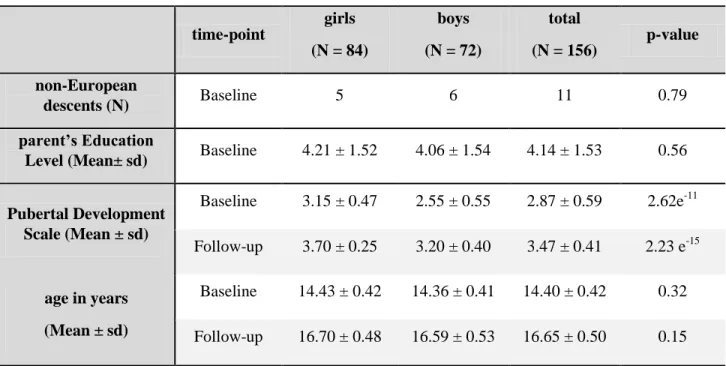
Within our sample of 156 subjects (84 girls and 72 boys) analyzed at both assessment times,
girls had higher PDS scores than boys but not significant difference in age (see Table 1 and
Supplementary Figure 2).
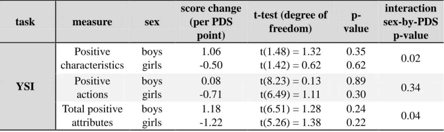
The YSI questionnaire yielded sex-by-PDS interaction with total positive attributes (p = 0.04,
see Table 2) and more specifically on the subscale “positive characteristics” (p = 0.02). “Positive characteristics” and “total positive attributes” increased in boys and decreased in girls with
No sex-by-PDS interaction was found in the SDQ or the DAWBA questionnaires (see
Supplementary Table 1).
Using age instead of PDS, no significant sex-by-age interactions were found for all behavioral
questionnaires but for the “positive characteristics” (p = 0.03, see Supplementary Table 2). We
did not find a favored model comparing “age” and “age plus PDS” models (See Supplementary
Table 3).
Imaging
Global measures
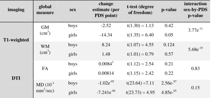
Global GM volume decreased along puberty, with a steeper rate in girls compared to boys (see
Figure 1, Table 3). Global WM volume increased with a steeper rate in boys compared to girls.
Global FA increase and global MD decrease were found for all subjects but no sex-by-PDS
interaction. Global GM and WM volumes followed similar changes when using age instead of
PDS (see Supplementary Table 4). Global diffusion indices displayed significant sex-by-age
interactions when using age instead of PDS. We did find favored models using “age plus PDS” instead of “age” only for global GM and WM volumes (See Supplementary Table 5).
Voxel-based and regional measures
The voxel-wise sex-by-PDS interaction showed a significant steeper GM volume decrease in girls in the prefrontal cortex, caudate, putamen, thalamus, Heschl’s gyrus and post-central gyrus,
while boys had a significant steeper GM volume increase in the amygdala-hippocampal complex,
precentral gyrus and parts of the occipital pole (see Figure 3, Supplementary Table 6). A steeper
WM volume increase was detected in boys compared to girls in most parts of the brain except in
bilateral external capsule, where the volume decreased more in girls than in boys. No voxel-wise
similar results in GM and WM volumes than the ones with PDS (see Supplementary Figure 3 and
Supplementary Table 7). Unlike the PDS, we found significant voxel-wise sex-by-age interaction
for FA and MD (see Supplementary Figure 4 and Supplementary Table 8).
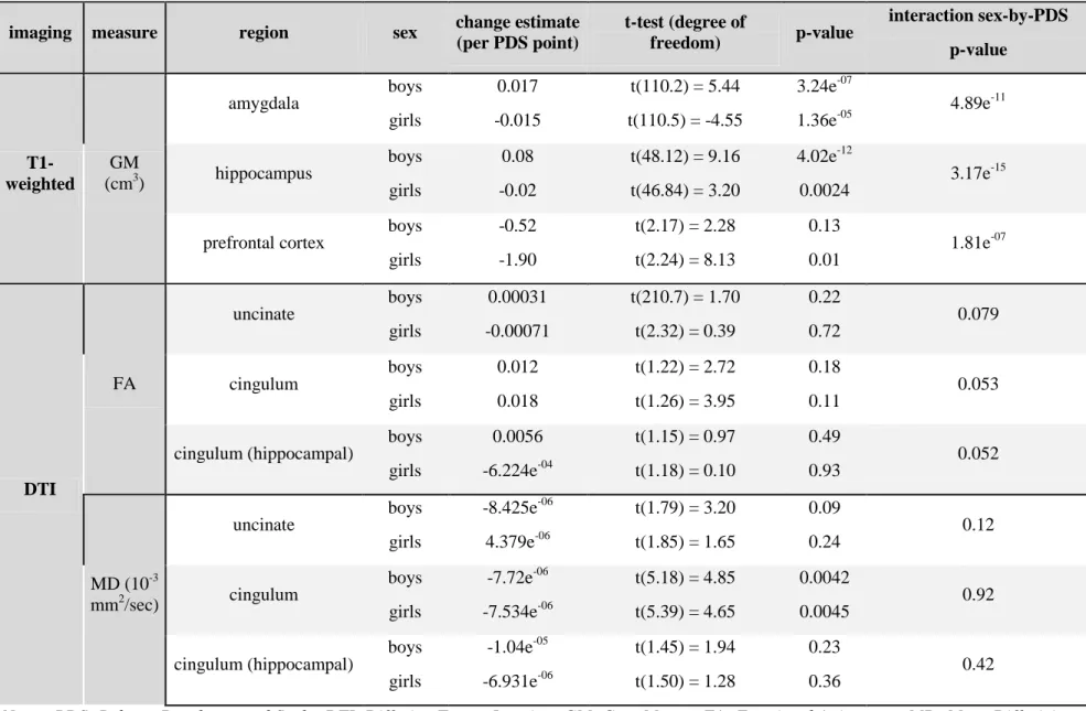
ROI investigations of macrostructure confirmed sex-by-PDS interactions in amygdala,
hippocampus and the prefrontal cortex, and concerning microstructure, we found only trends for
a sex-by-PDS interaction in the cingulum and the uncinate but with no significant change in boys
or girls taken separately (see Table 4). Boys displayed amygdala and hippocampus volumes
increases and a prefrontal cortex volume low decrease whereas girls displayed amygdala and
hippocampus volumes decreases and a prefrontal cortex volume low decrease. ROI investigations
of macro- and microstructure showed the same sex-by-age interactions (see Supplementary Table
9).
Mediation analyses
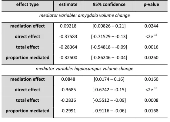
Mediation analyses showed that amygdala volume change accounted for 32.5% (p = 0.024)
and hippocampus volume change for 29.91% (p = 0.016) of the total effect between sex and “positive characteristics” along puberty (see Figure 3, Table 5). Amygdala and hippocampus
volumes increases in boys were related to “positive characteristics” increase, while amygdala and
hippocampus volumes decreases in girls were related to “positive characteristics” decrease.
No mediation effect of the prefrontal cortex volume or of the uncinate and cingulum
microstructural measures was found with YSI scores.
Discussion
Sexual differences of the brain maturation were identified in global GM and WM volumes and
approach in adolescents between 14 and 16 years. In contrast, no sexual difference of the
microstructure maturation was detected. Additionally, we found sex differences on emotional
regulation as measured by positive personality traits and this effect was related to the maturation
of regions of the limbic system.
The sex effects on the adolescents’ “positive characteristics” changes, that are a subscale of
the positive personality traits scale, were identified to be mediated by the hippocampus and
amygdala maturation. Positive attributes are meant to gather (1) positive character items (e.g.
how the adolescent feels generous, affectionate, caring, social, easy-going) and (2) positive action
items (e.g. how the adolescent is proud to be good at sport, well behaved, polite, helpful at
home). Globally, they are positively and closely related to current levels of adolescent’s
well-being (Gillham et al., 2011). “Positive characteristics” are assimilated to personality strengths
that promote connections to other people which increase positive affect, suggesting that
interpersonal interactions play an important role in the protection from depression (Gillham et al.,
2011; Peterson and Seligman, 2004). In our sample, the “positive characteristics” correlated
negatively with internalizing, externalizing and total difficulties scores (see Supplementary Table
11). Externalizing behaviors describe disruptive and dysregulated behaviors such as hyperactivity
or impulsivity whereas internalizing problems involve disturbances in emotion or mood (Graber,
2013; Perle et al., 2013; Yong et al., 2014). In this context, positive personality traits may contribute to a decreased risk of developing emotional disorders during early adulthood, as
demonstrated by (Bromley et al., 2006; Vidal-Ribas et al., 2015). The mediation by the
amygdalo-hippocampal complex, limbic structures largely involved in the emotional regulation
processing, has to be put in the light of the sex-related differences on the maturation of these
normal development, the amygdala and hippocampus continued to increase in volume during
puberty in both boys and girls with differential trajectories (Goddings et al., 2014; Herting et al.,
2018). Differences in the progression of brain structure could lead to important psychiatric
disorders in post-adolescence, which prevalence is notable during this period (Lebel and
Beaulieu, 2011; Paus et al., 2008). For example, variations of amygdala and hippocampus have
been involved in affective disorders, where volumes decreases were demonstrated in patients
with emotional symptomatology compared to controls (Blumberg HP et al., 2003; Rajmohan and
Mohandas, 2007). In summary, emotion dysregulation leading to emotional disorders is related to
limbic system maturation, in particular amygdala and hippocampus changes during adolescence.
According to our results, girls could be more sensitive to emotional disorders via positive
personality traits and limbic structures volumes decreases, suggesting that a faster and precocious
maturation during adolescence reflects a vulnerable framework for emotional dysregulation in
early adulthood. As an echo to that, we did find a significant PDS related increase of risk for
separation anxiety in girls only (See Supplementary Table 1). These elements taken together
seem to point out an increased risk for psychopathology in early maturation in girls. Graber
(2013) extended this relation in boys maturing too early or too late, which presented elevated
symptomatology of psychopathology. As for boys, we did find that amygdalo-hippocampal
complex increase was related to “positive characteristics” increase. Another study found that
amygdala-mPFC connectivity related to early life stress in adolescence was associated with
anxiety and depression in girls but again not in boys (Burghy et al., 2012). A long-standing
explanation has been that men's more active responses to their negative moods may be more
adaptive on average than women's less active, more ruminative responses (Nolen-Hoeksema,
1987). In our study, boys with no amygdalo-hippocampal complex increase could be considered
for developing psychopathology, contrary to boys with amygdalo-hippocampal complex and
positive attribute increases. Otherwise, no sex-by-PDS interaction was detected, neither in the
variables about affective disorders of the DAWBA questionnaire nor in the SDQ questionnaire.
As only healthy adolescents were recruited in this study, the lack of pathological subjects might
have decreased the statistical power of clinical variables to probe psychiatric dimensions.
The global patterns of brain maturation were confirmed in our study, with a global GM
volume decrease and a global WM volume increase in macrostructure (Giedd et al., 1997), that
might be an indication of a reduction in neuropil in the grey matter (e.g. synaptic pruning, glial
cell reduction) and an encroachment of white matter growth (K. L. Mills et al., 2014; Paus et al.,
2008). In microstructure, a global mean FA increase and a global mean MD decrease were found,
suggesting more organized fiber bundles (Schmithorst and Yuan, 2010; Wang et al., 2012).
We confirmed the sexual differences of brain maturation illustrated by a steeper global GM
volume decrease in girls and a steeper global WM volume increase in boys (Giedd et al., 1997;
Goddings et al., 2014; Lenroot and Giedd, 2010). Regionally, the sexual differences were also
confirmed in some specific regions as limbic regions and prefrontal cortex. These regions
highlighted a sexual differentiation in maturation rates, with differential decreasing trajectories in
prefrontal cortex volumes in boys and girls whereas trajectories were opposite in the
amygdalo-hippocampal complex. According to the dual systems model, the prefrontal cortex involved in
cognitive control follows a protracted development whereas limbic regions involved in
processing affect follow a more dynamic model (Casey et al., 2008; Gogtay et al., 2004; K. L.
Mills et al., 2014). In addition to confirming the differentiation in maturation rates between
cortical and subcortical structures across puberty, we demonstrated that the sex plays an
maturation in boys and an accelerated maturation in girls. From one perspective, the relation in
girls between what appears to be an accelerated amygdalo-hippocampal maturation and a decrease of “positive characteristics” could be interpreted as consistent with the dual system
model where heightened reactivity of the subcortical regions would lead to more affectively
driven behaviors and confer more risks for affective disorders (Casey et al., 2008). From another
perspective, we did not find the same relation for the prefrontal cortex, the second system of the
dual system model. In this case, our results could be consistent with an alternative model where
vulnerability to affective disorders is not due to a delayed prefrontal maturation and a failure of
regulation and controls over the subcortical system (Davey et al., 2008). From a general point of
view, we can only consider our data in the context that a delayed and protracted maturation
appears to be protective from emotional disorders.
No sex-by-PDS effect in the WM microstructural maturation between 14 and 16 was found,
neither in whole brain nor limbic regions. Some studies have found sex-by-age interactions in FA
from childhood to adulthood (Herting et al., 2017; Lebel and Beaulieu, 2011; Schmithorst et al.,
2008; Wang et al., 2012). These longitudinal studies had smaller sample size (Bava et al., 2010;
Wang et al., 2012) or larger age range (Lebel and Beaulieu, 2011), while others were
cross-sectional (Herting et al., 2012). With its longitudinal design on a large sample, our study should
have the computational strength to detect such changes. However, we did use pubertal
development scale instead of age, since it is more closely related to brain maturation and that our
age range is rather narrow (Goddings et al., 2014). As a confirmation, we did find sex differences
for WM microstructural maturation when using age instead of PDS, but these results may be
driven only by higher PDS increase for the same age range in boys as compared in girls giving in
return an artificially sex-differential pace of brain maturation. Furthermore, brain maturation can
in early–middle adolescence and an acceleration of growth again in late adolescence/early
adulthood (Simmonds et al., 2014). The limbic system appears to follow this pattern of
maturation, with cingulum and uncinate undergoing substantial changes after adolescence (Lebel
and Beaulieu, 2011). This period of little change that overlaps with our own study might account
for the absence of significant sexual differences detected here on the WM maturation.
The longitudinal image processing and use of linear mixed-models specifically designed for
repeated-measures constitute the main strengths of our study. Paired images underwent a
dedicated processing pipeline to measure individual changes before performing spatial
normalization and group analysis. In the first step of the model all time-points were registered to some form of within-subject average image, in order to avoid introducing an asymmetric bias and
to ensure all images undergo the same number of interpolations (Ashburner and Ridgway, 2013;
Reuter et al., 2012, 2010; Reuter and Fischl, 2011). This step is essential to guarantee the symmetry in the longitudinal processing. We were able to adapt our processing to diffusion
images in order to adjust precisely both modalities in the same space. We used also appropriate
statistical longitudinal models to take into account the dependence of repeated measurements
within subject, and by doing so, providing increased statistical power reducing the confounding
effect of between-subject variability (Bernal-Rusiel et al., 2013).
The findings of this study must be considered in the light of some limitations. First, we ran all
our analyses using the pubertal development scale (PDS). First, it is a self-report measurement
based on only five questions and can be prompted to subjectivity. Second, it measures not exactly
the same physical characteristics in both sexes (e.g. breast development, testis size) which can
bias the scale when comparing boys and girls brain maturations. In our study, we did not measure
(Marshall and Tanner, 1970, 1969). However, reliable studies have concluded that despite its
limitations, PDS still constitutes a suitable tool to measure the degree of puberty (Bond et al.,
2006; Dorn, 2006; Petersen et al., 1988) and remained useful and fundamental as predictor in
assessing longitudinal changes within subjects, much more precise than age (Brooks-Gunn et al.,
1987; Herting et al., 2017; Petersen et al., 1988). Given that girls in our sample have more
advanced pubertal development than boys for the same age, our strategic choice seems to be the
right one. Moreover, analyses conducted with age showed less significant results than with PDS
within behavioral questionnaire and model fitting for the global T1-weighted measures was
improved by adding the PDS.
In a second point regarding the temporal resolution, the current study had only two measures
per subject, allowing for only a linear model to be examined as an estimate of change within a
single individual (Herting et al., 2017). The two visits were close in time with a 2-year interval,
necessary to detect subtle changes during puberty but maybe quite too narrow in view of changes
during this period. Changes in brain maturation do not follow a linear curve; additional time
points will allow the testing of non-linear slopes at the individual level and to detect medium
effects of puberty. In the same vein considering the spatial resolution, we used a predefined set of
brain ROI and, for example, the different subparts of the prefrontal region were not specifically
considered in relation to their functions. Further investigations are needed to clarify the role of
each region in the maturational mismatch of the limbic system.
The third limitation of this study is the use of Youth Strength Inventory questionnaire to study
positive personality traits. Although part of the DAWBA, this questionnaire is often overlooked
and not studied in the literature for symptomatology. Indeed, it is interesting that positive
personality traits mirror emotional symptomatology in a study on healthy adolescents. Although
available data, the only scale currently existing to measure positive personality traits subjectively.
As an unexpected finding, externalizing and internalizing disorders and diagnoses scores of the DAWBA didn’t show any interaction between sex and puberty but correlated negatively with the
positive personality traits. This should be confirmed in future studies.
Conclusion
We demonstrated that the vulnerability of emotional disorders could be explained by the
mismatch of maturation rates of cortico/subcortical regions between sexes across puberty. The
delayed brain maturation in boys compared to girls showed to be related with positive personality
traits changes. These findings support that, beyond age, sex and puberty effects contribute to
neurodevelopmental trajectories and emotional regulation in girls and boys during adolescence.
Acknowledgments
This work received support from the European Union-funded FP6 Integrated Project IMAGEN
(LSHMCT-2007-037286), the French funding agency ANR (ANR-12-SAMA-0004), an
Eranet-Neuron grant (project AF12-NEUR0008-1-WN2NA) and the Fondation de France
(2012-00033703), the Fondation pour la Recherche Médicale (FRM; DPA20140629802;
DPP20151033945), the Fédération pour la Recherche sur le Cerveau (FRC Neurodon 2015), the
Fondation de l’Avenir (AP-RM-17-013), authors also thank the Strasbourg University for study
promotion in France, the Bundesministerium für Bildung und Forschung (BMBF Grants
01EV0711 and 01EE1406B), and the Deutsche Forschungsgemeinschaft (DFG SFB 940/1 and
SFB 940/2). PBF was supported by a doctoral fellowship from the Fondation de France
References
Achenbach, T., 1992. Manual for the Child Behavior Checklist/2-3 and 1992 Profile. VT: University of Vermont Department of Psychiatry, Burlington.
Angold, A., Costello, E.J., 2006. Puberty and Depression. Child Adolesc. Psychiatr. Clin. N. Am. 15, 919– 937. https://doi.org/10.1016/j.chc.2006.05.013
Angold, A., Costello, E.J., Erkanli, A., Worthman, C.M., 1999. Pubertal changes in hormone levels and depression in girls. Psychol. Med. 29, 1043–1053.
Angold, A., Costello, E.J., Worthman, C.M., 1998. Puberty and depression: the roles of age, pubertal status and pubertal timing. Psychol. Med. 28, 51–61.
Ashburner, J., Ridgway, G.R., 2013. Symmetric diffeomorphic modeling of longitudinal structural MRI. Brain Imaging Methods 6, 197. https://doi.org/10.3389/fnins.2012.00197
Aubert-Broche, B., Fonov, V.S., García-Lorenzo, D., Mouiha, A., Guizard, N., Coupé, P., Eskildsen, S.F., Collins, D.L., 2013. A new method for structural volume analysis of longitudinal brain MRI data and its application in studying the growth trajectories of anatomical brain structures in
childhood. NeuroImage 82, 393–402. https://doi.org/10.1016/j.neuroimage.2013.05.065 Bava, S., Thayer, R., Jacobus, J., Ward, M., Jernigan, T.L., Tapert, S.F., 2010. Longitudinal characterization
of white matter maturation during adolescence. Brain Res. 1327, 38–46. https://doi.org/10.1016/j.brainres.2010.02.066
Bernal-Rusiel, J.L., Greve, D.N., Reuter, M., Fischl, B., Sabuncu, M.R., 2013. Statistical Analysis of Longitudinal Neuroimage Data with Linear Mixed Effects Models. NeuroImage 0, 249–260. https://doi.org/10.1016/j.neuroimage.2012.10.065
Blakemore, S.-J., Burnett, S., Dahl, R.E., 2010. The Role of Puberty in the Developing Adolescent Brain. Hum. Brain Mapp. 31, 926–933. https://doi.org/10.1002/hbm.21052
Blumberg HP, Kaufman J, Martin A, et al, 2003. Amygdala and hippocampal volumes in adolescents and adults with bipolar disorder. Arch. Gen. Psychiatry 60, 1201–1208.
https://doi.org/10.1001/archpsyc.60.12.1201
Bond, L., Clements, J., Bertalli, N., Evans-Whipp, T., McMorris, B.J., Patton, G.C., Toumbourou, J.W., Catalano, R.F., 2006. A comparison of self-reported puberty using the Pubertal Development Scale and the Sexual Maturation Scale in a school-based epidemiologic survey. J. Adolesc. 29, 709–720. https://doi.org/10.1016/j.adolescence.2005.10.001
Bromley, E., Johnson, J.G., Cohen, P., 2006. Personality strengths in adolescence and decreased risk of developing mental health problems in early adulthood. Compr. Psychiatry 47, 315–324. https://doi.org/10.1016/j.comppsych.2005.11.003
Brooks-Gunn, J., Warren, M., Rosso, J., Gargiulo, J., 1987. Validity of self-report measures of girls’ pubertal status. Child Dev.
Burghy, C.A., Stodola, D.E., Ruttle, P.L., Molloy, E.K., Armstrong, J.M., Oler, J.A., Fox, M.E., Hayes, A.S., Kalin, N.H., Essex, M.J., Davidson, R.J., Birn, R.M., 2012. Developmental pathways to amygdala-prefrontal function and internalizing symptoms in adolescence. Nat. Neurosci. 15, 1736–1741. https://doi.org/10.1038/nn.3257
Casey, B.J., Jones, R.M., Hare, T.A., 2008. The Adolescent Brain. Ann. N. Y. Acad. Sci. 1124, 111–126. https://doi.org/10.1196/annals.1440.010
Dahl, R.E., 2004. Adolescent Brain Development: A Period of Vulnerabilities and Opportunities. Keynote Address. Ann. N. Y. Acad. Sci. 1021, 1–22. https://doi.org/10.1196/annals.1308.001
Davey, C.G., Yücel, M., Allen, N.B., 2008. The emergence of depression in adolescence: development of the prefrontal cortex and the representation of reward. Neurosci. Biobehav. Rev. 32, 1–19. https://doi.org/10.1016/j.neubiorev.2007.04.016
Davidson, R., Lewis, D., Alloy, L.B., Amaral, D.G., Bush, G., Cohen, J., Drevets, W.C., Farah, M., Kagan, J., McClelland, J., Nolen-Hoeksema, S., Peterson, B., 2002. Neural and behavioral substrates of mood and mood regulation. [WWW Document]. URL
https://www-ncbi-nlm-nih-gov.gate2.inist.fr/pubmed/12361665
Dennison, M., Whittle, S., Yücel, M., Vijayakumar, N., Kline, A., Simmons, J., Allen, N.B., 2013. Mapping subcortical brain maturation during adolescence: evidence of hemisphere- and sex-specific longitudinal changes. Dev. Sci. 16, 772–791. https://doi.org/10.1111/desc.12057
Dorn, L.D., 2006. Measuring Puberty. J. Adolesc. Health 39, 625–626. https://doi.org/10.1016/j.jadohealth.2006.05.014
Eliot, L., 2019. Neurosexism: the myth that men and women have different brains. Nature 566, 453–456. Eluvathingal, T.J., Hasan, K.M., Kramer, L., Fletcher, J.M., Ewing-Cobbs, L., 2007. Quantitative Diffusion
Tensor Tractography of Association and Projection Fibers in Normally Developing Children and Adolescents. Cereb. Cortex N. Y. N 1991 17, 2760–2768. https://doi.org/10.1093/cercor/bhm003 Fish, A.M., Nadig, A., Seidlitz, J., Reardon, P.K., Mankiw, C., McDermott, C.L., Blumenthal, J.D., Clasen,
L.S., Lalonde, F., Lerch, J.P., Chakravarty, M.M., Shinohara, R.T., Raznahan, A., 2019. Sex-biased trajectories of amygdalo-hippocampal morphology change over human development.
NeuroImage 204, 116122. https://doi.org/10.1016/j.neuroimage.2019.116122
Giedd, J.N., 2004. Structural Magnetic Resonance Imaging of the Adolescent Brain. Ann. N. Y. Acad. Sci. 1021, 77–85. https://doi.org/10.1196/annals.1308.009
Giedd, J.N., Castellanos, F.X., Rajapakse, J.C., Vaituzis, A.C., Rapoport, J.L., 1997. Sexual dimorphism of the developing human brain. Prog. Neuropsychopharmacol. Biol. Psychiatry 21, 1185–1201. https://doi.org/10.1016/S0278-5846(97)00158-9
Gillham, J., Adams-Deutsch, Z., Werner, J., Reivich, K., Coulter-Heindl, V., Linkins, M., Winder, B., Peterson, C., Park, N., Abenavoli, R., Contero, A., Seligman, M.E.P., 2011. Character strengths predict subjective well-being during adolescence. J. Posit. Psychol. 6, 31–44.
https://doi.org/10.1080/17439760.2010.536773
Giorgio, A., Watkins, K.E., Chadwick, M., James, S., Winmill, L., Douaud, G., De Stefano, N., Matthews, P.M., Smith, S.M., Johansen-Berg, H., James, A.C., 2010. Longitudinal changes in grey and white matter during adolescence. NeuroImage 49, 94–103.
https://doi.org/10.1016/j.neuroimage.2009.08.003
Goddings, A.-L., Mills, K.L., Clasen, L.S., Giedd, J.N., Viner, R.M., Blakemore, S.-J., 2014. The influence of puberty on subcortical brain development. NeuroImage 88, 242–251.
https://doi.org/10.1016/j.neuroimage.2013.09.073
Gogtay, N., Giedd, J.N., Lusk, L., Hayashi, K.M., Greenstein, D., Vaituzis, A.C., Nugent, T.F., Herman, D.H., Clasen, L.S., Toga, A.W., Rapoport, J.L., Thompson, P.M., 2004. Dynamic mapping of human cortical development during childhood through early adulthood. Proc. Natl. Acad. Sci. U. S. A. 101, 8174–8179. https://doi.org/10.1073/pnas.0402680101
Goodman, R., Ford, T., Richards, H., Gatward, R., Meltzer, H., 2000. The Development and Well-Being Assessment: Description and Initial Validation of an Integrated Assessment of Child and Adolescent Psychopathology. J. Child Psychol. Psychiatry 41, 645–655.
https://doi.org/10.1111/j.1469-7610.2000.tb02345.x
Goodman, R., Meltzer, H., Bailey, V., 2003. The Strengths and Difficulties Questionnaire: a pilot study on the validity of the self-report version. Int. Rev. Psychiatry 15, 173–177.
https://doi.org/10.1080/0954026021000046137
Graber, J.A., 2013. Pubertal timing and the development of psychopathology in adolescence and beyond. Horm. Behav. 64, 262–269. https://doi.org/10.1016/j.yhbeh.2013.04.003
Herting, M.M., Johnson, C., Mills, K.L., Vijayakumar, N., Dennison, M., Liu, C., Goddings, A.-L., Dahl, R.E., Sowell, E.R., Whittle, S., Allen, N.B., Tamnes, C.K., 2018. Development of subcortical volumes
across adolescence in males and females: A multisample study of longitudinal changes. Neuroimage 172, 194–205.
Herting, M.M., Kim, R., Uban, K.A., Kan, E., Binley, A., Sowell, E.R., 2017. Longitudinal changes in pubertal maturation and white matter microstructure. Psychoneuroendocrinology 81, 70–79.
https://doi.org/10.1016/j.psyneuen.2017.03.017
Herting, M.M., Maxwell, E.C., Irvine, C., Nagel, B.J., 2012. The impact of sex, puberty, and hormones on white matter microstructure in adolescents. Cereb. Cortex N. Y. N 1991 22, 1979–1992.
https://doi.org/10.1093/cercor/bhr246
Herting, M.M., Sowell, E.R., 2017. Puberty and structural brain development in humans. Front. Neuroendocrinol. 44, 122–137. https://doi.org/10.1016/j.yfrne.2016.12.003
Koolschijn, P.C.M.P., Crone, E.A., 2013. Sex differences and structural brain maturation from childhood to early adulthood. Dev. Cogn. Neurosci. 5, 106–118. https://doi.org/10.1016/j.dcn.2013.02.003 Lebel, C., Beaulieu, C., 2011. Longitudinal Development of Human Brain Wiring Continues from
Childhood into Adulthood. J. Neurosci. 31, 10937–10947. https://doi.org/10.1523/JNEUROSCI.5302-10.2011
Lenroot, R.K., Giedd, J.N., 2010. Sex differences in the adolescent brain. Brain Cogn. 72, 46–55. https://doi.org/10.1016/j.bandc.2009.10.008
Lenroot, R.K., Giedd, J.N., 2006. Brain development in children and adolescents: insights from anatomical magnetic resonance imaging. Neurosci. Biobehav. Rev. 30, 718–729.
https://doi.org/10.1016/j.neubiorev.2006.06.001
Lenroot, R.K., Gogtay, N., Greenstein, D.K., Wells, E.M., Wallace, G.L., Clasen, L.S., Blumenthal, J.D., Lerch, J., Zijdenbos, A.P., Evans, A.C., Thompson, P.M., Giedd, J.N., 2007. Sexual dimorphism of brain developmental trajectories during childhood and adolescence. NeuroImage 36, 1065– 1073. https://doi.org/10.1016/j.neuroimage.2007.03.053
Marshall, W.A., Tanner, J.M., 1970. Variations in the Pattern of Pubertal Changes in Boys. Arch. Dis. Child. 45, 13.
Marshall, W.A., Tanner, J.M., 1969. Variations in pattern of pubertal changes in girls. Arch. Dis. Child. 44, 291.
Menzies, L., Goddings, A.-L., Whitaker, K.J., Blakemore, S.-J., Viner, R.M., 2015. The effects of puberty on white matter development in boys. Dev. Cogn. Neurosci. 11, 116–128.
https://doi.org/10.1016/j.dcn.2014.10.002
Mills, K. L., Goddings, A.-L., Clasen, L.S., Giedd, J.N., Blakemore, S.-J., 2014. The developmental mismatch in structural brain maturation during adolescence. Dev. Neurosci. 36, 147–160.
https://doi.org/10.1159/000362328
Mills, Kathryn L., Goddings, A.-L., Clasen, L.S., Giedd, J.N., Blakemore, S.-J., 2014. The developmental mismatch in structural brain maturation during adolescence. Dev. Neurosci. 36, 147–160. https://doi.org/10.1159/000362328
Nolen-Hoeksema, S., 1987. Sex differences in unipolar depression: evidence and theory. Psychol. Bull. 101, 259–282.
Paus, T., Keshavan, M., Giedd, J.N., 2008. Why do many psychiatric disorders emerge during adolescence? Nat. Rev. Neurosci. 9, 947–957. https://doi.org/10.1038/nrn2513
Perle, J.G., Levine, A.B., Odland, A.P., Ketterer, J.L., Cannon, M.A., Marker, C.D., 2013. The Association Between Internalizing Symptomology and Risky Behaviors. J. Child Adolesc. Subst. Abuse 22, 1– 24. https://doi.org/10.1080/1067828X.2012.724289
Petersen, A.C., Crockett, L., Richards, M., Boxer, A., 1988. A self-report measure of pubertal status: Reliability, validity, and initial norms. J. Youth Adolesc. 17, 117–133.
Peterson, C., Seligman, M.E.P., 2004. Character Strengths and Virtues: A Handbook and Classification. American Psychological Association and Oxford University Press.
Rajmohan, V., Mohandas, E., 2007. The limbic system. Indian J. Psychiatry 49, 132–139. https://doi.org/10.4103/0019-5545.33264
Raznahan, A., Shaw, P.W., Lerch, J.P., Clasen, L.S., Greenstein, D., Berman, R., Pipitone, J., Chakravarty, M.M., Giedd, J.N., 2014. Longitudinal four-dimensional mapping of subcortical anatomy in human development. Proc. Natl. Acad. Sci. 111, 1592–1597.
https://doi.org/10.1073/pnas.1316911111
Reuter, M., Fischl, B., 2011. Avoiding asymmetry-induced bias in longitudinal image processing. NeuroImage 57, 19–21. https://doi.org/10.1016/j.neuroimage.2011.02.076
Reuter, M., Rosas, H.D., Fischl, B., 2010. Highly accurate inverse consistent registration: A robust approach. NeuroImage 53, 1181–1196. https://doi.org/10.1016/j.neuroimage.2010.07.020 Reuter, M., Schmansky, N.J., Rosas, H.D., 2012. Within-subject template estimation for unbiased
longitudinal image analysis. NeuroImage 61, 1402–1418. https://doi.org/10.1016/j.neuroimage.2012.02.084
Satterthwaite, T.D., Vandekar, S., Wolf, D.H., Ruparel, K., Roalf, D.R., Jackson, C., Elliott, M.A., Bilker, W.B., Calkins, M.E., Prabhakaran, K., Davatzikos, C., Hakonarson, H., Gur, R.E., Gur, R.C., 2014. Sex Differences in the Effect of Puberty on Hippocampal Morphology. J. Am. Acad. Child Adolesc. Psychiatry 53, 341-350.e1. https://doi.org/10.1016/j.jaac.2013.12.002
Schmithorst, V.J., Holland, S.K., Dardzinski, B.J., 2008. Developmental differences in white matter architecture between boys and girls. Hum. Brain Mapp. 29, 696–710.
https://doi.org/10.1002/hbm.20431
Schmithorst, V.J., Yuan, W., 2010. White matter development during adolescence as shown by diffusion MRI. Brain Cogn., Adolescent Brain Development: Current Themes and Future Directions 72, 16– 25. https://doi.org/10.1016/j.bandc.2009.06.005
Schumann, G., Loth, E., Banaschewski, T., Barbot, A., Barker, G., Büchel, C., Conrod, P.J., Dalley, J.W., Flor, H., Gallinat, J., Garavan, H., Heinz, A., Itterman, B., Lathrop, M., Mallik, C., Mann, K., Martinot, J.-L., Paus, T., Poline, J.-B., Robbins, T.W., Rietschel, M., Reed, J.-L., Smolka, M., Spanagel, R., Speiser, C., Stephens, D.N., Ströhle, A., Struve, M., IMAGEN consortium, 2010. The IMAGEN study: reinforcement-related behaviour in normal brain function and psychopathology. Mol. Psychiatry 15, 1128–1139. https://doi.org/10.1038/mp.2010.4
Seunarine, K.K., Clayden, J.D., Jentschke, S., Muñoz, M., Cooper, J.M., Chadwick, M.J., Banks, T., Vargha-Khadem, F., Clark, C.A., 2016. Sexual Dimorphism in White Matter Developmental Trajectories Using Tract-Based Spatial Statistics. Brain Connect. 6, 37–47.
https://doi.org/10.1089/brain.2015.0340
Simmonds, D.J., Hallquist, M.N., Asato, M., Luna, B., 2014. Developmental stages and sex differences of white matter and behavioral development through adolescence: A longitudinal diffusion tensor imaging (DTI) study. NeuroImage 92, 356–368.
https://doi.org/10.1016/j.neuroimage.2013.12.044
Sisk, C.L., Foster, D.L., 2004. The neural basis of puberty and adolescence. Nat. Neurosci. 7, 1040–1047. https://doi.org/10.1038/nn1326
Smith, S.M., Jenkinson, M., Johansen-Berg, H., Rueckert, D., Nichols, T.E., Mackay, C.E., Watkins, K.E., Ciccarelli, O., Cader, M.Z., Matthews, P.M., Behrens, T.E.J., 2006. Tract-based spatial statistics: voxelwise analysis of multi-subject diffusion data. NeuroImage 31, 1487–1505.
https://doi.org/10.1016/j.neuroimage.2006.02.024
Spear, L.P., 2000. The adolescent brain and age-related behavioral manifestations. Neurosci. Biobehav. Rev. 24, 417–463. https://doi.org/10.1016/S0149-7634(00)00014-2
Versace, A., Almeida, J.R.C., Quevedo, K., Thompson, W.K., Terwilliger, R.A., Hassel, S., Kupfer, D.J., Phillips, M.L., 2010. Right Orbitofrontal Corticolimbic and Left Corticocortical White Matter Connectivity Differentiate Bipolar and Unipolar Depression. Biol. Psychiatry 68, 560–567. https://doi.org/10.1016/j.biopsych.2010.04.036
Vidal-Ribas, P., Goodman, R., Stringaris, A., 2015. Positive attributes in children and reduced risk of future psychopathology. Br. J. Psychiatry 206, 17–25. https://doi.org/10.1192/bjp.bp.114.144519 Wang, Y., Adamson, C., Yuan, W., Altaye, M., Rajagopal, A., Byars, A.W., Holland, S.K., 2012. Sex
differences in white matter development during adolescence: A DTI study. Brain Res. 1478, 1–15. https://doi.org/10.1016/j.brainres.2012.08.038
Wierenga, L.M., Bos, M.G.N., Schreuders, E., Vd Kamp, F., Peper, J.S., Tamnes, C.K., Crone, E.A., 2018. Unraveling age, puberty and testosterone effects on subcortical brain development across adolescence. Psychoneuroendocrinology 91, 105–114.
https://doi.org/10.1016/j.psyneuen.2018.02.034
Wierenga, L.M., Langen, M., Oranje, B., Durston, S., 2014. Unique developmental trajectories of cortical thickness and surface area. NeuroImage 87, 120–126.
https://doi.org/10.1016/j.neuroimage.2013.11.010
Yong, M., Fleming, C.B., McCarty, C.A., Catalano, R.F., 2014. Mediators of the Associations Between Externalizing Behaviors and Internalizing Symptoms in Late Childhood and Early Adolescence. J. Early Adolesc. 34, 967–1000. https://doi.org/10.1177/0272431613516827
Table 1: Sample demographics. time-point girls (N = 84) boys (N = 72) total (N = 156) p-value non-European descents (N) Baseline 5 6 11 0.79 parent’s Education
Level (Mean± sd) Baseline 4.21 ± 1.52 4.06 ± 1.54 4.14 ± 1.53 0.56
Pubertal Development Scale (Mean ± sd) Baseline 3.15 ± 0.47 2.55 ± 0.55 2.87 ± 0.59 2.62e-11 Follow-up 3.70 ± 0.25 3.20 ± 0.40 3.47 ± 0.41 2.23 e-15 age in years (Mean ± sd) Baseline 14.43 ± 0.42 14.36 ± 0.41 14.40 ± 0.42 0.32 Follow-up 16.70 ± 0.48 16.59 ± 0.53 16.65 ± 0.50 0.15
Table 2: Effects of PDS by sex on psychometric measures.
task measure sex
score change (per PDS point) t-test (degree of freedom) p-value interaction sex-by-PDS p-value YSI Positive characteristics boys girls 1.06 -0.50 t(1.48) = 1.32 t(1.42) = 0.62 0.35 0.62 0.02 Positive actions boys girls 0.08 -0.71 t(8.23) = 0.13 t(6.49) = 1.11 0.89 0.30 0.34 Total positive attributes boys girls 1.18 -1.22 t(6.51) = 1.28 t(5.26) = 1.38 0.24 0.22 0.04
Table 3: Effects of PDS by sex on global imaging measures. imaging global measure sex change estimate (per PDS point) t-test (degree of freedom) p-value interaction sex-by-PDS p-value T1-weighted GM (cm3) boys girls -2.52 -14.34 t(1.30) = 1.13 t(1.35) = 6.40 0.42 0.05 3.77e-11 WM (cm3) boys girls 8.24 1.48 t(1.07) = 4.55 t(1.01) = 0.79 0.124 0.57 5.68e-16 DTI FA boys girls 0.00843 0.00814 t(1.12) = 2.54 t(1.15) = 2.42 0.21 0.22 0.83 MD (10-3 mm2/sec) boys girls -1.02e-05 -7.241e-06 t(23.64) =7.11 t(23.73) = 4.95 2.56e-07 4.85e-05 0.15
Notes: PDS: Puberty Developmental Scale; DTI: Diffusion Tensor Imaging; GM: Grey Matter; WM: White Matter; FA: Fractional Anisotropy; MD: Mean Diffusivity.
Table 4: Effects of PDS by sex on Region Of Interest measures.
imaging measure region sex change estimate
(per PDS point) t-test (degree of freedom) p-value interaction sex-by-PDS p-value T1-weighted GM (cm3) amygdala boys girls 0.017 -0.015 t(110.2) = 5.44 t(110.5) = -4.55 3.24e-07 1.36e-05 4.89e-11 hippocampus boys girls 0.08 -0.02 t(48.12) = 9.16 t(46.84) = 3.20 4.02e-12 0.0024 3.17e -15
prefrontal cortex boys girls -0.52 -1.90 t(2.17) = 2.28 t(2.24) = 8.13 0.13 0.01 1.81e -07 DTI FA uncinate boys girls 0.00031 -0.00071 t(210.7) = 1.70 t(2.32) = 0.39 0.22 0.72 0.079 cingulum boys girls 0.012 0.018 t(1.22) = 2.72 t(1.26) = 3.95 0.18 0.11 0.053
cingulum (hippocampal) boys girls 0.0056 -6.224e-04 t(1.15) = 0.97 t(1.18) = 0.10 0.49 0.93 0.052 MD (10-3 mm2/sec) uncinate boys girls -8.425e-06 4.379e-06 t(1.79) = 3.20 t(1.85) = 1.65 0.09 0.24 0.12 cingulum boys girls -7.72e-06 -7.534e-06 t(5.18) = 4.85 t(5.39) = 4.65 0.0042 0.0045 0.92
cingulum (hippocampal) boys girls -1.04e-05 -6.931e-06 t(1.45) = 1.94 t(1.50) = 1.28 0.23 0.36 0.42
Table 5: Mediation of brain volume changes of the amygdala and the hippocampus on the
relationship between sex and “positive characteristics” changes between 14 and 16 years using
causal mediation analysis.
effect type estimate 95% confidence p-value
mediator variable: amygdala volume change
mediation effect 0.09218 [0.00826 – 0.21] 0.0244 direct effect -0.37583 [-0.71529 – -0.13] <2e-16
total effect -0.28364 [-0.54818 – -0.09] 0.0016 proportion mediated -0.32500 [-0.86246 – -0.04] 0.0260
mediator variable: hippocampus volume change
mediation effect 0.0848 [0.0174 – 0.16] 0.0160 direct effect -0.3685 [-0.6742 – -0.15] <2e-16
total effect -0.2836 [-0.5512 – -0.09] 0.0008 proportion mediated -0.2991 [-0.9116 – -0.06] 0.0168
Captions to figures
Figure 1: Longitudinal effect of PDS on global GM and WM volumes, and global FA and MD
indices. Girls are in red and boys in blue; thin lines represent individual scores; thick lines
represent the linear mixed-effects model estimates. Sex-by-PDS interaction is only significant for
GM (p = 3.15 e-10; boys: b = -1.73, t(1.34) = -1.36, p = 0.36; girls: b = -8.31, t(1.39) = -6.47, p =
0.05) and WM volumes (p = 1.33 e-15; boys: b = 4.83, t(1.10) = 4.48, p = 0.12; girls: b = 0.91,
t(1.03) = 0.84, p = 0.55); PDS: Puberty Developmental Scale; GM: Grey Matter; WM: White
Figure 2: Voxel-based sex-by-PDS interaction for GM (top row) and WM (bottom row). Steeper
decreases in girls than boys in blue-light blue color scale and steeper increases in boys than girls
in red-yellow color scale are superimposed on the sample mean GM and WM images. Color
scales represent t-values (p < 0.05 FWE corrected). R: Right; PDS: Puberty Developmental
Scale; GM: Grey Matter; WM: White Matter
Figure 3: Mediation of brain volume changes of the amygdala (in green) and the hippocampus
(in red) on the relationship between sex and “positive characteristics” changes between 14 and 16