HAL Id: hal-02349679
https://hal.umontpellier.fr/hal-02349679
Submitted on 14 Nov 2020HAL is a multi-disciplinary open access archive for the deposit and dissemination of sci-entific research documents, whether they are pub-lished or not. The documents may come from teaching and research institutions in France or abroad, or from public or private research centers.
L’archive ouverte pluridisciplinaire HAL, est destinée au dépôt et à la diffusion de documents scientifiques de niveau recherche, publiés ou non, émanant des établissements d’enseignement et de recherche français ou étrangers, des laboratoires publics ou privés.
Synthesis, Crystal Structure, and Enthalpies of
Formation of Churchite-type REPO 4
×2H 2 O (RE =
Gd to Lu) Materials
Tamilarasan Subramani, Mohamed Ruwaid Rafiuddin, Anna Shelyug, Sergey
Ushakov, Adel Mesbah, Nicolas Clavier, Danwen Qin, Stephanie Szenknect,
Erik Elkaïm, Nicolas Dacheux, et al.
To cite this version:
Tamilarasan Subramani, Mohamed Ruwaid Rafiuddin, Anna Shelyug, Sergey Ushakov, Adel Mesbah, et al.. Synthesis, Crystal Structure, and Enthalpies of Formation of Churchite-type REPO 4×2H 2 O (RE = Gd to Lu) Materials. Crystal Growth & Design, American Chemical Society, 2019, 19 (8), pp.4641-4649. �10.1021/acs.cgd.9b00524�. �hal-02349679�
Synthesis, Crystal Structure and Enthalpies of
Formation of Churchite-type REPO
4
· 2 H
2
O (RE =
Gd to Lu) Materials.
Tamilarasan Subramani,† Mohamed Ruwaid Rafiuddin,‡ Anna Shelyug,† Sergey Ushakov,† Adel
Mesbah,‡,* Nicolas Clavier,‡ Danwen Qin,‡ Stephanie Szenknect,‡ Erik Elkaim,§, Nicolas
Dacheux,‡ Alexandra Navrotsky†,*
†
Peter A. Rock Thermochemistry Laboratory and NEAT ORU, University of California Davis, Davis CA 95616 USA
‡
ICSM, CEA, CNRS, ENSCM, Univ Montpellier, Site de Marcoule, Bat 426, 30207 Bagnols Sur Ceze, France
§ Synchrotron SOLEIL, L’Orme des Merisiers, Saint-Aubin, BP 48, 91192 Gif Sur Yvette, France
Abstract
Monazite- (REPO4; RE = La to Gd) and xenotime- (REPO4; RE = Dy to Lu & Y) type materials
have been proposed as host matrices for the immobilization of actinides. Aqueous alteration of
monazite and xenotime minerals could result in the formation of rhabdophane (REPO4 · 0.667
H2O; RE = La to Dy) and churchite (REPO4 · 2 H2O; RE = Gd to Lu & Y) phases, respectively.
Among these structure types, the structure and properties of churchite materials are not well understood and this study aims to bridge this gap by providing a comprehensive insight into the
structure and thermochemical properties of churchite materials. Churchite materials (REPO4 · 2
H2O; RE = Gd to Lu) were synthesized by a low temperature precipitation route and their crystal
structures were determined by powder X-ray diffraction (XRD). Examination of the powder XRD data has shown that the churchite materials crystallize in the monoclinic crystal system
(space group: C2/c). The enthalpies of formation (ΔH°f,ox) of churchite-type REPO4 · 2 H2O (RE
= Gd to Yb) determined by high temperature oxide melt solution calorimetry are more negative than their anhydrous counterparts (i.e., xenotime structure) and indicates that the formation of churchite is more exothermic than the xenotime phase. However, the churchite materials are
likely to have a more negative entropy of formation (ΔS°f,ox) due to the presence of water
molecules, resulting in a less negative Gibbs free energy of formation (ΔG°f,ox) than the
xenotime structure. Therefore, churchite materials are expected to be stable at lower
temperatures. For the unique case of GdPO4 and GdPO4 · n H2O materials which could adopt all
the above discussed structure types, the ΔG°f,ox of monazite from oxides was observed to be
more negative than those of xenotime, rhabdophane and churchite thereby suggesting the following order of stability: Gd-churchite < Gd-rhabdophane < Gd-xenotime < Gd-monazite.
Introduction
Rare earth (RE) phosphate materials have been the subject of numerous investigations particularly owing to their potential use as a host matrix for the storage of high level wastes
(HLW) arising from the reprocessing of spent nuclear fuel and dismantling of nuclear weapons.
1-6
RE phosphates are both compositionally and structurally diverse and occur in nature as
monazite (REPO4; RE = La to Gd), xenotime (REPO4; RE = Gd to Lu & Y), rhabdophane
(REPO4 · n H2O; RE = La to Dy; 0 ≤ n ≤ 0.667), and churchite (REPO4 · 2 H2O; RE = Gd to Lu
and Y).7-10 Among these minerals, monazite and xenotime minerals contain the highest weight
percent of rare earth elements. In addition, they also contain a large amount of U and Th thereby making these minerals radioactive. Even though, these minerals have been exposed to radiation
events over geological timescales,11-14 it should be noted that the monazite mineral, in particular,
is never completely found in the metamict state (i.e. amorphization due to radiation damages). Radiation studies on synthetic and natural monazite samples have shown that the monazite samples experience structural damage upon heavy-ion (e.g., Au ions) irradiation and can also recover completely from the structural damage upon annealing the irradiated samples at low
temperatures or via light-ion (e.g., He ions) irradiation of the damaged samples.15-20
Nevertheless, apart from a few radiation studies on synthetic xenotime samples, the structural
response of xenotime minerals to radiation is not well documented.17 In terms of chemical
durability, both monazite and xenotime minerals are reported to be chemically durable in
aqueous environments.21-25 Such mineralogical evidence suggests that the synthetic analogues of
monazite and xenotime minerals are promising matrices for the immobilization of high level
nuclear waste (HLW).2,26 Moreover, hydrated RE phosphates, namely rhabdophane is found to
stop or delay the release of actinides to the environment.8,27,28 Besides applications as nuclear waste forms, the rare-earth phosphates also have wide ranging applications in photonics, as
catalysts, as proton conductors, and in biolabeling.29-33
Rare earth phosphates can adopt the monazite-, xenotime-, rhabdophane-, and churchite-type structures depending on the ionic radius of the RE ions as well as the conditions of the synthesis. Using specific synthetic methods and conditions, it is possible to obtain all the above-mentioned crystal structures for the following rare-earths: Gd, Tb, and Dy. Anhydrous RE
phosphates adopting the monazite- (REPO4; RE = La to Gd) and xenotime structures (REPO4;
RE’ = Tb to Lu and Y) crystallize in the monoclinic (space group: P21/n) and tetragonal (space
group: I41/amd) crystal systems, respectively.7 Hydrated RE phosphates adopting the
rhabdophane structure (REPO4 · n H2O; RE = La to Dy; 0 ≤ n ≤ 0.667) crystallize in the
monoclinic crystal system (space group: C2) while the corresponding anhydrous form of
rhabdophane materials (REPO4; RE = La to Dy) adopt the trigonal structure (space group:
P3121).8,34 Detailed descriptions of the crystal structures of monazite, xenotime, and
rhabdophane can be found elsewhere.7,8,34 While the structures of monazite, xenotime, and
rhabdophane materials have been studied in much detail, the churchite (REPO4 · 2 H2O; RE =
Gd to Lu and Y) materials have been less explored. To the best of our knowledge, there has been
only two structural studies reported on churchite-type materials.9,10 Those were carried out on
synthetic (YPO4 · 2 H2O) and natural churchite (Y1-x(Gd,Dy,Er)xPO4 · 2 H2O) samples.9,10 Both
these studies have shown that the churchite materials adopt the gypsum structure (CaSO4 · 2
H2O) and crystallize in the monoclinic system (space group: C2/c).9,10 With the exception of
GdPO4 · 2 H2O, the synthesis and crystal structure of other members of the churchite group
thermodynamic properties of anhydrous RE phosphates adopting the monazite and xenotime structures are fairly well understood, those of hydrous RE phosphates adopting the churchite
structures are limited.36-38
This study reports the synthesis, structure, and thermochemical properties of the churchite
materials REPO4 · 2 H2O (RE = Gd to Lu). Members of the churchite series were prepared using
a precipitation route and their crystal structures were determined using laboratory and synchrotron powder X-ray diffraction (XRD). Thermal analysis was conducted using differential scanning calorimetry (DSC) and thermogravimetric analysis (TGA). The enthalpies of formation of the entire churchite series were determined by high temperature oxide melt solution calorimetry. The systematics of structure and thermodynamic stability are discussed. To the best of our knowledge, this represents the first study to carry out the synthesis and the structural and
thermochemical characterization of all members of the churchite (REPO4 ·2 H2O; RE = Gd to
Lu) series.
Experimental methods
Synthesis
The synthesis of REPO4 · 2 H2O materials was performed using the following starting
materials: GdCl3 · 6 H2O (Sigma Aldrich; 99 %), TbCl3 · 6 H2O (Sigma Aldrich; 99.9%), DyCl3
· 6 H2O (Sigma Aldrich; 99.99 %), HoCl3 (Sigma Aldrich, 99.9 %), ErCl3 · n H2O (Sigma
Aldrich; 99.9 %), TmCl3 (Sigma Aldrich; 99.9 %), YbCl3 (Sigma Aldrich; 99.9 %), LuCl3
(Sigma Aldrich; 99.9% ), H3PO4 (85% Normapur), and HCl (37 % Carbo Erba). The
hygroscopic nature of the lanthanide chloride salts prevents the accurate weighing of these powders. For this reason, these salts were dissolved in 1M HCl to prepare solutions whose
lanthanide concentration varied between 0.5 and 1M. The final concentrations of the dissolved lanthanides were further determined by ICP-AES.
Pure REPO4 2 H2O (RE = Gd, Tb, Dy, Ho, Er, Tm, Yb, Lu) materials were synthesized by
mixing lanthanide chloride solutions and H3PO4 (5 M) with 3 mol. % phosphate excess. Then,
each mixture was transferred in a glass container (100 mL) and left aside in a refrigerator at 4°C for two years. Afterwards, the resulting powder was washed twice with water, and once with ethanol, separated from the solution by centrifugation and then finally dried at room temperature in air. In order to determine the thermodynamic stability of monazite-, xenotime-, rhabdophane-, and churchite type structures for a given RE element, Gd-phosphates adopting these structure-types were also synthesized.
Powder X-Ray Diffraction (PXRD)
Synchrotron powder diffraction pattern of selected compounds REPO4 · 2 H2O (Ln = Gd,
Dy and Er) were collected from the beamline "Cristal" at Synchrotron Soleil, France in high angular resolution mode using the two-circle diffractometer equipped with twenty one Si(111) crystals rear analyzers. Scans were recorded at room temperature with the powder inside a glass capillary (diameter 0.3 mm) mounted on a spinner to improve particle statistics. A wavelength of
0.58403 Å was selected and calibrated using LaB6 standard NIST powder. With this setup, a
single scan was collected up to 2 = 60 ° in about two hours. In order to avoid the degradation of
the powder under irradiation, the capillary was shifted to a fresh zone every 15 minutes.
All the as-synthesized powders were also analyzed using laboratory PXRD and the data were collected in reflection geometry mode using a Bruker D8-Advance diffractometer
(LynxEye detector) employed with Cu Kα1,2 radiation (λ = 1.5418 Å) source. Data were recorded
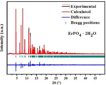
of about 2 hours per sample. All the collected powder patterns (see Figure 1) have been refined
by the Rietveld method using the Fullprof program suite.39 The refinement of the crystal
structure was performed using the Thompson-Cox-Hastings pseudo-Voigt function convoluted
with axial divergence asymmetry function.40 The instrumental function was extracted from LaB6
mentioned above. During the refinement, in addition of the usual parameters the broadening effect was simulated by using an anisotropic size effect in agreement with the Laue group.
Scanning Electron Microscopy
SEM images were collected from powders of churchite-type ErPO4 · 2 H2O material using
a Tescan Vega 3 electronic microscope. Powders of as-synthesized ErPO4 · 2 H2O were mounted
on a double-sided carbon tape and no metallization was performed prior to image collection. The images were collected in secondary electron mode from various locations of the sample under high vacuum conditions and using an accelerating voltage of 30 kV.
Thermal analysis
Differential scanning calorimetry (DSC) and thermogravimetric analysis (TGA) was performed using a Netzsch 449 TG/DSC instrument to determine the water content in the as-synthesized churchite samples. The instrument was calibrated using sapphire heat capacity
measurement. 10 - 15 mg of sample powder was heated in a Pt crucible under argon atmosphere
from room temperature to 1000 °C at 10 °C/min.
High temperature oxide melt solution calorimetry
High temperature oxide melt solution calorimetry for all the samples was performed
employing a custom built Tian-Calvet twin calorimeter.41-44 About 5 mg of pelletized samples
bubbled with the same gas at a flow rate of 5 mL/min throughout each measurement. At least 8-10 experiments were done per sample and the results report are average values with error being two standard deviations of the mean. The calorimeter was calibrated using the heat content of 5
mg pellets of α-Al2O3. The details of the calorimeter and procedures have been described
Results and discussion
Synthesis and Microstructure
According to the literature, the churchite-type REPO4 · 2 H2O materials were synthesized
via wet-chemistry methods at temperatures between room temperature and 40 °C and using excess phosphoric acid (molar ratio P/RE = 20). Initial attempts to synthesize phase-pure churchite-type materials by following the reported synthetic protocols resulted in a mixture of
churchite (major), rhabdophane (minor), and xenotime (minor) phases.9 For Gd, Tb and Dy
elements, rhabdophane was observed as the secondary phase and for the heavier rare-earth elements ranging from Ho to Lu, xenotime was observed as the secondary phase. Therefore,
several experiments were performed by varying the PO4/RE molar ratio (with RE= Gd, Er) and
the temperature of synthesis. Based on our investigations into the syntheses, it was found that the decrease of temperature had a direct influence on the phase purity of the churchite. In this way, the temperature of the synthesis was decreased to 4 °C and pure churchite-type materials were synthesized.
The microstructure of the churchite materials was determined by collecting the SEM
images from one of the members of the churchite series (ErPO4 · 2 H2O). The SEM images of
ErPO4 · 2 H2O obtained at different magnifications from two different locations are presented in
Figure 2. It can be observed in Figure 2, that the churchite materials exhibit a rod-like
Structure
The crystal structure of churchite materials was determined by performing a Rietveld
refinement of the synchrotron powder XRD pattern of ErPO4 · 2 H2O material using the
monoclinic structural model (YPO4 · 2 H2O; Space group: C2/c) reported by Ivaskevich et al.9
As is observed in Figure 1, the calculated XRD pattern matches well with the experimental powder XRD pattern which confirms that the as-synthesized churchite-type materials adopt the
monoclinic system. The lattice constants and the refined structural parameters of ErPO4 · 2 H2O
material are presented in Table 1. The synchrotron powder XRD patterns of other members of
the churchite series (REPO4 · 2 H2O; RE = Gd, Dy) were also refined using the monoclinic
structural model (See Figure S1 in the Supporting Information and Table 1). It should be noted
that the as-synthesized GdPO4 · 2 H2O material was found to crystallize in both churchite-type
(major) and rhabdophane-type (minor) structures (Figure S1a). The laboratory powder XRD
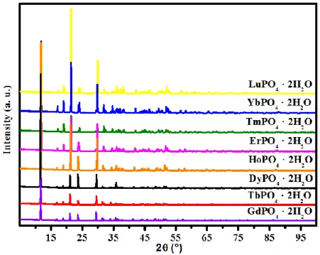
patterns were also collected from REPO4 · 2 H2O (RE = Gd to Lu) materials and are shown in
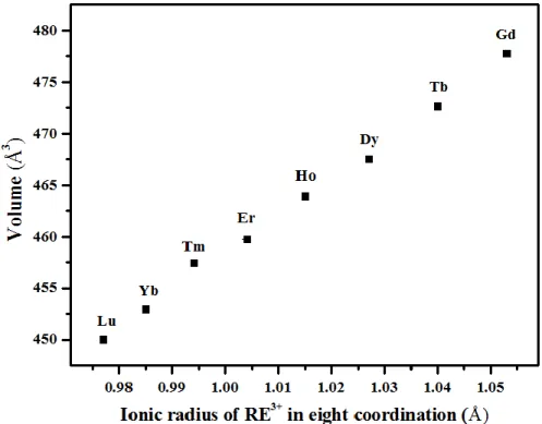
Figure 3. It can be observed from this figure that on moving from Gd to Lu, the XRD peaks shifted toward higher 2θ, which indicates the contraction of the unit cell dimension (Figure 3 and Table 3). This observation is consistent with the decreasing ionic radius of RE ion on moving
from Gd to Lu. Similarly, the unit cell volumes of the REPO4 · 2 H2O materials were found to
decrease with decreasing ionic radii (Table 3 and Figure 4).
The obtained churchite phases crystallize in the gypsum structure type (CaSO4 · 2 H2O) in
the monoclinic system in the C2/c space group with four formula units per cell. The asymmetric units contain one RE atom (2, symmetry site), one P atom (2, symmetry site) and three oxygen atoms located in a general position (1, symmetry site). The crystal structure of representative
The Er atom in the ErPO4 · 2 H2O is coordinated to eight oxygen atoms. Six of them are
provided by the phosphate anions whereas the remaining two oxygen atoms are provided by
water molecules. The resulting ErO8 polyhedron adopts a highly distorted square antiprism.9 The
churchite phase exhibits a layered structure and the layers consist of edge-sharing
one-dimensional chains of alternating ErO8 and PO4 polyhedra running along the [100] direction
(Figure 5a). The 1D chains in the structure are arranged in a zig-zag fashion along the [001]
direction and these chains in turn are connected to one another via edge-sharing of ErO8
polyhedra (Figure 5b). The connection of those chains leads to the formation of two-dimensional (2D) layers, which are parallel to each other in the [010] plane (Figure 5b). The structure of churchite is built by stacking these 2D layers along the b axis and with the water molecules occupying the interlayer regions (Figure 5b). The individual 2D layers are held together via formation of H-bonds between the hydrogen atoms of the water molecules present in one of the 2D layers and the oxygen atoms of the neighboring 2D layer. This results in the formation of a three-dimensional (3D) framework. The H-bonds are also formed with the oxygen atoms present within the same layer. The oxygen atom of the water molecules also forms a part of the coordination environment of the erbium atom through bonding of oxygen atom with the erbium atom.
Selected interatomic bond distances for REPO4 · 2 H2O (RE = Gd, Dy, Er) which were
refined using synchrotron data are shown in Table 2. It can be observed in Table 2, that there are
4 unique RE-O bond distances (4[2 RE-O]) in the REO8 polyhedra. The longest RE-O bonds are
due to bidentate bonding with the adjacent phosphate anions (See Table 2 and Figure 5). In the
case of PO4 tetrahedra, two distinct P-O bond distances (2[2 P-O]) were observed. The average
the PO4 tetrahedra exhibit a considerable deviation from the ideal tetrahedral angle thereby
indicating the distortion of the PO4 tetrahedra (Table 2).9 Two of the four O-P-O bond angles are
considerably lower than the ideal tetrahedral angle as a result of the participation of O atoms in
the bidentate bonding with the erbium atom (Table 2).9
Thermal analysis (TG-DSC)
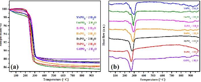
In order to understand the dehydration process and to determine the total water content in
the as-synthesized churchite materials, TG-DSC experiments were performed on REPO4 · 2 H2O
(RE = Gd to Yb) samples (Figure 6 and Table 4). The results show that all samples dehydrate endothermically between 210 and 230 °C (Figure 6b). The dehydration temperature increases gradually with decreasing RE ionic radius and unit cell volume. In the structure, water molecules reside in the interlayer between the 2D layers. As the RE ion radius decreases, the interlayer becomes shorter and water molecules are held more strongly between interlayers, resulting in a gradual increase in the dehydration temperature. Thus, the dehydration reaction appears to be kinetically controlled. The laboratory PXRD analysis of post TG-DSC samples show that all the
churchite REPO4 · 2 H2O phases have dehydrated and transformed to the xenotime structure
(Figure 7). It should be noted that the churchite-type GdPO4 · 2 H2O material transforms to
metastable xenotime-type GdPO4 upon dehydration. Thus, dehydration of churchite-type GdPO4
· 2 H2O appears to be one of the ways to produce xenotime-type GdPO4.46 TG-DSC analysis
reveal the presence of slight excess amounts of water (more than 2 moles) in REPO4 · 2 H2O
(Figure 6 and Table 4), which could be due to the presence of surface adsorbed water. Taking into account the surface adsorbed water, the chemical formulae of the as-synthesized
enthalpies of drop solution of churchite materials were calculated using the molecular weight of this corrected molecular formula.
High temperature oxide melt solution calorimetry
Churchite phosphates - REPO4 · (2+n) H2O
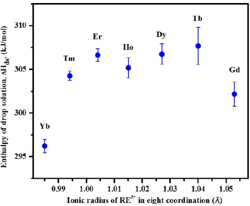
The enthalpies of drop solution (∆Hds) of REPO4 · (2+n) H2O materials are shown in
Figure 8 and Table 5. The enthalpies of formation from oxides (∆H°f, ox) were calculated for all
compounds (Table 5) from the enthalpies of drop solution of the compounds and their respective
rare earth oxides (RE2O3),36,41 phosphorus pentoxide (P2O5)36,41 and water.47-50 The
thermochemical cycle employed to calculate the enthalpies of formation from oxides is given in Table 6.
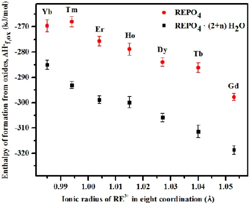
Figure 9 shows that the enthalpy of formation from oxides of churchite compounds
REPO4 · (2+n) H2O becomes more exothermic with increase of the ionic radius of RE3+ in 8-fold
coordination.51 This trend has been already observed for anhydrous REPO4 compounds.36 A
comparison of the enthalpies of formation from oxides of churchites REPO4 · (2+n) H2O and
anhydrous REPO4 phosphates (Figure 9) confirms that the enthalpies of formation of REPO4 ·
(2+n) H2O compounds are always more exothermic than their corresponding anhydrous phases.
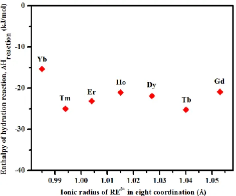
Therefore, the reaction,
REPO4(s) + (2+n) H2O(l) → REPO4 · (2+n) H2O(s)
is exothermic at room temperature by 15.0 to 25.5 kJ/mol for all churchite compounds, with no obvious dependence on ionic radius (Figure 10).
Gadolinium phosphates - GdPO
4· n H
2O
Gd-phosphates adopting the monazite-, xenotime-, rhabdophane, and churchite-type structures were synthesized to determine and compare the thermodynamic stability of each
Gd-xenotime, Gd-rhabdophane, and Gd-churchite, obtained at 25 °C are reported in Table 7. The
enthalpies of formation of rhabdophane GdPO4 · 0.533 H2O and churchite GdPO4 · 2.03 H2O are
-303.67 ± 1.36 kJ/mol and -318.67 ± 1.67 kJ/mol, respectively. Thus the enthalpy of formation of rhabdophane is more exothermic than that of monazite, in agreement with the data reported by
Shelyug et al.52 In that study, standard Gibbs free energies from oxides (∆G°f,ox) of rhabdophane
(-278.80 ± 6.10 kJ/mol) and monazite (-291.80 ± 1.05 kJ/mol) calculated from solubility data,53
confirmed that rhabdophane was metastable with respect to monazite plus water. That work52
also found that standard entropy of formation from oxides (∆S°f,ox) of Gd-rhabdophane (-86.00 ±
7.20 J mol-1 K-1), calculated by combining Gibbs free energy and enthalpy of formation at 25 °C,
is more negative than that of Gd-monazite (-8.30 ± 7.60 J mol-1 K-1)due to the confinement of
liquid water in the rhabdophane structure. Thus, the metastability of Gd-rhabdophane with respect to Gd-monazite plus water is driven by the significant negative entropy associated with hydration, which is not compensated by the negative enthalpy of hydration, even at room temperature. Similarly, churchite, which has more water than rhabdophane, is likely to have entropy of formation from oxides more negative than that of rhabdophane, monazite, and
xenotime. Hence, churchite (GdPO4 · 2.03 H2O) materials is also expected to be metastable with
respect to rhabdophane plus water, monazite plus water, and xenotime plus water.
The enthalpy of formation of xenotime GdPO4, which was formed by dehydration of
churchite GdPO4 · 2.03 H2O is -284.07 ± 0.95 kJ/mol. Since, there are no water molecules in the
xenotime structure, the entropy of formation from oxides of Gd-xenotime can be assumed to be
very similar to that of Gd-monazite (-8.30 ± 7.60 J mol-1 K-1). Then, the standard Gibbs free
energy of formation from oxides of Gd-xenotime is calculated to be -281.59 ± 0.95 kJ/mol. It is less negative than that of Gd-monazite (-291.80 ± 1.05 kJ/mol) reflecting the difference in
enthalpy and confirming the metastability of Gd-xenotime. Thus, Gd-monazite is the thermodynamically most stable phase followed by xenotime, rhabdophane and Gd-churchite (Figure 11 and Table 7).
The more negative enthalpies of formation for churchite REPO4 · 2 H2O compared to
anhydrous xenotime REPO452 imply that the enthalpies of formation of churchite phases are
more exothermic than those of anhydrous xenotime phases. However, the presence of water
molecules in churchite REPO4 · 2 H2O means that their entropies of formation are likely to be
more negative than those for anhydrous REPO4 (xenotime), which would significantly affect the
Gibbs free energy of formation and stability. Thus, if churchite phases have any stability field relative to xenotime plus water, it would be limited to low temperature (probably room temperature or lower). In environmental conditions, the transformation of xenotime to churchite may be thermodynamically favorable at low temperatures. Such thermodynamic stability may explain why the precipitation of churchite materials is easier than their xenotime counterparts at lower temperatures when starting from heavy RE and phosphate ions in solution. Additionally, even if churchite is metastable, its precipitation may be kinetically favored as long as the free energies of the hydrous and anhydrous phases are similar.
Such interplay between churchite and xenotime phases may be similar to that already
observed for rhabdophane and monazite.22,28 Indeed, for light RE elements, it was found that
rhabdophane was rapidly precipitated at the surface of monazite materials during leaching under weathering conditions and low temperatures. Rhabdophane precipitation then acts as a very efficient process in delaying the elemental releases in solution from the initial radwaste ceramic. Moreover, monazite materials have high chemical stability since associated normalized dissolution rates are found to be very low thereby reducing the source term in the field of an
underground repository of radioactive waste. The kinetic and thermodynamic stabilization of monazites can be proved by their common occurrence in natural assemblies and the use of such phases for geochronology (closed system over time). Thus, one could consider that the churchite-xenotime system could follow the same trend for heavy RE: the churchite precipitates as a neoformed phase on the surface of the xenotime mineral during the dissolution at lower temperatures. Therefore, the thermodynamic data of churchite obtained in this work are essential,
as they contribute to the better understanding about the stability of REPO4 phases in the presence
of water.
Conclusions
Materials adopting the churchite structure (REPO4 · 2 H2O; RE = Gd to Lu) were
synthesized and studied using PXRD and high temperature oxide melt solution calorimetry.
Rietveld refinement of the synchrotron PXRD patterns of the REPO4 · 2 H2O (RE = Gd, Dy, Er,
Tb) showed that the churchite materials crystallize in the monoclinic crystal system with a
layered structure similar to gypsum (CaSO4 · 2 H2O). Enthalpies of formation of the churchite
materials were determined using high temperature oxide melt solution calorimetry. The
enthalpies of formation from the oxides of churchite-type REPO4 · (2+n) H2O compounds were
always found to be more exothermic than those of xenotime-type REPO4 without significant
dependence on the RE3+ ionic radius. From the obtained data, churchite materials appear of
significant interest in the field of radioactive waste management. Under a failed container situation in the geological repository, the churchite materials which could form at the surface of the xenotime wasteform during aqueous alteration (including repository relevant conditions) could play an important role in delaying the release of actinides (e.g., americium and curium) to
ASSOCIATED CONTENT
Supporting Information.
Crystallographic files in CIF format for ErPO4 · 2 H2O, DyPO4 · 2 H2O and GdPO4 · 2
H2O have been deposited with the Cambridge Crystallographic Data Centre as CCDC# 1909900,
CCDC# 1909899, CCDC# 1909901. These data may be obtained free of charge by contacting CCDC at (https://www.ccdc.cam.ac.uk/).
AUTHOR INFORMATION
Corresponding Author
*E-mail: adel.mesbah@cea.fr (A. M.)
*E-mail: anavrotsky@ucdavis.edu (A. N.)
ACKNOWLEDGMENT
The calorimetric studies at UC Davis were supported by the U.S. Department of Energy grant DE-FG02-03ER46053. The work in France also benefited from financial support of the French
Figures
Figure 1. Rietveld refined synchrotron powder XRD pattern of churchite-type ErPO4 · 2 H2O
Figure 3. Laboratory powder XRD patterns of the churchite-type REPO4 · 2 H2O (RE = Gd to
Figure 4. Evolution of the refined unit cell volume in the REPO4 · 2 H2O based compounds with
Figure 5. Crystal structure of churchite-type ErPO4 · 2 H2O (Space group: C2/c) oriented along
the (a) ‘c’ and (b) ‘a’ direction. The crystal structures were generated using the VESTA
Figure 7. PXRD patterns of the churchite-type REPO4 · 2 H2O after TG-DSC at 1000°C
Figure 9. Enthalpies of formation from oxides (∆H°(f, ox)) of REPO4 and REPO4 · (2+n) H2O vs
Figure 10. Enthalpies of hydration reaction REPO4(s) + (2+n) H2O(l) → REPO4 · (2+n) H2O(s) vs
Figure 11. Standard Gibbs free energy of formation from oxides (∆G°f,ox) obtained for GdPO4 ·
n H2O versus the number of water molecules, n, in GdPO4 · n H2O (*∆G°f,ox of churchite phase
when ∆S°f,ox is considered twice that of rhabdophane phase)
0.0 0.5 1.0 1.5 2.0 -290 -285 -280 -275 -270 GdPO4.nH2O G ° f,o x ( k J /m o l) No of moles of H2O (n) in GdPO4.nH2O n = 0, monazite n = 0.533, rhabdophane n = 2.030, churchite* n = 0, xenotime
Tables
Table 1. Crystallographic data and Structure Refinement of REPO4 · 2 H2O (RE = Er, Dy, Gd)
Sample ErPO4 · 2 H2O DyPO4 · 2 H2O GdPO4 · 2 H2O
Formula weight
(g/mol) 298.26 293.50 288.25
T (°C) 25 25 25
Crystal System Monoclinic Monoclinic Monoclinic
Space group C 2/c C 2/c C 2/c a 6.1527(1) 6.1889(1) 6.2359(1) b 15.0177 (1) 15.0797(1) 15.1777(1) c 5.5857(1) 5.6106(1) 5.6409(1) Β 115.47 (1) 115.22(1) 114.95(1) V (Å3) 465.94 (1) 473.71 (1) 484.07(1) Z 4 4 4 Wavelength (Å) 0.58403 0.58403 0.58403 Rp 0.099 0.104 0.095 Rwp 0.127 0.137 0.122 RBragg 0.072 0.062 0.046 RF 0.073 0.057 0.044
Table 2. Selected interatomic bond distances (Å) and bond angles (°) of REPO4 · 2 H2O (RE =
Er, Dy, Gd)
ErPO4 · 2 H2O DyPO4 · 2 H2O GdPO4 · 2 H2O
Bond distance (Å) (Ln-O1) × 2 2.439 (4) 2.458(6) 2.480(4) (Ln-O2) × 2 2.453 (4) 2.465(5) 2.491(3) (Ln-O2) × 2 2.289 (5) 2.311(5) 2.333(3) (Ln-O3) × 2 2.397 (5) 2.409(4) 2.431(3) (P-O1) × 2 1.557 (4) 1.550(4) 1.543(3) (P-O2) × 2 1.527 (4) 1.525(6) 1.536(4) Bond angle (°) O1-P-O2 103.7 (2) 103.7(5) 111.2(4) O1-P-O2 112.9(5) 111.2(5) 103.9(3) O1-P1-O1 113.2 (3) 114.7(4) 114.1(3) O2-P-O2 110.6 (4) 112.7(6) 112.9(5)
Table 3. Lattice constants of REPO4 · 2 H2O (RE = Gd - Lu)
Table 4 Summary of weight loss measurements from TG-DSC studies
Table 5. Enthalpies of drop solution (∆Hds) of churchite REPO4 · 2 H2O in 3 Na2O · 4 MoO3
solvent at 700 °C and enthalpies of formation from oxides (∆H°f,ox)
a
Error is two standard deviations of the mean and number in () is number of individual experiments performed. Sample a (Å) b (Å) c (Å) β (°) V (Å3) GdPO4 · 2 H2O 6.2103 (1) 15.1105 (24) 5.6163 (1) 114.96 (1) 477.82(1) TbPO4 · 2 H2O 6.1868 (1) 15.0437 (3) 5.6071 (1) 115.08 (1) 472.67(2) DyPO4 · 2 H2O 6.1671 (7) 15.0123 (5) 5.5844 (4) 115.26 (8) 467.58(6) HoPO4 · 2 H2O 6.1466 (2) 14.9854 (3) 5.5743 (2) 115.36 (2) 463.96(2) ErPO4 · 2 H2O 6.1265 (2) 14.9509 (3) 5.5602 (2) 115.47 (2) 459.79(2) TmPO4 · 2 H2O 6.1175 (8) 14.9114 (2) 5.5588 (8) 115.56 (6) 457.46(1) YbPO4 · 2 H2O 6.1000 (8) 14.8712 (2) 5.5401 (9) 115.66 (8) 453.00(1) LuPO4 · 2 H2O 6.0879 (2) 14.8546 (5) 5.5289 (2) 115.82 (2) 450.08(2) Sample Number of H2O molecules
corresponding to weight loss
Number of excess (surface adsorbed) H2O molecules 'n' GdPO4 · 2 H2O 2.03 0.03 TbPO4 · 2 H2O 2.05 0.05 DyPO4 · 2 H2O 2.04 0.04 HoPO4 · 2 H2O 2.08 0.08 ErPO4 · 2 H2O 2.07 0.07 TmPO4 · 2 H2O 2.06 0.06 YbPO4 · 2 H2O 2.07 0.07
Sample ∆Hds ( kJ/mol) ∆H°(f, ox) ( kJ/mol)
GdPO4 · 2.03 H2O 302.17 ± 1.39 (8) a -318.67 ± 1.67 TbPO4 · 2.05 H2O 307.69 ± 2.14 (7) -311.33 ± 2.50 DyPO4 · 2.04 H2O 306.81 ± 1.16 (9) -305.79 ± 1.67 HoPO4 · 2.08 H2O 305.19 ± 1.15 (9) -299.83 ± 2.22 ErPO4 · 2.07 H2O 306.64 ± 0.74 (8) -298.74 ± 1.52 TmPO4 · 2.06 H2O 304.27 ± 0.53 (8) -292.99 ± 1.39 YbPO4 · 2.07 H2O 296.23 ± 0.75 (7) -284.93 ± 1.82
Table 6. Thermochemical cycle for the calculation of enthalpy of formation from oxides for
REPO4.(2+n) H2O churchite phase
Table 7 Enthalpies of drop solution (∆Hds) in 3 Na2O · 4 MoO3 solvent at 700 °C and associated
enthalpies, entropies and free Gibbs energies of formation from oxides, ΔH°f,ox, ΔS°f,ox and
ΔG°f,ox obtained for Gd-xenotime, Gd-monazite, Gd-rhabdophane and Gd-churchite. ΔG°f,ox
values were calculated at 298.15 K (25 °C)
a
Error is two standard deviations of the mean and number in () is number of individual experiments performed.
b
Determined in this study
c
Determined by Shelyug et al(Ref. 52)
Reaction ∆H(kJ/mol) REPO4 · (2 + n) H2O(s,25) → ½ RE2O3(sln,700) + ½ P2O5(sln,700) + (2 + n) H2O(g,700) [1] ∆Hds(REPO4 · (2+n) H2O) [Table 5] RE2O3(s,25) → RE2O3(sln,700) [2] ∆Hds(RE2O3) [36, 41] P2O5(s,25) → P2O5(sln,700) [3] ∆Hds(P2O5)=-164.60±0.85 [36] H2O(l,25) → H2O(g,700) [4] ∆H(H2O)=69.00±0.10 [47-50] ½ RE2O3(s,25) + ½ P2O5(s,25) + (2+n) H2O(l,25) → REPO4 · (2 + n) H2O(s,25) [5] ∆H°f,ox(REPO4 · (2+n) H2O) [Table 5] ∆H[5] = -∆H[1] + ½ ∆H[2]+ ½ ∆H[3] + (2 + n) ∆H[4] Sample Number of water molecules 'n' ∆Hds (kJ/mol)a ∆H°f, ox (kJ/mol)b ∆S°f,ox (J mol-1 K-1)c ∆G°f,ox (kJ/mol) GdPO4 – xenotime 0 127.47±0.29 (4) -284.07±0.95 -8.30±7.60 -281.59±0.95 GdPO4 – monazite 0 137.71±0.53 (7) -294.28±1.05 -8.30±7.60 -291.80±1.05 GdPO4 · 0.533 H2O – rhabdophane 0.533 183.88±0.83 (8) -303.67±1.36 -86.00±7.20 -278.03±1.36 GdPO4 · 2.03 H2O – churchite 2.030 302.17±1.39 (8) -318.67±1.67 - -
REFERENCES
(1) Ewing, R. C.; Wang, L. Phosphates as Nuclear Waste Forms. Rev. Mineral. Geochem.
2002, 48, 673–699.
(2) Oelkers, E. H.; Montel, J-M. Phosphates and Nuclear Waste Storage. Elements 2008, 4,
113–116.
(3) Montel, J-M. Minerals and design of new waste forms for conditioning nuclear waste. C R
Geosci. 2011, 343, 230–236.
(4) Achary, S. N.; Bevara, S.; Tyagi, A. K. Recent progress on synthesis and structural
aspects of rare-earth phosphates. Coord. Chem. Rev. 2017, 340, 266–297.
(5) Rafiuddin, M. R.; Grosvenor, A. P. A Structural Investigation of Hydrous and Anhydrous
Rare-Earth Phosphates. Inorg. Chem. 2016, 55, 9685–9695.
(6) Clavier, N.; Mesbah, A.; Szenknect, S.; Dacheux, N. Monazite, rhabdophane, xenotime &
churchite: Vibrational spectroscopy of gadolinium phosphate polymorphs. Spectrochim.
Acta Part A Mol. Biomol. Spectrosc. 2018, 205, 85–94.
(7) Ni, Y.; Hughes, J. M.; Mariano, A. N. Crystal chemistry of the monazite and xenotime
structures. Am. Mineral. 1995, 80, 21–26.
(8) Mesbah, A.; Clavier, N.; Elkaim, E.; Gausse, C.; Ben Kacem, I.; Szenknect, S.;
Dacheux, N. Monoclinic form of the rhabdophane compounds: REEPO4.0.667H2O. Cryst.
Growth Des. 2014, 14, 5090–5098.
(10) Kohlmann, M.; Sowa, H.; Reithmayer, K.; Schulz, H.; Krüger, R. R.; Abriel, W.
Structure of a Y1−x(Gd,Dy,Er)xPO4.2H2O microcrystal using synchrotron radiation. Acta
Crystallogr. Sect. C Cryst. Struct. Commun. 1994, 50, 1651–1652.
(11) Zepf, V. Rare Earth Elements: What and Where They Are. In: Rare Earth Elements
Springer Theses (Recognizing Outstanding Ph.D. Research); Springer: Berlin, 2013;
Heidelberg
(12) Voncken, J. H. L. The Ore Minerals and Major Ore Deposits of the Rare Earths, in: The
Rare Earth Elements; SpringerBriefs in Earth Sciences. Springer: Cham, 2016.
(13) Emden, B.; Thornber, M. R.; Graham, J.; Lincoln, F. J. The incorporation of actinides in monazite and xenotime from placer deposits in Western Australia. Can. Mineral. 1997, 35, 95-104.
(14) Spear, F. S.; Pyle, J. M. Apatite, Monazite, and Xenotime in Metamorphic Rocks. Rev.
Mineral. Geochemistry. 2002, 48, 293-335.
(15) Karioris, F. G.; Gowda, K. A.; Cartz, L. Heavy ion bombardment of monoclinic ThSiO4,
ThO2 and Monazite. Radiat. Eff. Lett. 1981, 58, 1–3.
(16) Meldrum, A.; Boatner, L. A.; Weber, W. J.; Ewing, R. C. Radiation damage in zircon and monazite. Geochim. Cosmochim. Acta.1998, 62, 2509–2520.
(17) Rafiuddin, M. R.; Grosvenor, A. P. Probing the effect of radiation damage on the structure of rare-earth phosphates. J. Alloys Compd. 2015, 653, 279–289.
radiation-Acta Mater. 2013, 61, 2984–2992.
(19) Meldrum, A.; Boatner, L.; Ewing, R. Displacive radiation effects in the monazite- and zircon-structure orthophosphates. Phys. Rev. B. 1997, 56, 13805–13814.
(20) Seydoux-Guillaume, A-M.; Deschanels, X.; Baumier, C.; Neumeier, S.; Weber, W. J.; Peuget, S. Why natural monazite never becomes amorphous: Experimental evidence for alpha self-healing. Am. Mineral. 2018, 103, 824-827.
(21) Gausse, C.; Szenknect, S.; Mesbah, A.; Clavier, N.; Neumeier, S.; Dacheux, N.
Dissolution kinetics of monazite LnPO4 (Ln = La to Gd): A multiparametric study. Appl.
Geochem. 2018, 93, 81–93.
(22) Dacheux, N.; Clavier, N.; Podor, R. Monazite as a promising long-term radioactive waste matrix: Benefits of high-structural flexibility and chemical durability. Am. Mineral. 2013, 98, 833–847.
(23) Arinicheva, Y.; Neumeier, S.; Brandt, F.; Bosbach, D.; Deissmann, G. Dissolution
kinetics of synthetic LaPO4-monazite in acidic media. MRS Adv. 2018, 3, 1133–1137.
(24) Rafiuddin, M. R.; Grosvenor, A. P. An investigation of the chemical durability of hydrous and anhydrous rare-earth phosphates. J. Nucl. Mater. 2018, 509, 631-643.
(25) Arinicheva, Y.; Gausse, C.; Neumeier, S.; Brandt, F.; Rozov, K.; Szenknect, S.; Dacheux, N.; Bosbach, D.; Deissmann, G. Influence of temperature on the dissolution kinetics of
synthetic LaPO4-monazite in acidic media between 50 and 130 °C. J. Nucl. Mater. 2018,
monazite. J. Nucl. Mater. 2011, 409, 221–224.
(27) Berger, A.; Gnos, E.; Janots, E.; Fernandez, A.; Giese, J. Formation and composition of rhabdophane, bastnäsite and hydrated thorium minerals during alteration: Implications for geochronology and low-temperature processes. Chem. Geol. 2008, 254, 238–248.
(28) Du Fou de Kerdaniel, E.; Clavier, N.; Dacheux, N.; Terra, O.; Podor, R. Actinide solubility-controlling phases during the dissolution of phosphate ceramics. J. Nucl. Mater.
2007, 362, 451–458.
(29) Li, H.; Zhu, G.; Ren, H.; Li, Y.; Hewitt, I. J.; Qiu, S. The Synthesis of Multiwalled Rare-Earth Phosphate Nanomaterials Using Organophosphates with Upconversion Properties.
Eur. J. Inorg. Chem. 2008, 2008, 2033–2037.
(30) Wang, L.; He, D.; Feng, S.; Yu, C.; Hu, L.; Qiu, J.; Chen, D. Phosphate ytterbium-doped single-mode all-solid photonic crystal fiber with output power of 13.8 W. Sci. Rep. 2015, 5, 8490, 1-4.
(31) Kitamura, N.; Amezawa, K.; Tomii, Y.; Yamamoto, N. Protonic conduction in rare earth orthophosphates with the monazite structure. Solid State Ionics 2003, 162–163, 161–165.
(32) Onoda, H.; Nariai, H.; Moriwaki, A.; Maki, H.; Motooka, I. Formation and catalytic characterization of various rare earth phosphates. J. Mater. Chem. 2002, 12, 1754–1760.
(33) Meiser, F.; Cortez, C.; Caruso, F. Biofunctionalization of Fluorescent Rare-Earth-Doped Lanthanum Phosphate Colloidal Nanoparticles. Angew. Chemie Int. Ed. 2004, 43, 5954– 5957.
rhabdophane crystal structure: from the hydrated monoclinic LnPO4.0.667H2O to the
hexagonal LnPO4 (Ln = Nd, Sm, Gd, Eu and Dy). J. Solid State Chem. 2017, 249, 221–
227.
(35) Assaaoudi, H.; Ennaciri, A.; Rulmont, A.; Harcharras, M. Gadolinium orthophosphate weinschenkite type and phase change in rare earth orthophosphates, Phase Transitions.
2000, 72, 1–13.
(36) Ushakov, S. V.; Helean, K. B.; Navrotsky, A.; Boatner, L. A. Thermochemistry of rare-earth orthophosphates. J. Mater. Res. 2001, 16, 2623–2633.
(37) Hirsch, A.; Kegler, P.; Alencar, I.; Ruiz-Fuertes, J.; Shelyug, A.; Peters, L.; Schreinemachers, C.; Neumann, A.; Neumeier, S.; Liermann, H.-P.; Navrotsky, A.; Roth, G.; Structural, vibrational, and thermochemical properties of the monazite-type solid
solution La1–xPrxPO4. J. Solid State Chem. 2017, 245, 82–88.
(38) Neumeier, S.; Kegler, P.; Arinicheva, Y.; Shelyug, A.; Kowalski, P. M.; Schreinemachers,
C.; Navrotsky, A.; Bosbach, D.; Thermochemistry of La1−xLnxPO4-monazites (Ln=Gd,
Eu). J. Chem. Thermodyn. 2017, 105, 396–403.
(39) Dinnebier, R.; Rietveld Refinement from Powder Diffraction Data, 2001.
(40) Finger, L. W.; Cox, D. E.; Jephcoat, A. P.; Correction for powder diffraction peak asymmetry due to axial divergence J. Appl. Crystallogr.1994, 27, 892–900.
(41) Navrotsky, A. Progress and New Directions in Calorimetry: A 2014 Perspective. J. Am.
(42) Navrotsky, A. Mineralogy, materials science, energy, and environment: A 2015 perspective. Am. Mineral. 2015, 100, 674–680.
(43) Navrotsky, A. Progress and new directions in high temperature calorimetry. Phys. Chem.
Miner. 1977, 2, 89–104.
(44) Navrotsky, A. Progress and new directions in high temperature calorimetry revisited.
Phys. Chem. Miner. 1997, 24, 222–241.
(45) Momma, K.; Izumi, F. VESTA: a three-dimensional visualization system for electronic and structural analysis. J. Appl. Crystallogr. 2008, 41, 653–658.
(46) Rodriguez-Liviano, S.; Becerro, A. I.; Alcantara, D.; Grazu, V.; de la Fuente, J.
M.; Ocana, M. Synthesis and properties of multifunctional tetragonal
Eu:GdPO4 nanocubes for optical and magnetic resonance imaging applications. Inorg.
Chem. 2013, 52, 647–654.
(47) Maloney, J. O. Perry’s Chemical Engineers Handbook 8th Edition - Section 1, 8th Edition; McGraw-Hill: New York, 2008.
(48) Barin, I. Thermochemical Data of Pure Substances; VCH: Weinheim, 1995.
(49) Chase, M. W.; Davies, C. A.; Frurip, D. J.; McDonald, R. A.; Syverud, A. N.; NIST-
JANAF Thermochemical Tables, Third Edition; The American Chemical Society and the
American Institute of Physics for the National Institute of Standards and Technology: Gaithersburg, 1985.
(50) Bale, C.; Bélisle, E.; Chartrand, P.; Decterov, S.; Eriksson, G.; Gheribi, A.; Hack, K.;
J; Spencer, P.; VanEnde, M.-A. FactSage thermochemical software and databases,
2010–2016. CALPHAD: Comput. Coupling Phase Diagrams Thermochem.
2016, 54, 35–53. doi: 10.1016/j.calphad.2016.05.002.
(51) Shannon, R. D. Revised effective ionic radii and systematic studies of interatomic distances in halides and chalcogenides, Acta Crystallogr. Sect. A. 1976, 32, 751–767. doi:10.1107/S0567739476001551.
(52) Shelyug, A.; Mesbah, A.; Szenknect, S.; Clavier, N.; Dacheux, N.; Navrotsky, A.
Thermodynamics and stability of rhabdophanes, hydrated rare earth phosphates
REPO4.n H2O. Front. Chem. 2018, 6, 604. doi: 10.3389/fchem.2018.00604
(53) Gausse, C.; Szenknect, S.; Qin, D. W.; Mesbah, A.; Clavier, N.; Neumeier, S.; Bosbach,
D.; Dacheux, N. Determination of the Solubility of Rhabdophanes LnPO4.0.667H2O (Ln
0.99 1.00 1.01 1.02 1.03 1.04 1.05 -320 -310 -300 -290 -280 -270 H °f , ox , ( k J /m o l ) Gd
Ionic radius of RE3+ in eight coordination (Å)
REPO4 REPO4.(2+n)H2O Yb Tm Er Ho Dy Tb 0.0 0.5 1.0 1.5 2.0 -290 -285 -280 -275 -270 GdPO4.nH2O G °f, o x ( k J /m o l )
No of moles of H2O (n) in GdPO4.nH2O n = 0, monazite n = 0.533, rhabdophane n = 2.030, churchite* n = 0, xenotime
FOR TABLE OF CONTENTS USE ONLY
Authors: Tamilarasan Subramani, Mohamed Ruwaid Rafiuddin, Anna Shelyug, Sergey Ushakov,
Adel Mesbah, Nicolas Clavier, Danwen Qin, Stephanie Szenknect, Erik Elkaim,, Nicolas
Dacheux, Alexandra Navrotsky
Synopsis:
Churchite-type materials (REPO4 ·2H2O; RE = Gd to Yb) adopt the gypsum structure and were
synthesized using a precipitation route. The enthalpies of formation from oxides (ΔHf,ox) of
churchite were determined using high temperature oxide melt solution calorimetry. The ΔHf,ox of
churchite materials was more exothermic than their anhydrous counterparts adopting the
xenotime structure. The Gibbs free energy of formation from oxides (ΔG°f,ox) of Gd-phosphates