HAL Id: hal-03173221
https://hal.laas.fr/hal-03173221
Submitted on 18 Mar 2021HAL is a multi-disciplinary open access archive for the deposit and dissemination of sci-entific research documents, whether they are pub-lished or not. The documents may come from teaching and research institutions in France or abroad, or from public or private research centers.
L’archive ouverte pluridisciplinaire HAL, est destinée au dépôt et à la diffusion de documents scientifiques de niveau recherche, publiés ou non, émanant des établissements d’enseignement et de recherche français ou étrangers, des laboratoires publics ou privés.
DNA nano-engineering and DNA driven nanoparticle
assembly
Alain Estève, Carole Rossi
To cite this version:
Alain Estève, Carole Rossi. DNA nano-engineering and DNA driven nanoparticle assembly. Biological Soft Matter Fundamentals, Properties and Applications, Wiley, inPress, 978-3-527-34348-5. �hal-03173221�
1
DNA nano-engineering and DNA driven nanoparticle assembly
Alain Estève, Carole Rossi
University of Toulouse, LAAS-CNRS, 7 avenue du colonel Roche, 31031 Toulouse, France
Table des matières
1 Introduction ____________________________________________________________ 1 2 From the DNA molecule to nanotechnologies _________________________________ 4 3 DNA nanostructures: from Holliday junctions to 3D origami _____________________ 5 4 DNA-directed assembly of particles: from concepts to the realization of ordered assemblies _________________________________________________________________ 8
4.1 DNA/nanoparticle assembly: primary functionalization strategies ______________ 9 4.2 Towards high-order crystalline structures ________________________________ 11 4.3 Crystallization of heterogeneous systems _________________________________ 13 4.4 DNA/nanoparticle assembly: applications ________________________________ 15
5 Nano-engineering of DNA self-assembled Al/CuO nanothermite _________________ 16
5.1 Fundaments and characterization of DNA/surface chemistry and grafting strategies 17
5.1.1 DNA/alumina interaction evaluation through infrared spectroscopy and first principles calculations ___________________________________________________ 18 5.1.2 Functionalization protocol and colloidal characterization _________________ 20 5.1.3 Quantification of streptavidin and DNA surface densities __________________ 21 5.2 Kinetics of DNA-directed assembly of Al and CuO nanoparticles ______________ 23 5.2.1 Design and impact of the DNA coding sequence _________________________ 24 5.3 Structural and energetic properties of the Al/CuO bionanocomposite __________ 26
6 Conclusion ____________________________________________________________ 29 7 Bibliography ___________________________________________________________ 30
1 Introduction
In much the same way that the use of silicon did in the 1970s, leading to the modern information technology industry, the development of advanced functional nanomaterials will fuel many of the emerging industries that will address energy, healthcare, and environmental challenges as well as those in other areas. Therefore, a current challenge in the field of nanotechnology and materials science is the development of strategies to control the structuring and fabrication of functional materials and devices with potentially atomic scale precision on a large length scale, i.e. up to micro and even centimetre scales. These new advanced functional materials aim at improving performances and developing new functionalities.
2
derived from the microelectronics industry. These methods are primarily dedicated to semiconductor materials and are currently based on powerful and efficient techniques for structuring matter that are essentially operated by standard and industrial machines. These top-down technologies face a number of issues, including cost and the need for machinery modifications and manufacturing lines to promote miniaturization and mass production. Importantly, these technologies also face the intrinsic physical limitations inherent to the miniaturization of classical materials, which necessitates fundamental modifications to the overall device architecture. For instance, the concept of planar metal oxide semiconductor (MOS) transistor was proposed and developed since the mid 1960s and is one reference of the road mapping in microelectronics.1
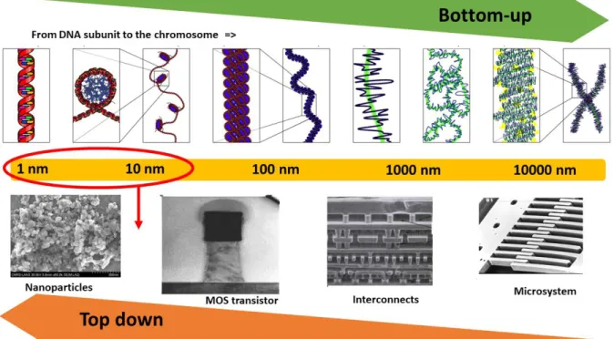
Self-assembly and, at large scales, “bottom-up” technologies are game changers in the way that materials can be envisaged. Along with the era of nanotechnology launched in the US in the 1990s, inspiration from the living world, where the directed assembly of molecules allows the production of organisms, was a disruptive technological alternative.2-3 If we compare the dimensions of the devices reachable by the “top-down” technologies to those of the living world (see Figure 1), two technological paths emerge that may lead to a re-foundation of the classical concepts of technologies in favour of systems that are highly heterogeneous in chemical and structural nature.
Figure 1: Perspective of the relative scaling of the synthetic and biological worlds with respect to multiscale DNA architecture.
To date, numerous bottom-up strategies have been investigated.4-5 These approaches fully rely on self-assembly, which consists of using the basic physical and chemical attractive/repulsive interactions (van der Waals, electrostatic, hydrogen or covalent bonding) as well as the basic processes of molecular recognition in biology. The goal of these strategies is to interface and link various nano-objects, such as nanoparticles, nanotubes, biological molecules and other organic objects,6 without external intervention.
Among these self-assembly strategies, a particularly interesting and versatile technique is to take advantage of the exquisite properties of deoxyribonucleic acid (DNA), i.e. the permutations and complementarity of nucleotide sequences, to programme and direct the
3
assembly of nano-objects in potentially extremely complex architectures, which is not feasible by any other state-of-the-art technique. This idea was first proposed in 1982 in the seminal work by Nadrian Seeman,7 who proposed the construction of nanometric “tiles” from elementary DNA bricks and to build regular networks of tiles for the formation of larger 2D or 3D structures.
This initial revolutionary concept to deviate from the original biological function of DNA and use it as a versatile and programmable technological brick was rapidly extended in many different directions.8 More complex hybrid structures were produced by combining DNA with other materials, inorganic or not, while also taking full advantage of the recognition capability of folded DNA, so-called aptamers, and finally, of modified DNA strands (chemical terminations, for peptide nucleic acid (PNA), etc.) to extend its ability to marry other materials and fabrication techniques.9
These studies led to a technologically wide and active field of study called DNA nanotechnology in which DNA is considered to be an engineering material that can be used as an architectural material, functional building blocks and devices for applications non strictly dedicated to biology; in other words, a field in which DNA is no more utilized as the carrier of the genetic information in the cell.10 The use of DNA out of its biological context is now widely accepted by the scientific community, which has been accompanied by the massive industrial production of artificial DNA. This concept opens unparalleled perspectives for the nano-engineering of a multitude of “new advanced materials” for various applications such as Energy, Environment, Health ...11-13
In 2017, there were 400 published articles (source: Web of Science) on DNA nanotechnology in high impact journals. Section 2 will give a brief overview on the DNA biomolecule and its primary features and properties, which will be followed by a description of the founding studies on DNA nanotechnologies in section 3. Then, sections 4 and 5 will focus on nanoparticle DNA-directed assembly, accompanied with state-of-the-art techniques and a case study by the authors on Al/CuO assembly. DNA nanotechnologies can be split into two fields:
- DNA nanostructures (section 3): As proposed by Nadrian Seeman,7 relatively small DNA strands are assembled to constitute elementary “bricks”, which can be further used to fabricate larger structures that essentially comprise DNA and have one, two or even three dimensions. The folding of much longer strands, such as the genome of the M13 virus, with the help of DNA staples (short DNA strands having partitioned complementarity with the longer DNA strand) is another way of generating structures any shape one can imagine.14 Notably, small-sized DNA strands can also fold into specific conformations, making it possible to have very specific interactions of the lock and key type, as proteins typically do in biology. These DNA strands are called aptamers.15
- DNA as technological tool to drive nanoparticle assembly. DNA can be used as a “cement” to guide in the self-assembly of various organic and non-organic nano-objects. This pathway, directly inspired by the work of Nadrian C. Seeman, was first demonstrated in 1996 by Chad A. Mirkin, who implemented the directed assembly of gold nanoparticles by DNA strands.16 This research field has become very active, accounting for approximately 10% of all published articles on DNA nanotechnology over the past five years (source: Web of Science), whereas it represented less than 1% in the 2000’s. A wide variety of different building-block shapes as well as different strategies for assembling them have been reported thus far, as summarized in section 5. To date, nanoparticle and colloidal particle assemblies using synthetic DNA have allowed for the production of mostly plasmonic, photonic or phononic metamaterials.
4
Interestingly, another new application of DNA nanotechnologies is the synthesis of highly ordered metal/oxide nanoparticle structures for energy-generating materials. The properties of these materials, also called nanothermites, are highly dependent on the size of nanoparticles and their distribution in the 3D structure. In this context, the use of DNA strands to direct self-assembly shows great potential for the synthesis of nanothermites with outstanding energetic performances. Section 5 focusses on the nano-engineering of DNA to self-assemble Al/CuO nanothermite to illustrate the potential of DNA nanotechnologies in obtaining high performance nanothermite for practical applications.
2 From the DNA molecule to nanotechnologies
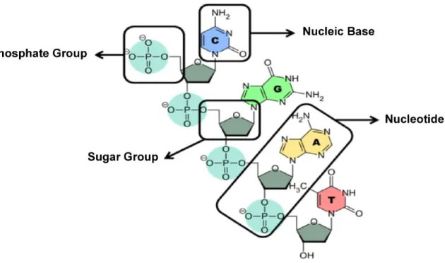
Deoxyribonucleic acid (DNA) is a core constituent of all living cells, containing the genetic information from which all the functions of an organism are programmed. The composition and properties of DNA have been extensively studied, and the DNA double helix is certainly one of the best-known structures in biology. DNA is composed of sequences of nucleotides that are themselves made up of three components (Figure 2): a phosphate group bonded to a sugar (deoxyribose) to which a nitrogenous base is linked. It is therefore a true modular framework consisting of sugars and phosphate groups as a backbone that can harbour one of four different nitrogenous bases, including adenine (A), thymine (T), cytosine (C) and guanine (G), ensuring the sequential signature of the DNA molecule as well as the complementarity required to form the double helix.
Figure 2: Schematic of the DNA molecular substructure
The nitrogenous bases are complementary, where adenine (A) and guanine (G) can specifically interact with thymine (T) and cytosine (C), respectively, due to hydrogen bonds (two for the first “couple” and three for the second). Hybridization is the pairing of two complementary single-stranded DNA sequences, which results in the formation of a double helix. The energy gain resulting from the association of two single strands of DNA is, in the first approximation, the sum of the association energies of each pair of base pairs, with 2 and 3 kBT (where kBT is
the thermal stirring energy) for A-T and G-C, respectively. Note that hybridization is a complex process characterized by the interplay of mutual attraction forces, including hydrogen bonds, solvent, and counter ions to screen the negatively charged backbone.
5
The four primary characteristics that make DNA an excellent technological brick for nanoconstruction are:
- Reversible Watson-Crick pairing, which is based on the complex interplay between the solvent, DNA backbone and hydrogen bonds, including inter-base hydrogen bonds. External stimuli, such as the temperature, pH or ionic strength of the solution can denature the DNA double helix, where it is separated into its single strand components. In addition, DNA can be repaired by DNA modification enzymes, particularly ligases. This specificity is especially used for the nanoconstruction of complex DNA patterns. - Its size is ideally suited to nanoconstruction. The diameter of the double helix is
approximately 2 nm, the distance between bases is 0.34 nm, and the helical pitch is composed of 10.5 base pairs, which corresponds to approximately 3.5 nm.
- The stability and flexibility of an assembly can be adjusted. The persistence length of double stranded DNA is ~ 50 nm (or 150 base pairs). Below this length, the strand can be considered as a rigid rod, whereas it becomes very flexible beyond this length. There is also a difference in flexibility between double- and single-stranded DNA, with the latter being much more flexible, with a persistence length of only 1 nm, allowing for the formation of true loops or curved structures.
- Finally, 50 years of molecular biology research on DNA has produced a consistent palette of tools for DNA synthesis, manipulation, and modification. Many companies specifically offer synthetic DNA with a great variety of chemical modifications. It is now possible to order or create, in the laboratory, single or double DNA strands consisting of the desired nitrogenous base sequence with a wide variety of chemical or biological functions that can be further integrated at the strand termini or at a specific locations within the strand.
In addition to the remarkable intrinsic properties of DNA, all of these features lay the foundation for important expectations regarding DNA nanotechnologies to control the structuring and fabrication of functional materials and devices. The following examples shall illustrate the implementation of these technologies.
3 DNA nanostructures: from Holliday junctions to 3D origami
As mentioned earlier, the development of DNA nanotechnologies began with the seminal work by Nadrian Seeman in the 1980s.7 He envisaged associating several double strands of DNA with the help of the hybridization of so-called “sticky-ends” to build more complex DNA networks. “Sticky-ends” refers to DNA termini that remain single stranded, offering hybridization possibilities with other basic DNA elements with complementary single-stranded termini.
In doing so, he faced a number of issues due to DNA helices being highly flexible, and the resulting assemblies did not exhibit the desired structural stability. To address this problem, the Seeman team came up with the idea of building complex structures of branched DNA, strands nicknamed “tiles”, the simplest configuration of which was called DX (for Double Crossover, Figure 3). These structures are highly rigid owing to their double points of attachment and four points of external sticky-end attachments. They were the first building blocks used for the construction of more complex networks based on DNA, and the first two-dimensional networks were created by his team in 1998.17
6
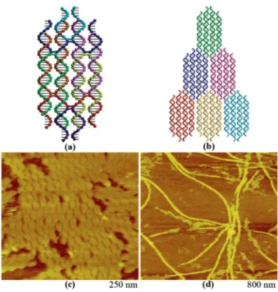
Figure 3: double-double crossover (DDX) DNA-based structure (top schematics) and AFM image of a
2D crystal made of branched DDX motifs (lower view). (Reprinted with permission from J Am Chem Soc 2005, 127 (50), 17590-17591. Copyright (2005) American Chemical Society)
Several studies followed based on these DX structures (Figure 3),18-20 giving rise to more complex structures, such as TX-type tiles (triple-crossover),21-23 and type PX (paranaemic-crossover).24-25
In the mid 2000s, another key step in the emergence of DNA nanotechnology was the development of DNA origami by Paul Rothemund.14 This breakthrough concept significantly increased the complexity, versatility, and size of DNA structures. The term “origami” refers to the Japanese art of making various unique 3D forms with a paper sheet through folding techniques. In DNA origami, the paper sheet is a long DNA strand (obtained from DNA extraction of the M13 virus), which can be considered to be a scaffold that is folded into the desired shape by means of hundreds of small oligonucleotides [see Figure 4 (a)] called “staples”. Each staple is designed to bind to different locations of the long strand to induce local folding, which collectively cause the overall DNA strand to acquire a defined shape. Rothemund illustrated the versatility of this concept by creating a number of different structures [Figure 4 (b)]. A key advantage of this technology is that it is not necessary to precisely control the stoichiometry and purification of oligonucleotides, because the use of a long scaffold makes the assembly highly selective. The minimum of energy is reached when all the staples are associated, regardless of the number of staples by unit volume. DNA origami achieved immediate success thanks to the overall reliability in reproducing complex 2D and 3D shapes and because of its conceptual and experimental simplicity. Progress in this technique has resulted in the formation and control of uniform structures with unprecedented complexity. Importantly, this method is associated with an automated computer design strategy. Indeed, all DNA folding and associated staples must have their base sequences designed depending on each other and must be preprogramed with the help of computer-aided design procedures. Therefore, the staples are determined before the manufacturing of origami and be ordered from
7
DNA synthesis companies.
In addition, DNA staples can be obtained with chemical or biological modifications, the subtle positioning of which within the origami structure can yield a chemically or biologically functional structure. Similarly, by including thiol termini at specific locations of the origami, Zhang et al. managed to create a periodic network of gold nanoparticles on a DNA-modified surface [Figure 4 (c)].26
(a) (b)
(c)
(d)
Figure 4: (a) Design of a DNA origami. (b) Different shapes obtained using the origami technique: a, square; b, rectangle; c, star; d, smiley; e and f, triangles. The first two lines correspond to design images from computer simulation, while the two other lines correspond to experimental validations. (Reprinted with permission from Nature 2006, 440 (7082), 297-302, Copyright (2006), Springer Nature); (c) DNA nanostructures used as templates for the assembly of a 2D network of gold nanoparticles. (Reprinted with permission from Nano Lett 2006, 6 (2), 248-251, Copyright (2006), American Chemical Society) (d) Full origami DNA box. The box may be actuated for closing/opening through the introduction of an oligonucleotide (the top image reconstructed from transmission electron cryomicroscopy experiment, and the bottom image is a schematic of the origami box. (Reprinted with permission from Nature 2009, 459 (7243), Copyright (2009), Springer Nature)
After numerous studies and publications on two-dimensional DNA origami, the concept has been extended to three-dimensional DNA origami,27 which appeared for the first time in 2009. The Gothelf and Kjems team at the Center for DNA Nanotechnology (CDNA) at the University of Aarhus in Denmark demonstrated the connection of several 2D origami planar structures at their edges. In their report, they described in detail the formation and characterization of a DNA box with a controllable lid [Figure 4 (d)]. Two sides of the box were hinged to one side and
8
held closed along the opposite edge by a pair of “staple” DNA strands named “locks”. Each lock can be opened by adding a DNA strand complementary to the strands constituting the locks to the solution, releasing the lid of the DNA box.
In summary, since the initial vision of Seeman in the early 1980s, fundamental steps in the programming and engineering of DNA nanostructures have succeeded one another, and the invention of the DNA origami technique has allowed for the considerable development of these technologies, making possible to create complex structures with any shape and precise properties. However, it is important to note that production yields tends to fall as soon as the complexity and density of structures increases. Furthermore, the lack of detailed information on the folding process remains an obstacle to the development of DNA nanotechnologies. Thus, extensive studies on the fundamental thermodynamic versus kinetic aspects of the folding processes should be pursued.
4 DNA-directed assembly of particles: from concepts to the realization of
ordered assemblies
An interest in using DNA nanotechnology to programme the assembly of nanoparticles and colloids into larger hierarchical structures emerged in the 1990s (Alivisatos et al.,28 and Mirkin
et al.)16. Using the controlled chemistry of sulfur on gold surfaces, both groups had the idea of grafting single-ended thiol-terminated DNA strands (-SH) onto gold nanoparticles.
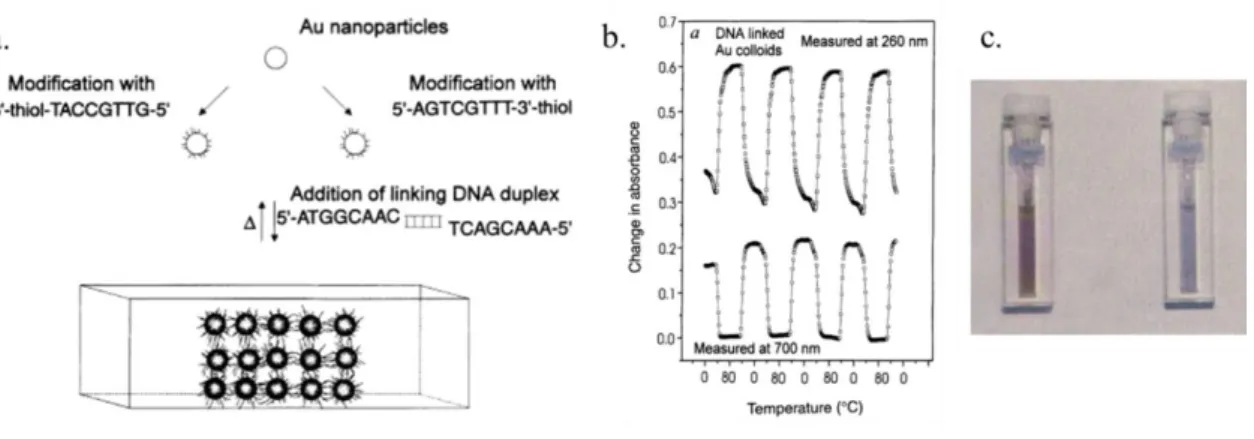
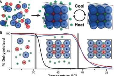
Figure 5: (a) Schematic diagram of the linker self-assembly of 13-nm diameter gold nanoparticles
(left figure). (b) Measuring the absorbance at 260 and 700 nm of the colloidal solution of nanoparticles as a function of temperature (0 and 80 °C) and demonstrating the reversibility of the hybridization of the DNA strands. (c) Image of the colloidal solution heated to 80 °C (red), and cooled
to 0 °C (blue). (Reprinted with permission from Nature 1996, 382 (6592), 607-609, Copyright (1996), Springer Nature)
The Mirkin team made use of thiol-modified oligonucleotides (-SH) at one end of the strand for chemical grafting onto the surface of 13-nm gold nanoparticles.16 Two separate solutions of
gold NPs modified with non-complementary strands were made and mixed in equal amounts. A single-stranded DNA sequence that was complementary to both strands was then added in excess to the above mixture. This additional DNA strand acted as a “linker” and induced the assembly of the nanoparticles into aggregates of several microns by hybridization of the linker strands with both strands grafted onto the nanoparticles. The colour of the nanoparticle solution gradually changes from pink (dispersed particles) to blue (aggregated particles). An interesting aspect of this type of assembly is its reversibility. Indeed, at ~ 55 °C, the so-called melting temperature, dehybridization of the strands takes place, and the redispersion of the nanoparticles causes the solution to return to its initial pink colour (see Figure 5). Alivisatos chose to work
9
on particles that were 10-fold smaller, onto which a single oligonucleotide sequence was covalently attached using alternating strand with distinct sequences. Then, hybridization was performed at different defined positions of a long single strand that resulted in the nanoscale-controlled positioning of the NPs along the one-dimensional DNA strand. Undoubtedly, the controlled interplay of complementary and non-complementary DNA strands makes DNA nanotechnology one of the most powerful bottom-up approaches for building hierarchical architectures of nanoobjects (noble metals, semiconductors, oxides, and polymers) with an almost infinite variety of high-performance programmable DNA/nanoparticle hybrid materials. Since the seminal work by Alavisatos and Mirkin on gold nanoparticles, many DNA/nanoparticle assembly processes have been reported, mostly by varying the DNA length and processing conditions and to generate materials for applications in biodiagnostics,8–11 therapeutic agents,12,13 plasmon-enhanced spectroscopy,14–18 magneto-optical sensors,19 and catalysis20 and energetic materials, which will be presented in detail in section 5.
4.1 DNA/nanoparticle assembly: primary functionalization strategies
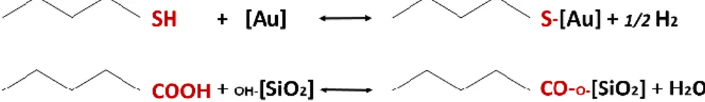
The most successful and widespread technique for gold surface functionalization technique used to graft DNA strands uses thiol chemistry, taking advantage of the well-known and controlled reaction between thiol and metal surfaces (such as gold, see Figure 6).29 An
alternative method using antigen/antibody interactions has been reported to immobilize DNA strands on surfaces, notably oxide surfaces,30 extending the possible applications of the organized and controlled heterogeneous structures of nanoparticles.
Figure 6: Typical thiol-terminated species for a gold surface (in brackets) chemical reaction (top
chemical reaction equation) and carboxylic acid (COOH)-terminated species to a hydroxylated (OH) silicon dioxide surface (in brackets) chemical reaction (bottom equation).
The biotin/streptavidin interaction is recognized as one of the strongest non-covalent interactions in nature, having a complex dissociation constant of 4 10-14 mol.L-1 and a formation enthalpy of 30 kBT.31 This strong interaction is results from a perfectly adjusted
protein pocket for biotin such that its binding is insensitive to pH, salinity and temperature. Streptavidin is a protein consisting of four tetrameric fragments, each of which can accommodate four biotin species, which are immobilized onto streptavidin and form hydrogen, electrostatic and hydrophobic bonds with the aromatic amine groups of streptavidin. The use of this interaction has attracted a great deal of interest because of its versatility, and it can be applied to any type of surface, even gold, provided that the surface is sufficiently reactive to ensure that the protein is immobilized. For example, Cobbe et al. proposed an aggregation of gold nanoparticles previously functionalized with a biotin group using streptavidin and single-stranded DNA.32 The two strategies used in this study are schematically shown in Figure 7 a and b respectively. The self-assembly of heterogeneous nanoparticles by protein-DNA interactions and their differences from gold were also assayed by Oleg Gang and colleagues.33
10
11
Figure 7: a. Direct biotin/streptavidin-mediated nanoparticle aggregation; b. Protocol for the
biotin/streptavidin-mediated DNA-driven assembly of nanoparticles.32(Reprinted with permission from
J Phys Chem B 2003, 107 (2), 470-477. Copyright (2003) American Chemical Society)
Finally, we should mention the use of other chemical functions, such as carboxylic acid (-COOH), which is more appropriate for metal oxide surfaces, or other oxides, such as silicon dioxide (see Figure 6). While the literature commonly reports on organic coatings to inhibit further oxidation of metallic nanoparticles, to the best of our knowledge, no mention of these possible chemical species has been addressed with respect to DNA.
4.2 Towards high-order crystalline structures
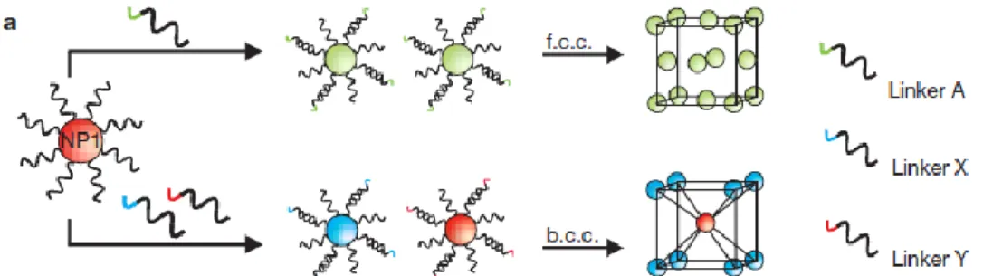
In practice, the overall optimization process for controlling DNA-directed assembly in the liquid phase is a highly complex task. Over the last several decades, substantial progress has been achieved in understanding and controlling the wet chemical technical parameters for the patterning of matter at the nanoscale, especially the effect of salt concentration on both the grafting density and hybridization of DNA strands34-36 or the heterogeneity of DNA grafted on anisotropic nanoparticles as demonstrated very recently.37 Highly ordered face centre cubic (fcc) and body centre cubic (bcc) structures of 5- or 10-nm gold nanoparticles have been described with different DNA strand lengths and a programmable interparticle distance (as illustrated in Figure 8)38-39 and have more recently been reported with the crystallization of nanoparticles into more complex structures (VB23, 24).
Figure 8: Diagram summarizing the influence of the linker on the crystalline structure obtained after
the self-assembly of gold nanoparticles. (Reprinted with permission from Nature 2008, 451 (7178),
553-556, Copyright (2008), Springer Nature)
It is now known that several key parameters have a large effect on the assembly kinetics and architecture of the final structures:
- The ionic concentration in solution, such as the salt concentration (NaCl, MgCl2, ...).
Park et al. have shown that the salt concentration tends to decrease the inter-particle distance by decreasing the repulsive interactions between nanoparticles.
- The spacer nature of the oligonucleotide also influences the inter-particle distance. Indeed, a “slump” of an oligonucleotide on the nanoparticle was observed with a spacer composed of repeated adenine bases, reducing the inter-particle distance by 2.5-fold because of a greater affinity of this base with gold compared to that observed with other bases.40-41
- The DNA strand length impacts crystallinity, where an excessively long strand lowers crystallinity because nanoparticles are less constrained, whereas a short link with respect to the size of the nanoparticle does not allow crystallization because of the size polydispersity of nanoparticles.38, 42-43 Experimental phase diagrams showing the crystallinity of gold nanoparticles as a function of these parameters have been
12
established.44 Owing to the flexibility of their structure, DNA strands are also reprogrammable, allowing a crystal structure to be directly altered in solution by selecting the appropriate oligonucleotide sequence45 or by introducing DNA interlayers, modifying the stiffness and temperature sensitivity of the DNA bond.46 For example,
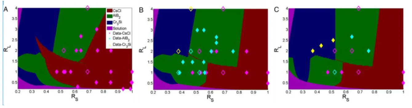
Gang et al. recently determined gold nanoparticle phase diagrams based on stoichiometry (i.e. the relative proportion of two populations of gold nanoparticles with complementary DNA strands), the size of nanoparticles, and the length of DNA linker, both experimentally and theoretically, using coarse grain models.47 These phase diagrams are shown in Figure 9 below, where gold nanoparticles can adopt the crystal phase of CsCl, AlB2 or Cr3Si depending on the size of the nanoparticles or the linker.
Figure 9: Gold nanoparticle phase diagrams as a function of nanoparticle size (RS) or linker
length (RL) for three different stoichiometries (in a binary mixture, the ratio in terms of the population of nanoparticles functionalized with a given sequence versus another set of nanoparticles with the complementary sequence): (A) 1:1, (B) 2:1, (C) 3:1. The symbols represent the experimental points:
the solid symbols represent the pure structures, while the voids represent polymorphic structures.
Overall, there is good agreement between experimental points and theoretical results.47
- The temperature of the solvent also influences crystallinity. By aggregating at a temperature close to the melting temperature of DNA, a transition can take place at which nanoparticles are allowed to rearrange into well-defined crystalline structures and on a larger scale. Figure 10 shows the principles of this rearrangement and gives the ingredients, thermal treatment and spacer length required for obtaining crystals. 38, 48-49
Figure 10: Diagram showing the behaviour of nanoparticle aggregates formed upon DNA
self-assembly versus temperature and the length of DNA strands used.(Reprinted with permission from
Nature 2008, 451 (7178), 549-552, Copyright (2008), Springer Nature)
13
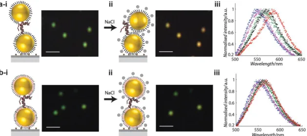
Cooling the colloidal solution at a sufficiently slow rate, which can take from two to three days, was recently shown to lead to the formation of micrometric rhombic dodecahedron “super crystals”.50 In contrast, performing self-assembly at a constant temperature below the melting temperature results in the polycrystallization of the nanoparticles, which are then less organized despite having undergone the annealing process.51 Importantly, the formation of these dodecahedra is independent of the size of nanoparticles, showing that this structure is thermodynamically the most stable that is achievable for such systems. Finally, post-crystallization modifications have also made it possible to increase the temperature ranges or solvents in which these structures can be used by adding organic complexes or encapsulating DNA in silica.51-52 Notably, it is also possible to control the number of functionalized strands on the surface of gold nanoparticles with one, two or more strands by “sorting” a colloidal solution of nanoparticles by electrophoresis. This technique allows for the hybridization of two or three nanoparticles alone (dimers or trimers) that have optical properties dependent on the inter-particle binding, which is governed by the nature of the DNA binding and can be dynamically altered in solution45, 53 by ionic interactions and the chemical nature of the substrate.54 Thus, the chemical nature of the surface of the gold nanoparticles depends on the ligands used to stabilize the colloids. Lermusiaux et al. investigated the optical properties of dimers immobilized on the surface of gold nanoparticles as a function of the ionic concentration and the hydrophobicity of the surfaces by varying the nature of the ligand. The results presented in Figure 11 show that the use of an amphiphilic ligand (i.e. having both a hydrophobic and hydrophilic group) makes it possible to improve stability in terms of decreasing the unwanted aggregation of nanoparticles or in the conformation between two assembled nanoparticles.
Figure 11: Comparison of the variation in the interparticle distance of 40-nm diameter gold
nanoparticles organized into dimers according to the use of hydrophilic (a) or amphiphilic (b) ligands for their stabilization after increases in the saline concentration (i: 5 mM NaCl; ii: 800 mM NaCl) by
dark field microscopy and spectral absorbance analysis. (Reprinted with permission from Small 2015,
11 (42), 5696-5704, Copyright (2015), Wiley)
4.3 Crystallization of heterogeneous systems
The vast majority of work has focused on the self-assembly of gold or silver nanoparticles, owing to their good colloidal stability, controlled synthesis and subsequent controlled thiol chemistry. However, extending their potential applications requires manipulating other materials. Gang et al. have proposed a general strategy of nanoparticle functionalization by DNA that is theoretically applicable for a wide range of materials,33 which they applied to three model materials: gold nanoparticles with palladium of three different shapes (cube, octahedron and dodecahedron), iron oxide (Fe2O3), and quantum dots (CdSe/CdTe/ZnS and CdSe/ZnS).
14
They identified three primary parameters that are crucial in establishing heterogeneous superstructures: (i) the role of the shape of the nanoparticle on crystallinity, (ii) the influence of non-specific interactions related to the presence of DNA, and (iii) the emergence of disorder when manipulating multi-elemental structures. This work follows the pioneering work by Severac et al., who first demonstrated the possibility of using DNA as an assembly vector in the context of the synthesis of high energy performance materials from Al and CuO nanoparticles.55 In addition, Fan et al. developed assemblies of five nanoparticles of different
sizes involving a small nanoparticle surrounded by four larger nanoparticles.56 Such anisotropic assemblies have the advantage of having better defined optical properties. In addition, Tan et al. developed a general and simple single-step method for the functionalization and crystallization of anisotropic systems from 2 nanoparticle sizes.57 Beyond the grafting densities that strongly change with anisotropy, a predominant effect of the nanoparticle curve, which is dependent on the size of the nanoparticle, has been observed.58-59 Jones et al. have studied the crystallization of non-spherical nano-objects, such as octahedra, prisms and rods.60 In general,
the resulting structures of these objects remains the one that allows for the maximum hybridization of DNA strands61 and involves a superposition of nanoprisms,62 a 1D arrangement of nanorods, or a classical crystallization in Cubic Face Centered and Cubic Centered.60
However, only small aggregates are obtained, and it remains difficult to obtain crystals on large scales. From a theoretical perspective, Travesset and Knorowski have studied the self-assembly of nanocubes,63 and a face-to-face orientation of the cubes was obtained when short strands were used, while several other structures could be obtained using longer DNA strands with application of osmotic constraints. Finally, Macfarlane et al. demonstrated the possibility of obtaining complex ternary systems by inserting a third size of nanoparticles into a pre-existing binary structure (see Figure 12).64 This process has made it possible to establish a wide range
of crystalline structures in a completely reversible manner owing to the use of DNA, opening the way for the development of a method that can be generalized to other materials.
Figure 12: Diagram of the self-assembly of nanoparticles by DNA in ternary crystalline structures. A
binary structure is first synthesized prior to insertion of a third size nanoparticles into a
predetermined site (A). In (B), the thermal analysis of the ternary structure is presented.(Reprinted
with permission from Science 2013, 341 (6151), 1222-1225, Copyright (2013), Science American Association for the Advance of Science)
The previously described work primarily focuses on the aggregation of nanoparticles, the diameter of which can vary from 10 to 150 or even 200 nm. However, other teams have observed different behaviours using micrometric-sized nanoparticles, specifically with respect to their ability to stabilize colloidal solutions with increase in temperature. Indeed, interactions between particles vary with their size, especially with respect to weak Van der Waals interactions that can lead to non-specific and irreversible aggregation.65 Therefore, crystallization is much more difficult to achieve, and the aggregates are amorphous gels with a fractal structure.65-67 Crocker et al. demonstrated the possibility of crystallizing microspheres
15
into short hexagonal crystals under extremely precise experimental conditions with respect to temperature and especially surface preparation.68-70 Indeed, only a specific method of functionalization by swelling-deflating micrometric organic particles (such as polystyrene) immersed in organic solvent in the presence of polyethylene glycol (PEG) was sucessful.68
Beyond this example, the phenomenon of reversibility with temperature is rarely observed and strongly depends on the length of the DNA bond and the temperature, which must be sufficient to de-hybridize double-stranded DNA without irreversible aggregation of the nanoparticles. Although a recent review did not report significant progress in the self-assembly of micrometric particles,71 a team from the United States recently demonstrated the possibility of functionalizing micrometric organic particles [polystyrene or poly(methyl methacrylate) (PMMA)] or inorganic (titanium oxide or silicon oxide) with single-stranded DNA by a generic method of alkyne-azide cyclo-addition.72 This method enables higher DNA grafting densities to be obtained than methods using the biotin-streptavidin complex, consequently allowing for crystallization of the particles at room temperature or optimized rearrangement after annealing.
4.4 DNA/nanoparticle assembly: applications
The most intriguing applications of DNA nanotechnology, those that best take advantage of the small size, biocompatibility and programmability of DNA-based systems, lie at the interface with biology. Below, we highlight key successes in the development of DNA-based imaging probes for biomolecular detection and prototypes of smart therapeutics and drug delivery systems. Several dynamic plasmonic systems have also been demonstrated for applications in energy harvesting, nanophotonics and imaging.73
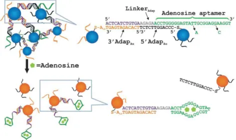
Biomolecular detection – One of the first applications of DNA nanotechnology put into practice was biomolecular detection based on the colorimetric principle. For example, gold nanoparticles initially functionalized with complementary strands may be disintegrated in the presence of a target species, hybridizing with a portion of the DNA strands (see Figure 13). 74-75 Disintegration caused by the de-hybridization of the two complementary strands is associated
with a colour change from blue to red in the colloidal solution. This principle has been used by many teams to detect several species, such as thrombin,76 adenosine and cocaine,75 or cysteine.74 The possibilities are numerous but limited by the relatively high detection
concentrations (in the nanomolar range) compared with the higher sensitivity that is attainable with fluorescence-based methods.77
16
Figure 13: Schematic diagram of the detection of adenosine using the colorimetric method.
Functionalized gold particles with DNA strands are aggregated owing to the presence of a complementary aptamer with adenosine. Thus, in the presence of the target molecule, the aptamer hybridizes to the molecule, causing particle de-aggregation and a colorimetric change in the solution
from blue to red. (Reprinted with permission from Angew Chem Int Edit 2006, 45 (1), 90-94,
Copyright (2006), Wiley)
Optical and plasmonic devices - Crystals composed of specific inorganic and semi-conductive nanoparticles, also called “quantum dots”, have the advantage of having luminescence energies that have for biomedical applications to replace the traditional organic fluorophores used for imaging, exhibiting lower luminescence and greater sensitivity to the spectrum of white light.78 Their association with a metal such as gold allows for further increase in the photoluminescence released by the quantum dots. Thus, the organized assembly of gold nanoparticles and quantum dots allows the photoluminescence of the structure to be controlled according to several parameters, such as the size of the constituents and the inter-particle distance, allowing for the monitoring of biological events and the development of improved plasmonic spectroscopies. Catalysis - Finally, control of the size, composition and density of particles is crucial for catalysis, where DNA can provide novel possibilities. A drawback is that DNA requires a specific aqueous environment, limiting catalysis by passivating the surface of nanoparticles, making them less reactive. To overcome these difficulties, Auyeung et al. developed a three-step process, where after synthesizing a superstructure of gold nanoparticles, they freeze the structure in the silica and then calcine the whole structure, resulting in an organized porous structure.79 In summary, the authors predict a real utility for catalytic reactions using gold, such as the oxidation of alcohol or carbon monoxide, or reduction via plasmonic effects.
5 Nano-engineering of DNA self-assembled Al/CuO nanothermite
While much of the literature describes the DNA-directed assembly of gold nanoparticles, achieving the assembly of oxide nanoparticles is a technically complex task due to increased surface chemistry issues. This issue constitutes a formidable scientific challenge that well illustrates all possible routes to optimization of practical and efficient technological solutions and the promise of novel and practical applications in energy-generating materials. This last section is devoted to such an application example, namely, thermite energetic materials. Interestingly, we present a series of methodological steps that have been performed to visualize and optimize the DNA-directed assembly of Al and CuO nanoparticles, which can be generalized to many other materials.
17
Thermites are substances that store chemical energy that can be released after being submitted to an external electrical, optical, or pressure wave stimulus... These substances are composed of a fuel (most of the time aluminium) mixed with an oxidizer, which can be varied to monitor reaction properties, and this metastable mixture undergoes exothermic oxidation-reduction reactions80-81. The aluminium/copper oxide (Al/CuO) system is among the most interesting as it offers high energy density and reactivity, particularly when mixing Al and CuO nanoparticles, compared to their micronic counterpart82-83. Energetic materials are used in widespread applications, including in the space, military, automotive and civil security industries84-91. In the early 1990s, taking advantage of Al and CuO compatibility with microelectromechanical systems (MEMS) fabrication techniques92, we proposed that they be integrated into silicon microsystems for the realization of local micro-actuations in extremely small volumes (less than a cubic millimetre) and relatively substantial forces (~ 0.1 N). In this context, DNA nanotechnologies open perspectives to create ad hoc materials that have high-performance, are safe and are REACH legislation compatible.93-99 The exothermic oxidation/reaction process, in which oxygen atoms are liberated from their CuO matrix (reduction until pure copper) to oxidize aluminium, with formation of alumina, can be represented by the following overall chemical reaction:
3CuO + 2Al →3Cu + Al2O3
The properties of these materials are highly dependent on the sizes of components and their distribution in the composite. The use of nanoparticles allows for a significant improvement of energetic properties, but there remain important issues with respect to controlling the mixing of nanoparticles at the nanoscale. In this context, the use of DNA self-assembly shows great potential for the synthesis of new types of nanothermites with exquisite control of the contact surfaces between oxidizer and reducer, thereby enabling the optimization and control of energetic performances.
Our approach to using DNA for the controlled assembly of Al and CuO NPs into thermite aggregates is outlined in Figure 14.
Figure 13. Illustration of the functionalization process developed to enable the DNA-directed
self-assembly of core-shell Al (Al covered with Al2O3 shell) and CuO nanoparticles.
In the following section, we will successively detail and discuss: 1. The fundaments and characterization of DNA interactions and overall grafting protocol; 2. The characterization of DNA-directed assembly; and 3. The performances of DNA-assembled Al/CuO energetic nanomaterials compared with those of nanopowder mixtures.
5.1 Fundaments and characterization of DNA/surface chemistry and grafting strategies
18
To guide technological steps, we provided an atomic scale understanding of intrinsic physical and chemical interactions taking place between DNA and oxide surfaces. To allow for experimental and atomic scale simulations of synergistic associations, we proposed to use a representative dTMP molecule that includes all DNA subunits instead of a full DNA fragment (dTMP: 2-desoxi-thymidine-5’-monophosphate). Then, we studied the chemical interaction of dTMP molecules with controlled alumina surfaces through XPS and infrared spectroscopy. The experimental results were supported by density functional theory (DFT) calculations. In a second stage, we developed, characterized and optimized the overall grafting protocol by quantifying all processing steps.
5.1.1 DNA/alumina interaction evaluation through infrared spectroscopy and first principles calculations
The dTMP molecule, as shown in Figure 15, is a rather small molecule compared to DNA strands with multiple bases, but it still harbours all DNA subunits, including a phosphate group, a sugar and a base, and in this case study harbours a thymine group. The alumina oxide surface is chosen as it is much simpler to manipulate and characterize at the atomic scale than CuO. Notably, to provide well-controlled model surfaces, we used flat alumina surfaces deposited by atomic layer deposition, which allows the thickness of alumina to be adjusted at the monolayer level.
Figure 15. Schematic of the dTMP molecule with its subunits equivalent to those of DNA.
We performed first principles calculations to evaluate the different possible chemical reactions taking place at surfaces, which was one of the objectives of the quantification of all dTMP vibrational modes of the different and most pertinent configurations (either in the liquid or in contact with the surface) for assignation of the complex infrared IR spectra obtained experimentally. One such calculation is shown in Figure 16, corresponding to the most stable configuration of all trials, indicating chemical reactions at both the phosphate and base levels, with an energy gain in the order of 1-2 eV compared to dTMP in the liquid phase.100
19
Figure 16: Chemisorption of dTMP on a hydrated Al2O3 surface (with an -OH termination) in the
most stable case, i.e. with formation of covalent bonds at the level of phosphate and thymine groups.100
Based on theoretical calculations, we first experimentally validated the irreversible nature of the interaction of dTMP with alumina. After performing rinsing and ultra-vacuum pumping steps, XPS measurements clearly indicated the presence of dTMP on the surface, as shown in Figure 17. The results showed the appearance of nitrogen and phosphorus signals, demonstrating the presence of DNA. A detailed analysis of these spectra with respect to the influence of the initial dTMP concentration indicates a chaotic and/or multilayered coverage, as well as the quantification of carbon extra-contamination that probably result from the overall deposition procedure [atomic layer deposition (ALD), dTMP, and rinsing].
Figure 17: XPS spectra for three primary elements in dTMP deposited on alumina surface. The black
curve corresponds to the signal of the reference (alumina substrate), while the red curve
corresponds to the alumina surface with deposited dTMP.100
To identify the bonding nature of dTMP with the alumina surface, we made use of infrared spectroscopy and extensively investigated deposition under multiple conditions (concentration, temperature, nature of solvent …). Using first principles quantification of vibrational modes to assign different and reproducible peaks, we confirmed the creation of covalent bonds and multiple interactions on the surface, particularly through phosphate and thymine dissociation. Such assigned spectra are shown in Figure 18, in which the nature of the solvent was modified (water and methanol).
20
Figure 18: Influence of the nature of the solvent on the IR absorbance spectra of an alumina surface
after dTMP deposition. The incubation of the surface was performed using methanol (top curve) and water (bottom curve) with the same concentration of dTMP to a clean alumina substrate in both cases.
Related dTMP regions are reported on the top of the figure: T for thymine, S for sugar, and P for
phosphate according to assignments resulting from DFT calculations.100
To direct the assembly of Al and CuO, we decided to use a biotin/streptavidin grafting protocol. With the knowledge that direct, irreversible and chaotic bonding would take place between DNA and alumina or CuO nanoparticle surfaces, screening them as much as possible through streptavidin coverage would lead to subsequent specific interaction of the antigen/antibody type with biotin terminated DNA strands. In the following section, we describe this grafting protocol and the process of generating the Al and CuO colloidal solutions.
5.1.2 Functionalization protocol and colloidal characterization
The overall DNA functionalization procedure, which is conceptually equivalent for both the Al and CuO nanoparticles, is shown in Figure 14. The first step of the process consists of the dispersion and stabilization of Al and CuO nanopowder. For the experiment, we used 15 mg of 50-nm CuO and 80-nm Al nanopowders of nominal size as provided by the suppliers. These nanoparticles were dispersed in ultra-pure deionized water containing a surfactant, Tween-20 (or polyoxyethylene (20) sorbitan monolaurate), which acts as a stabilizing agent, and the solutions were buffered to neutral pH (pH = 7). Tween-20 is a molecule comprising hydrophilic and hydrophobic regions on opposing sides. In solution with nanoparticles, these molecules nonspecifically interact with nanoparticles and prevent aggregation. During their dissolution, nanopowders do not spontaneously disperse, yielding numerous large aggregates, necessitating an ultrasound step followed by a sedimentation step. The solutions are ultrasonicated for 8 min at 200 W, while maintaining the temperature below 40 °C. The resulting solutions are then left overnight at room temperature, allowing large aggregates that have not been broken down to settle. The supernatant of the solutions is then recovered and characterized by dynamic light scattering (DLS). The results obtained for unmodified particles can be seen in Figure 19 (black curves). We observed hydrodynamic diameters (ZH) values of ~ 224 ± 7 and 187 ± 5 nm for Al and CuO, respectively. Scanning electron microscopy results revealed that these colloidal solutions are composed of small indivisible aggregates of 2 to 4 nanoparticles, with diameters that are stable over hours. Note that a shell layer of approximately 3 nm of oxide covers the pure Al core of the Al nanoparticles. This protective shell allows the particles to remain unoxidized and stable in solution under neutral pH conditions.
0 250 500 750 1000 1250 0 2 4 6 8 10 12 14 CuO Inten sité (%) Diamètre hydrodynamique (nm) 0 2 4 6 8 140 160 180 200 220 240 260 Dia mè tre h yd ro. (n m) Temps (h) 0 250 500 750 1000 1250 0 2 4 6 8 10 12 14 Al Inten sité (%) Diamètre hydrodynamique (nm) 0 2 4 6 8 160 180 200 220 240 Dia mè tre h yd ro. (n m) Temps (h) 200 190 0 250 500 750 1000 1250 0 2 4 6 8 10 12 14 CuO CuO-Strep Inten sité (%) Diamètre hydrodynamique (nm) 0 2 4 6 8 140 160 180 200 220 240 260 Dia mè tre h yd ro. (n m) Temps (h) 0 250 500 750 1000 1250 0 2 4 6 8 10 12 14 Al Al-Strep Inten sité (%) Diamètre hydrodynamique (nm) 0 2 4 6 8 160 180 200 220 240 Dia mè tre h yd ro. (n m) Temps (h) 0 250 500 750 1000 1250 0 2 4 6 8 10 12 14 Al Al-Strep Al-ADN Inten sité (%) Diamètre hydrodynamique (nm) 0 2 4 6 8 160 180 200 220 240 Dia mè tre h yd ro. (n m) Temps (h) 0 250 500 750 1000 1250 0 2 4 6 8 10 12 14 CuO CuO-Strep CuO-ADN Inten sité (%) Diamètre hydrodynamique (nm) 0 2 4 6 8 140 160 180 200 220 240 260 Dia mè tre h yd ro. (n m) Temps (h)
21
Figure 19: Schematic overview of primary DNA grafting steps and strategy for the Al/CuO directed
assembly.
Then, the grafting of streptavidin was performed after determining the optimal quantity that can be generated, which is derived from the total nanoparticle surface available in solution, considering a sphere approximation, and knowing that streptavidin covers approximately 5 nm2. Finally, for both Al and CuO colloidal solutions, an excess of streptavidin was added. In both cases, an incubation time of at least 4 h was used, and the solutions were rinsed multiple times to remove the excess of ungrafted streptavidin. Between each rinsing step, the solution was centrifuged and further dispersed in an ultrasonic bath after changing the solvent. Rinsing was performed out in a 0.1% diluted phosphate buffered saline (PBS) solution containing 0.05% vol. Tween 20, although the grafting of streptavidin tends to stabilize the colloids. Again, DLS measurements were performed, shown as green curves in Figure 19, and could be compared to non-grafted particle diameters. We observed a slight increase in the mean diameters due to the addition of streptavidin molecules. Notably, for the CuO nanoparticles, the inflation was more pronounced, which could be attributed to undesired interparticle interactions mediated by streptavidin or their accumulation of multiple layers.
The next step was to achieve biotin-terminated DNA grafting owing to the biotin/streptavidin interaction. For streptavidin, the first step was to estimate the amount of DNA needed to functionalize all the available proteins on the surface of the nanoparticles. For both colloidal solutions of Al and CuO streptavidin-functionalized particles, which were previously prepared, we added the DNA solution at a concentration that was 5-fold the amount necessary for the biotin-terminated DNA to complement the optimal surface density of streptavidin sites. Again, after incubation, rinsing and centrifugation to remove excess DNA in solution, the colloidal solution was redispersed after each centrifugation in an aqueous solution of 0.1× PBS and 0.05% Tween in an ultrasonic bath. Subsequently, the hydrodynamic diameter and Zeta potential of the obtained functionalized nanoparticles were characterized by DLS, the results of which are shown in Figure 19 (red curves, obtained with 30-base DNA strands). Shorter (15 bases) and longer (45 bases) DNA strands have also been used, showing coherent results for CuO nanoparticles.
5.1.3 Quantification of streptavidin and DNA surface densities
Based on previous fluorescence based methodologies that were exclusively used for quantifying the grafting of DNA strands on gold nanoparticles, we have derived the following quantification strategy, which allows the measurements of streptavidin and DNA surface densities.30, 101 The end of the subsection describes the quantification of the grafted DNA that is still functional for hybridization, which we termed the “hybridization efficiency”. Note that in this section, hybridization is performed with DNA strands that are nanoparticle free. Because of the
22
permanent immobilization of streptavidin on the surfaces of the nanoparticles, we developed a method for quantifying the amount of streptavidin grafted onto the nanoparticle surfaces (Al or CuO). This process was performed in three steps, shown schematically in Figure 20 that quantify a. streptavidin coverage, b. DNA coverage and c. hybridization efficiency, respectively, which are the primary principles detailed in the following section. For more technical details, see ref.30.
Figure 20. Schematic representation of the process developed for the determination of (a) streptavidin
surface density on CuO or Al nanoparticles, (b) the DNA surface density on CuO or Al nanoparticles and (c) the hybridization efficiency on CuO or Al nanoparticles.
Quantification of streptavidin surface density - Cy3-labelled streptavidin is added to the colloidal suspensions. After incubation, the solutions are centrifuged, and the fraction of unbound streptavidin [Figure 20 (a)] is measured in the supernatant. The difference in the streptavidin concentration before and after centrifugation is used to deduce the amount of bound streptavidin, which is then normalized by the quantity of nanoparticles determined by atomic absorption spectrometry (AAS).
Quantification of DNA surface density – Fluorescent FAM-oligonucleotides are added to 𝐶𝑢𝑂𝑆𝑡𝑟𝑒𝑝and 𝐴𝑙𝑆𝑡𝑟𝑒𝑝 colloidal suspensions. After incubation, the solutions are centrifuged, and the fluorescence intensity of the supernatant is measured. The quantity of DNA grafted onto the nanoparticles is then determined from the difference between the initial DNA concentration (using a control sample) and the DNA concentration in the supernatant, which is then
23
normalized by the quantity of nanoparticles determined using AAS. This protocol is summarized in Figure 20 (b). Note that the reversibility of the streptavidin/biotin interaction can be evaluated by heating/cooling the solutions containing functionalized Al and CuO nanoparticles.
Quantification of DNA hybridization efficiency - FAM-labelled oligonucleotides are added to Al and CuO colloidal suspensions previously functionalized with non-fluorescent strands. The NaCl concentration is set to enable the hybridization of the FAM-labelled strands with the non-fluorescent ones, grafted on the Al and CuO nanoparticles. After a few hours, the colloids are centrifuged and re-suspended in solution (0.1× PBS, Tween 0.05% and NaCl) to remove excess DNA strands. After rinsing, the solutions are finally heated to 80 °C for 5 min to de-hybridize DNA double strands. The fluorescence intensity of the supernatant containing the released fluorescent strands is then measured, and the quantity of DNA strands grafted on the nanoparticles is calculated [see Figure 20 (c)].
Table 1: Quantification of streptavidin coverage, grafted DNA, and hybridization efficiency during the
assembly protocol.
Overall, the results indicate that a careful handling of selected process parameters is required to maximize the DNA surface density. Particularly, we observe that 15-30 mM NaCl is required to avoid irreversible nanoparticle aggregation and that gentle sonication during the early stage of streptavidin incubation allows for increased streptavidin loading by approximately 175 and 135% for the CuO and Al nanoparticles, respectively. The DNA strand length has no noticeable impact on the DNA grafting density. We showed that direct grafting of DNA onto Al and CuO nanoparticles largely dominates the overall functionalization process, with DNA immobilization on streptavidin sites representing less than 5-10% of all DNA strands (see Table 1). As streptavidin covers only a small portion of the Al and CuO nanoparticle surfaces, a large surface area is prone to non-specific interactions with DNA. We experimentally confirmed the strong chemical affinity of DNA bases with both copper oxide and alumina surfaces and the lack of control of the conformation of the immobilized strands onto the oxide surfaces. The higher density of grafted DNA observed for CuO suggests a partially spread-out conformation compared to the completely spread-out strands on Al. Finally, we quantitatively proved the crucial role of the antigen/antibody protocol to preserve the ability of DNA to hybridize, improving the hybridization efficiency by 2-fold, ensured by the specific DNA strands grafting onto streptavidin sites (~1.2 and 0.9 strands by streptavidin for CuO and Al nanoparticles, respectively).
24
Many parameters (length and nature of the spacer, NaCl concentration ...) clearly affect the assembly and crystallinity of nanoparticle aggregates. However, the role of the coding sequence selected to enable hybridization upon aggregation has hardly been addressed, despite being a crucial factor in determining the thermodynamics and kinetics of aggregation. Various hybridization strategies involving strands of variable coding sequences have been described in the literature. In addition, the role of the length of the spacer has been investigated. However, the choice of the oligonucleotide sequence exclusively follows a priori empirical design and has not been studied nor commented on in an extensive manner. In this section, we propose to design and optimize the sequence that will be used to obtain our energetic nanobiocomposites in silico, which will be validated experimentally in a second stage. These results were published in ref.102.
5.2.1 Design and impact of the DNA coding sequence
Referring to the pioneering research by Oleg Gang,33 presenting a generic method for the functionalization of different types of nanoparticles (gold, palladium, iron oxide or quantum dots), the chosen DNA strand is composed of a repeated T-base spacer to separate the coding portion that will be further hybridized to the surface of the particle, with a coding portion of 15 bases. A close examination of the sequence allowed us to notice that partial hybridization between two complementary strands was possible due to the repetition of the four AGGT bases twice in the sequence (AAT-AGG-TGA-AGG-TTA). In addition, folding of the strand on itself is possible when the spacer is composed of T bases, where the sequence becomes [(T)12-TTT-AAT-AGG-TGA-AGG-TTA]. This possible folding is crucial because it could prevent any hybridization with the complementary strand. In addition, this folding can result in “stranded” interactions when a large number of strands of identical composition are in solution. These parasitic interactions are schematically shown in Figure 21 and should be avoided or minimized.
Figure 21: schematic of the constraints used in the programme. Oligonucleotides are represented by
bars: red bars correspond to spacers (for example, repeated thymine bases), blue bars correspond to the sequence designed by the algorithm, and green bars correspond to the complementary strand of the designed oligonucleotide. (a) Avoidance of self-interactions for a single strand through the folding
process, (b) hindered interactions (total or partial hybridization) between two strands with the same sequences, and (c) undesired partial hybridization of sticky-end sequences in the presence of their complementary counterpart. (d, e) Detailed and local sequence constraint: (d) a maximum of three
25
adjacent bases are allowed to hybridize and (e, f) mismatched sequences (noted as M) that are
forbidden when considering interactions.102
Given the limited number of studies on the generation of an optimized DNA strand sequence of a length close to standard requirements, we have developed an algorithm to define an optimized sequence by removing 4 types of undesired interactions:
- The generated DNA strand must not fold back on itself to maintain its coding specificity. - Non-complementarity of the strand, total or partial, with a strand of identical composition. Based on the use of the DNA strand in solution, it is indeed inevitable to identify many identical strands capable of interacting with each other.
- The considered strand must hybridize over the entire coding portion of the other strand to better control the hybridization process.
- The fourth type of constraints is based on the definition of hybridization criteria, where the user first chooses to consider the minimum number of adjacent bases allowing for hybridization (denoted NM) (d), with the possibility of including one or two non-complementary bases (f) between these adjacent bases. The value of NM is set to 3 by default, according to the Gibbs energies reported in the literature.103
Note also that constraint (b) is more restrictive than constraint (a), because it does not require a loop for hybridization such that the application of constraint (b) includes constraint (a). Considering all restrictions, the number of solutions drastically decreases from the intrinsic combination of DNA coding possibilities, which is tractable by computer analysis. The full details of our algorithm and obtained sequences are available in 102. This optimization process allows the sequences to be selected according to the type of constraint applied and for the results to be sorted according to the melting temperature of each selected DNA strand. Such calculation results are shown in Figure 22 (a,c). For each sequence length, the table presents the best sequence in term of melting temperature that also fits the restriction requirements. The curve (a) of Figure 22 shows the number of available sequences for each DNA strand length that the operator wishes to implement in the assembly protocol and the associated average melting temperature. Note that while melting temperature continuously increases, it starts to become saturated at a nominal level for a DNA length of approximately 6 to 10 bases.
Figure 22: (a) Number of sequences obtained according to the number of bases by considering three
![[PDF] Cours Merise Pas à Pas](data:image/gif;base64,R0lGODlhAQABAIAAAP///wAAACH5BAEAAAAALAAAAAABAAEAAAICRAEAOw==)