HAL Id: hal-02947272
https://hal.archives-ouvertes.fr/hal-02947272
Submitted on 23 Sep 2020HAL is a multi-disciplinary open access archive for the deposit and dissemination of sci-entific research documents, whether they are pub-lished or not. The documents may come from teaching and research institutions in France or abroad, or from public or private research centers.
L’archive ouverte pluridisciplinaire HAL, est destinée au dépôt et à la diffusion de documents scientifiques de niveau recherche, publiés ou non, émanant des établissements d’enseignement et de recherche français ou étrangers, des laboratoires publics ou privés.
Internal Temperature Measurements by X-Ray
Diffraction on Magnetic Nanoparticles Heated by a
High-Frequency Magnetic Field
Stéphane Faure, N Mille, S Kale, J.-M Asensio, J Marbaix, P Farger, D
Stoian
2, W van Beek
2, Pier-Francesco Fazzini, Aikaterini Soulantika, et al.
To cite this version:
Stéphane Faure, N Mille, S Kale, J.-M Asensio, J Marbaix, et al.. Internal Temperature Measure-ments by X-Ray Diffraction on Magnetic Nanoparticles Heated by a High-Frequency Magnetic Field. Journal of Physical Chemistry C, American Chemical Society, In press, �10.1021/acs.jpcc.0c03350�. �hal-02947272�
Internal Temperature Measurements by X-Ray Diffraction on
Magnetic Nanoparticles Heated by a High-Frequency Magnetic Field
S. Faure*1, N. Mille*1, S. Kale1, J.-M. Asensio1, J. Marbaix1, P. Farger1, D. Stoian², W. Van Beek²,
P.-F. Fazzini1, K. Soulantica1, B. Chaudret1, J. Carrey1*
1 Laboratoire de Physique et Chimie des Nano-Objets (LPCNO), Université de
Toulouse-INSA-UPS, 135 Avenue de Rangueil, F-31077 Toulouse, France. ² Swiss Norwegian Beamlines – ESRF – Grenoble – France
To whom correspondence should be addressed. E-mail: s_faure@insa-toulouse.fr;
julian.car-10
rey@insa-toulouse.fr
Abstract: There is a theoretical and experimental controversy on the possibility for magnetic
nanoparticles (MNPs) heated by high-frequency magnetic fields to reach a temperature much larger than the one of their environments. Here the internal temperature of magnetically heated magnetite MNPs is measured using the temperature dependence of their lattice pa-rameter, and compared to the one of their environments, measured from reference non-mag-netic particles. Within the uncertainty of our experimental methods, which is estimated to be below 5°C, the MNP temperature is the same as the one of their environments.
Keywords: X-rays diffraction; Local temperature; Inductive heating; magnetic nanoparticles;
Synthetic natural gas;
20
1. Introduction
The fact that magnetic nanoparticles (MNPs) generate heat when they are excited by a high-frequency magnetic field permits various potential applications like tumor therapy1,
catalysis2 or water electrolysis3. This diversification has been made possible thanks to
contin-ued progresses in the fabrication of chemically-synthesized MNPs, improving the control over their size, shape (core-shell structure, surface morphology), chemical composition and heating power4. Magnetic heating allows transferring the energy directly to the MNPs without the
need to heat the surrounding (catalysis reactor, human body, electrolyte) and has also the advantage of a fast heat transfer with a quick start-stop, which is an advantage for applications
30
aiming at storing excess energy from renewable sources2.
Another potential advantage of magnetic heating is the subject of a controversy, which started in the field of magnetic hyperthermia. In 2002, it was proposed that allowing MNPs to enter inside tumor cells could be more efficient because the heat generation as well as the temperature rise, being intracellular, would be more efficient5. A few years later, it was
con-vincingly argued, using standard heat diffusion equations, that the temperature gradient around magnetically heated single MNPs or micron-size assemblies of MNPs should not ex-ceed the mK. It means that, at the cell level, the temperature has to be considered as homo-geneous6. However, in 2010, Villanueva et al. showed for the first time that, during in vitro
experiments, tumor cells could be killed without any global rise of the temperature, renewing
the hypothesis that localized heating occurred and could be efficient to induce cell death7.
These biology results have since then been confirmed by several groups, including us8, 9, 10, 11, 12, 13, 14.
Most probes used during standard experiments measure temperatures at the millime-ter scale, much larger than the scale where the local thermal gradients are hypothesized to occur. Several groups have developed ingenious strategies to map temperature gradients in the vicinity of the MNP surfaces using molecular15, 16, 17, 18, 19 or fluorescent20, 21, 22, 24, 25 probes.
All of them led to the conclusion that, indeed, NPs in liquid media produce and sustain a sig-nificant temperature gradient (T = [6°C-90°C]) in their immediate vicinity when heated by radiofrequency magnetic fields. In a recent review, Cazares-Cortes26 summarizes in a figure
50
the results issued from the aforementioned experiments and interprets the general trend as showing a strong thermal gradient from the surface of the nanoparticles to the surrounding medium.
On the theoretical side, things are less unanimous. A comparison with theoretical heat transfer mechanisms would suggest that, with dimension similar to that of the phonons’ mean free path, heat transport around MNPs is not diffusive but ballistic, and Fourier law is not valid anymore15, 27. On the other side, Keblinski et al. suggest that the heat flow in liquids can be
well described by the diffusive heat equations even at the nanoscale6. Adding to the confusion,
some authors give theoretical arguments against nanoscale thermal phenomena 5, 28. A recent
paper from Chiu-Lam and al.29 summarizes the clash between theoretical predictions and
ex-60
perimental observations. Current investigations on nanoscale thermal transport rather focus on NP-fluid interfacial properties30, 31, 32.
The convincing experimental results coming from the biology side have led our group − which also works on the catalysis side −, to hope that, in chemical reactions where MNPs are both the heating agents and the catalysts, strong nanoscale gradients could also be ob-served in the vicinity of MNPs. In applications where energy efficiency matters, such as the Sabatier reaction used for power-to-gas, this would permit to heat the catalyst without heat-ing the support and the reactor, permittheat-ing energy savheat-ings, a potentiality that we have named “cold catalysis” 33.
In the present article, we report on the measurements, in solid-state samples where
70
MNPs are grafted on a support, of the gradient occurring between the MNPs and their sur-roundings. To probe the MNP temperature, we have used an original approach, which consists in deducing the MNP temperature from the temperature dependence of their lattice param-eter, using X-Ray diffraction (XRD) on a synchrotron beamline. This approach permits, for the first time, to measure the internal temperature of magnetically heated MNPs, and not the temperature in their vicinity. To measure the mean temperature of the medium, a reference material (i.e. with a strong temperature dependence of its lattice parameter) was added to the sample under the form of micron-size boron nitride (BN) particles, the temperature of which was also deduced using the same method. We did not detect any temperature gradient between the MNPs and the medium; if the latter exists, it is within the error bar of our method,
which is estimated to be around 5°C. The potential reasons for this absence of temperature gradient are discussed.
2. Methods
2.1. Magnetic field source
The electromagnet was a toroidal ferrite (Ferroxcube 3C95) winded with Litz wire (RUPALIT Safety V155 480x0.071) and encapsuled in a plastic jacket. The gap has to be as small as pos-sible to enhance the field, but required to be at least 3 mm wide to fit a 2 mm diameter ca-pillary and to avoid to cut the diffracted beam. The gap was opened with a diamond wheel saw and beveled at 38° to permit to get the largest possible portion of the diffracted beam. The electromagnet was mounted in series with a rotary variable air capacitor that is able to
90
support high alternating currents without heating, contrary to chemical capacitors. At maxi-mum power, the setup was fed with a voltage Vrms= 56.6 V and an alternating current Irms =
2.5 A (high-speed bipolar amplifier HSA 4052) at frequency f = 270 kHz, for a total power of 140 W. The magnetic field generated in the gap had a maximal intensity µ0Hmax = 45 mT. The
electromagnet was cooled down by a forced air flow through the tubular arm structure. 2.2. Beamline detection
The detector usually used for XRPD, a CMOS 2D detector, was perturbed by the electromag-netic field of the electromagnet. It was replaced by an image plate detector MAR 345. How-ever, the reference diffractogram for the SiRAlOx powder (see below) was measured using the first detector.
100
3. Results and discussion
The initial studied samples were SiRAlOx-supported (γ-Al2O3) catalyst consisting of
10wt% BN and 13 wt% FeC MNPs that corresponds to 1% volume fraction. As will be explained below, the final samples on which nanoscale temperature measurements have been per-formed are samples where the NPs have been oxidized. NP synthesis is detailed in the Supple-mentary Information. TEM structural analysis shows that the NPs are well dispersed with a size distribution of 14.6 1.1 nm. The measured SAR value in a toluene solution was of ca. 2000 W·g-1 under 47 mT magnetic field and 100 kHz frequency. For a typical sample prepared
in the glove box, FeC NPs (60 mg, Fe content ca. 75%) were dispersed in toluene (10 ml). Then, the catalyst (SiRAlOx, 400 mg) was added to the dispersion and the mixture sonicated for 20
110
min. At the end, the excess of toluene was removed under vacuum. The yield is ca. 420 mg of FeC / SiRAlOx and the Fe content determined by Inductively Coupled Plasma (ICP) is 9.15 wt%.
In situ X-Ray powder diffraction (XRPD) measurements were performed at the BM31
line of the Swiss-Norwegian Beamlines located at the European Synchrotron Radiation Facility (ESRF) in Grenoble, France. A scheme of the experimental configuration under magnetic field can be seen in Figure 1. Special attention was given to the geometry of the coil to fit the beam-line configuration as well as the field intensity requirements. High-resolution-XRPD data were collected with the standard BM31 setup, using wavelengths of = 0.49417 Å. Powder samples were put inside a quartz capillary which had a length of 60 mm, a diameter of 2 mm and a wall
thickness superior to 0.02 mm34. Capillaries with 1 mm outer diameter were also used, but the
120
volume of powder was not sufficient to observe a sufficient heating under rf field excitation. The catalyst was diluted with the required amount of hexagonal boron nitride (h-BN) (BNhcp Alfa Aesar 99.5% CAS: 10043- 11- 5), loaded into the quartz capillary and sealed with a high temperature epoxy glue. The sample was heated by a vertical hot air blower for calibration experiments or by the electromagnet.
The sample was originally designed to make on the same sample the experiments de-scribed in this article as well as an in-situ monitoring by XRD and EXAFS of the Sabatier reaction under a high-frequency magnetic field. This explains the structure of our sample, which is
130
composed of FeC MNPs as heating agent supported on SiRAlOx powder.
Figure 2 shows the XRD diffraction of our sample. The SiRAlOx diffractogram shows that the powder is almost amorphous (cf. black curve on Figure 2) and is responsible of the heavy background. The diffraction patterns of the sample show the contributions of the FeC and h-BN phases, broaden by the SiRAlOx background. As a preliminary test to confirm the heating power of the NPs, the sample was heated by the magnetic field in absence of a gas flow. The temperature of the capillary surface, measured by an infrared camera, was 121 °C. Then, we measured the evolution of the sample heated by the hot air blower during an overnight temperature cycle. The sample was heated up to 225°C (blower temperature) before cooling down back to room temperature. The diffraction patterns obtained after
140
heating clearly show an oxidation (green curve on Figure 2) with a phase transformation of the FeC to magnetite (Fe3O4) or maghemite (-Fe2O3). The oxidation was not expected and we
Figure 1. Representation of the X-rays set-up configuration with alternating magnetic field ex-citation.
guess it is due to a leak from the capillary’s seal. The fit made with the software Maud35
highlights the different phases of the species present inside the sample. The presence of the Fe3O4 phase is consistent with the standard pattern for JCPDS Card No. (79 - 0417) Magnetite
– synthetic36 and permit to completely fit the diffractogram, suggesting that the FeC was fully
oxidized and disappeared below the detection level inside the background. The SiRAlOx alone was fitted with Maud and included as a background baseline. The fit was performed using only the Fd-3m:1 cubic phase for the Fe3O4 and the P-6 hexagonal phase for the BN. We reasonably
assumed that no further evolution subsists at the issue of the oxidation that could affect the
150
lattice parameter and impair the results. The peak resolution is strongly degraded by the SiRAlOx background and we can only base the analysis of the diffraction angle shift in temperature on high intensity peaks. Hence, we focus on the small angles part of the diffractogram, namely the range 2𝜃 = [4°, 12.5°], where different Fe3O4 planes, in addition
to h-BN peak, are clearly identifiable after oxidation (the positions of the peak of interest are indicated by grey vertical dotted lines on Figure 2).
The evolution of the diffraction pattern in the temperature range T = [23°C - 190°C] is presented in Figure 3. A zoom in on each peak helps the visibility. There are four Fe3O4 peaks
corresponding to the [111], [220], [311] and [222] crystallographic planes centered at 5.85°, 8.5°, 9.55°, 11.2° and 11.8° respectively. The Fe3O4 [222] plane is merged with a SiRAlOx [110]
160
plane. The peak centered at 8.52° corresponds to the h-BN [002] plane. Clearly the shift of the Figure 2. Diffractogram of SiRAlOx support (black curve), FeC supported sample before (blue curve) and after (green curve) heating. The positions of the different phases of FeC, Fe3O4,
SiRAlOx and h-BN determined by the fit are indicated on the bottom panel. Vertical dotted lines are guide for the eyes to see the Fe3O4 phases corresponding to peaks in the oxidized
boron nitride peak in temperature is more important than for the other peaks. It will be used as a reference to know the mean temperature inside the sample.
In a material the lattice parameter a is given by Bragg’s law: 𝑎 = 𝑛𝜆
2
√ℎ²+ 𝑘²+ 𝑙²
𝑠𝑖𝑛 𝜃 , (1)
with h, k, l the Miller’s indices characterizing a plane, the X-ray wavelength and θ the dif-fraction angle. The lattice parameter variation is related to the temperature variation ∆𝑇 = 𝑇 − 𝑇0 by the thermal expansion coefficient αc (~10−5𝐾−1 in our case):
𝑎 = 𝑎0(1 + 𝛼𝑐∆𝑇) , (2)
The calibration law ∆(2θ) = 2θ𝑇− 2θ𝑇0 = f(∆T) of each plane is presented in Figure
4. The 2θ position of each peak is determined from a fit with a gaussian function and is related to the lattice parameter via equation (1). The error bars on the heating T are obtained from
170
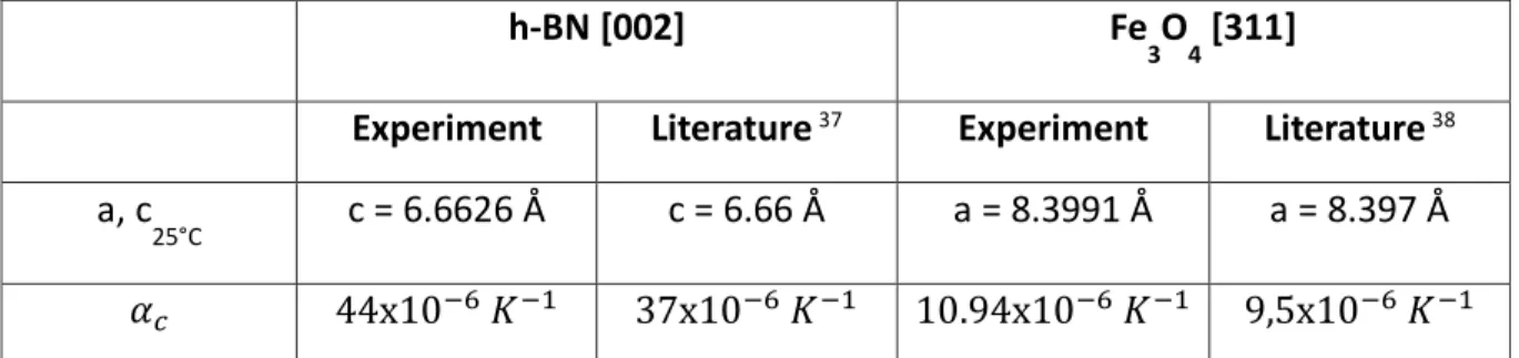
the covariance matrix of the least squares gaussian fit function. The standard deviation on the temperature, related to the measurement error using the blower, was arbitrarily fixed to an overestimated value of 1°C. The lattice parameter is calculated by an orthogonal distance re-gression fit of equation (2) that includes the standard deviation (error bars). The R-square parameter corresponding to the variance of the data with a linear regression fit is given in the legend. An agreement better than 1% is obtained for the h-BN[002] and Fe3O4 [311] planes.
It corresponds to a thermal expansion coefficient of the c-axis 𝛼𝑐= 44 10−6 𝐾−1 and a-axis
Figure 3. Evolution of Fe3O4 [111], [220], [311], [222] and BN [002] planes in
𝛼𝑎 = 10.94 10−6 𝐾−1 respectively which are in good agreement with the literature (cf. Table
1). We will focus on these two peaks to probe the temperature variation under magnetic field with the best accuracy.
180
h-BN [002] Fe
3O4 [311]
Experiment Literature37 Experiment Literature38
a, c
25°C c = 6.6626 Å c = 6.66 Å a = 8.3991 Å a = 8.397 Å
𝛼𝑐 44x10−6 𝐾−1 37x10−6 𝐾−1 10.94x10⁻⁶ 𝐾−1 9,5x10⁻⁶ 𝐾−1
Table 1. Lattice parameter and thermal expansion coefficient of BN [002] and Fe3O4 [311]
planes calculated from the linear fit of the data shown in Figure 4 and comparison with the literature.
The hot air blower was replaced by the electromagnet for experiments described thereafter. X-rays diffraction pattern was recorded at regular interval of time before turning on the electromagnet (room temperature regime), under magnetic field application (heating regime) and after the field was turned off (cooling temperature regime). Due to the time scales of the measuring technique, only stationary temperature gradients could be measured. The possibility to measure transient temperature gradient profile and how it may change the re-sult of the experiment will be discussed at the end of the article.The angle shift of the different
190
peaks was measured and converted into a temperature value using the calibration law previ-ously obtained. The heating of the BN and Fe3O4 NPs is shown in Figure 5. They are at the
same temperature (no heating) before applying the magnetic field. Then, the field is turned on and the particles heating evolves similarly. Finally, when the field is turned off, the temper-ature inside the NPs decreases at the same speed.
Using the initial slope method, the temperature sweeping rate permits to estimate a lower limit for the SAR of the particles: 0.5 W/g. One reason for this low value is that particles are supported on a solid matrix and thus not able to form chains39. Part of the heat is also
afforded to the sample from the electromagnet. It affects h-BN and iron peaks, similarly, act-ing as an offset in the measurements. The heatact-ing measured by the IR camera at t = 1142 sec
200
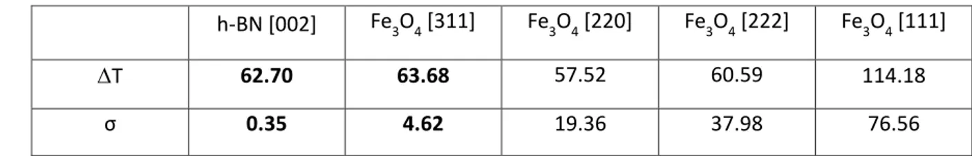
(Figure 5) is T = 95 °C on the surface of the capillary and T = 6 °C on the surface of the electromagnet. The thermal inertia of the electromagnet (with a maximal heating of T = 49 °C) is responsible for the residual heating observed after the magnetic field was turned off”. We measure a maximum heating for the MNPs of T = 63.7 ± 4.6°𝐶 and for the h-BN particles of T = 62.7 ± 0.3°C. The maximum heating T obtained from the other peaks corresponding to different Fe3O4 crystallographic planes is similar even though the
measure-ment uncertainty is more important (cf. Table 2) due to a poorer resolution. These results show that there is no significant temperature gradient between the Fe3O4 MNP core and the
surrounding powder.
h-BN [002] Fe3O4 [311] Fe3O4 [220] Fe3O4 [222] Fe3O4 [111]
T 62.70 63.68 57.52 60.59 114.18
σ 0.35 4.62 19.36 37.98 76.56
Table 2. Maximum heating and standard deviation calculated from the BN and Fe3O4 peak 210
shifts under RF magnetic field.
At first sight, this result could appear in contradiction with experimental results of the literature 15, 17, 18, 19, 21, 23. However, two important differences between previous experimental
results and ours should be noted, and might be at the origin of the difference: i) all other Figure 5. Heating inside the FeC MNPs and BN NPs in presence (H field ON area) and absence (H field OFF area) of a RF magnetic field. The temperature variation is calculated from the lattice parameter variation of Fe3O4 [311] and BN [002] planes respectively.
groups evaluated the temperature using the physical properties of an organic agent external to the MNPs: the thermal degradation of a ligand15, 17, 18, 19or the fluorescence of a lanthanide
complex21, 23. ii) all other groups performed their experiments on colloidal solutions, i.e. with
MNPs in liquid phase.
With respect to i), we wonder if it would be possible that the large temperatures meas-ured by organic entities in the vicinity of the magnetically excited MNPs could be due to
pow-220
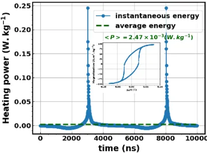
erful heat impulses. Indeed, the total heat power delivered by single-domain NPs during one cycle of the field equals the loop area. Nevertheless, the heat power is not delivered continu-ously along time, but presents strong intensity pulses when magnetization jumps, as illus-trated in a typical example in Figure 6. The most extreme case occurs when the magnetic field is aligned with the NP anisotropy axis. In this case, the hysteresis loop is a rectangle so all the energy is concentrated in two intense power peaks occurring at the coercive field of the NP. A hypothesis to consider is that the energy released this way would be high enough to break thermosensitive covalent bonds of ligands or decrease the fluorescence of molecules, for ex-ample, whereas it would not if the energy was released continuously (green dotted curve in Figure 6). In this case, experiments would conclude to a local heating even though there is
230
none.
Figure 6 – Instantaneous (blue line) and time averaged (green dotted line) power released by one NP during one hysteresis loop (shown in inset).
With respect to ii), it is a well-known fact that, in liquid phase, MNPs excited by an external magnetic field form chains, which are sometimes several tens or hundreds of microns wide, depending on the MNP properties, their functionalization, the magnetic field amplitude, and so forth39, 40, 41, 42. When such chains form, the measured “local” temperature is not the
temperature at the surface of an isolated nanoscale object, but the temperature at the surface and/or inside a micron-scale magnetically heated assembly of MNPs. Since the temperature
240
gradient at the surface or inside an object heated from inside scales as the volume/surface ratio of this object, one can easily conclude that the temperature gradient might be much larger in these chains than in isolated MNPs. For instance, our group has recently shown that,
when millimeter chains are formed, gradients which are probably of the order of several tens of degrees build up42. Although this hypothesis is appealing and casts doubts on the
experi-mental results obtained by other groups, a detailed reading of these works make, in our opin-ion, this hypothesis unlikely to explain all, if any, experimental results. In Clerc and al.20, MNPs
are internalized in lysosomes so only assemblies with a maximum size of one micron could form, which is insufficient to generate a 14°C gradient using standard diffusion equations. In the case of Piñol and al.23, the fact that small (10 nm) superparamagnetic particles are
embed-250
ded in a rather thick polymer matrix makes unlikely the appearance of strong dipolar interac-tions between beads and thus the formation of thick chains. In Riedinger et al., particles are 15 nm in diameter, a size where significant dipolar interactions could build up, but significant temperature gradients are observed even when a small magnetic field of 9mT is applied15.
A last hypothesis related to ii) is that, in our case, heat diffusion through the solid sup-port would be much more efficient than in a liquid environment. However, to explain the dis-crepancy between our results and the ones from the literature, the difference in the diffusion coefficient should have to be of several orders of magnitude, as already discussed in several previous experimental results15, 23. Experiments similar to the ones described here but
per-formed on particles in liquid could permit to eliminate or reinforce the hypotheses formulated
260
above. For now, the problem remains open to new experimental and theoretical investiga-tions.
4. Conclusions
We have measured the inner temperature of MNPs heated by a rf magnetic field. It is a turnabout in the methodology compare to all previous experiments that consist in designing probes that get closer to the surface of the nanoparticle. Our method is a powerful tool to measure the gradient between the internal temperature of the MNPs and the mean temper-ature of the sample. It could be used to measure the tempertemper-ature gradient of particles sup-ported on different media, or into water, since the gradient could strongly vary depending on the nature of the MNP-substrate link. Our results show that, if a gradient appears at the
inter-270
face between the MNPs and the solid-state catalytic bed, it is of rather small amplitude. Our work thus brings a new brick on the active debate on the nanoscale gradient of magnetically heated MNPs.
Conflict of Interest: The authors declare no competing financial interest.
Supporting Information Available: Detailed synthesis of the nanoparticles.
Acknowledgements: The authors thank ERC Advanced Grant (MONACAT 2015-694159) for
financial support. The personnel of the Swiss-Norwegian Beamlines are acknowledged for ex-perimental assistance (experiment MA-4029BM31@SNBL).
References
[1] Ortgies, D.H. ; Teran, F.J.; Rocha, U.; de la Cueva, L.; Salas, G.; Cabrera, D.; Vanetsev, A.S.; Rähn, M.; 280
Sammelselg, V.; Orlovskii, Y.V.; and al. ; Optomagnetic Nanoplatforms for In Situ Controlled Hyperther-mia. Adv. Funct. Mater. 2018, 28, 1704434-1704444. https://doi.org/10.1002/adfm.201704434.
[2] Bordet, A. ; Lacroix, L.-M.; Fazzini, P.-F.; Carrey, J.; Soulantica, K.; Chaudret, B. ; Magnetically In-duced Continuous CO2 Hydrogenation Using Composite Iron Carbide Nanoparticles of Exceptionally High Heating Power, Angew. Chem., Int. Ed. 2016, 55, 15894-15898. https://doi.org/10.1002/anie.201609477.
[3] Niether, C. ; Faure, S.; Bordet, A.; Deseure, J.; Chatenet, M.; Carrey, J.; Chaudret, B. ; Improved Water Electrolysis Using Magnetic Heating of FeC–Ni Core–Shell Nanoparticles, Nat. Energy 2018, 3, 476-483. https://doi.org/10.1038/s41560-018-0132-1.
[4] Lacroix, L.-M.; Lachaize, S.; Falqui, A.; Respaud, M.; Chaudret, B.; Iron Nanoparticle Growth in Or-290
ganic Superstructures, J. Am. Chem. Soc. 2009, 131, 549-557. https://doi.org/10.1021/ja805719c. [5] Rabin, Y.; Is Intracellular Hyperthermia Superior to Extracellular Hyperthermia in the Thermal Sense?”, Int. J. Hyperthermia 2002, 18, 194-202. https://doi.org/10.1080/02656730110116713. [6] Keblinski, P.; Cahill, D.G.; Bodapati, A.; Sullivan, C.R.; Taton, T.A.; Limits of Localized Heating by Electromagnetically Excited Nanoparticles, J. Appl. Phys. 2006, 100, 054305-054309. https://doi.org/10.1063/1.2335783.
[7] Villanueva, A.; de la Presa, P.; Alonso, J.M.; Rueda, T.; Martínez, A.; Crespo, P.; Morales, M.P.; Gon-zalez-Fernandez, M.A.; Valdés, J.; Rivero, G.; Hyperthermia HeLa Cell Treatment with Silica-Coated Manganese Oxide Nanoparticles, J. Phys. Chem. C, 2010, 114, 1976-1981. https://doi.org/10.1021/jp907046f.
300
[8] Hamad-Schifferli, K.; Schwartz, J. J.; Santos, A. T.; Zhang, S.; Jacobson, J. M.; Remote Electronic Con-trol of DNA Hybridization through Inductive Coupling to an Attached Metal Nanocrystal Antenna, Na-ture 2002, 415, 152–155. https://doi.org/10.1038/415152a.
[9] Creixell, M.; Bohorquez, A.C.; Torres-Lugo, M.; Rinaldi, C.; EGFR-Targeted Magnetic Nanoparticle Heaters Kill Cancer Cells without a Perceptible Temperature Rise, ACS Nano 2011, 5, 7124-7129. https://doi.org/10.1021/nn201822b.
[10] Sanchez, C.; El Hajj Diab, D. ; Connord, V.; Clerc, P.; Meunier, E.; Pipy, B.; Payré, B.; Carrey, J.; Gigoux V.; Fourmy, D. ; Targeting a G-protein Coupled Receptor Overexpressed in Endocrine Tumors by Magnetic Nanoparticles to Induce Cell Death. ACS Nano 2014, 8, 1350-1363. https://doi.org/10.1021/nn404954s.
310
[11] Bastus, N. G.; Kogan, M. J.; Amigo, R.; Grillo-Bosch, D.; Araya, E.; Turiel, A.; Labarta, A.; Giralt, E.; Puntes, V. F. ; Gold Nanoparticles for Selective and Remote Heating of β-amyloid Protein Aggregates. Mater. Sci. Eng. 2007, 27, 1236-1240. https://doi.org/10.1016/j.msec.2006.08.003.
[12] Xu, Y.; Mahmood, M.; Li, Z.; Dervishi, E. Trigwell, S.; Zharov, V. P.; Ali, N.; Saini, V.; Biris, A. R.; Lupu, D.; and al. ; Cobalt Nanoparticles Coated with Graphitic Shells as Localized Radio Frequency Absorbers for Cancer Therapy. Nanotechnology 2008, 19, 435102-435111. https://doi.org/10.1088/0957-4484/19/43/435102.
[13] del Rosal, B.; Carrasco, E.; Ren, F.; Benayas, A.; Vetrone, F. Sanz-Rodríguez, F.; Ma, D.; Juarranz, A.; Jaque, D. ; Infrared-Emitting QDs for Thermal Therapy with Real-Time Subcutaneous Temperature Feedback, Adv. Funct. Mater. 2016, 26, 6060-6068. https://doi.org/10.1002/adfm.201601953. 320
[14] Huang, H.; Delikanli, S.; Zeng, H.; Ferkey, D. M.; Pralle, A.; Remote Control of Ion Channels and Neurons Through Magnetic-Field Heating of Nanoparticles. Nat. Nanotechnol. 2010, 5, 602–606. https://doi.org/10.1038/nnano.2010.125.
[15] Riedinger, A.; Guardia, P.; Curcio, A.; Garcia, M. A.; Cingolani, R.; Manna, L.; Pellegrino, T.; Subna-nometer Local Temperature Probing and Remotely Controlled Drug Release Based on Azo-Functional-ized Iron Oxide Nanoparticles. Nano Lett. 2013, 13, 2399−2406. https://doi.org/10.1021/nl400188q. [16] Lee, J.; Govorov, A.O.; Kotov, N.A.; Nanoparticle Assemblies with Molecular Springs: A Nanoscale Thermometer. Angew. Chem. 2005, 44, 7439-7442. https://doi.org/10.1002/anie.200501264.
[17] Dias, J.T.; Moros, M.; del Pino, P.; Rivera, S.; Grazú, V.; de la Fuente, J.M.; DNA as a Molecular Local Thermal Probe for the Analysis of Magnetic Hyperthermia. Angew. Chem. Int. 2013, 52, 11526-11529. 330
https://doi.org/10.1002/anie.201305835.
[18] N'Guyen, T.T.T. ; Duong, H.T.T. ; Basuki, J.; Montembault, V.; Pascual, S.; Guibert, C.; Fresnais, J.; Boyer, C.; Whittaker, M.R.; Davis, T.P.; and al. ; Functional Iron Oxide Magnetic Nanoparticles with Hyperthermia-Induced Drug Release Ability by Using a Combination of Orthogonal Click Reactions. An-gew. Chem. Int. 2013, 52, 14152-14156. https://doi.org/10.1002/anie.201306724.
[19] Griffete, N.; Fresnais, J.; Espinosa, A.; Wilhelm, C.; Bee, A.; Menager, C. ; Design of Magnetic Mo-lecularly Imprinted Polymer Nanoparticles for Controlled Release of Doxorubicin under an Alternating Magnetic Field in Athermal Conditions. Nanoscale, 2015, 7, 18891-18896. https://doi.org/10.1039/C5NR06133D.
[20] Clerc, P.; Jeanjean, P.; Halalli, N.; Gougeon, M.; Pipy, B.; Carrey, J.; Fourmy, D.; Gigoux, V. ; Tar-340
geted Magnetic Intra-lysosomal Hyperthermia Produces Lysosomal Reactive Oxygen Species and Causes Caspase-1 Dependent Cell Death”, J. Control Release 2018, 270, 120-134. https://doi.org/10.1016/j.jconrel.2017.11.050.
[21] Dong J.; Zink, J. I.; Taking the Temperature of the Interiors of Magnetically Heated Nanoparticles. ACS Nano 2014, 8, 5199-5207. https://dx.doi.org/10.1021%2Fnn501250e.
[22] Brites, C.D.S.; Lima, P.P.; Silva, N.J.O.; Millán, A.; Amaral, V.S.; Palacio, F.; Carlos, L.D.; A Lumines-cent Molecular Thermometer for Long-Term Absolute Temperature Measurements at the Nanoscale. Adv. Mater. 2010, 22, 4499-4504. https://doi.org/10.1002/adma.201001780.
[23] Piñol, R.; Brites, C. D. S.; Bustamante, R.; Martínez, A.; Silva, N. J. O.; Murillo, J. L.; Cases, R.; Carrey, J.; Estepa, C.; Sosa, C.; and al.; Joining Time-Resolved Thermometry and Magnetic-Induced Heating in 350
a Single Nanoparticle Unveils Intriguing Thermal Properties”, ACS Nano 2015, 9, 3134-3142. https://doi.org/10.1021/acsnano.5b00059.
[24] Polo-Corrales, L.; Rinaldi, C.; Monitoring Iron Oxide Nanoparticle Surface Temperature in an Alter-nating Magnetic Field Using Thermo Responsive Fluorescent Polymers. J. Appl. Phys. 2012, 111, 07B334. https://doi.org/10.1063/1.3680532.
[25] Gupta, A.; Kane, R.S.; Borca-Tasciuc, D.A.; Local Temperature Measurement in the Vicinity of Elec-tromagnetically Heated Magnetite and Gold Nanoparticles. J. Appl. Phys. 2010, 108, 064901-064908. https://doi.org/10.1063/1.3485601.
[26] Cazares-Cortes, E.; Cabana, S.; Boitard, C.; Nehlig, E.; Griffete, N.; Fresnais, J.; Wilhelm, C.; Abou-Hassan, A.; Ménager, C. ; Recent Insights in Magnetic Hyperthermia: From the “Hot-Spot” Effect for 360
Local Delivery to Combined Magneto-Photo-Thermia Using Magnetoplasmonic Hybrids”, Adv. Drug Deliv.Rev. 2019, 138, 233-246. https://doi.org/10.1016/j.addr.2018.10.016.
[27] Chen, G.; Nonlocal and Nonequilibrium Heat Conduction in the Vicinity of Nanoparticles. J. Heat Transfer 1996, 118, 539-545. https://doi.org/10.1115/1.2822665.
[28] Kozissnik, B.; Bohorquez, A. C.; Dobson, J.; Rinaldi, C.; Magnetic Fluid Hyperthermia: Advances, Challenges, and Opportunity. Int. J. Hyperthermia 2013, 29, 706-714. https://doi.org/10.3109/02656736.2013.837200.
[29] Chiu-Lam, A.; Rinaldi. C.; Nanoscale Thermal Phenomena in the Vicinity of Magnetic Nanoparticles in Alternating Magnetic Fields. Adv. Funct. Mater. 2016, 22, 3933-3941. https://dx.doi.org/10.1002%2Fadfm.201505256.
370
[30] Cahill, D. G.; Braun, P. V.; Chen, G.; Clarke, D. R.; Fan, S.; Goodson, K. E.; Keblinski, P.; King, W. P.; Mahan, G. D.; Majumdar, A.; Maris, H. J.; Phillpot, S. R.; Pop, E.; Shi, L; and al.; Nanoscale Thermal Transport. II. 2003–2012. Appl. Phys. Rev. 2014, 1, 011305-011350. https://doi.org/10.1063/1.4832615.
[31] Chiavazzo, E.; Fasano, M.; Asinari P.; Decuzzi, P. ; Scaling Behaviour for the Water Transport in Nanoconfined Geometries. Nat. Commun., 2014, 5, 3565-3575. https://doi.org/10.1038/ncomms4565. [32] Shenogina, N.; Godawat, R. Keblinski P.; Garde, S. ; How Wetting and Adhesion Affect Thermal Conductance of a Range of Hydrophobic to Hydrophilic Aqueous Interfaces. Phys. Rev. Lett., 2009, 102, 156101-156104.
https://ui.adsabs.harvard.edu/link_gateway/2009PhRvL.102o6101S/doi:10.1103/PhysRevLett.102.15 380
6101.
[33] Meffre, A. Mehdaoui, B.; Connord, V.; Carrey, J.; Francesco Fazzini, P.; Lachaize, S.; Respaud, M.; Chaudret, B.; Complex Nano-objects Displaying Both Magnetic and Catalytic Properties: A Proof of Concept for Magnetically Induced Heterogeneous Catalysis. Nano Lett. 2015, 15, 3241-3248. https://doi.org/10.1021/acs.nanolett.5b00446.
[34] Tsakoumis, N. E.; Voronov, A.; Ronning, M.; van Beek, W.; Borg, O.; Rytter E.; Holmen, A. ; Fischer– Tropsch Synthesis: An XAS/XRPD Combined In Situ Study from Catalyst Activation to Deactivation. J. Catal., 2012, 291, 138-148. https://doi.org/10.1016/j.jcat.2012.04.018.
[35] Lutterotti, L.; Campostrini, R.; Gialanella, S.; Di Maggio, R.; Microstructural Characterisation of Amorphous and Nanocrystalline Structures Through Diffraction Methods. JMNM 2000, 8, 657– 390
664. https://doi.org/10.4028/www.scientific.net/JMNM.8.657.
[36] El Ghandoor, H.; Zidan, H.; Khalil, M.; Ismail, M.I.M.; Synthesis and Some Physical Properties of Magnetite (Fe3O4) Nanoparticles. Int. J. Electrochem. Sci., 2012, 7, 5734-5745. http://www.electrochemsci.org/papers/vol7/7065734.pdf
[37] Hamdi I.; Meskini, N.; Ab Initio Study of the Structural, Elastic, Vibrational and Thermodynamic Properties of the Hexagonal Boron Nitride: Performance of LDA and GGA. Physica B, 2010, 405, 2785 - 2794. https://doi.org/10.1016/j.physb.2010.03.070.
[38] Touloukian Y.S.; Kirby, R.K.; Thermophysical Properties of Matter 1977, 13, 247-250.
[39] Serantes, D.; Baldomir, D.; Martinez-Boubeta, C.; Simeonidis, K.; Angelakeris, M.; Natividad, E.; Castro, M.; Mediano, A.; Chen, D.-X.; Sanchez, A.; and al.; Influence of Dipolar Interactions on Hyper-400
thermia Properties of Ferromagnetic Particles. J. Appl. Phys. 2010, 108, 073918-073923. https://doi.org/10.1063/1.3488881.
[40] Mehdaoui, B.; Meffre, A.; Carrey, J.; Lachaize, S.; Lacroix, L.M.; Gougeon, M.; Chaudret, B.; Res-paud, M.; Increase of Magnetic Hyperthermia Efficiency Due to Dipolar Interactions in Low-Anisotropy Magnetic Nanoparticles: Theoretical and Experimental Results. Adv. Funct. Mater., 2011, 21, 4573-4582.
https://arxiv.org/ct?url=https%3A%2F%2Fdx.doi.org%2F10.1103%2FPhysRevB.87.174419&v=d94ac4 5a.
[41] Saville, S.L.; Qi, B. ; Baker, J.; Stone, R.; Camley, R.E.; Livesey, K.L.; Ye, L.; Crawford, T.M.; Thomp-son; Mefford, O.; The Effect of Magnetically Induced Linear Aggregates on Proton Transverse Relaxa-410
tion Rates of Aqueous Suspensions of Polymer Coated Magnetic Nanoparticles. J. Colloid Interface Sci., 2014, 424, 2152-2163. https://doi.org/10.1039/C3NR32979H.
[42] Asensio, J.M. ; Marbaix, J.; Mille, N.; Lacroix, L.-M.; Soulantica, K.; Fazzini, P.-F.; Carrey, J.; Chaudret, B.; To Heat or not to Heat: a Study of the Performances of Iron Carbide Nanoparticles in Magnetic Heating. Nanoscale 2019, 11, 5402-5411. https://doi.org/10.1039/C8NR10235J.
420