HAL Id: hal-02954842
https://hal.inrae.fr/hal-02954842
Submitted on 6 Nov 2020
HAL is a multi-disciplinary open access
archive for the deposit and dissemination of
sci-entific research documents, whether they are
pub-lished or not. The documents may come from
teaching and research institutions in France or
abroad, or from public or private research centers.
L’archive ouverte pluridisciplinaire HAL, est
destinée au dépôt et à la diffusion de documents
scientifiques de niveau recherche, publiés ou non,
émanant des établissements d’enseignement et de
recherche français ou étrangers, des laboratoires
publics ou privés.
Distributed under a Creative Commons Attribution| 4.0 International License
bioinsecticide on non-target Drosophila flies
Aurélie Babin, Marie-Paule Nawrot-Esposito, Armel Gallet, Jean-Luc Gatti,
Marylène Poirié
To cite this version:
Aurélie Babin, Marie-Paule Nawrot-Esposito, Armel Gallet, Jean-Luc Gatti, Marylène Poirié.
Dif-ferential side-effects of Bacillus thuringiensis bioinsecticide on non-target Drosophila flies. Scientific
Reports, Nature Publishing Group, 2020, 10 (1), �10.1038/s41598-020-73145-6�. �hal-02954842�
Differential side‑effects of Bacillus
thuringiensis bioinsecticide
on non‑target Drosophila flies
Aurélie Babin
1, Marie‑Paule Nawrot‑Esposito
1, Armel Gallet
1, Jean‑Luc Gatti
1,2*&
Marylène Poirié
1,2Bioinsecticides based on Bacillus thuringiensis (Bt) spores and toxins are increasingly popular alternative solutions to control insect pests, with potential impact of their accumulation in the environment on non‑target organisms. Here, we tested the effects of chronic exposure to commercial
Bt formulations (Bt var. kurstaki and israelensis) on eight non‑target Drosophila species present in Bt‑treated areas, including D. melanogaster (four strains). Doses up to those recommended for field
application (~ 106 Colony Forming Unit (CFU)/g fly medium) did not impact fly development, while no fly emerged at ≥ 1000‑fold this dose. Doses between 10‑ to 100‑fold the recommended one increased developmental time and decreased adult emergence rates in a dose‑dependent manner, with species‑ and strain‑specific effect amplitudes. Focusing on D. melanogaster, development alterations were due to instar‑dependent larval mortality, and the longevity and offspring number of adult flies exposed to bioinsecticide throughout their development were moderately influenced. Our data also suggest a synergy between the formulation compounds (spores, cleaved toxins, additives) might induce the bioinsecticide effects on larval development. Although recommended doses had no impact on non‑ target Drosophila species, misuse or local environmental accumulation of Bt bioinsecticides could have side‑effects on fly populations with potential implications for their associated communities.
The world’s population is expected to reach more than 9.7 billion people by 20501, increasing the demand for
food. This requires fighting pests, especially insect pests that cause more than 30% of agricultural losses2.
Nowa-days, their management relies heavily on chemical insecticides. However, their use starts to be reduced due to the emergence of resistance, the appearance of secondary pests, the adverse side-effects on non-target species (pests’ natural enemies, pollinators)3,4, and importantly the impacts on human health and biodiversity5,6. Developed as
more specific and safer alternatives, bioinsecticides represent 5% of the pesticide market, the large majority being microbial insecticide formulations based on viable spores and toxins of the bacterium Bacillus thuringiensis (Bt) (over 400 registered formulations)4,7.
Bt is a Gram-positive endospore-forming bacterium that synthesizes a wide range of toxins with different
chemical structures, modes of action and biological targets. The most abundant and studied are Cry δ-endotoxins encoded by genes located on large plasmids and produced as parasporal crystalline inclusions during the station-ary growth phase8,9. Bt produces other insecticidal toxins, the Cyt (cytolytic δ-endotoxins) and Vip (secreted
Vegetative Insecticidal Proteins) that synergize their effects with Cry toxins, virulence factors such as β-exotoxins (or thuringiensin), a secreted nucleotide toxic for almost all tested life forms thus prohibited in commercial formulations10, and anti-fungal factors11. Bt subspecies and strains can differ in their plasmid number and in the
synthesized toxins cocktail responsible for their biological activity, which determine potential target insects12.
For instance, Bt var. kurstaki (Btk), used mainly against lepidopteran larvae, produces 5 Cry toxins (Cry1Aa, Cry1Ab, Cry1Ac, Cry2Aa and Cry2Ab)13, while Bt var. israelensis (Bti), used mainly against mosquitoes and
black flies, produces a combination of Cry4Aa, Cry4Ba, Cry10Aa, and Cry11Aa14. The different toxin cocktails
produced by some Bt subspecies can also be detrimental to non-insect organisms such as nematodes, protozoa, and even molluscs12.
The bioinsecticide formulations based on spores and toxin crystals of Btk and Bti are the most sprayed in organic and conventional farming, and in natural areas (e.g. forests, swamps). It is generally accepted that once
open
1Institut Sophia Agrobiotech, Université Côte D’Azur, INRAE, CNRS, ISA, 400 route des chappes, 06903 Sophia
Antipolis, France. 2These authors contributed equally: Jean-Luc Gatti and Marylène Poirié. *email: jean-luc.gatti@
ingested by insect larvae, the toxin crystals are dissolved by the midgut alkaline pH, releasing ~ 130 kDa pro-toxins that are then processed by digestive proteases into smaller, soluble, active toxin fragments of ~ 60–70 kDa15,16. Active toxins bind to specific receptors of midgut epithelial cells, eliciting pores formation in the cell
membrane, cell lysis and gut epithelium disorganization17. This allows gut bacteria, including Bt, to colonize the
hemocoel, and leads to rapid septicaemia and death18.
Numerous impact studies of field application rates and acute intoxications have shown that Bt bioinsec-ticides are safe or have a limited impact on non-target vertebrates and invertebrates, and associated species communities19,20. However, the increasing use of bioinsecticides based on Bt spores and toxins has recently
raised concern21 and led to the assessment of their potential effects on non-target species, such as auxiliary
insects of biological control22, pollinators23 and species communities which share their habitat with Bt-targeted
insect pests24–26. There is growing evidence of direct and indirect cross-effects of Bt bioinsecticide formulations
and Bt- Cry and Cyt toxins across insect species and orders, or even across phyla, suggesting that Bt targeting is only partly specific12,26,27. Data also showed that almost all of the applied Btk formulation dose was still present
on the leaves surface 72 h after spraying28, its amount returning close to environmental levels only 28 days after
treatment29. Finally, Bt spores can survive in the soil and on different supports for months and even years after
application30–33. Bt formulations contain also different compounds to protect spores and crystals and aggregate
them into a wettable form, surfactants to facilitate spraying and dispersion on plants, and phagostimulants34,35.
Nevertheless, since toxin crystals, and to a much lesser extent spores36, are somewhat sensitive to abiotic
condi-tions (e.g. UV, pH, rainfall), repeated spraying with a minimum delay of 3 to 8 days is often recommended over the period of pest occurrence to achieve the required pest control level35,37 (https ://www.certi seuro pe.fr; https
://www.certi susa.com). All these can potentially lead to Bt accumulation in the environment, thus raising the rarely addressed issue of potential side-effects of chronic exposure (i.e. continuous and increasing exposure dose for an extended period) of non-target species to doses unexpectedly above those that are recommended.
Diptera are worldwide distributed insects, most of which are not targets of commercial Bt formulations. This is the case of the genus Drosophila, represented by ~ 1500 described species38, including the model organism
Dros-ophila melanogaster. In the field, most of these flies feed and reproduce mainly on ripening or rotting/fermenting
fruits and are therefore naturally present in areas treated with Bt such as orchards, vineyards and gardening areas. Unable to disperse between food patches, early developmental stages of Drosophila eat intensively and grow exponentially39 and may thus ingest high doses of Bt bioinsecticides that have accumulated during the
treat-ment periods. Surprisingly, despite the presence of many Drosophila species in Bt-treated areas, their role in the decomposition of organic matter, and the ease of study of some species, only a few studies have focused on these flies. However, most of them suggested susceptibility to Btk, but they used mainly late 3rd instar larvae preparing for pupation, which do not feed much. In addition, these studies used Bt preparations, especially field isolates, that possibly contained highly toxic β-exotoxins, which are not authorized in commercial Bt formulations40–47.
So far, no study addressed the effects of chronic exposure to commercial Bt formulations on developing stages of these Dipterans that are present in Bt-treated areas.
Here, we tested the chronic side-effects of commercial formulations of Btk and, to a lesser extent of Bti, on non-target Drosophila flies (D. melanogaster and seven other Drosophila species), with doses starting from mean recommended spray doses up to ~ 1000 times this dose (i.e. below acute intoxication doses used in most stud-ies). We mainly focused on developmental traits (developmental time, emergence rate), but also on two fitness-related traits (longevity and offspring number) of adult flies that developed from the egg under Btk formulation exposure. Our study would be a first-step in the exploration of potential implications of chronic exposure to Btk formulation on Drosophila flies.
Results
Btk formulations adversely impact the development of D. melanogaster.
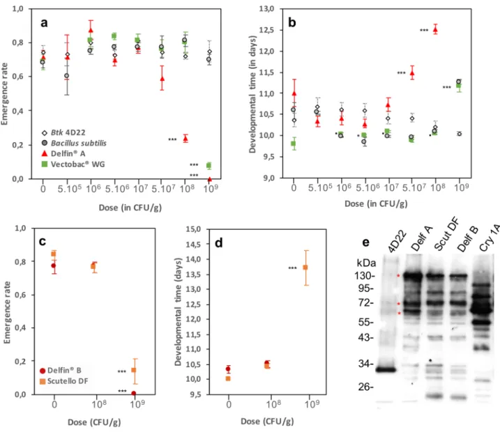
In a dose–response assay, emergence rates (ER) and developmental times (DT) of wild-type D. melanogaster Canton S flies exposed to doses up to 107 CFU/g of DELFIN A in a standard low-protein/high-sugar fly medium were similar to those of the control unexposed group (Fig. 1a,b; Table 1). At higher doses, both ER and DT were affected in a dose-dependent manner: ER was reduced by 17% at 5 × 10⁷ CFU/g (although not statistically significant), up to 100% at 109 CFU/g, at which no individual reached the pupal stage. The lethal dose 50 (LD50) was estimated between 5 × 10⁷ and 108 CFU/g (Fig. 1a). DT was increased of about 0.5 day at 5 × 10⁷ CFU/g (+ 4% versus controls) to up to 1.5 days (+ 14%) at 108 CFU/g (Fig. 1b; Table 1). The sex-ratio at emergence (SR, proportion of males) was strongly biased towards males at 108 CFU/g, with 58% more males compared to the control (Supplementary information S2).We observed no change in ER using the same dose range of the Btk Cry-free strain 4D22 (Fig. 1a,e; Table 1) and the non-pathogenic Bacillus subtilis (Fig. 1a, Table 1), two controls for the effect of ingestion of high loads of spores. In contrast, addition of the formulation of Bt var. israelensis VectoBac WG reduced ER by 89% only at 109 CFU/g (~ 2000 times the recommended dose; Fig. 1a; Table 1; Supplementary information S1). DT varied with the dose of Btk 4D22, the differences being mainly between doses but not with the control. DT increased by ~ 1.5 days at the highest dose of VectoBac WG (Fig. 1b; Table 1) and showed a similar trend with B. subtilis (P = 0.06; Fig. 1b; Table 1). None of these treatments influenced dramatically the SR (Supplementary informa-tion S2).
To test whether these effects are generic to Btk formulations, the fly development was evaluated on two other formulations, DELFIN B (same brand) and Scutello DF (brand Dipel), at the critical doses 108 and 109 CFU/g. As DELFIN A, these formulations contain spores and Cry toxins such as Cry-1A as pro-toxins of ~ 130 kDa, activated toxins of ~ 60–70 kDa, but also as smaller fragments20 (Fig. 1e, red asterisks). ER remained unchanged at
the adult stage on Scutello DF, DT being increased by more than 2 days (Fig. 1c,d; Table 1). No significant bias in SR was observed for either formulation (Supplementary information S2).
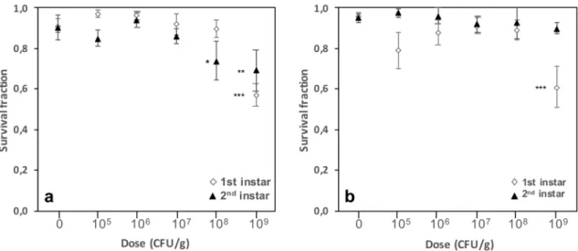
Btk formulation strongly impacts survival during the larval stages.
Cumulative exposure toDELFIN A from the egg to late stages of the 1st and 2nd instars did not influence larval survival at 107 CFU/g but reduced it for both instar larvae above this dose to reach up to 37% mortality at 109 CFU/g (Fig. 2a). Reduced survival tended to occur at a lower dose when cumulative survival was measured later in the development, i.e. 109 CFU/g for late 1st instar larvae and 108 CFU/g for late 2nd instar larvae (Fig. 2a; Table 1). For both instars, larvae surviving at 109 CFU/g were noticeably smaller and less active than those surviving at lower doses, and none of these individuals reached the pupal stage (see results above). A 24-h exposure of the 1st or 2nd instar larvae resulted in a 36% decrease in survival of 1st instar larvae at 109 CFU/g, whereas survival of 2nd instar larvae was unchanged (Fig. 2b, Table 1).
Figure 1. Development of D. melanogaster Canton S flies on Btk and Bti commercial formulations. (a)
Emergence rate and (b) developmental time (mean ± s.e.m.) of 20 initial eggs on increasing doses of Btk
DELFIN A (red triangles), the Cry-free Btk 4D22 (open lozenges), the mosquito-targeting Bti VectoBac WG
(green squares) and the non-pathogenic Bacillus subtilis (light grey circles). For VectoBac WG and B. subtilis,
N = 4–7 per dose; for DELFIN A and Btk 4D22, N = 9–12 for the control, N = 3 for 5.105 and 109, N = 4–9 for 106,
N = 7–14 from 5.106 to 108. (c) Emergence rate (mean ± s.e.m.) and (d) developmental time (mean ± s.e.m.) on increasing doses of the two Btk formulations DELFIN B (dark red circles) and Scutello DF (orange squares).
N = 4 replicates of 20 eggs per dose and formulation, except for controls and 108 CFU/g of DELFIN B (9–10 replicates of 20 eggs). Results of post hoc comparisons of each dose to the control: •0.05 < P < 0.1; *0.01 < P < 0.05; **0.001 < P < 0.01; ***P < 0.001. (e) Immunoblotting with an anti-Cry1A polyclonal antibody on proteins from a suspension of laboratory-produced spores of Cry-free Btk 4D22, the three Btk formulations DELFIN A, B,
Scutello DF and a suspension of laboratory-produced Cry1A toxins. Red asterisks indicate the Cry protoxins
Source of variation/data χ2/deviance d.f P value
Development on Btk DELFIN A, Btk 4D22, Bti VectoBac WG, Bacillus subtilis
Emergence rate
Dose × treatment 285.7 20 < 0.0001
Dose for each treatment:
- DELFIN A 237.5 6 < 0.0001 - 4D22 7.0 7 0.40 - VectoBac WG 165.8 5 < 0.0001 - B. subtilis 1.9 6 0.93 Developmental time Dose × treatment 220.8 19 < 0.0001
Dose for each treatment:
- DELFIN A 68.8 6 < 0.0001
- 4D22 16.08 7 0.024
- VectoBac WG 37.5 6 < 0.0001
- B. subtilis 13.5 7 0.060
Development on Btk DELFIN B and Scutello DF (dose effect)
Emergence rate - DELFIN B 151.2 2 < 0.0001 - Scutello DF 105.1 2 < 0.0001 Developmental time - DELFIN B 2.5 1 0.12 - Scutello DF 30.9 2 < 0.0001 Role of formulation components in the development alterations (dialysis)
Dose effect:
Emergence rate 459.8 3 < 0.0001
Developmental time 13.7 2 0.0011
Survival of larval stages on DELFIN A
Cumulative survival
Dose × larval instar 16.2 5 0.0063
Dose for each instar:
- Late 1st instar 87.4 5 < 0.0001
- Late 2nd instar 25.7 5 0.0001
24-h survival
Dose × larval instar 15.9 5 0.007
Dose for each instar:
- Late 1st instar 55.9 5 < 0.0001
- Late 2nd instar 3.76 5 0.58
Adult fitness-related traits after development on DELFIN A
Longevity
Experiment 20.1 1 < 0.0001
- 1st experiment
Dose 12.3 3 0.0065
Sex (eβ coefficient males vs. females ± se: 0.55 ± 0.16) 35.0 1 < 0.0001
Dose × sex 20.4 3 0.00014
Sexes analyzed separately
Females (eβ coefficients vs. control ± se: 5 × 10⁶: 1.05 ± 0.17, 5 × 10⁷: 0.71 ± 0.16, 108: 0.60 ± 0.21) 12.0 3 0.0073
Males (eβ coefficients vs. control ± se: 5 × 10⁶: 0.80 ± 0.16, 5 × 10⁷: 0.66 ± 0.16, 108: 1.53 ± 0.18) 20.4 3 0.00014
Adult fitness-related traits after development on DELFIN A
- 2nd experiment
Dose 16.5 3 0.00090
Sex (eβ coefficient males vs. females ± se: 0.45 ± 0.22)(eβ coefficient males vs. females ± se: 0.45 ± 0.22) 31.5 1 < 0.0001
Dose × sex 0.69 3 0.88
Sexes analyzed separately
Females (eβ coefficients doses vs. control ± se: 5 × 10⁶: 0.92 ± 0.22, 5 × 10⁷: 0.63 ± 0.21, 108:
0.51 ± 0.21) 13.2 3 0.0043
Males (eβ coefficients doses vs. control ± se: 5 × 10⁶: 1.02 ± 0.22, 5 × 10⁷: 0.70 ± 0.22, 108: 0.64 ± 0.22) 7.01 3 0.072 Total numbers of offspring
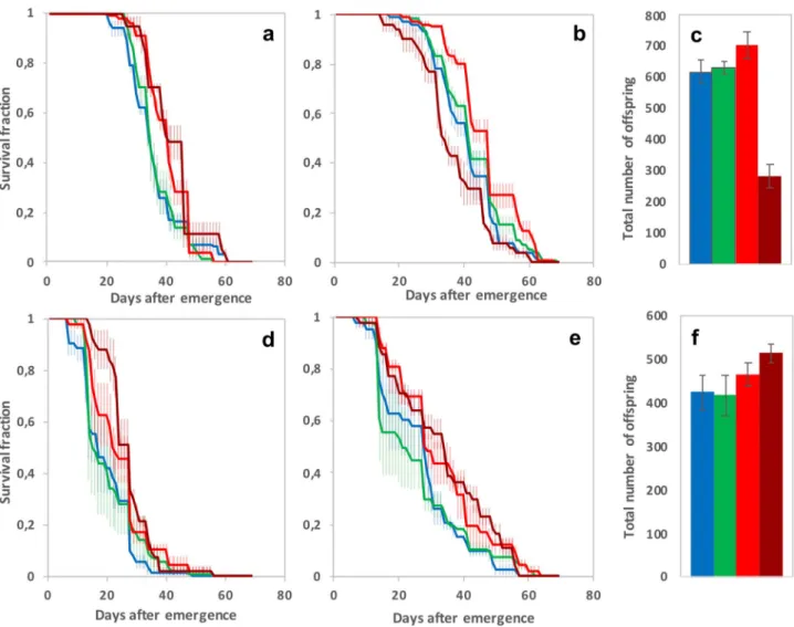
Developmental exposure to Btk formulation does not strongly influence fitness‑related traits
in adults.
Despite a large variation between the two independent sets of experimental replicates (Table 1), the longevity of adults reared on 5 × 10⁶ CFU/g of DELFIN A in low-protein/high-sugar medium was similar to that of non-exposed controls (Fig. 3). Males and females which developed on the two highest doses showed a moderate longevity benefit, higher in females for 108 CFU/g (Fig. 3a,b,d,e; Table 1). Males generally survived better than females (Table 1) but their longevity benefit of developing on 108 CFU/g was only observed in one experiment (Fig. 3b,e).The number of offspring produced by the 15 females of each fly group during the longevity experiment varied depending on both the experiment and the DELFIN A dose (Table 1). In the 1st experiment, adults from larvae reared on 108 CFU/g had fewer offspring compared to the controls and to adults developed on the other doses whereas the total offspring number varied regardless of the DELFIN A dose in the 2nd experiment (Fig. 3c,f, Table 1).
Table 1. Results of statistical analyses to assess the effect of the dose of formulation/spore production and its
interaction with the treatment, the larval instar, the experiment, the sex, the fly strain and the fly species when appropriate. See figures for post hoc comparisons of the doses with the control dose. Significant statistical differences are indicated in bold
Source of variation/data χ2/deviance d.f P value
Dose × experiment 28.1 3 < 0.0001
Dose for each experiment:
- 1st experiment 26.3 3 < 0.0001
- 2nd experiment 4.1 3 0.25
Development of other strains of D. melanogaster on DELFIN A (including Canton S)
Emergence rate
Dose × fly strain 105.5 15 < 0.0001
Dose for each fly strain:
- Canton S 588.6 5 < 0.0001
- Nasrallah 745.3 5 < 0.0001
- Sefra 900.7 5 < 0.0001
- YW1118 636.9 5 < 0.0001
Developmental time
Dose × fly strain 9.3 12 0.68
Dose for each fly strain:
- Canton S 40.3 4 < 0.0001
- Nasrallah 18.0 4 0.0012
- Sefra 27.2 4 < 0.0001
- YW1118 28.9 4 < 0.0001
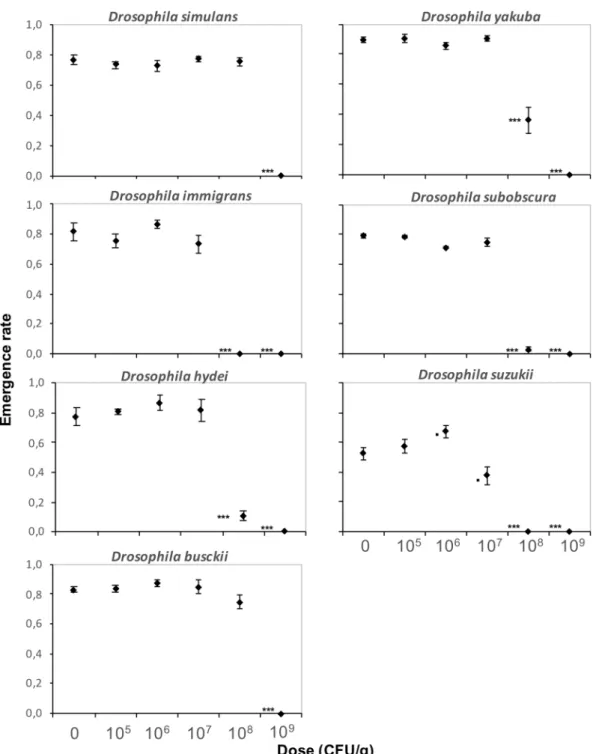
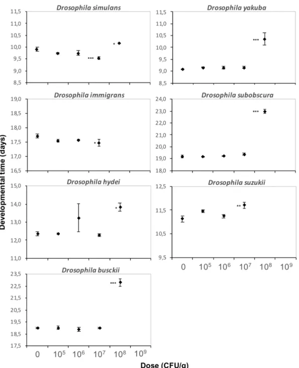
Development of other Drosophila species on DELFIN A
Emergence rate
Dose × fly species 538.2 30 < 0.0001
Dose for each species:
- D. simulans 461.0 5 < 0.0001 - D. yakuba 750.7 5 < 0.0001 - D. hydei 596.8 5 < 0.0001 - D. immigrans 726.3 5 < 0.0001 - D. subobscura 729.6 5 < 0.0001 - D. suzukii 725.0 5 < 0.0001 - D. busckii 586.0 5 < 0.0001 Developmental time
Dose × fly species 59.9 22 < 0.0001
Dose for each species:
- D. simulans 25.9 4 < 0.0001 - D. yakuba 34.7 4 < 0.0001 - D. hydei 11.5 4 0.022 - D. immigrans 6.01 3 0.11 - D. subobscura 68.8 4 < 0.0001 - D. suzukii 11.7 3 0.0085 - D. busckii 58.8 4 < 0.0001
Developmental alterations dependent on Btk formulation dose are not specific to D.
mela-nogaster Canton S.
As with D. melanogaster Canton S, the development of three other D. melanogaster strains (wild-type Nasrallah and Sefra, and double mutant YW1118) was not impacted at doses up to 107 CFU/g of DELFIN A in a high-protein/sugar-free medium. In contrast, the ER of each strain was greatly reduced and DT was increased at higher doses (Fig. 4a,b, Table 1), with no individual reaching the pupal stage at 109 CFU/g (LD50 between 108 and 109 CFU/g). At 108 CFU/g, the magnitude of effects on Canton S flies was lower than that observed on the low-protein/high-sugar medium (see Fig. 1a,b). At this dose, the ER varied between strains, the largest reduction being observed for Sefra (Table 1). We observed no dose-dependent bias in SR (Supplementary information S3).Btk formulation affects differently other Drosophila species.
For seven other Drosophila species from different phylogenetic clades that co-occur in the field48–51, doses up to 106 CFU/g of DELFIN A in a high-protein/sugar-free medium had no effect on ER and DT, whereas all individuals failed to reach the pupal stage at 109 CFU/g (Figs. 5, 6). The amplitude of development alterations at 107 and 108 CFU/g varied among species (Figs. 5, 6; Table 1). All species were affected at 108 CFU/g as was D. melanogaster (see Fig. 4a for comparison).D. simulans and D. busckii had unchanged ER, but DT was slightly increased for D. simulans (although slightly
reduced at 107 CFU/g; similar results with a Japanese strain, data not shown) and strongly increased for D.
busckii (by 20%, i.e. ~ 4 days) (Figs. 5, 6, Table 1). D. yakuba ER and DT were similar to those of D. melanogaster, with an LD50 around 108 CFU/g and a moderate DT increase of ~ 1 day (Figs. 5, 6, Table 1; similar results with a strain from Sweden, data not shown). The ER of D. hydei and D. subobscura were very low at 108 CFU/g (LD50 below this dose), with a high DT (Figs. 5, 6; Table 1), while D. immigrans did not survive. No D. suzukii indi-vidual emerged at 108 CFU/g and development was already moderately impacted at 107 CFU/g (Figs. 5, 6). No dose-dependent bias in SR was detected for either species (Supplementary information S5).
Development alterations may result from a synergy between formulation compo‑
nents.
Because some additives of commercial formulations might contribute to the observed effects, aDELFIN A suspension was dialyzed to remove low molecular weight additives, resuspended, and mixed with
low-protein/high-sugar fly medium. At 107 CFU/g, the suspension did not affect ER and DT, while no individual pupated at 109 CFU/g (Fig. 7a; Table 1). At 108 CFU/g, ER was not modified but DT increased in one experi-mental set by ~ 1 day, partially reproducing the changes observed without dialysis (Fig. 7a,b; see also Fig. 1a,b, Table 1; 3 independent experiments for ER, 2 independent experiments for DT).
The Cry1A profiles of DELFIN A suspensions (dialyzed or not), included a band for the 130-kDa pro-toxins and a band at 60–70 kDa likely representing the activated toxins, but also smaller fragments resulting from the degradation of Cry1A (Fig. 7c). We further explored the respective roles of Btk toxin fragments and spores in the alterations of D. melanogaster development through dialysis experiments followed by successive centrifuga-tions to remove most of the spores and toxin crystals. Despite variation between experiments, ER was strongly affected only in one of the three experiments while DT was always significantly increased in the presence of centrifuged supernatants (Supplementary information S6). Noteworthy, the D. melanogaster development was not impacted in the presence of a homemade production of the Btk strain 4D1 containing spores, toxins, but no additives, even at the highest dose (Supplementary information S7).
Figure 2. Survival of D. melanogaster Canton S larval stages on increasing doses of Btk DELFIN A. (a)
Proportion of surviving larvae (mean ± s.e.m.) upon Btk formulation exposure from the egg to late 1st instar (open lozenges) and late 2nd instar (black triangles). (b) Proportion of surviving larvae (mean ± s.e.m.) upon 24-h Btk formulation exposure of early 1st instar larvae (open lozenges) and 2nd instar larvae (black triangles).
N = 5–7 replicates of 20 individuals per dose. Results of post hoc comparisons of each dose with the control:
Discussion
Our study tested the side-effects of ingestion of Bt bioinsecticide commercial formulations (mainly made of Bt
kurstaki strains (Btk) but also of Bt israelensis (Bti)) during the development of eight non-target species of Dros-ophila naturally present in treated areas. Although the recommended doses for one formulation field spray did
not affect the Drosophila development, those 10 and 50 times higher markedly induced mortality and/or devel-opmental delay in at least two of the species tested. We can extrapolate from our data that these doses may affect six of the eight tested species and the four strains of D. melanogaster. The development alterations were already strong at these doses, suggesting an occurrence from lower ingested doses but not visible in our experimental set-up. In addition, in our experimental conditions, a single Drosophila larva could probably not process 1 g of medium during its development. Further analyses, maybe at molecular level, would be required to determine the minimal dose affecting the fly larva. Furthermore, all the tested species except D. simulans were strongly affected at a 100 times the field spray dose, and no or very limited fly development occurred at the highest tested dose, equivalent to 1000 times the maximum field dose but far below the acute intoxication doses classically used in numerous studies5. The recommended doses for each spraying of stabilized formulation are given for a
homo-geneous and dry area, without overlapping. In the field, recommended repeated sprays and post-spray rainfall washouts may increase the concentration of Bt spores and toxins in both space and time. While a dose 1000 times the recommendations would be hardly reached in the field, the minimum doses at which the fly develop-ment was impacted and the lower doses from which developdevelop-mental changes appeared could be reached. Our data also identified a first developmental window of susceptibility to Btk formulation during the 1st larval instar mainly explaining the adverse effects, while a second event of mortality seemed to occur at the pupation period.
Figure 3. Fitness-related traits of adults (longevity and total offspring number) after development on
Btk DELFIN A. (a,d) Female longevity (mean survival fraction over time ± s.e.m.), (b,e) Male longevity
(mean ± s.e.m.), and (c,f) total offspring number (mean ± s.e.m.), measured on individuals that developed without Btk formulation (blue items) and on 5 × 10⁶ CFU/g of Btk DELFIN A (green items), 5 × 10⁷ CFU/g (red items), and 108 CFU/g (dark red items). Data from 2 experiments (a–c, experiment 1; d–f, experiment 2). For each trait, N = 3–5 replicates of 15 males and 15 females per dose in experiment 1, N = 3 replicates of 15 males and 15 females in experiment 2. Results of post hoc comparisons of each dose with the control: *0.01 < P < 0.05; **0.001 < P < 0.01; ***P < 0.001.
In testing for generic side effects of Bt formulations, we observed similar patterns of developmental alterations on D. melanogaster but only at higher doses with two other Btk formulations and one Bti formulation (1000–2000 times the recommended spray dose). The three Btk formulations, based on two different bacterial strains, have similar profiles of Cry1A protoxins and activated toxins but differ in their efficient spore content. Thus, the type of formulation and probably the additives, may explain the observed variation in the dose effect.
The impacts of Btk formulations on the development of D. melanogaster are consistent with growing evi-dence suggesting partial specific targeting of Bt12,26,27.The consensus on the mode of action of Bt after
inges-tion by insects relied until recently on the key steps of the specific binding of proteolyzed Bt toxins to midgut epithelial cell receptors, defining targets for each Bt subspecies12,15,17. Several primary and secondary types of
toxin receptors have been identified in the Lepidoptera and Diptera mosquitoes such as cadherin-like proteins, aminopeptidases, GPI-anchored alkaline phosphatases8, and more recently the ATP dependent binding
cas-sette reporter C252. No orthologues of the Lepidoptera cadherin-like Cry receptors were found in Drosophila52,
supporting the idea of the lack of effect of Btk toxins on these flies. Yet, Drosophila flies may have other types of Cry receptors, therefore explaining the developmental impacts observed, but this remains to be investigated. In addition, the possible lack of solubilization of the protoxin crystals and of proteolytic activation of toxins by proteases in the fly gut, both required for Cry activity in insects’ larvae15, would be possibly compensated by the
substantial amounts of active Cry1A toxin fragments in Btk formulations. Other Btk-synthesized toxins present in the formulations could also be players in the observed cross-order activity since some, like Cry2A, have an insecticidal effect on both Lepidoptera and Diptera53.
Since ingestion of Bacillus subtilis or Btk Cry-free does not affect the development of D. melanogaster, the observed development alterations cannot result solely from a severe disturbance of digestion and nutrient absorp-tion/competition due to the presence of high loads of spores/bacteria in the larval gut throughout development. This suggests a synergistic action of Btk spores and Cry toxins, consistent with the Bt action models on insect larvae, i.e. the breach of the intestinal epithelium allowing colonization of the hemocoele by the gut bacteria, including Bt spores15,17,18. The partial mimicry of mortality rates and developmental delays in preliminary dialysis
assays would also support a contribution of diffusible low molecular weight compounds in Btk formulations (e.g. residues of culture media, salts, additives) to these development alterations. Furthermore, there is no impact on the development of D. melanogaster of the ingestion of homemade spores and Cry toxins of the Btk strain 4D1 used without additives even at the highest dose (or HD1, a reference strain used also as a control). Unlike commercial Btk formulations, Btk 4D1 culture contains few activated Cry toxins and smaller toxin fragments, advocating the possible contribution of such fragments to the cross-order activity of Btk formulations on
Dros-ophila. Completion of these preliminary tests is required to further investigate the mechanisms of the harmful
effects of Btk formulations on the development of Drosophila and unravel the respective roles of the synergy spores/toxins/crystals and of formulation additives.
As reported for D. suzukii exposed to Btk cultures45, D. melanogaster mortality on the Btk formulation
occurred mainly during early development. Only ∼40% of the 1st and 2nd instar larvae died at the highest dose tested (Fig. 2) while no individual reached the pupal stage, the remaining mortality likely occurring during, or at the end of, the 3rd larval instar, possibly due to the delayed action of the gut accumulated Btk spores and tox-ins at the onset of pupation. Interestingly, developmental alterations (mortality, delayed emergence) mimicked those typically caused by nutritional stress in insect larvae54,55. Accordingly, the developmental alterations were
partially rescued on a protein rich fly medium, probably by a compensatory protein intake, as in other arthropod species55–57. Also, the sex ratio of flies was strongly biased towards males after development on the Btk formulation Figure 4. Development of four D. melanogaster strains on increasing doses of Btk DELFIN A. (a) Emergence
rate and (b) developmental time (mean ± s.e.m.) of the strains Canton S (blue lozenges), Nasrallah (yellow triangles), Sefra (green squares), and YW1118 (red circles). N = 4 groups of 50 eggs per dose and fly strain for each trait. Results of post hoc comparisons of each dose to the control: **0.001 < P < 0.01; ***P < 0.001.
dose affecting fly emergence and under protein restriction. This highlights the importance of nutritional con-ditions such as protein restriction, added to sex-specific differences in larval susceptibility to environmental stressors, here the accumulation of Btk formulation, as already reported previously in D. melanogaster58.
The development on sublethal doses of Btk formulation did not dramatically affect the longevity of D.
mela-nogaster adults, nor their lifetime offspring number. Exposure during development to doses of Btk formulation
that slightly and strongly reduced the likelihood of reaching the adult stage even provided a dose-dependent longevity benefit to the surviving flies and tended to increase their offspring number. Exposure to the Btk formulation throughout development probably selected resistant and/or tolerant individuals, reminding the increased longevity of adult insects having survived a nutritional stress during development59,60, or withstood
environmental stressors61.
Figure 5. Emergence rate of seven Drosophila species on increasing doses of Btk DELFIN A. Mean emergence
rate (± s.e.m.). N = 4 replicates of 50 eggs per dose for D. simulans, D. yakuba, D. subobscura and D. busckii, N = 4 replicates of 30 eggs per dose for D. hydei, D. suzukii, and D. immigrans. Results of post hoc comparisons of each dose with the control: •0.05 < P < 0.1; ***P < 0.001.
The origin of Drosophila (species, population/strain) influenced the amplitude of the development alterations induced by the Btk formulation. For D. melanogaster, all the tested strains were equally susceptible, but with variation in the dose effect amplitudes. These differences in susceptibility suggest a possible spatial and temporal heterogeneity of the potential impacts of Btk spraying among natural D. melanogaster populations. Among the other seven species tested, differences occurred in the susceptibility to the Btk formulation, in terms of nature of development alterations and effect amplitudes, regardless of their phylogenetic distances. For the subgenus
Drosophila, D. simulans was less sensitive than its sister species D. melanogaster, while the African D. yakuba
experienced similar development impacts as D. melanogaster. Although phylogenetically close, D. melanogaster and D. simulans would respond very differently to Btk formulations, with a possible advantage for D. simulans in case of competition. D. immigrans, D. subobscura and D. hydei were similarly more sensitive than D.
mela-nogaster. The phylogenetically distant D. busckii (subgenus Dorsilopha) was the least affected of all the species
in terms of developmental mortality, but its development was strongly delayed. The species D. melanogaster, D.
Figure 6. Developmental time of seven Drosophila species on increasing doses of Btk DELFIN A. Mean
developmental time (± s.e.m.). N = 4 replicates of 50 eggs per dose for D. simulans, D. yakuba, D. subobscura and D. busckii, N = 4 replicates of 30 eggs per dose for D. hydei, D. suzukii and D. immigrans. Results of post hoc comparisons of each dose with the control: *0.01 < P < 0.05; **0.001 < P < 0.01; ***P < 0.001.
simulans, D. hydei, D. immigrans, and D. busckii belong to the guild of cosmopolitan domestic Drosophila species, D. subobscura is a sub-cosmopolitan species, and D. busckii is an opportunistic frugivorous species62,63,64. They all
coexist frequently and compete on the same discrete and ephemeral rotting fruit patches, with seasonal variations in the composition of the fly community47–49,62. Differences in species susceptibility to Btk formulations could
modify the conditions of larval competition, therefore adding local and temporal variations in the composition of Drosophila communities. The potential side-effects of Bt sprays on non-target Drosophila communities would be hardly predictable as they would depend on spatial patterns of Bt accumulation. A formal mesocosm study of
Drosophila community dynamics under exposure to Btk formulation, at least under semi-field conditions, would
help to identify the consequences of Bt accumulation on species competition and community composition. The exposure to Btk formulation also impacted the development of the invasive D. suzukii, as recently reported45, this
species being the most susceptible with effects already clearly detectable at only 10 times the recommended spray dose. Compared to the other tested species living on rotten fruits, D. suzukii threatens fruit production since it feeds and lay eggs inside healthy ripening soft fruits63–65, colonizing orchards and vineyards earlier during the
fruit season. The higher susceptibility of D. suzukii to the accumulation of Btk formulation in the environment might reduce the possible ecological burden of its invasion for local communities of fruit-eating Drosophila in orchards. Alternatively, since D. suzukii attacks on fruits can accelerate their decomposition, its increased sus-ceptibility may reduce the number of fruits available for the rotting fruit-eating Drosophila species.
Overall, our data show that the ingestion of Btk bioinsecticides above the recommended spray doses can potentially impact non-target Drosophila flies, with an effect amplitude depending on both the formulation and the fly species. Although our study was carried out under controlled laboratory conditions, which may consid-erably differ from those of the field (e.g. temperature, pH, humidity, food availability, presence of predators/ parasites/pathogens, etc.…), standard laboratory strains and flies derived from recently collected populations exhibited similar patterns of developmental alterations, suggesting our results are likely generalizable. Recent studies have reported similar adverse side-effects due to repeated spraying of the Bti formulation on non-target organisms25, and indirectly on predators via food webs66. From these studies and our data here, care should
clearly be taken when using Bt bioinsecticides to avoid, or at least minimize, potential side-effects on non-target organisms and therefore on biodiversity. At last, D. melanogaster could serve as a model species to identify the mechanisms underlying these side effects and/or the potential emergence of resistance to these bioinsecticides.
Methods
Commercial formulations, Bacillus productions and Colony Forming Unit.
Btk brands (serotype3a, b, c67) were DELFIN (two formulations named A and B; strain SA-11; wettable granules, Valent BioSciences,
AMM 9200482, 32,000 UI/mg) and Scutello DF (a Dipel sub-brand; strain ABTS-351; wettable granules, Biobest, AMM 2010513, 540 g/kg). Bti brand (strain HD-14; serotype 1467) was VectoBac WG (wettable
gran-ules, Bayer, AMM 2020029, 3000 UTI/mg). For each formulation, the number of viable spores was estimated by counting Colony Forming Units (CFUs) developing on LB agar after overnight incubation at 30 °C from serial dilutions of a suspension (Colony Forming Units (CFU) per mg of product). Estimations were 5 × 10⁷ CFU/mg for DELFIN A; 2.5 × 10⁷ CFU/mg for DELFIN B; 2.2 × 10⁷ CFU/mg for Scutello DF; 6 × 10⁷ CFU/mg for
Vecto-Figure 7. Evaluation of the role of small molecular weight components of Btk DELFIN A (dialysis; membrane
cut-off: 8–10 kDa) in the altered development of D. melanogaster Canton S. (a) Emergence rate and (b) developmental time (mean ± s.e.m.) on increasing doses of dialyzed DELFIN A. N = 3 experiments of 4 replicates with 20 eggs per dose for the emergence rate, N = 2 experiments of 4 replicates per dose for the developmental time. Results of post hoc comparisons of each dose with the control: ***P < 0.001. (c) Anti-Cry1A probed immunoblot of non-dialyzed (ND) and dialyzed (D) suspensions showing the decrease in the amount of ~ 130/140 kDa migrating protoxins and the increase in that of the potential ~ 60/70 kDa activated toxins after dialysis.
Bac WG, and were stable during the experiments time frame. Our CFU estimations agree with those indicated
for the formulations, between 1–5 × 10¹³ CFU/kg. Manufacturer-recommended DELFIN doses for one spraying range from 0.15 to 1.5 kg/ha depending on the crop. Based on our CFU estimations, this corresponds to 7.5 × 10⁴ to 7.5 × 10⁵ CFU/cm2 of DELFIN A, and 3.75 × 10⁴ to 3.75 × 10⁵ CFU/cm2 of DELFIN B. Scutello DF is used at 0.1 to 1 kg/ha, equivalent to 2.2 × 10⁴ to 2.2 × 10⁵ CFU/cm2. VectoBac WG is used at 0.125 to 1 kg/ha, equivalent to 7.5 × 10⁴ to 6 × 10⁵ CFU/cm2.
The acrystalliferous Btk 4D22 strain (depleted for the Cry toxin-encoding plasmids68; Bacillus Genetic Stock
Center, https ://bgsc.org, Columbus, USA), and a Drosophila non-pathogenic Bacillus subtilis (from Dr. E. Bremer, University of Marburg, Germany) were grown at 30 °C in the sporulation-specific medium (Bactopeptone 7.5 g, KH2PO4 3.4 g, K2HPO4 4.35 g, glucose 7.5 g, PGSM salts (MgSO4·7H2O, MnSO4·H2O, ZnSO4·7H2O, FeSO4·7H2O) 5 mL, CaCl2 0.25 M, distilled water qsp 1L, pH 7.2) until sporulation (about 14 days). Vegetative cells were eliminated (1 h at 70 °C) and after centrifugation (4500 rpm, 20 min, 4 °C), a spore pellet was collected, washed with sterile water, and lyophilized. Production CFUs was estimated as described above.
Fly stocks.
Drosophila melanogaster strains (phylogenetic subgroup: melanogaster) were wild-type CantonS (Bloomington Drosophila Centre) used as a reference strain, “Nasrallah” from Tunisia (strain 1333, Gif-sur-Yvette), a French field-collected strain “Sefra” (Southern France, 2013), and the double mutant standard strain YW1118 (white and yellow mutations; gift from Dr. B. Charroux, IBD, Marseille-Luminy). Other Drosophila species were D. simulans (Gif strain 1132; phylogenetic subgroup: melanogaster), D. yakuba (Gif strain 1880; phylogenetic subgroup: melanogaster), D. hydei (phylogenetic subgroup: hydei) and the invasive D. suzukii (phylogenetic subgroup: immigrans) (both kindly provided by Dr. R. Allemand, LBBE, University Lyon 1), D.
immigrans (phylogenetic subgroup: immigrans), D. subobscura (phylogenetic subgroup: obscura), and D. busckii
(Dorsilopha subgenus). The populations of the last three species were initiated from individuals collected in South-East of France in Spring 2015.
All the flies were maintained at controlled densities (150–200 eggs/40 ml of fly medium) under standard laboratory conditions (25 °C, or 20 °C for recently collected species, 60% relative humidity, 12:12 light/dark cycle), on a high-protein/sugar-free fly medium (10% cornmeal, 10% yeast, 0% sugar). In our laboratory, the D.
melanogaster Canton S was also reared on a standard low-protein/high-sugar medium (8% cornmeal, 2% yeast,
2.5% sugar). For each experiment, eggs, larvae and flies were maintained under standard conditions.
General method of intoxication and dose–response assay.
For the dose–response assays, formula-tions and spore producformula-tions were serially diluted in buffer, and 100 µl of each dilution was homogenized thor-oughly with 1 g of fly medium (100 µl/g doses). Eggs and defined larval instar were collected from stock vials and transferred to the intoxication vials and dishes as described below. They were then reared until the fly emergence, and, for larval susceptibility tests, until the desired development stage was reached, or for 24 h for early larvae of the 1st and 2nd instars. Equivalent control groups were transferred on fly medium homogenized with the same volume of buffer alone.Development‑related traits and larval survival.
Developmental traits upon intoxication throughout the entire development of the D. melanogaster strains and the other Drosophila species were evaluated on a precise number of viable eggs collected from mass oviposition and transferred to intoxication vials containing fly medium (high sugar/low protein or high protein/sugar free as indicated) mixed with formulations or spore productions at doses ranging from 1 × 10⁵ or 5 × 10⁵ CFU/g (mean equivalent to the manufacturer recommenda-tions; Supplementary information S1) to 109 CFU/g. Eggs were let to develop until fly emergence. Egg density was set at 8–10 eggs/g of medium (10 eggs/g on 2 g medium in small vials Ø 3.3 cm, surface ~ 8.5 cm2, 0.24 g/cm2 for tests on D. melanogaster Canton S; 8 eggs/g on 6 g medium in large vials Ø 4.6 cm, surface ~ 16 cm2, 0.37 g/ cm2 for strains and species comparisons), except for D. hydei, D. suzukii and D. immigrans for which the egg density was reduced by half because of their reproductive biology (5 eggs/g on 6 g). Numbers and sex of emerg-ing flies were recorded once a day until the day on which the first pupae of the next generation forms. The emer-gence rate (proportion of flies emerged from the initial eggs), the developmental time (mean number of days for development completion) and the sex-ratio (proportion of male flies) were calculated for each replicate vial.For the larval susceptibility tests, survival was measured on 20 eggs/larvae of D. melanogaster Canton S at a suitable instar collected from a 4-h mass oviposition and transferred to small dishes (Ø 3 cm, surface ~ 7 cm2) containing 1 g of high-protein/sugar-free fly medium (less limiting for early larval development) homogenized with DELFIN A doses ranging from 105 CFU/g to 109 CFU/g. First and 2nd instar larvae were used since growth is exponential during these two instars39,69 and larvae are more likely to be heavily exposed to the bioinsecticide.
Proportion of surviving larvae was measured at the indicated developmental stage for the cumulative survival test, or 24 h later. For cumulative survival, unhatched eggs were discarded from the counting. The pH of the fly medium was measured for the Btk dose-responses; neither the presence of Btk formulation nor the dose altered it (Supplementary Information S4).
Adult fitness‑related traits.
For longevity and total offspring number measurements, D. melanogaster Canton S eggs from mass oviposition were transferred to several vials with low-protein/high-sugar medium mixed with 5 × 10⁶, 5 × 10⁷ or 108 CFU/g of DELFIN A. For each dose, flies from the vials were pooled 2 days after emergence and groups of 15 males and 15 females were transferred on the same medium without DELFIN A. Flies were transferred to a new vial every 3–4 days and previous vials were incubated for the offspring to develop. Mortality and sex of dead flies were recorded daily until the last fly died. Offspring numbers were counted from the first emergence until pupae of the next generation appeared. Two experimental blocks were set. Due todif-ferences in the duration of the experiment, the offspring numbers of all vials of each dose of DELFIN A were summed for each experimental block.
Dialysis and cry toxin analysis.
For some products, additives can be more harmful than the active ingredient70. To eliminate low molecular weight additives present in the formulation, a DELFIN A suspension at2 × 1010 CFU was dialyzed against PBS (KH
2PO4 1 mM, Na2HPO4(2H2O) 3 mM, NaCl 154 mM, pH 7.2), over-night at 4 °C, using an 8–10 kDa cut-off membrane (ZelluTrans, Roth). CFUs of the dialyzed suspension were estimated as above. The effects on the emergence rate (ER) and developmental time (DT) of D. melanogaster Canton S were analysed on 20 eggs, at 107, 108 and 109 CFU/g of dialyzed and also centrifuged suspension mixed with 2 g of low-protein/high-sugar fly medium. The dialyzed suspension was subject to a 12.5% SDS-PAGE and compared to the non-dialyzed suspension after silver staining. The presence of Cry1A pro-toxins, activated tox-ins and toxin fragments was probed by Western-blot using an in-house anti-Cry1A rabbit polyclonal antibody.
Data analysis.
Data were analysed with mixed-effects models with replicates as random effects. Dose ofBtk formulation/spore production, D. melanogaster strain, Drosophila species or developmental stage,
experi-mental block where necessary, and appropriate 2-way interactions between these factors, were included as fixed effects. Main fixed effects and their interactions were tested with log-likelihood ratio tests. Post hoc pairwise comparisons were made for D. melanogaster strains, formulation/spore treatments, and between the control and the other doses.
Data of emergence rate, sex-ratio, and larval survival were analysed with generalized linear models with binomial distribution and logit link; for emergence rate data, the model was also bias-corrected to correct for multiple 0 values, and with replicate as random effect. To run the post-hoc analysis, the same model including replicates as fixed effect was applied to the data of emergence rate and provided similar results. Developmental time (1/x transformed) and offspring number were analysed with linear models. Adult longevity data were ana-lysed with proportional hazard Cox regression models, including fly sex and formulation dose as fixed effects, and replicate as a random effect. Analyses were performed in R71 using the packages lme472, brglm73, multcomp74,
survival75, and coxme76.
Received: 31 December 2019; Accepted: 7 September 2020
References
1. United Nations, Department of Economic and Social Affairs, Population Division. World Population Prospects 2019—Data Booklet
(ST/ESA/ SER.A/377), (2019). https ://popul ation .un.org/wpp/Publi catio ns/Files /WPP20 19_DataB ookle t.pdf
2. Pimentel, D. & Burgess, M. Environmental and economic costs of the application of pesticides primarily in the United States. In
Integrated Pest Management: Innovation-Development Process (eds Peshin, R. & Dhawan, A. K.) 47–71 (Springer, Dordrecht, 2014). https ://doi.org/10.1007/978-1-4020-8992-3_4
3. Devine, G. J. & Furlong, M. J. Insecticide use: Contexts and ecological consequences. Agric. Hum. Values 24(3), 281–306. https :// doi.org/10.1007/s1046 0-007-9067-z (2007).
4. Sanchis, V. & Bourguet, D. Bacillus thuringiensis: Applications in agriculture and insect resistance management. A review. Agron.
Sustain. Dev. 28(1), 11–20. https ://doi.org/10.1051/agro:20070 54 (2008).
5. WHO report. WHO specifications and evaluations for public health pesticides: Bacillus thuringiensis subspecies israelensis strain AM65-52. (World Health Organization, Geneva, 2007).
6. Rizzati, V., Briand, O., Guillou, H. & Gamet-Payrastre, L. Effects of pesticide mixtures in human and animal models: An update of the recent literature. Chem. Biol. Interact. 254, 231–246. https ://doi.org/10.1016/j.cbi.2016.06.003 (2016).
7. Lacey, L. A. et al. Insect pathogens as biological control agents: Back to the future. J. Invertebr. Pathol. 132, 1–41. https ://doi. org/10.1016/j.jip.2015.07.009 (2015).
8. Adang, M. J., Crickmore, N. & Jurat-Fuentes, J. L. Diversity of Bacillus thuringiensis Crystal Toxins and Mechanism of Action. Adv.
Insect Physiol. 47, 39–87. https ://doi.org/10.1016/B978-0-12-80019 7-4.00002 -6 (2014).
9. Crickmore, N. Bacillus thuringiensis toxin classification. In Bacillus thuringiensis and Lysinibacillus sphaericus. (eds Fiuza, L.M. et
al.) ISBN 978-3-319-56677-1, 41-52, (Spinger, Cham, 2017).
10. WHO report. Guideline specification for bacterial larvicides for public health use. WHO document WHO/CDS/CPC/ WHOPES/99.2 (World Health Organization, Geneva, 1999).
11. Bravo, A., Pacheco, S., Gomez, I., Garcia-Gomez B., Onofre, J., Soberon, M. Insecticidal Proteins from Bacillus thuringiensis and their Mechanism of Action. In Bacillus thuringiensis and Lysinibacillus sphaericus (eds Fiuza, L.M. et al.) ISBN 978-3-319-56677-1, 53–66, (Spinger, Cham, 2017).
12. Palma, L., Muñoz, D., Berry, C., Murillo, J. & Caballero, P. Bacillus thuringiensis toxins: An overview of their biocidal activity.
Toxins 6(12), 3296–3325. https ://doi.org/10.3390/toxin s6123 296 (2014).
13. Ben-Dov, E. et al. Extended screening by PCR for seven cry-group genes from field-collected strains of Bacillus thuringiensis. Appl.
Environ. Microb. 63(12), 4883–4890. https ://doi.org/10.1128/aem.63.12.4883-4890.1997 (1997).
14. Berry, C. et al. Complete sequence and organization of pBtoxis, the toxin-coding plasmid of Bacillus thuringiensis subsp. israelensis.
Appl. Environ. Microbiol. 68(10), 5082–5095. https ://doi.org/10.1128/aem.68.10.5082-5095.2002 (2002).
15. Bravo, A., Gill, S. S. & Soberon, M. Mode of action of Bacillus thuringiensis Cry and Cyt toxins and their potential for insect control.
Toxicon 49, 423–435. https ://doi.org/10.1016/j.toxic on.2006.11.022 (2007).
16. Wei, J. et al. Activation of Bt protoxin Cry1Ac in resistant and susceptible cotton bollworm. PLoS ONE 11(6), e0156560. https :// doi.org/10.1371/journ al.pone.01565 60 (2016).
17. Bravo, A., Likitvivatanavong, S., Gill, S. S. & Soberon, M. Bacillus thuringiensis: A story of a successful bioinsecticide. Insect Biochem.
Mol. Biol. 41(7), 423–431. https ://doi.org/10.1016/j.ibmb.2011.02.006 (2011).
18. Caccia, S. et al. Midgut microbiota and host immunocompetence underlie Bacillus thuringiensis killing mechanism. Proc. Natl.
Acad. Sci. USA 113(34), 9486–9491. https ://doi.org/10.1073/pnas.15217 41113 (2016).
19. Glare, T.R., O’Callaghan, M. Bacillus thuringiensis: Biology, Ecology and Safety. ISBN: 9780471496304, 350, (Wiley, New York, 2000).
20. Rubio-Infante, N. & Moreno-Fierros, L. An overview of the safety and biological effects of Bacillus thuringiensis Cry toxins in mammals. J. Appl. Toxicol. 36, 630–648. https ://doi.org/10.1002/jat.3252 (2016).
21. EFSA Panel on Biological Hazards (BIOHAZ). Risks for public health related to the presence of Bacillus cereus and other Bacillus spp. including Bacillus thuringiensis in foodstuffs. EFSA J. https ://doi.org/10.2903/j.efsa.2016.4524 (2016).
22. Amichot, M., Curty, C., Benguettat-Magliano, O., Gallet, A. & Wajnberg, E. Side effects of Bacillus thuringiensis var. kurstaki on the hymenopterous parasitic wasp Trichogramma chilonis. Environ. Sci. Pollut. Res. Int. 23, 3097–3103. https ://doi.org/10.1007/ s1135 6-015-5830-7 (2016).
23. Renzi, M. T. et al. Chronic toxicity and physiological changes induced in the honey bee by the exposure to fipronil and Bacillus
thuringiensis spores alone or combined. Ecotoxicol. Environ. Saf. 127, 205–213. https ://doi.org/10.1016/j.ecoen v.2016.01.028 (2016). 24. Caquet, T., Roucaute, M., Le Goff, P. & Lagadic, L. Effects of repeated field applications of two formulations of Bacillus thuringiensis
var. israelensis on non-target saltmarsh invertebrates in Atlantic coastal wetlands. Ecotoxicol. Environ. Saf. 74, 1122–1130. https :// doi.org/10.1016/j.ecoen v.2011.04.028 (2011).
25. Duguma, D. et al. Microbial communities and nutrient dynamics in experimental microcosms are altered after the application of a high dose of Bti. J. Appl. Ecol. 52, 763–773. https ://doi.org/10.1111/1365-2664.12422 (2015).
26. Venter, H. J. & Bøhn, T. Interactions between Bt crops and aquatic ecosystems: A review. Environ. Toxicol. Chem. 35(12), 2891–2902.
https ://doi.org/10.1002/etc.3583 (2016).
27. van Frankenhuyzen, K. Specificity and cross-order activity of Bacillus thuringiensis pesticidal proteins. In Bacillus thuringiensis and Lysinibacillus sphaericus (eds Fiuza, L.M. et al.) ISBN 978-3-319-56677-1, 127–172, (Springer, Cham, 2017).
28. Bizzarri, M. F. & Bishop, A. H. The ecology of Bacillus thuringiensis on the phylloplane: Colonization from soil, plasmid transfer, and interaction with larvae of Pieris brassicae. Microb. Ecol. 56(1), 133–139. https ://doi.org/10.1007/s0024 8-007-9331-1 (2008). 29. Raymond, B., Wyres, K. L., Sheppard, S. K., Ellis, R. J. & Bonsall, M. B. Environmental factors determining the epidemiology and
population genetic structure of the Bacillus cereus group in the field. PLoS Pathog. 6(5), e1000905. https ://doi.org/10.1371/journ al.ppat.10009 05 (2010).
30. Hendriksen, N. B. & Hansen, B. M. Long-term survival and germination of Bacillus thuringiensis var. kurstaki in a field trial. Can.
J. Microbiol. 48(3), 256–261. https ://doi.org/10.1139/w02-009 (2002).
31. Hung, T. P. et al. Persistence of detectable insecticidal proteins from Bacillus thuringiensis (Cry) and toxicity after adsorption on contrasting soils. Environ. Pollut. 208, 318–325. https ://doi.org/10.1016/j.envpo l.2015.09.046 (2016).
32. Hung, T. P. et al. Fate of insecticidal Bacillus thuringiensis Cry protein in soil: Differences between purified toxin and biopesticide formulation. Pest Manag. Sci. 72, 2247–2253. https ://doi.org/10.1002/ps.4262 (2016).
33. Enger, K. S. et al. Evaluating the long-term persistence of Bacillus spores on common surfaces. Microb. Biotechnol. 11(6), 1048–1059.
https ://doi.org/10.1111/1751-7915.13267 (2018).
34. Couch, T.L. Industrial fermentation and formulation of entomopathogenic bacteria. In Entomopathogenic Bacteria: From
Labora-tory to Field Application (eds Charles, J.-F. et al.) ISBN 978-90-481-5542-2, 297–316.43, (Springer, Dordrecht, 2000).
35. Brar, S. K., Verma, M., Tyagi, R. D. & Valéro, J. R. Recent advances in downstream processing and formulations of Bacillus
thur-ingiensis based biopesticides. Process Biochem. 41(2), 323–342. https ://doi.org/10.1016/j.procb io.2005.07.015 (2006).
36. Setlow, P. Spore resistance properties. Microbiol. Spectr. 2(5), TBS-0003-2012. https ://doi.org/10.1128/micro biols pec.TBS-0003-2012 (2014).
37. European Food Safety Authority. Conclusion on the peer review of the pesticide risk assessment of the active substance Bacillus
thuringiensis subsp. Kurstaki (strains ABTS 351, PB 54, SA 11, SA 12, EG 2348). EFSA J. 10(2), 2540. https ://doi.org/10.2903/j. efsa.2012.2540 (2012).
38. Bächli, G. TaxoDros: The database on Taxonomy of Drosophilidae: Database 2020/1.https ://www.taxod ros.uzh.ch. (1999–2020). 39. Tennessen, J. M. & Thummel, C. S. Coordinating growth and maturation—Insights from Drosophila. Curr. Biol. 21(18), R750–R757.
https ://doi.org/10.1016/j.cub.2011.06.033 (2011).
40. Benz, G. & Perron, J. M. The toxic action of Bacillus thuringiensis “exotoxin” on Drosophila reared in containing and yeast-free media. Experientia 23(10), 871–872 (1967).
41. Saadoun, I., Al-Moman, F., Obeidat, M., Meqdam, M. & Elbetieha, A. Assessment of toxic potential of local Jordanian Bacillus
thuringiensis strains on Drosophila melanogaster and Culex sp. (Diptera). J. Appl. Microbiol. 90, 866–872. https ://doi.org/10.104 6/j.1365-2672.2001.01315 .x (2001).
42. Khyami-Horani, H. Toxicity of Bacillus thuringiensis and B. sphaericus to laboratory populations of Drosophila melanogaster (Diptera: Drosophilidae). J. Basic Microbiol. 42(2), 105–110. https ://doi.org/10.1002/1521-4028(20020 5)42:2<105::AID-JOBM1 05>3.0.CO;2-S (2002).
43. Obeidat, M. Toxicity of local Bacillus thuringiensis isolates against Drosophila melanogaster. WJAS 4(2), 161–167 (2008). 44. Obeidat, M., Khymani-Horani, H. & Al-Momani, F. Toxicity of Bacillus thuringiensis β-exotoxins and δ-endotoxins to Drosophila
melanogaster, Ephestia kuhniella and human erythrocytes. Afr. J. Biotechnol. 11(46), 10504–10512 (2012).
45. Cossentine, J., Robertson, M. & Xu, D. Biological activity of Bacillus thuringiensis in Drosophila suzukii (Diptera: Drosophilidae).
J. Econ. Entomol. 109(3), 1–8. https ://doi.org/10.1093/jee/tow06 2 (2016).
46. Biganski, S., Jehle, J. A. & Kleepies, R. G. Bacillus thuringiensis serovar israelensis has no effect on Drosophila suzukii Matsumura.
J. Appl. Entomol. 142, 33–36. https ://doi.org/10.1111/jen.12415 (2017).
47. Haller, S., Romeis, J. X. R. & Meissle, M. Effects of purified or plant-produced Cry proteins on Drosophila melanogaster (Diptera: Drosophilidae) larvae. Sci. Rep. 7(1), 11172. https ://doi.org/10.1038/s4159 8-017-10801 -4 (2017).
48. Benado, M. & Brncic, D. An eight-year phenological study of a local drosophilid community in Central Chile. J. Zool. Syst. Evol.
Res. 32, 51–63. https ://doi.org/10.1111/j.1439-0469.1994.tb004 70.x (1994).
49. Nunney, L. The colonization of oranges by the cosmopolitan Drosophila. Oecologia 108, 552–561. https ://www.jstor .org/stabl e/42214 51 (1996).
50. Mitsui, H. & Kimura, M. T. Coexistence of drosophilid flies: Aggregation, patch size diversity and parasitism. Ecol. Res. 15, 93–100. https ://doi.org/10.1046/j.1440-1703.2000.00328 .x (2000).
51. Withers, P. & Allemand, R. Les drosophiles de la région Rhône-Alpes (Diptera, Drosophilidae). Bull. Soc. Entomol. Fr. 117(4), 473–482. https ://www.perse e.fr/doc/bsef_0037-928x_2012_num_117_4_3076 (2012).
52. Stevens, T., Song, S., Bruning, J. B., Choo, A. & Baxter, S. W. Expressing a moth abcc2 gene in transgenic Drosophila causes susceptibility to Bt Cry1Ac without requiring a cadherin-like protein receptor. Insect Biochem. Mol. Biol. 80, 61–70. https ://doi. org/10.1016/j.ibmb.2016.11.008 (2017).
53. George, Z., Crickmore, N. Bacillus thuringiensis applications in agriculture. In Bacillus thuringiensis Biotechnology (ed Sansinenea, E.) 392, (Springer, Dordrecht, 2012).
54. Nepoux, V., Haag, C. R. & Kawecki, T. J. Effects of inbreeding on aversive learning in Drosophila. J. Evol. Biol. 23, 2333–2345. https ://doi.org/10.1111/j.1420-9101.2010.02094 .x (2010).
55. Vantaux, A., Ouattarra, I., Lefèvre, T. & Dabiré, K. R. Effects of larvicidal and larval nutritional stresses on Anopheles gambiae development, survival and competence for Plasmodium falciparum. Parasite. Vector. 9, 226. https ://doi.org/10.1186/s1307 1-016-1514-5 (2016).
56. Moret, Y. & Schmid-Hempel, P. Survival for immunity: The price of immune system activation for bumblebee workers. Science