DEVELOPMENT OF A NOVEL IN VITRO MODEL TO STUDY THE
TRYPTIC: ENDOTHELIAL CELLS, MONOCYTES AND FLOW
k104M
MASSACHUSETTS INGTMUITE
By OF TECHNOLOGY
Alexis S. Turjman
SEP
12
201
M.S. in Mechanical Engineering and Materials Science
LIBRARIES
from Arts & Metiers ParisTech, 2009
SUBMITTED TO THE DEPARTMENT OF MATERIALS SCIENCE AND ENGINEERING IN PARTIAL FULFILLMENT OF THE REQUIREMENTS FOR THE DEGREE
OF
DOCTOR OF PHILOSOPHY
AT THE
MASSACHUSETTS INSTITUTE OF TECHNOLOGY
SEPTEMBER 2014
@ 2014 Massachusetts Institute of Technology. All rights reserved.
Signature redacted
Signature of author...
Department of orials Science and Engineering August 12, 2014
Signature redacted
Certified by... .... ... ...
Elazer R. Edelman, M.D., PhD. Thomas and Virginia Cabot Professor of Health Sciences and Technology, MIT Thesis supervisor
Signature redacted
Certified by... ... ... Guillermo Garcia-Cardefia, PhD. Associate Professor of Pathology, Harvard Medical School Thesis supervisor
Signature redacted
Accepted by...
Gerbrand Ceder
R. P. Simmons Professor of Materials Science and Engineering, MIT Chair, Departmental Committee on Graduate Students
DEVELOPMENT OF A NOVEL IN VITRO MODEL TO STUDY THE
TRYPTIC: ENDOTHELIAL CELLS, MONOCYTES AND FLOW
By
Alexis S. Turjman
Submitted to the Department of Materials Science and Engineering on August 5, 2014 in Partial Fulfillment of the Requirements for the Degree of Doctor of
Philosophy in Materials Science and Engineering.
Advisors: Elazer R. Edelman, Thomas and Virginia Cabot Professor of Health Sciences and Technology, MIT.
Guillermo Garcfa-Cardefia, Associate Professor of Pathology, Harvard Medical School
Thesis committee:
1. Michael
J.
Cima, David H. Koch Professor of Engineering, Sumitomo Electric Industries Professor of Engineering, MIT.2. Darrell J. Irvine, Professor of Materials Science and Engineering & Biological Engineering, MIT.
ABSTRACT
This thesis describes the development of a novel in vitro model of monocytes transmigration under flow and use in the study of early molecular events of atherogenesis. In this work, we focused on how endothelial dysfunction, specifically mediated by disturbed flow from atherosusceptible regions of the vasculature, is both communicated to recruited monocytes as they reside in the subendothelial
matrix, and how reciprocally, monocytes may exacerbate the endothelial dysfunctional state.
We built and integrated our in vitro model to a unique flow apparatus that can precisely replicate atheroprone and atheroprotective shear stress waveforms. We carefully characterized the model that relies on a fibronectin-coated collagenous matrix seeded with a confluent monolayer of endothelial cells co-cultured with
THP-1 monocytes under flow. We used the model to draw biological insight from
endothelial:monocyte co-cultures under flow. We found that monocytes
preferentially accumulate on endothelial monolayers exposed to atheroprone flow. We also observed the upregulation of IL-1p in endothelial cells exposed to atheroprone flow when co-cultured with monocytes but not in endothelial cells alone, in each of three independent experiments; yet the aggregated results are not statistically relevant due to variability.
Flow-driven dysfunctional endothelium recruits and interacts with monocytes that soon after transmigration become dysfunctional foam cells. Our novel in vitro model that congregates endothelial cells, monocytes and flow responds to the pressing need to understand the interplay between these protagonist cells during
atherogenesis, and allowed us to define further monocyte- and flow-mediated transition of endothelium from normal to dysfunctional to diseased states. Harnessing the power of a versatile platform of transmigration under flow may foster the discovery of novel targets for atherogenesis and the development of original therapeutic strategies.
To my parents, Carole and Francis, and my brother, Cl6ment
ACKNOWLEDGEMENTS
I express my deepest gratitude and appreciation to my mentors and research
advisors Dr. Elazer R. Edelman and Dr. Guillermo Garcia-Cardefia for their exceptional guidance, for sharing their exquisite knowledge with patience and building with me an exciting project at the frontier of vascular biology and engineering. I also want to thank them for their kindness, openness and understanding throughout the program. Both Elazer and Guillermo are bright scholars with exceptional minds and incredibly broad knowledge in biology and medicine but more importantly great family men. Each one in their own way has shaped my vision of the field and has helped me grow as a scientist, yet, it is their many teachings about life, wisdom and moral compass that helped me grow as a person and will remain the most profoundly anchored inside me. For all of these reasons, I am forever indebted to them and I wish that one day I can resemble them.
I want to genuinely thank the other members of my thesis committee, namely Dr.
Michael Cima and Dr. Darrell Irvine, for keeping me focused on my goals and bringing their invaluable perspective to the project. Their continued encouragements and technical expertise has catalyzed the refinement of my thesis subject and sped up the research. In addition, Darrell and Michael are generous people with whom it is always possible to have a good laugh, which makes working with them a real pleasure.
Next, I want to acknowledge the outstanding teaching of my professors in the Department of Materials Science and Engineering: Dr. Gerbrand Ceder whose great sense of humor is only second to his mastery of thermodynamics and quantum mechanics, Dr. Michael Demkowicz who knows how to communicate his passion for the rigorous application of mechanics to students and Dr. Samuel Allen. I also would like to thank Dr. Michael Gimbrone (always ready to suggest a good Italian restaurant in Boston!) and Dr. Roger Kamm who both provided encouragements and with whom I had fascinating scientific discussions. I thank Dr. Ajay Wakhloo and
Dr. Lionel Collet for their support.
This project would not have been possible without the support of my colleagues and
friends. The Edelman and Garcfa-Cardefia laboratories are stimulating
environments that foster scientific discovery through collaborative work and camaraderie. I dearly thank Ayumi Miyakawa for her caring mentorship inside and outside the laboratory and for training me as a vascular biologist. Ayumi is one of the kindest and most generous person I have met during my Ph.D. and an
extraordinary woman who I am proud to be friends with. I thank Joseph Frances and Konstantinos Koskinas for their "big-brother's" advice and continued friendship.
I am grateful for the patient training and scientific advice from Andrew Koo, William
Adams and Hiroshi Kohara. I also want to fondly thank my cheerful lab mates Pauline De Jong, Bendix Slegtenhorst and Floris Voskuil for teaching me rudimentary Dutch and sharing their love for pannekoeken, stroopwafel and ice-skating. I also thank Kumaran Kolandaivelu Vijaya Kolachalama, Juliana Dreyfuss,
Laura Indolfi, and Tarek Shazly. In addition I wish to thank people who made our lives at the laboratory a safe and efficient place: Melissa St-Pierre, Yuzhi Zhang, Kay Case, Vannessa Davis and Luz Montilla. The 7h floor at NRB would not be the same without namnobs, the famous and affectionate George Stavrakis who grumpiness is second to none. We shared amazing moments of laughter.
As life outside the laboratory is at least as important as life inside the laboratory, my friends' support was instrumental in keeping a balanced life and good spirit. In this regard, I must say UXaptrTd no?,5 to the Scrivanos family especially Peter, Nectaria, George, Spiros and their parents for providing a home far away from home, feeding me with delicious Greek food and inviting me to many joyous celebrations. Peter is an extraordinary friend: he is a truly gifted person with a big heart. Only him would make me laugh so hard that I could forget any bad day or experiment that failed. Philipp Robbel and Hussam Busfar are also two exceptional friends. They are loyal and cheerful friends who brought much excitement to my life and with whom I share the best memories. All four of us had the best time at Liberty (almost to the point that I should give credit to Liberty for my well-being...). My dear friends Thibault Pr6vost-Monfort, Jonathan-Lionel Kluberg, Maxime "Maxou" Cohen, Audren Cloitre, Michael "Michel" Chemama, Elizabeth Rapoport, Jonah Bernstein, Alexandre "Boubou" Bouaziz, David "DPZ" Puyraimond-Zemmour, Luis Voloch, Gal Shulkind, Eleni and Yiannis Granas, Eleni and loannis Zervantonakis, Sacha and Ora Zyto, Veronica Azcutia Criado, Eva Ieronimaki and Spyros Zoumpoulis, Sara Agosta, Egle Spano and so many others are invaluable people that I keep close to my heart. I will not forget our many celebrations together and your sincere affection and enthusiastic support. I must thank the Panamanians at large for inviting me to so many Shabbat dinners and parties in Etna: you rock, chuchonitos; their kindness and generosity is infinite. The list of people is too long to spell out entirely but I want to thank in particular Jaime Cohen and his family for their encouragements and treating me graciously while in Panama. The help, advice and cheers from Jacques Bojin and David Znaty were a real motivation to come to MIT. Last, I need to thank Rabbis Dov Berish Ganz, Aaron Hamaoui, Hirschy Zarchi and Shmuel Posner and their families for their spiritual guidance and for always opening their doors during Jewish Holidays.
As always in my life, everything circles back to my family: my mom Carole, my dad Francis and my brother Clement. They deserve my deepest gratitude for their unconditional support and love. They have educated me with righteous moral values and covered me with affection. I thank them for being by my side during the tough times as well as the festive moments of my life. This work is dedicated to them; I could not have done it without them. I address a special and loving thank you to my grandmother "Mamie Suzy" who brightened my day so many times by calling me and sending me periodically packages filled with homemade pastries, presents or tomato-sauce tuna cans that I could not find at the local grocery store!
TABLE OF CONTENTS
1 INTRODUCTION
12
1.1 Arterial structure and function 12
1.1.1 Physiology of the cell 12
1.1.2 Carriage of oxygen and nutrient and disposal of by products 12
1.1.3 Morphology of the vessels 12
1.1.4 Distinct hemodynamic environments throughout the body 15
1.2 Endothelial cells 23
1.2.1 Discovery 23
1.2.2 Characteristics 24
1.2.3 Historical perspective of endothelial function in vascular homeostasis 25
1.3 Atherosclerosis 33
1.3.1 Incidence 33
1.3.2 Historical perspective 34
1.3.3 Morphological description of atherosclerosis 36
1.3.4 Co-localization of atherosclerotic lesions and regions of disturbed flow 39
1.3.5 Atheroprone and atheroprotective flow 40
1.3.6 Pathogenesis of atherosclerosis 41
1.3.7 In vitro models of monocyte transmigration 57
1.4 Interactions between EC and monocytes 57
1.5 Objective of the project 57
2 ESTABLISHMENT AND CHARACTERIZATION OF A NOVEL
IN VITRO MODEL OF TRANSMIGRATION UNDER FLOW
61
2.1 Introduction 61
2.2 Materials and methods 62
2.3 Results 68
2.3.2 Fibronectin-coated gels 71
2.3.3 Human Umbilical Vein Endothelial Cells 75
2.3.4 THP-1 78
2.3.5 Transmigration 82
2.4 Partial conclusion 1 91
3 BIOLOGICAL INSIGHT DRAWN FROM THE MODEL
94
3.1 Introduction 94
3.2 Materials and Methods 95
3.3 Hypothesis 1 100
3.4 Monocyte recruitment: atheroprotective versus atheroprone flow 100
3.5 KLF2 suppresses monocyte recruitment to endothelial cells. 103
3.6 Gene analysis of THP-1 104
3.7 Hypothesis 2 106
3.8 Results 107
3.8.1 Gene analysis of EC 107
3.8.2 Expression of cell adhesion molecules 108
3.9 Partial conclusion 2 110
4 DISCUSSION
114
TABLE OF FIGURES
FIGURE 1.1: ULTRASTRUCTURE OF THE HUMAN AORTA. 15
FIGURE 1.2: PHYSICAL DEFINITION OF WALL SHEAR STRESS 17
FIGURE 1.3: MESHING OF A RECONSTRUCTED ARTERY BEFORE CFD ANALYSIS. 19
FIGURE 1.7: THE LOWER CURVATURE OF THE AORTA AND THE ADJACENT BRANCHES ARE
SUBJECTED TO DISTURBED BLOOD FLOW. 22
FIGURE 1.8: CFD ANALYSIS OF THE WALL SHEAR STRESS IN THE HUMAN CAROTID ARTERY. 23
FIGURE 1.9: SUMMARY OF THE TRANSENDOTHELIAL MIGRATION OF LEUKOCYTES PROCESS. 31 FIGURE 1.10: ENGRAVING ON WOOD OF ONE OF THE EARLIEST HISTOLOGICAL SECTION OF AN
ATHEROSCLEROTIC LESION. 37
FIGURE 1.11: THE INFLAMMATORY CASCADE IN ATHEROSCLEROSIS. 38
FIGURE 1.12: LOCALIZATION OF ATHEROGENESIS OBSERVED THROUGH LIPID STAINING OF THE
MOUSE AORTA. 39
FIGURE 1.13: FLOW DISTURBANCE CORRELATE WITH THE LOCALIZATION OF ATHEROGENESIS. 40
FIGURE 1.14: ATHEROPROTECTIVE AND ATHEROPRONE WAVEFORMS. 41
FIGURE 1.15: DISTURBED FLOW ELICITS ENDOTHELIAL MORPHOLOGICAL CHANGES AND
DYSFUNCTION. 44
FIGURE 1.16: ATHEROPRONE REGIONS ARE PREDISPOSED TO INFLAMMATION. 45
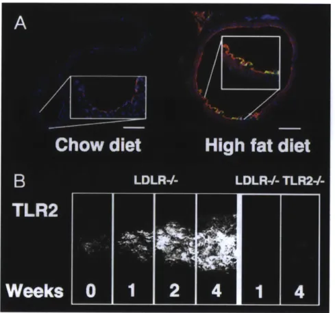
FIGURE 1.17: TOLL-LIKE RECEPTOR-2 IS EXPRESSED IN ATHEROPRONE REGIONS OF THE MOUSE
AORTA AND AUGMENTED BY HIGH FAT DIET. 46
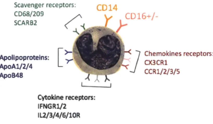
FIGURE 1.19: MAJOR SURFACE RECEPTORS OF HUMAN MONOCYTES. 53
FIGURE 1.20: PRIMARY BLOOD MONOCYTES CONSTITUTE AN HETEROGENEOUS POPULATION
IDENTIFIED BY THEIR CD14 AND CD16 EXPRESSION. 54
FIGURE 1.21: MONOCYTE TRANSMIGRATION IS AN EARLY EVENT IN ATHEROGENESIS. 55 FIGURE 1.22: CD68+ CELLS ACCUMULATE IN ATHEROPRONE REGIONS OF THE VASCULATURE. 56 FIGURE 1.23: STUDY OF THE INTERACTIONS OF ENDOTHELIAL CELLS AND MONOCYTE UNDER
ATHEROPRONE AND ATHEROPROTECTIVE FLOW. 59
FIGURE 2.1: FLOW APPARATUS. 66
FIGURE 2.2: DESCRIPTION OF THE MODEL. 69
FIGURE 2.3: WALL-SHEAR STRESS IS QUASI-HOMOGENOUS ACROSS OUR FLOW CHAMBER. 70 FIGURE 2.4: PICTURE OF A SHEAR PLATE COATED WITH THE POLYMERIZED GEL. 71
FIGURE 2.5: METHOD OF ACTION OF AN AFM APPARATUS. 73
FIGURE 2.6: DETERMINATION OF THE STIFFNESS OF THE CANTILEVER PROBE. 74
FIGURE 2.7: DETERMINATION OF THE YOUNG'S MODULUS OF OUR GELS. 74
FIGURE 2.9: MORPHOLOGY OF ENDOTHELIAL CELLS AFTER 24H OF CULTURE IN STATIC
CONDITIONS ON A FIBRONECTIN-COATED COLLAGEN GEL. 76
FIGURE 2.10: EXPRESSION OF CELL ADHESION MOLECULES IN ENDOTHELIUM CULTURED ON GEL
AND PLASTIC. 77
FIGURE 2.11: EXPRESSION LEVELS OF CD14 AND CD16 IN THP-1 CELLS COMPARED TO ISOTYPE
CONTROLS. 79
FIGURE 2.12: Mi-MACROPHAGE SECRETIONS CAN ACTIVATE ENDOTHELIAL CELLS THROUGH IL-1
PRODUCTION. 82
FIGURE 2.13: ADHESION AND TRANSMIGRATION OF THP-1 MONOCYTE THROUGH ENDOTHELIUM
CULTURED IN STATIC CONDITIONS. 83
FIGURE 2.14: TRANSMIGRATED MONOCYTES CAN PHAGOCYTOSE LATEX BEADS. 83 FIGURE 2.15: ADHERENT MONOCYTE CAPTURED BY SCANNING ELECTRON MICROSCOPY. 84
FIGURE 2.16: ELECTRON MICROGRAPH SHOWING A TRANSMIGRATED MONOCYTE. 85 FIGURE 2.17: EVIDENCE OF FOAM CELL DIFFERENTIATION IN GELS CONTAINING MODIFIED LIPIDS.
86
FIGURE 2.18: KLF2 EXPRESSION ON GEL AND ON PLASTIC. 87
FIGURE 2.19: EXPRESSION OF CAM OF ENDOTHELIAL CELLS UNDER ATHEROPROTECTIVE AND
ATHEROPRONE FLOW. 88
FIGURE 2.20: THP-1 CELLS TRANSMIGRATION THROUGH ENDOTHELIUM EXPOSED TO FLOW IN
OUR IN VITRO SYSTEM. 90
FIGURE 3.1: GRAPHICAL HYPOTHESIS: PHENOTYPIC MODULATION OF TRANSMIGRATING
MONOCYTES. 100
FIGURE 3.2: DIFFERENTIAL RECRUITMENT OF THP-1 MONOCYTES UNDER PROTECTIVE AND
PRONE FLOW. 102
FIGURE 3.3: QUANTIFICATION OF RECRUITED MONOCYTES. 102
FIGURE 3.4: KLF2 EXPRESSION PROTECTS THE ENDOTHELIUM FROM THP-1 MONOCYTE
RECRUITMENT IN THE ABSENCE OF HEMODYNAMIC FORCES. 103
FIGURE 3.5: GENE EXPRESSION OF FREE FLOATING, ADHERENT AND TRANSMIGRATED THP-1
MONOCYTES. 105
FIGURE 3.6: GRAPHICAL HYPOTHESIS: MONOCYTES EXACERBATE THE ATHEROPRONE
ENDOTHELIAL PHENOTYPE. 106
FIGURE 3.7: GENE EXPRESSION ANALYSIS OF ENDOTHELIAL CELLS EXPOSED TO ATHEROPRONE
FLOW CULTURED WITH OR WITHOUT THP-1 MONOCYTES. 108
FIGURE 3.8: EXPRESSION OF CAM IN ENDOTHELIAL CELLS EXPOSED TO ATHEROPRONE FLOW
1
Introduction
1.1 Arterial structure and function 1.1.1 Physiology of the cell
Cells are living organisms that are delimited into space by a membrane and perform certain functions such as self-replication, energy generation, the performance of various chemical reactions using and producing compounds. They are the building units of complex organisms and require energy in order to maintain a viable state. Energy in organisms is drawn from catabolism and cell respiration, the breakdown and oxidation of large molecules of the chemical reactions of catabolism. These biochemical reactions provide the cell with the energy needed to perform cellular functions, maintain its integrity and respond to environmental cues.
1.1.2 Carriage of oxygen and nutrient and disposal of by products During development, complex organisms accrue their number of cells through cell division, grow in size and in mass. Although the delivery of nutrients and oxygen can be achieved through diffusion until a certain size, the critical value of ~200 pm [1, 2] is quickly attained and diffusion-based growth is no longer compatible with the metabolism of the organism that transitions to a convection plus diffusion mechanism: this is the role of the circulatory system. As an order of magnitude, the body of an adult human is constituted of about 100 trillion cells, which all depend on a convoluted network of arteries and veins to provide critical nutrients and oxygen to the tissue while remove waste.
1.1.3 Morphology of the vessels
Microstructure Large arteries are tubes composed of three coaxial layers that assume a myriad of roles in the sustenance of life. One of the most important and obvious ones is to contain and channel the blood to the various organs, but this obvious function has been gradually overshadowed by the more complex and global machinery that probes the biochemical and hemodynamic environments and, adapt and repairs the circulatory system, as vascular research
advanced. The three layers of the large and medium size arteries are: 1) the tunica
intima 2) the tunica media and 3) the adventitia (Figure 1.1).
The tunica intima is a one-cell thick layer that lines the inner surface of blood vessel and is composed of tightly linked adjacent endothelial cells that pave the vessel wall. The endothelial cells sit on the basement membrane, extracellular matrix and the internal elastic lamina that circumscribes the intima from the media. The tunica
media is a muscular layer composed essentially of phalanges of smooth muscle cells
grouped in packets surrounded by connective tissue that control the tone of the vessel. Under stimulation with nitric oxide (NO), prostacyclin or prostaglandins, the media can relax within seconds and allow vasodilation when tissues need increased blood flow. The third and outmost layer of arteries is the tunica adventitia that is mainly composed of collagen and fibroblasts and that anchors the artery to the surrounding tissue of the viscera. The adventitia is separated from the media by the external elastic lamina. The cells of large vessels themselves need perfusion, supplied by the vasa vasorum, literally "vessels of the vessels". The vasa vasorum either originates from the outer tunic in which case it is referred to vasa vasorum
externa, or inner tunic, giving rise to the vasa vasorum interna. In addition to
capillaries, the vessels are innervated by the nervi vascularis.
As we will see in later in this Chapter, the microenvironment of the cells, especially the endothelial cells has profound implications on its state and phenotype. The extracellular matrix separating the endothelium from the media is composed of several major macromolecules such as elastin and collagen. The internal elastic lamina of the aorta is dominated by collagen type III and I. Yet, the extracellular matrix of vessels is a dynamic interface modulated by many environmental cues that affect the state of the surroundings cells. This is extremely relevant to the study of cardiovascular diseases because as the lesion develops, the microstructure of the vessel is remodeled. Throughout the pathogenesis of atherosclerosis the composition, abundance and structure of the extracellular matrix changes; endothelial cells are sensitive to these changes, which influence their state.
Exchanges The blood is the fluid used by the human body to carry the oxygen and the nutrients necessary to the cell functions. The exchanges between the blood and the tissue are mainly carried through diffusion. Hence to be most efficient, the interface between blood and tissue should be maximal. The surface-to-volume ratio of a cylinder of radius R and height h is 21T*R*h/(*RA2*h), which varies with the inverse of the radius of the cylinder. Thus vessels with large surface-to-volume ratios are particularly suited for this task since they provide a high interfacial density. Theses vessels are called the capillaries and are composed of endothelial cells surrounded by pericytes. They are typically in the range of 5 to 10 im in diameter. This is small enough that the erythrocytes that carry oxygen have to deform when going through the capillaries. The network of capillaries is highly parallel meaning that large arteries branch out many times before becoming capillaries and connecting to the venous circulation. The development of a parallel circulatory system is supported by energy and safety considerations. Indeed, if the circulatory system was linear then any disruption of the flow would cause all tissues and organs distal to the blockage to suffer from ischemia. The analogy with the design of the electrical grid illustrates well the point. The electrical grid is designed in parallel from the power source to avoid massive disruption of service, similarly, a semi-parallel vascular network secures the supply of blood to parallel patent branches even if one of them is occluded.
Tunica intima Elastic fibers Tunica media Smooth muscle Tunica adventitia jVasa vasorum 1001A Human, 10% FormalinWeigert's elastic tissue stain and phlox ine,162 x.
Figure 1.1: Ultrastructure of the human aorta.
Histological section showing the trilaminate architecture of the human aorta. From the lumen to the periphery, three tunics can be delineated: the intima, the media and the adventitia [3].
1.1.4 Distinct hemodynamic environments throughout the body The circulatory system is a complex network of vessels that branch out at numerous bifurcations creating variable hemodynamic profiles throughout the body. In the previous paragraphs, we explained some of the evolutionary reasons that lead to this complex circuitry. Here we combine this list and summarize.
Humans have developed with one organ, the heart, to propel blood - a liquid - to the organs. As the vast majority of animals, humans have a high degree of bilateral symmetry - however some internal organs such as the heart, liver or digestive track are not symmetrical with respect to the sagittal plane - and their delivery system of oxygen and nutrients is based on convection and diffusion. Those two considerations, combined to the safety advantages of a parallel circulatory system over a linear one, have forced the evolution towards an anatomy dominated by a central vessel, the aorta, that ramifies into large arteries, medium arteries and capillaries to irrigates organs.
Characteristics offlows Flowing blood applies three types of stress on the vascular bed: pressure, which acts normal to the lumen and can affect the physiology of the endothelium and the tension of the smooth muscle cells (SMC) creating at the same time cyclic stress due to the increase in diameter of the vessel during the cardiac cycle, and wall shear stress or endothelial shear stress. Wall shear stress corresponds to the force per unit area applied by the blood in a direction tangential to the wall; it is also defined as the product of viscosity, the friction between layers of the flow with different velocities, and shear rate (Figure 1.2). In general and when parabolic flow dominates blood flow can be considered as a series of parallel flow lines whose velocity is maximal at the centerline and zero at the walls. The change in velocity follows inversely, maximum at the walls and zero at the centerline. Shear rate describes the rate by which the velocity changes as a distance from the wall. Wall shear stress and rate computed from the derivation of velocities throughout the fluid domain are particularly interesting in the study of vascular diseases as endothelial cells respond to variations in wall shear stress, eliciting different phenotypes and biological processes.
x
dv WSS
=--dy 4
y
Figure 1.2: Physical definition of wall shear stress
Blood flood applies two types of stress on the vessels; pressure directed perpendicular to the vessel wall and wall shear stress that is tangential to the wall and is the consequence of frictional forces of the fluid onto the surface due to viscosity. The diagram illustrates the calculation of wall shear stress (WSS). It shows a typical velocity profile for a straight vascular segment. The slope of the tangent of the velocity distribution at the origin (grey line) is proportional to the WSS. Flows that are highly variable in velocity close to the wall have a high wall shear stress, whereas shallow velocity distributions will correspond to low WSS. Of notice, WSS is a dynamic value that evolves with time similarly to velocities during the cardiac cycle.
Means of quantifying flow parameters Computational fluid dynamics
(CFD) is one of the major techniques used to quantify the motion, frictional forces
and other characteristics of flows. Compared to direct measurements CFD has the advantage of being non-invasive and once the geometry and boundary conditions are measured the computational time does not require the patient to be present.
CFD emerged in the 1930s as a branch of fluid mechanics to solve problems too
complex for analytical solutions. CFD is based on the strategic partition of a fluid domain into small elements, usually tetrahedrons or hexahedrons. The equations that define fluid mechanics in each element provide neighboring elements with boundary conditions for their equations of state and when coupled, provide an integrated view of the fluid dynamics of the system. The quality of the results depend on many parameters including the partitioning strategy and number of
elements used to divide the fluid domain, the original boundary conditions, the algorithm used to solve the equations and the time steps between each calculation, in the case of a time-dependent simulation. As discussed in detail later in this section, the geometry of the vasculature, the fluid boundary conditions, the constitutive laws describing the vessel rigidity and blood, collectively determine the degree of precision of the solution and extra care should be taken to generate a solution that matches the level of understanding of the biological phenomenon studied.
Parameters and sensitivity offlow simulation in biology The overall
relevance of CFD depends on the definition of the boundary conditions: the geometry describing the arterial wall, the inflow and outflow conditions and the model used to describe the behavior of the blood. CFD is a tool and one should not expect that the precision of the simulation will be greater than the precision of the input values or constitutive models. The CFD procedure to model flow in a vessel begins with the reconstruction of the geometry of the lumen obtained from a range of medical imaging modalities. The gold standard is rotational angiography, which produces a series of contrast maps defining the fluid volume. Angiography is often used in concert with iodine-based contrast agents that increase the quality and contrast of the images. Software is used to segment and stack together sequential images, and then clean and smooth the volume. The most delicate steps in setting up a CFD analysis are adequately meshing the fluid domain and prescribing the fluid boundary conditions. See the example of a mesh used to compute the flow characteristics in branches of the internal carotid artery (ICA, Figure 1.3). An ideal mesh should be fine enough such that increasing the number of elements does not affect the solution of the problem. Detailed procedures and information are available in Finite element procedure by K.
J.
Bathe [4].The precision and relevance of the waveforms/flow rates imposed at inlets and outlets are the subject of debate. Options include phase-contrast MRI, Doppler or intravascular ultrasound (IVUS) velocimetry. MRI gives the most precise information as it yields a map of velocities in a section of the vessel. Practically, phase-contrast MRI can difficult to obtain in aclinical setting because it often extends the diagnostic time, so it is not always available. Alternatively, one can compute the Womersley profiles at the inlet from the waveform of the flow rate, which correspond to the analytic solution for this specific waveform in a straight pipe of the same diameter than the inlet.
Figure 1.3: Meshing of a reconstructed artery before CFD analysis.
Example of meshed volume with tetrahedron, pentahedron and hexahedron elements. Sequential images of the bifurcation of the ICA were acquired with angiography, segmented and reconstructed. The volume was meshed with ANSYS Fluent (Canonsburg, PA).
The reconstruction of the arterial geometry is crucial as real images rarely delineate a clean border between tissue and blood; thus, image segmentation and smoothing is left to the judgment of the operator. This is all the more important as such considerations freeze the geometry, impacting subsequent results. If the artery is distensible, the use of a model that correctly takes into account the deformation of the vessel during the cardiac cycle is important. For the in vitro model that we will describe later, we used flow waveforms derived from 1) the carotid sinus and 2) a straight segment of the internal carotid artery. To account for the deformation the vessel, the vessel was modeled with a nonlinear, isotropic, hyperelastic material. This class of computational simulations that takes both into account the dynamics of the fluid and the boundaries are called Fluid Structure Interaction (FSI) models. The
solving algorithm not only iterates on the equations ruling fluid dynamics but also considers the fluid forces deforming the walls of the model and adjusts the geometry and iterates back on the equations of fluid mechanics. FSI is currently the most accurate way to derive flow parameters such as the velocity vector field, shear stress or shear gradient.
Fluid boundary conditions include evaluation of the flow inlets and outlets. Classically, inlets are set to drive the simulation by specifying the velocity or flow rate in a cross-section, while outlets insure the continuity of the flow through setting the exit pressure. Ideally, phase contrast-MRI at the inlet and pressure measurements at the outlet, in the same patient, should be used for maximal precision. Yet, phase contrast-MRI is not a panacea, it is highly dependent on the parameters specified for acquisition and the range of velocities that can be tracked is limited. Thus, heterogeneous flows have larger uncertainty and may require extrapolation of the velocity distribution near the wall. Fortunately, the characteristics of the flow change as it develops in the vasculature, decreasing its dependence on the boundary condition away from the inlet; consequently, it is key to keep enough length before the region of interest
Blood is a viscous fluid, described with classical Newtonian physics, but more appropriately considered with more complex models [5]. Carreau-Yasuda or Casson approximations offer a more general framework, allowing viscosity to vary with the shear rate [6]. Rheological effects can be important depending on the geometry. Newtonian models can degrade the results of the simulation, while Casson models do not significantly burden computational cost
The trend in CFD is to bring additional physiological relevance and complexity to the models and boundary conditions, in particular, to circumvent the rigid wall assumption. FSI takes into account a model of deformation of the wall to constantly iterate on the mechanical model of the artery to make the geometry current - pulses of flow induce the dilation of the vasculature. The challenges that remain are to
obtain more precise and appropriate experimental data on the mechanics and patient-specific topology of the wall to drive the models proposed.
CFD is a valuable tool because of the dearth of adequate in vivo imaging that can
reliably measure flow parameters such as WSS, but as a sensitive tool, requires careful selection of boundary conditions. General mechanistic processes, such as mechanotransduction, need only low precision and averages, as they seek to reach global conclusions. Case-specific problems, such as the destabilization of the atheromatous plaque or aneurysmal cavities, are in contrast highly dependent on the level of details. In conclusion, the precision of CFD should match the level of understanding required to derive mechanistic insight.
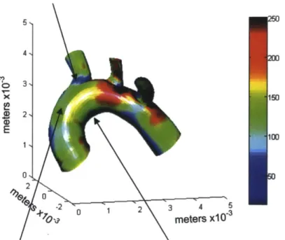
The hemodynamic stress applied to the vessel drastically change throughout the vasculature. The diagram in Figure 1.4 shows the computed average WSS in the aortic arch of a mouse [7]. The color-coded map highlights clearly the spatial variations in WSS, especially between the lower curvature of the arch or parts of the bifurcations (blue, low intensity) and the median or higher curvature (green/red, high intensity).
5 N 4, 3% 115 E 100 01 2 4 -2 02 3 4 5 OV meters x103
Figure 1.4: The lower curvature of the aorta and the adjacent branches are subjected to disturbed blood flow.
Map of the time-average intensity of wall shear stress in a mouse aorta quantified
from CFD. The colors indicate the level of wall shear stress in dynes/cm2. The
arrows show regions of the arch with distinct flow patterns [7].
A similar observation can be made in the human vasculature: shear stress varies both spatially and with time. Figure 1.5 shows a CFD analysis of a healthy volunteer with no history of cardiovascular diseases. The map of WSS at the bifurcation of the common carotid illustrates the variations in intensity of the shear forces in straight segments of the carotid arteries compared to the carotid sinus.
The heterogeneity in hemodynamics are particularly important to the development of cardiovascular diseases such as atherosclerosis because endothelial cells are prime sensors and responders to fluid forces. In the following sections, we will give some background on the endothelium and its multiple functions, notably in relationship to flow and leukocytes and we will explain how it relates to atherogenesis.
Ezturual Carotd Iftarnal Carotid Carotid Sinus lime Averaged Wall Shear Strs (dynicm2) 32 y 16 11 8 2 Common Carotid
Figure 1.5: CFD analysis of the wall shear stress in the human carotid artery. CFD analysis of the WSS of the carotid of a healthy male. Similar to the previous
figure, this diagram shows a clear heterogeneity in the level of WSS between the straight parts of the arteries i.e. common carotid and internal and external carotids, and the carotid sinus (adapted from [8])
1.2 Endothelial cells
1.2.1 Discovery
Three British scientists, Henry Hyde Salter, Robert Bentley Todd and Sir William Browman, first observed and isolated the endothelial cells. The trio isolated for the first time in 1852 what they named epithelial particles - pointy and elongated cells with a large nucleus that align with the axis of the vessel - from the aortae of an ox and horse and published there work in PhysiologicalAnatomy and Physiology of Man in 1856. Yet, Wilhelm His is credited for coining the term "endothelium" as he posited in 1865 that epithelium and endothelium have different functions and origins.
1.2.2 Characteristics
Endothelial cells pave the innermost layer of vessels as they form a monolayer that plays multiple roles in vascular homeostasis. The appearance of the endothelium is often compared to cobblestone as the cells form a confluent monolayer made from elongated cells. Each individual endothelial cell measures around 10 pm in length,
by 3 pm in width and 0.5-1 gm in thickness [9] (endothelial cells are sensitive to mechanical forces such as flow, which accentuate their elongated shape as they align with the direction of the flow) and is connected to its neighboring cells by junctional molecules. The endothelium is found in three different configurations: 1) "continuous" like in most of the vasculature of large vessels or in the brain-blood barrier (central nervous system) that strictly regulates insudation of plasma molecules and circulating cells, 2) "fenestrated", characterized by small disruptions
(0.1-1 pm) between endothelial cells but an intact basement membrane, like in the
kidney glomeruli or 3) "discontinuous" when disruptions occur in both, endothelial junctions cells and basement membrane e.g. in the liver, spleen or bone marrow. The state of the monolayer reflects the functional requirements of the local vascular bed in terms of permeation, filtration or secretion, yet fenestrations are also observed at bifurcations, loci of intense hemodynamics stresses and gradients [10,
11]. This is all the more important that vascular lesions often occur at branching
points where fenestration allows for increased exchanges between lumen and tissue.
Endothelial cells possess the same set of major organelles than other cells: a plasma membrane that delimits a cytoplasm, a nucleus anchored to the cells membrane by an actin skeleton, endoplasmic reticula, ribosomes, Golgi apparatus, mitochondria as well as some endothelial-specific organelles such as the Weibel-Palade bodies that store granules of von Willebrand factor and P-selectin and vesiculo-vacuolar organelles (VVO). In addition, a network of polysaccharides covers the apical surface of endothelial cells, in the same way that the kelp covers the seafloor. The glycocalyx is 0.2-4.5 pm thick and is composed of three major glycosaminoglycans: heparin sulfate, chondroitin sulfate and hyaluronic acid that are attached to the
transmembrane proteins. This organelle that is a charged interface, plays a key role in regulating what binds to the endothelial surface.
1.2.3 Historical perspective of endothelial function in vascular homeostasis
1.2.3.1 The endothelium as a container
The human endothelium constitutes an impressive interfacial surface of about 350 m2
[12], yet because of its feeble thickness it only accounts for a negligible -110g compared to the -70kg of body mass of an adult [9]. Until the 1950, the endothelium is mainly regarded as a coating or envelope that acts as a container for the blood. Part of the disinterest for the endothelial layer was due, in part, to the limited visualization tools available at the time. To put things into perspective with the technological advances at the time, the first electron microscope was made in
1926 but only commercialized at the end of the 30s. The other hurdle that muted
investigational ardor was the unavailability of an in vitro model. As we will see in the next paragraphs, these two limitations were overcome in the following years.
1.2.3.2 Barrier function
In 1966, Lord Henry Florey published a seminal paper that challenged the assumption of the endothelium as an inert liner between the vascular space and the interstitium but rather recognized the variety of endothelial cells states and functions and conceptualized it as an active barrier that has a major role in transporting molecules from the blood to the underlying tissue. He concludes the article by writing:
"In the time available, I have been able to show you a little of the current knowledge of the morphology of cells which fifteen years ago were thought toform little more than a sheet of nucleated cellophane.
The caveolae intracellulares, which are prominent in some but not all endothelium, may facilitate the passage of substances through
endothelium even if they function only by furnishing a greater area through which diffusion may occur. The fine structure of the interendothelial junctions may hold the key to understanding how small quantities of plasma proteins may leak out of even the most impermeable blood-vessels." Lord H. Florey [13].
This paradigm shift - from a simple wrapper to an active barrier - was one of the motors for the nascent field of vascular biology because it opened a new field of investigation that would unveil the many characteristics and functions of endothelial cells, which emerged as the central actor of vascular health or disease. Yet, another discovery was missing before the field could really develop; a practical
in vitro model of endothelial cell was required. This milestone was achieved in the
70s when Jaffe, Nachman, Becker and Minic [14] and Gimbrone, Cotran and Folkman
[15] independently reported the isolation, prolonged culture and identification of
Human Umbilical Cord Endothelial Cells (HUVEC).
These concurrent discoveries propelled the field to great extent and permitted its exponential growth. A PubMed search with the terms "endothelial cells" returned respectively 229 and 487 until 1965 and 1973 when the same query returns an astonishing 150,000+ publications when the time range is up to 2014. We know today, that the endothelium is involved in virtually all the diseases either directly or indirectly [16, 17]. In the next paragraphs, we briefly highlight the major functions performed by the endothelium and explain its involvement in atherosclerosis.
1.2.3.3 Active paracrine signaling
The endothelium regulates many of the processes that maintain homeostasis, such as controlling permeability, adjusting the vascular tone, providing an anti-thrombogenic surface to the blood, creating new vessels, namely angiogenesis or mounting an inflammatory response, by paracrine secretions. Here, we summarize the major proteins that mediate these endothelial functions.
1.2.3.3.1 Permeability
The first hypotheses of the endothelium seen as a permeable surface originate from functional studies by Starling in 1896 [18], followed by the exquisite drawings and insight by Ramon y Cajal (Museum Ram6n y Cajal, Madrid; Spain), and later Kohn and Cowdry and Pappenheimer [19], who laid the foundations of modern views on endothelial functions. Many of the major advances on the ultrastructure of the endothelium and the vessels could not have happened without the technical prowess and deep scientific insight of Karnovsky [20, 21], notably through his drastic improvement of cytochemical analysis of intact tissue and avant-garde methods to prepare scanning electron microscope samples. Permeability is a physical trait of a barrier or interface that quantifies the amount of matter allowed to cross the barrier. In the context of the endothelium it is characterized by the propensity of the monolayer to let plasma components intravasate or vice versa. The exchanges occur principally either in between the cells, via the paracellular route, or through the cell itself, via transcellular mechanisms. Both molecules and cells can be channeled through the endothelium. For molecules such as cholesterol, caveolae, which are lipid raft invagination at the surface of endothelial cells, bundles of caveolae or vesicles can regulate their passage. Concomitantly, cellular junctions that anchor one cell to its neighbors, also regulate the passage of molecules between them by building pores. The junctional molecules include claudins, occuldins, nectins, junctional adhesion molecules and Vascular Endothelial-Cadherin (VE-Cadherin). The transport of plasma cells requires a different set of molecules and will be discussed later. During atherogenesis, the increase in endothelial
permeability causes lipid insudation and accumulation in the subendothelial space that is characteristic of the beginning of the lesion.
Three classes of junctional molecules are the major regulator of permeability: 1) tight junctions, including junctional adhesion molecules (JAM), occulinds and claudins, 2) gap junctions, mainly the connexins and 3) adherens junctions such as VE-Cadherin. Several secreted factors including thrombin, bradykinin, histamine and Vascular Endothelial Growth Factor (VEGF) can disrupt the organization of cell
junctions and therefore increase permeability. On the contrary, molecules such as cyclic AMP (cAMP) can improve the endothelial barrier function [22].
1.2.3.3.2 Vascular tone
The vasculature controls blood pressure by dilating or constricting the lumen of the vessels in a process that we know now is tightly regulated by the endothelium. At the beginning of the 70s however, little was known about the effectors of vasodilation. To fill this void, many vascular biologists would try to understand what causes the relaxation or constriction of smooth muscle cells. The following 15 years of research produced quantum leaps in characterizing these mechanisms, in particular, the discovery of two puissant vasodilators: prostacyclin and nitric oxide
(NO), profoundly transformed the field by the breadth of their implication in
vascular homeostasis.
Two years after the establishment of HUVEC as a model for endothelial cells, Gimbrone and Alexander showed that the endothelium is a source for prostaglandin, a hormone-like lipid that has implications in vasodilation, platelet aggregation and permeability [23]. Later, Sir Moncada and colleagues isolated the most potent form of prostaglandin, prostacyclin (PGI2) [24, 25] while Furchgott and Zawadzki showed that the endothelium was necessary to the vasomotility and discovered and
characterized the Endothelium-Derived Relaxation Factor as the active
substance[26]. Rich from the knowledge accumulated by the forefathers of modern vascular biology, we now know that cyclooxygenase (COX), especially COX-2, catalyzes the conversion of arachidonic acid into prostaglandins; in parallel, Nitric Oxide Synthase (NOS) - and for the matter of interest to us, endothelial NOS (eNOS)
- catalyzes the synthesis of NO from L-arginine in endothelial cells [27, 28]. The scope of the biology of NO is extremely wide. It ranges from vasodilation to leukocyte adhesion and is also produced by the endothelium subjected to healthy
1.2.3.3.3 Anti-thrombogenic surface
Healthy endothelium provides an anti-thrombogenic surface that prevents the aggregation of plasma cells on its surface and thus is responsible for keeping the vessel patent by acting on four major mechanisms: 1) the endothelium displays tissue factor pathway inhibitors that block Factor VIIa, a serine protease involved in the initiation of the coagulation cascade; 2) the heparin sulfate of the glycocalyx tethers anti-thrombin III that inhibit thrombin molecules, another serine protease that cleaves soluble fibrinogen into insoluble fibrin; 3) the display of cell surface thrombomodulin, which binds thrombin, change its substrate from fibrinogen to Protein C and activates Protein C. Activated Protein C cleaves factors of clot formation; 4) the sequestration of von Willebrand Factor (vWF) into Weibel-Palade Bodies (WPB) that prevents vWF to bind to Factor VIII, collagen or platelets. In endothelial cells, vWF is present as multimers of over 20,000 kDa that bundle in helical arrangements. This helical packing is responsible for the cigar shape of the mature WPB that contain vWF. Upon activation of the endothelial cells, for example with thrombin, histamine, epinephrine or vasopressin, the WBP vesicles release vWF in the plasma, which acts as a carrier for Factor XIII and triggers the coagulation cascade. Exocytosis proceeds through two mechanisms: 1) lingering-kiss exocytosis where WPB vesicles merge with the plasma membrane or 2) multi-granular exocytosis, where several WBP fuse together into secretory pods before
releasing amalgamate vWF [29].
1.2.3.3.4 Angiogenesis
Angiogenesis is one of the major functions of the endothelium. It is the process leading to the formation of new vessel from existing ones. Several stimuli including hypoxia can trigger angiogenesis, which goal is to restore hemostasis by forming collaterals. The endothelium is equipped to sense disturbance in oxygen supply and hypoxia-inducible factor and can activate the angiogenic cascade if new vessel are needed for tissue perfusion. The main stimuli for angiogenesis are the VEGF family, the Fibroblast Growth Factor (FGF) family, Angiopoietin-2 and chemokines.
Angiogenesis is very relevant to myocardial infarction caused by the occlusion of coronary arteries because it provides new vessels to perfuse the heart. A complete review of angiogenesis can be found elsewhere [30].
1.2.3.3.5 Inflammation
Inflammation is a biological process triggered by harmful stimuli with the aim of eradicating it. It begins with acute inflammation, a rapid response of the vascular system, which builds up through the synergistic combination of leukocyte chemoattraction, activation, adhesion, and transmigration into the arterial wall. The successful removal of the noxious stimulus restores homeostasis and depending on the magnitude and intensity of the inflammation the initial vascular architecture is either recovered or replaced by connective scar tissue. If however the stimulus persists, inflammation becomes chronic and set a new set of interactions between endothelium and plasma cells.
The four cardinal signs of inflammation, rubor (redness), calor (feeling of heat),
dolor (pain) and tumor (swelling), sometimes complemented by a fifth mousquetaire, fonctio laesa (loss of function; although not unique to inflammation) are the direct
result of the alteration of the endothelial state and its interaction with the immune system. Rubor and calor are the consequences of vasodilation through an increase of cytosolic Ca2
+ that mediates the release of PGI2 via the COX pathway and NO.
Additionally, the influx of Ca2+ and synergetic activation of the RHO-pathway phosphorylates myosin light chain, which results in the contraction of the actin cytoskeleton and promotes vascular leakiness. The increase in permeability of the vessel induces the leakage of plasma exudate, which accounts for the tumor. The secretions of inflammatory cytokines by leukocyte cause dolor [31].
The infiltration of leukocytes, in particular the early recruitment of neutrophils and monocytes, sustains inflammation by the secretion of the potent pro-inflammatory cytokines Tumor Necrosis Factor-a (TNF-a) and Interleukin-1 (IL-1) [32, 33]. These cytokines activate the master regulator NF-KB that in turn trigger the expression of myriad of pro-inflammatory mediators including COX2 involves in increased
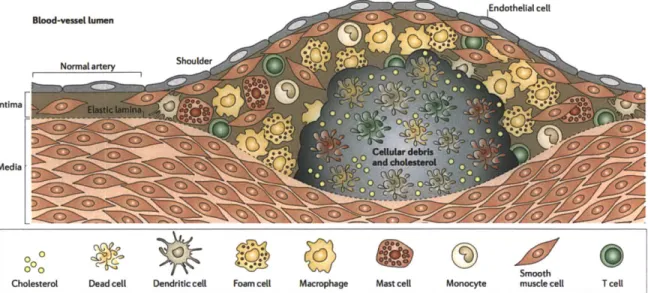
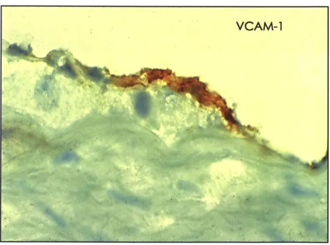
permeability, chemokines such as Monocyte Chemoattractant Protein-1 (MCP-1) and Interleukin-8 (IL-8) that augment the affinity of the adhesions molecules for their cognate receptor, and adhesion molecules like Intercellular Cell Adhesion Molecule-1 (ICAM-1), Vascular Cell Adhesion Molecule-1 (VCAM-1), E-selectin that are directly responsible for the recruitment of leukocytes.
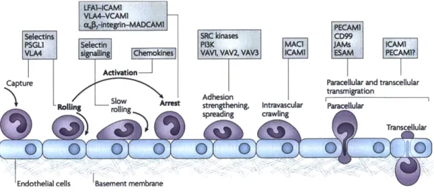
The recruitment of leukocytes is a complex process involving many molecules at the surface of the endothelium and leukocytes. Together, these molecules interact to signal and create bonds between the cells. Endothelial cells are quasi-immobile in the frame of reference of the body, whereas leukocytes are in motion, propelled by blood flow. In order to decrease their momentum, bonds have to be created with the vascular bed, which further allows leukocytes to change direction and intravasate despite fluid forces. Transmigration is described as a multiple step process consisting of capture, rolling, slow rolling during which leukocyte find the best "spot" to transmigrate, arrest of activated leukocytes, adhesion strengthening and spreading, crawling and finally the actual transmigration that can occur either paracellularly or transcellularly. At each step, various surface molecules are involved both on the endothelium and the leukocytes reviewed in great details by Ley and colleagues [34] (Figure 1.6).
LFA1--CAM1
VLA4-VCAM
,-integrln-MADCAM PEcAM1
Selectins SRC kinases CD99
PSGL1 Selecttn P13K MACi JAMs ICAMN
VLA s g dek, VAVI, VAV2, VAV3 ICAMI ESAM PECAM1?
Activation
Capture Paracellular and transcellular
Adhesion transmigration
SRoong Arlst strengthening, Intravascular Paracellular
O spradin cralingTranscellular
Endothelial cells Basement membrane
Transmigration is a multiple-step process during which activated leukocytes are stopped before they can cross the endothelial barrier at determined loci such as cell junctions or thin cell membranes. PSGL1: P-selectin glycoprotein ligand-1; VLA4: Very Late Antigen 4; LFA1: Lymphocyte function-associated antigen 1; MADCAM1: mucosal vascular addressin cell-adhesion molecule 1; P13K: phosphoinositide 3-kinase; MAC1: macrophage antigen 1; PECAM1: platelet/endothelial-cell adhesion molecule 1; JAMs: junctional adhesion molecules; ESAM: endothelial cell-selective adhesion molecule [34].
1.2.3.4 Mechanosensor
The endothelial monolayer is the center of command of vascular health and a fine sensor of its microenvironment. Its endothelial cells are the first line of exposure to luminal elements. Residing at the privileged vantage point at the frontier between vessel and blood on the one hand and smooth muscle from the opposing side, the endothelium is ideally suited to orchestrate the sensing of complex forces and flows, surveillance of circulating elements, and right of passage and defense of the vessel against these entities. Changes in endothelial connectivity, in endothelial cell shape and cytoskeleton are followed by dynamic alterations to the cell's secretome and with that regulation of state and environment [35, 36]. For example under laminar flow, endothelial cells become spindoidal and align with the flow but lose a preferential orientation and turn cuboidal under disturbed or turbulent flow due to F-actin reorganization [37], and while the former inhibit thrombosis, leukocyte adhesion, smooth muscle proliferation and vasoconstriction that latter phenotype promotes these processes.
The transmission of force to the cell is carried through intracellular tension created
by the contraction of cytoskeletal filaments influenced by flow via the anchorage of
the cell membrane to the substrate [38], the nucleus [39] or neighboring cells [40]. Intracellular forces act on the conformation of intracellular proteins [41] to modify the physiology of the cell and consequently the extracellular milieu, including the mechanical properties of the substrate, indirectly influences through this
mechanism, the biology of the cell [42]. Several mechanosensors have been identified at the apical and basal surfaces of the endothelium, such as ion channels [43], integrins [44], cell adhesion molecules [45], G-protein coupled receptors [46] or others cellular organelles [47] and can lead to the activation of inflammation mediators such as the master regulator Nuclear Factor-KB (NF-KB) or potent anti-inflammatory factor like Kruppel-Like Factor-2 [8] depending on the waveform
applied to the cells. Further details on endothelial flow-mediated
mechanotransduction are summarized in an exhaustive review by Davies [48].
1.3 Atherosclerosis
Atherosclerosis is the most prevalent disease in developed countries [49]. It slowly and often silently mutates into localized lesions of the large and medium vessels to culminate in myocardial infarction or stroke [50-52]. Together these two diseases claim a staggering 15 million lives in the U.S. each year despite the innumerable advances made in the understanding of the risk factors, the pathobiology of the disease, the general biology of vessels, and the deployment of a panel of small molecules to counteract the progression of the disease, and devices to prevent and protect patients during the acute phase [53, 54].
1.3.1 Incidence
The pathological evolution of atherosclerosis, namely plaque rupture or stenosis can causes myocardial infarction, angina pectoris, heart failure and stroke. All together cardiovascular diseases are the first cause of mortality in the world, especially in the Western world where cholesterol-rich diet promotes the disease. Eighty-three millions Americans have at least one cardiovascular disease and in 2010 alone, more than 1.3 million died because of cardiovascular maladies [53]. Despite great advances in our understanding of the pathogenesis of the disease and technological breakthroughs like the invention of stents, drug-eluding stents or cholesterol lowering drugs such as statins, the prevalence of atherosclerosis keeps rising notably in oriental countries adopting the Western diet. The conclusion of an acute cardiovascular event is often tragic: death and disabilities are still very frequent and
the colossal economic burden - $315+ billions in 2010, predicted to triple by 2030
-adds to the immense emotional strain [53). The onus is on today's society to bend the macabre course of cardiovascular diseases. It is with this in mind that we have developed new tools to investigate atherosclerosis.
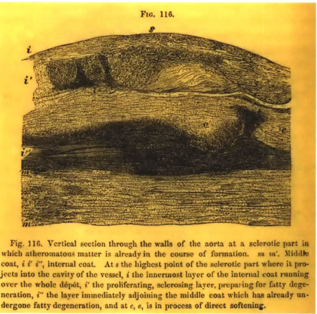
1.3.2 Historical perspective
With the earliest evidence of atherosclerotic lesions tracing back to Egyptian mummies and early histological data dating back to Gabrielle Falloppio's first reports of calcified arteries during the sixteenth century, the origin and mechanisms underlying atherosclerosis is a long-standing issue and the subject of intense speculation, hypotheses and investigation. Contrary to what is often read, the French pathologist Pierre Rayer and the British physician Joseph Hodgson were the first to postulate the inflammatory nature of atherosclerotic lesions, which to this day, still influences our modern vision of the disease. Rayer writes:
"Morbid ossification of the arteries [is] the result of the inflammation of their fibrous layer. It is accompanied by bright redness of its internal layer, [...] and frequently surrounded by a yellow matter, soft and solid,
non transparent ". P. Rayer [55].
Yet, another great scientist, the Prussian Rudolph Virchow, is credited with drawing the finest insight in the pathogenesis of atherosclerosis. In his 1856 treaty entitled
"Thrombose und Embolie. Gefassentzundung und septische Infektion" [56], Virchow
induced pulmonary embolism in dogs by seeding foreign material such as elderberry cores into their venous system, noting that the occlusion would induce thrombosis. He surmised that the consequences of thrombosis could be due to 1) irritation of the vessel, 2) coagulation of the blood or 3) interruption or disturbance of the flow. We now know that the concept is flipped on its head and that it is inflammation/endothelium dysfunction, blood coagulability and disturbed flow that are the three pillars of thrombosis, remembered as Virchow's Triad [57]. Nevertheless, Virchow's observations and analyses are brilliant and still shape our conception of atherogenesis. In 1858, two years after writing his opus on
thrombosis, he explains in lectures to the Pathological Institute of Berlin his agreement with the "old view" in which inflammation of the endothelium precedes the "fatty metamorphosis". He writes:
"The state of the matter here also is more or less very simply this, that two processes must be distinguished in the vessels, which are very analogous in their ultimate results ; first, the simple fatty metamorphosis, which sets in without any discoverable preliminary stage, and in which the existing histological elements pass directly into a state of fatty degeneration and are destroyed, so that a larger or smaller proportion of the constituents of the walls of the vessel perishes; and, in the next place, a second series of changes, in which we can distinguish a stage of irritation preceding the fatty metamorphosis, comparable to the stage of swelling, cloudiness, and enlargement which we see in other inflamed parts. I have therefore felt no hesitation in siding with the old view in this matter, and in admitting an inflammation of the inner arterial coat to be the starting point of the so-called atheromatous degeneration ; and I have moreover endeavoured to show that this kind of inflammatory affection of the arterial coat, is in point offact exactly the same as what is universally termed endocarditis, when it occurs in the parietes of the heart. There is no other difference between the two processes than that the one more
frequently
runs an acute, the other a chronic, course." R. Virchow [58].Another conceptual milestone was reached at the turn of the nineteenth century when Nikolai Anitschkow provided the first evidence linking dietary cholesterol to the development of atheroma in what is considered the germinal model of atherosclerosis in a rabbit [59, 60]. Yet, systemic elevated levels of cholesterol alone could not explain the confinement of inflammation and atheromatous lesions to specific regions of the vasculature, such as the lower curvature of the aortic arch, the carotid sinus or broadly bifurcations. Modern theories have emerged to attempt to reconcile the systemic and local components of atherogenesis, which gave rise to the