Coreceptor affinity for MHC defines peptide specificity
requirements for TCR interaction with coagonist peptide-MHC
The MIT Faculty has made this article openly available.
Please share
how this access benefits you. Your story matters.
Citation
Hoerter, J. A. H., J. Brzostek, M. N. Artyomov, S. M. Abel, J. Casas,
V. Rybakin, J. Ampudia, et al. “Coreceptor Affinity for MHC Defines
Peptide Specificity Requirements for TCR Interaction with Coagonist
Peptide-MHC.” Journal of Experimental Medicine 210, no. 9 (August
12, 2013): 1807–1821.
As Published
http://dx.doi.org/10.1084/jem.20122528
Publisher
Rockefeller University Press
Version
Final published version
Citable link
http://hdl.handle.net/1721.1/89124
Terms of Use
Creative Commons Attribution-Noncommercial-Share Alike
The Rockefeller University Press $30.00
The vast majority of the peptides presented by MHC molecules are derived from self-proteins and do not activate mature T cells. Antigen recognition and T cell activation must thus be CORRESPONDENCE
Nicholas R.J. Gascoigne: [email protected] Abbreviations used: CHO, Chinese hamster ovary; pMHC,
peptide–MHC. J.A.H. Hoerter’s present address is Genomics Institute of the Novartis Research Foundation, San Diego, CA 92121. J. Brzostek, J. Casas, V. Rybakin, and N.R.J. Gascoigne’s present address is Dept. of Microbiology, Yong Loo Lin School of Medicine, National University of Singapore, Singapore 117545. M.N. Artyomov’s present address is Dept. of Pathology and Immunology, Washington University in St. Louis, St. Louis, MO 63110.
S.M. Abel’s present address is Dept. of Chemical and Biomolecular Engineering, University of Tennessee, Knoxville, TN 37996.
J. Ampudia’s present address is Takeda Pharmaceutical Company, San Diego, CA 92121.
C. Lotz’s present address is Actelion Pharma, 5400 Baden, Switzerland.
Coreceptor affinity for MHC defines peptide
specificity requirements for TCR interaction
with coagonist peptide–MHC
John A.H. Hoerter,
1Joanna Brzostek,
1Maxim N. Artyomov,
7Steven M. Abel,
3,4,5Javier Casas,
1Vasily Rybakin,
1Jeanette Ampudia,
1Carina Lotz,
1Janet M. Connolly,
8Arup K. Chakraborty,
3,4,5,6Keith G. Gould,
2and Nicholas R.J. Gascoigne
11Department of Immunology and Microbial Science, The Scripps Research Institute, La Jolla, CA 92037 2Department of Immunology, Wright-Fleming Institute, Imperial College London, London W2 1PG, England, UK 3Department of Chemical Engineering, 4Department of Chemistry, and 5Department of Biological Engineering,
Massachusetts Institute of Technology (MIT), Cambridge, MA 02139
6Ragon Institute of MGH, MIT, and Harvard, Cambridge, MA 02139 7Broad Institute of MIT and Harvard, Cambridge, MA 02142
8Department of Pathology and Immunology, Washington University in St. Louis School of Medicine, St. Louis, MO 63110
Recent work has demonstrated that nonstimulatory endogenous peptides can enhance T cell recognition of antigen, but MHCI- and MHCII-restricted systems have generated very differ-ent results. MHCII-restricted TCRs need to interact with the nonstimulatory peptide–MHC (pMHC), showing peptide specificity for activation enhancers or coagonists. In contrast, the MHCI-restricted cells studied to date show no such peptide specificity for coagonists, sug-gesting that CD8 binding to noncognate MHCI is more important. Here we show how this dichotomy can be resolved by varying CD8 and TCR binding to agonist and coagonists coupled with computer simulations, and we identify two distinct mechanisms by which CD8 influences the peptide specificity of coagonism. Mechanism 1 identifies the requirement of CD8 binding to noncognate ligand and suggests a direct relationship between the magnitude of coagonism and CD8 affinity for coagonist pMHCI. Mechanism 2 describes how the affinity of CD8 for agonist pMHCI changes the requirement for specific coagonist peptides. MHCs that bind CD8 strongly were tolerant of all or most peptides as coagonists, but weaker CD8-binding MHCs required stronger TCR binding to coagonist, limiting the potential coagonist peptides. These findings in MHCI systems also explain peptide-specific coagonism in MHCII-restricted cells, as CD4–MHCII interaction is generally weaker than CD8–MHCI.
© 2013 Hoerter et al. This article is distributed under the terms of an Attribution– Noncommercial–Share Alike–No Mirror Sites license for the first six months after the publication date (see http://www.rupress.org/terms). After six months it is available under a Creative Commons License (Attribution–Noncommercial– Share Alike 3.0 Unported license, as described at http://creativecommons.org/ licenses/by-nc-sa/3.0/).
tuned to allow for recognition of the small minority of disease-associated peptide–MHC (pMHC) “needles in the haystack” of nonstim-ulatory endogenous pMHC (Davis et al., 2007; Gascoigne, 2008; Gascoigne et al., 2010). Sev-eral experiments have shown that T cell activa-tion by small amounts of antigen is enhanced by the presence of endogenous peptides (Irvine et al., 2002; Yachi et al., 2005). Although this activation enhancement or coagonist phenom-enon has been reported for both MHC class I
The Journal of Experimental Medicine
on September 27, 2013
jem.rupress.org
Downloaded from
Supplemental Material can be found at:
on September 27, 2013 jem.rupress.org Downloaded from on September 27, 2013 jem.rupress.org Downloaded from on September 27, 2013 jem.rupress.org Downloaded from on September 27, 2013 jem.rupress.org Downloaded from on September 27, 2013 jem.rupress.org Downloaded from on September 27, 2013 jem.rupress.org Downloaded from on September 27, 2013 jem.rupress.org Downloaded from on September 27, 2013 jem.rupress.org Downloaded from on September 27, 2013 jem.rupress.org Downloaded from on September 27, 2013 jem.rupress.org Downloaded from on September 27, 2013 jem.rupress.org Downloaded from on September 27, 2013 jem.rupress.org Downloaded from on September 27, 2013 jem.rupress.org Downloaded from on September 27, 2013 jem.rupress.org Downloaded from
MHC, TCR, and coreceptor interactions. Here, an extension of this model allows us to describe coagonism enabled by self-peptides, taking into account the distinct activation states of Lck (Nika et al., 2010; Stirnweiss et al., 2013).
Because of the glaring discrepancies in the requirements for TCR discrimination between coagonist peptides in MHCI- and MHCII-restricted systems, there is a need for a unifying concept to explain activation enhancement for both T cell lineages. In this paper, we used H-2Kb and H-2Db single
chain (sc)–pMHCs (Yu et al., 2002; Choudhuri et al., 2005; Palmowski et al., 2009), which allowed us to dissect the dis-tinct contributions of CD8 affinity and of TCR affinity for
both antigenic and nonstimulatory pMHCs. Using H-2Kb–
and H-2Db–restricted TCRs and stochastic computer
simula-tions of the kinetics of T cell activation, we describe two distinct mechanisms by which CD8 affinity for pMHC can influence the requirements for coagonists. Mechanism 1 de-scribes CD8 binding to nonstimulatory pMHC as an absolute requirement for coagonism and shows that higher-affinity CD8–pMHC interactions can mitigate peptide specificity re-quirements for coagonists and increase the magnitude of en-hancement. Mechanism 2 describes how the affinity of CD8 for agonist pMHC influences the requirements for TCR in-teraction with coagonist pMHC. A relatively simple kinetic model of T cell activation is sufficient to account for all coag-onist phenomena, thus unifying disparate observations from CD4 and CD8 T cells.
RESULTS
Development of inducible, antigenic, sc-MHC Chinese hamster ovary (CHO) cell expression systems
sc-pMHCI molecules are produced by recombinant con-structs in which the MHC-binding peptide is linked to the N terminus of 2-microglobulin, which in turn is linked to
the N terminus of the MHCI heavy chain (Yu et al., 2002; Choudhuri et al., 2005; Palmowski et al., 2009). When ex-pressed on the cell surface, these sc-MHCI constructs are very stable and can act as potent agonists for T cells. sc-KbOVA
(OVA peptide 257–264, SIINFEKL, on H-2Kb, recognized
by OT-I TCR; Hogquist et al., 1994) and sc-DbNP68
(influ-enza A/NT/60/68 nucleoprotein 366–374, ASNENMDAM, on H-2Db, recognized by F5 TCR; Townsend et al., 1986;
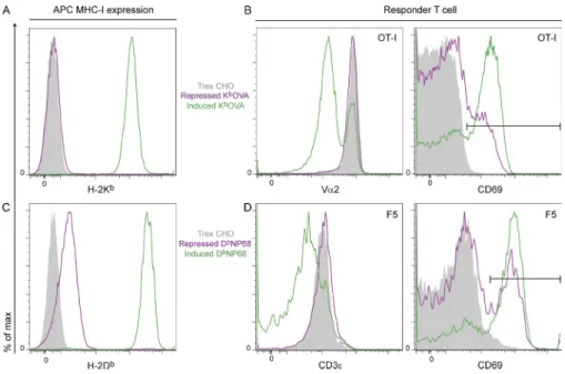
Mamalaki et al., 1992) in doxycycline-inducible vectors were transfected into CHO Trex cells (expressing tetracycline re-pressor). Doxycycline strongly up-regulated cell surface ex-pression of sc-MHCI, allowing the CHO cells to stimulate antigen-specific T cell activation, as measured by CD69 up-regulation or TCR endocytosis (Fig. 1). In the repressed state (absence of doxycycline), “leaky” expression of sc-KbOVA or
sc-DbNP68 presented enough agonist to the CD8 OT-I or
F5 T cells to induce some CD69 up-regulation but not enough to induce significant TCR down-regulation (Fig. 1, B and D). Expression of sc-KbOVA in the repressed state was
virtually undetectable by flow cytometry with a KbOVA-specific
antibody (Fig. 1 A; and not depicted; Porgador et al., 1997), (MHCI)–restricted T cells and thymocytes (Yachi et al., 2005,
2007; Anikeeva et al., 2006; Juang et al., 2010) and for MHCII-restricted T cells (Irvine et al., 2002; Li et al., 2004; Krogsgaard et al., 2005), the relative importance of TCR recognition of the endogenous pMHC appears to be very different for CD4 and CD8 T cells (Davis et al., 2007; Gascoigne, 2008; Gascoigne et al., 2010).
The number of potential coagonist peptides for a given CD4 T cell are very limited (Krogsgaard et al., 2005; Ebert et al., 2009; Lo et al., 2009), whereas coagonism for CD8 T cells or thymocytes occurs with a wide range of different non-stimulatory peptides (Yachi et al., 2005, 2007; Juang et al., 2010). This evidence thus suggests that MHCII-restricted TCRs discriminate between endogenous peptides, whereas MHCI-restricted TCRs do not. However, recent data indicate that nonstimulatory pMHCI ligands show a very weak but possibly biologically significant interaction with TCR (Juang et al., 2010). This suggested that TCRs might play a role in coagonism in MHCI-restricted cells but that its specificity is only evident for very weakly stimulatory TCR ligands such as those in-volved in positive selection.
The CD8 coreceptor’s interaction with nonstimulatory MHCI has been suggested to be important for coagonism in MHCI-restricted cells (Yachi et al., 2005; Gascoigne, 2008; Gascoigne et al., 2010). Nonstimulatory pMHC alone can recruit CD8 to the T cell–APC interface (Yachi et al., 2005; Rybakin et al., 2011). Also, coagonist pMHCs became antag-onists in CD8-negative cells (Stone et al., 2011). These results, along with the lack of peptide specificity for coagonists, sug-gest that non-cognate CD8 coreceptor binding to nonsti-mulatory pMHC is the dominant mechanism of activation enhancement for MHCI-restricted T cells. In addition, CD8 affinity for the MHC presenting the antigenic peptide (ago-nist) plays a direct role in signaling through the TCR, where increasing the affinity of CD8 can increase ligand potency and even bypass peptide specificity requirements altogether (Laugel et al., 2007; Wooldridge et al., 2007, 2010). Because there is a range of affinities for CD8 binding to different MHCI molecules (Cole et al., 2012), the relative require-ments for CD8, or for TCR interaction with the nonstimula-tory ligand, might be expected to vary with the strength of CD8–MHC binding. Interestingly, the two mouse MHCI-restricted TCR models that have been analyzed in coago-nism experiments (OT-I [Yachi et al., 2005, 2007; Juang et al., 2010] and 2C [Stone et al., 2011]) recognize H-2Kb or Ld,
which show relatively high-affinity CD8 binding (Cole et al., 2012).
A stochastic, computational model has been used to inves-tigate the role of coreceptors in TCR triggering, and results suggest that CD8 plays a dual role of stabilizing the TCR– pMHC interaction and of delivering the CD8-associated kinase Lck to the TCR to initiate signaling, with the latter ef-fect being the more important (Artyomov et al., 2010). This model explicitly combined two key features, membrane-protein mobility and protein–protein interactions (Lis et al., 2009), which allowed incorporation of many biophysical measurements for
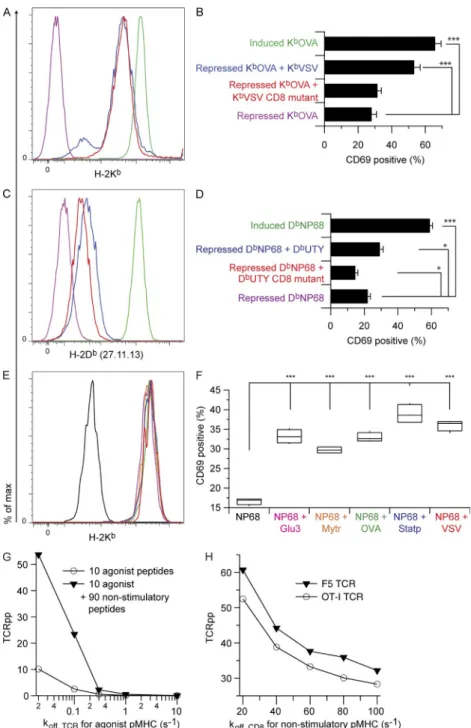
Anikeeva et al., 2006; Gascoigne, 2008). To test this hypothesis, we generated D227K-E229K mutations, which are known to abrogate CD8 binding (Connolly et al., 1988, 1990; Potter et al.,
1989), in the sc-KbVSV and sc-DbUTY 3 domains. When
super-transfected into inducible KbOVA-expressing cells
(Fig. 3 A), we found that the sc-KbVSV CD8-binding mutant
(sc-KbVSV-CD8m) did not provide coagonism to OT-I T cells
(Fig. 3 B) or preselection thymocytes (not depicted).
Simi-larly, sc-DbUTY-CD8m expressed with repressed sc-DbNP68
did not provide coagonism for F5 cells (Fig. 3, C and D). Un-expectedly, the sc-DbUTY-CD8m had a mild and statistically
significant inhibitory effect on F5 activation. These results re-vealed that the intact CD8-binding site is absolutely required for coagonism in both OT-I and F5 systems.
We also tested the ability of known H-2Kb–binding
pep-tides (Santori et al., 2002; Yachi et al., 2005) to act as coago-nists for F5 T cells. We used RMA-S cells as APCs, as these cells express very few MHCI molecules unless exogenous peptide is provided (Ljunggren et al., 1990; Yachi et al., 2005, 2007).
When H-2Kb–binding peptides Slc2a3 (VNTIFTVV), Nmt1
(AAYSFYNV), OVA, Stat3 (ATLVFHNL), and VSV were loaded on RMA-S cells pretreated with trace NP68 agonist peptide, we found that all peptides acted as strong coagonists for F5 CD8+ T cells (Fig. 3, E and F).
A computational model of T cell activation describes coagonism
Various computational and conceptual studies have dealt with the principles behind coagonism (Li et al., 2004; Wylie et al., 2007; Feinerman et al., 2008), but there has not yet been a treatment that simultaneously incorporates the diffu-sion, binding, and reactions of proteins in space and time with many available biophysical measurements. However, such ki-netic models have been used to describe the different depen-dencies of T cell activation on CD4 and CD8 coreceptors
whereas repressed sc-DbNP68 was detectable above
back-ground (Fig. 1 C).
Both H-2Kb and H-2Db nonstimulatory pMHCs
can be coagonists
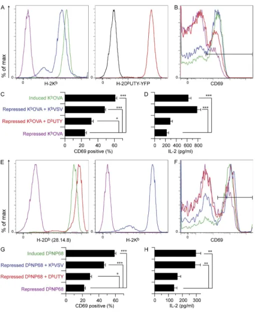
CHO cells expressing inducible sc-KbOVA or sc-DbNP68
were super-transfected with nonstimulatory sc-MHCI con-structs, enabling coexpression of agonist and nonstimulatory pMHC on the same cell (Fig. 2). This allowed us to probe co-agonism by nonstimulatory peptides presented on H-2Kb and
H-2Db, with both H-2Kb– and H-2Db–restricted TCRs.
KbVSV (RGYVYQGL) and DbUTY (WMHHNMDLI) are
nonstimulatory pMHCs for both OT-I and F5 T cells. We found that sc-KbVSV provided strong coagonism for both
OT-I and F5 T cells, measured as CD69 up-regulation or IL2 secretion (Fig. 2, B–D and F–H, respectively), or by phos-phorylation of the NF-B precursor p-p105 (not depicted). sc-DbUTY also provided activation enhancement for both
TCR transgenic T cells, but this was much weaker than the sc-KbVSV effect and did not show statistically significant
co-agonism of IL2 secretion. Nonstimulatory sc-DbNP68 also
gave weak coagonism for OT-I T cells (not depicted). Analo-gous activation results were obtained with preselection OT-I thymocytes stimulated with KbOVA-expressing CHO cells
(not depicted). These data demonstrate that both H-2Kb
and H-2Db nonstimulatory pMHC molecules were capable
of being coagonists for TCRs restricted by the same or
an-other MHCI, but H-2Kb provided much stronger coagonism
than H-2Db.
CD8 binding to nonstimulatory pMHC is directly related to the magnitude of coagonism
The lack of peptide specificity in coagonism in MHCI- restricted T cells could be the result of noncognate CD8 inter-action with nonstimulatory MHCI molecules (Yachi et al., 2005;
Figure 1. Expression of inducible sc-MHCI
agonist molecules KbOVA and DbNP68 in
CHO cells and activation of corresponding OT-I and F5 CD8 T cells. (A) Trex CHO cells
expressing dox-inducible sc-MHC were stained with anti-Kb and analyzed by flow cytometry.
Gray shading: untransfected Trex CHO cells. Purple: repressed (no dox) sc-KbOVA. Green:
induced (+dox) sc-KbOVA. (B) OT-I T cells were
incubated with the indicated CHO cell APCs for 3 h, and TCR endocytosis (anti-V2; left) and CD69 expression (right) were assessed by flow cytometry. The horizontal black line indicates the CD69hi gate. The color coding is the same
as in A. (C and D) As in A and B except CHO cells expressing dox-inducible sc-DbNP68
were stained with anti-Db or exposed to F5
responder T cells. TCR staining was by anti-CD3. Data are representative of three inde-pendent experiments.
model to be consistent with these binding data. The TCR on rate for nonstimulatory peptides for both OT-I and F5 simu-lations was set to the OT-I value for KbOVA so that we could
directly compare nonstimulatory peptides with equal affinity and kinetics. We developed this model (see Supplemental text) that describes signal enhancement caused by the coexis-tence of agonist and nonstimulatory peptides on the APC surface (Fig. 3 G). This model also describes increased TCR signaling in response to nonstimulatory ligands with higher CD8-binding affinities in both F5 and OT-I model systems (Fig. 3 H). This computational result is consistent with the following data: Fig. 2 showing a greater magnitude of
en-hancement by H-2Kb compared with H-2Db nonstimulatory
pMHC, Fig. 3 (B and D) showing the absolute dependence of (Artyomov et al., 2010). We applied this formalism to a
descrip-tion of coagonism, incorporating separate on and off rates for TCR binding to agonist versus nonstimulatory pMHCI. The OT-I TCR has approximately twofold higher affinity for ago-nist pMHCI than F5 (Kd of 5.9–6.5 µM for OT-I [Alam et al.,
1996, 1999] versus 11 µM for F5 [Willcox et al., 1999]). The kinetics describing these equilibrium constants are markedly different. The on and off rates for OT-I TCR–binding KbOVA
are well characterized (3,720/M/s and 0.022/s, respectively; Alam et al., 1999). We calculated the on rate for the F5 TCR for DbNP68 based on the off rate and equilibrium affinity
reported in the literature (koff = 0.8/s, Kd = 11 µM, kon =
72,727/M/s, calculated; Willcox et al., 1999). Therefore, we varied the TCR kinetics for agonist pMHC in the kinetic
Figure 2. Coagonism of OT-I and F5 CD8
T cells by both sc-H-2Kb and sc-H-2Db
nonstimulatory sc-MHCI. Inducible
sc-KbOVA or sc-DbNP68 CHO cells were
super-transfected with constitutive sc-KbVSV or
sc-DbUTY and used as APCs to assess
coago-nism of OT-I and F5 T cells. CHO cell APCs were stained for MHCI expression and in parallel exposed to responder T cells whose activation status was measured either by flow cytometry or IL2 ELISA. Inducible sc-KbOVA APCs and OT-I
responder T cells are described in the top pan-els, whereas inducible sc-DbNP68 APC and F5
responder T cells are described in the bottom panels. (A and E) Analysis of Kb or Db
expres-sion on CHO cell APCs. Color coding is as shown in the y-axis labels of the bar graphs. In A, sc-DbUTY-YFP fusion construct was used
to transfect cells expressing repressed KbOVA,
and the YFP fluorescence was used here to report on sc-DbUTY expression relative to
untransfected CHO cells (black). In the re-maining panels, sc-MHCI expression was analyzed by antibody staining. (B and F) OT-I (B) and F5 (F) CD8 T cell expression of CD69 after incubation with the correspond-ing CHO APCs from A or E, respectively. Horizontal black lines indicate the CD69hi
gate. (C and G) Bar graphs represent mean ± SEM values of CD69hi cells for OT-I (C) and
F5 (G). (D and H) IL2 production after 8 h of exposure to CHO cell APCs as indicated for OT-I (D) and F5 (H). Statistical significance (*, P < 0.05; **, P < 0.01; ***, P < 0.001) results are from ANOVA analysis with Dunnett’s post-test, referencing the repressed agonist sample as the standard. For the OT-I CD69 bar graph in C, n = 8 (individual mice) after
removing outliers as defined by values >1.25 times the interquartile distance. For D, n = 4;
G, n = 6 (individual mice); H, n = 6. Each
data panel is representative of three inde-pendent experiments except C and G, which report the combined results from three inde-pendent experiments.
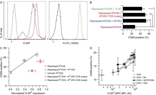
nonstimulatory Kb ligand (Juang et al., 2010). To probe the
role of TCR interaction with the nonstimulatory ligands in coagonism using the sc-MHCI system, we used several strat-egies. Residues in the 12-helices of MHCI comprise the main recognition structures for TCR CDR1 and CDR2 (Sun et al., 1995; Sim et al., 1998; Garcia et al., 2009). The E166K mutation disrupts recognition of KbVSV by VSV-specific CTL
(Sun et al., 1995) and of KbOVA by OT-I CTL (not depicted).
Second, we used a chimera between the 12 domains of
the MHCIb molecule H-2TL (which cannot bind or present
peptide to TCRs; Devine et al., 2002; Weber et al., 2002;
Liu et al., 2003) and the 3 from H-2Kb, enabling normal
CD8 binding (Attinger et al., 2005). Both KbVSV with the
coagonism on CD8 binding to the nonstimulatory pMHCI, and Fig. 3 F showing the lack of F5 coagonist peptide speci-ficity for peptides bound to the high CD8-binding affinity
MHCI protein H-2Kb.
Minimal contribution of OT-I TCR recognition of nonstimulatory pMHCI to coagonism
Previous studies showed no peptide dependence for coago-nism by nonstimulatory pMHCI, with both OT-I preselec-tion thymocytes and peripheral T cells (Yachi et al., 2005, 2007), nor was there any discrimination between endogenous nonstimulatory peptides in signaling for negative selection by
covalent pMHC dimers containing one Kb-OVA with one
Figure 3. The effect of CD8 affinity for
coago-nist pMHC on coagonism. Inducible sc-KbOVA
or sc-DbNP68 CHO cells were super-transfected
with constitutive sc-KbVSV CD8-binding mutant
(sc-KbVSV-CD8m) or sc-DbUTY CD8-binding mutant
(sc-DbUTY-CD8m), respectively, and used as APCs to
assess coagonism of OT-I and F5 T cells. CHO cell APCs were stained for MHCI expression and in paral-lel exposed to responder T cells whose activation status was measured by flow cytometry. (A) Anti-Kb
staining of CHO cells expressing sc-MHCI. Color cod-ing as shown in B. (B) Percentage of CD69hi OT-I
T cells (from n = 10 individual mice) after 3-h exposure
to the CHO APCs in A. (C) Anti-Db staining of CHO
cells. Color coding as shown in D. (D) Percentage of CD69hi F5 T cells (n = 6 individual mice) after 3-h
exposure to the CHO APCs in C. RMA-S cell were also used as APCs to examine coagonism of F5 T cells. (E) Anti-Kb staining of RMA-S APCs loaded
with combinations of NP68 agonist peptide and various H-2Kb–binding peptides: purple, Slc2a3;
orange, Nmt1; green, OVA; blue, Stat3; and red, VSV. (F) Percentage of CD8+ F5 cells expressing CD69hi
after 3.5-h incubation with the RMA-S cells in E (n = 4).
A kinetic model of T cell activation reproduces the effect of coagonism and describes a direct relationship between CD8 affinity for coagonist pMHC and TCR phosphorylation. (G) The kinetic model discriminates based on antigen quality and exhibits enhanced TCR phosphorylation (TCRpp) in the presence of non-stimulatory peptides. Results are shown for 10 ago-nist peptides alone and for 10 agoago-nist plus 90 nonstimulatory peptides using OT-I kinetic param-eters. (H) Results of computer simulations of the kinetic model describing coagonism as a function of CD8 affinity for nonstimulatory pMHC. Results are shown for F5 and OT-I kinetic parameters. Statistical significance (*, P < 0.05; ***, P < 0.001) results are from ANOVA analysis with Dunnett’s post-test, refer-encing the repressed agonist sample (B and D) or the sample with agonist alone (F) as the standard. Data in A, C, E, and F are representative of three independent experiments. Data in B and D are the combined re-sults of three independent experiments. Error bars represent SEM.
To probe CD8 T cell coagonism with a similar strategy, RMA-S cells were first loaded with a very small amount of OVA pep-tide followed by loading with a titration of peppep-tides including the OVA-derived antagonist/positively selecting peptide R4 (SIIRFEKL), the nonstimulatory peptide VSV (RGYVYQGL), and the engineered peptide poly-serine (SSYSYSSL), which should lack any TCR contact residues (Hogquist et al., 1994). Each of these peptides acted as a coagonist in an H-2Kb
ex-pression–dependent manner (Fig. 4 D), indicating that for the OT-I TCR, any discrimination based on the quality of the non-stimulatory peptide is at best very small.
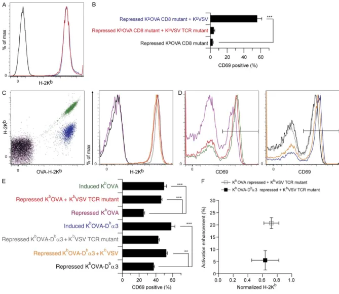
Reduction in CD8-binding affinity for agonist pMHCI enforces requirement for intact TCR interaction with nonstimulatory pMHC
To test the impact of CD8 affinity for agonist pMHCI on
co-agonism, we tested sc-KbOVA-CD8m (Fig. 5, A and B). As
expected from previous work (Connolly et al., 1988, 1990; Potter et al., 1989), this mutation minimized direct activation of OT-I T cells by sc-KbOVA (Fig. 5 B and not depicted).
Ac-tivation of OT-I T cells was rescued by sc-KbVSV but not by
E166K TCR-binding site mutation (sc-KbVSV-TCRm) and
the TL/Kb chimera provided significant coagonism for
rec-ognition of sc-KbOVA (Fig. 4, A and B), indicating that the
TCR interaction with nonstimulatory pMHCI was at most a minor determinant of coagonism for OT-I T cells. We also noted that recruitment of CD8 to the immunological syn-apse (Yachi et al., 2005; Rybakin et al., 2011) was
unper-turbed by the sc-KbVSV-TCRm mutation (not depicted).
When we plotted CD69 expression data for OT-I T cells versus MHCI expression on CHO cells for repressed and induced sc-KbOVA, or repressed sc-KbOVA plus sc-KbVSV,
we found a linear relationship (Fig. 4 C). Repressed sc-KbOVA
plus sc-KbVSV-TCRm revealed a minor defect in coagonism
when TCR binding to nonstimulatory pMHC was attenu-ated. In contrast, the repressed sc-KbOVA plus the sc-Kb
VSV-CD8m showed no coagonism.
A recent study showed that the OT-I TCR has a very weak but biologically relevant interaction with endogenous nonstimulatory ligands. Removal of this TCR interaction, for example with a peptide designed to bind Kb but to lack TCR
interaction residues, abrogated coagonism (Juang et al., 2010).
Figure 4. Minor contribution of OT-I TCR binding to nonstimulatory pMHC in coagonism. Inducible sc-KbOVA CHO cells were super-transfected
with constitutive sc-KbVSV TCR-binding mutant (sc-KbVSV-TCRm) or H-2TL-Kb3 (TL/Kb) and used as APCs to assess coagonism of OT-I T cells. CHO cell
APCs were stained for MHCI expression and in parallel exposed to responder T cells whose activation status was measured by flow cytometry. (A) CHO cell staining for either H-2Kb or H-2TL expression as indicated. Black indicates TL-negative staining control (Trex CHO cells). Other colors are as indicated in B.
(B) Percentage of CD69hi OT-I T cells (n = 10 mice) after 3-h exposure to the CHO APCs in A. (C) Percentage of CD69hi OT-I T cells plotted as a function of
the corresponding CHO APC H-2Kb expression, as indicated (n = 10 mice for CD69 data and n = 3 for H-2Kb expression on CHO cell APCs). A correction
factor of 2.8 is applied to the H-2Kb staining of the repressed KbOVA plus KbVSV-CD8m to account for attenuated anti-Kb staining resulting from the
CD8-binding site mutation (not depicted). RMA-S cell APCs were also used to test coagonism of OT-I T cells. (D) Percentage of CD69hi OT-I T cells plotted
as a function of Kb expression of RMA-S cells loaded with increasing concentrations of nonstimulatory peptides R4, VSV, and SSYSYSSL, as indicated
(n = 2 for both CD69 and H-2Kb expression). Statistical significance (*, P < 0.05; ***, P < 0.001) results are from ANOVA analysis with Dunnett’s post-test,
ref-erencing the repressed agonist sample as the standard. Data in A are representative of three independent experiments, and data in D are representative of two independent experiments. Data in B and C are the combined results of three independent experiments. Error bars represent SEM (B and C) or SD (D).
binds CD8 similarly to the native H-2Kb molecule (Moody
et al., 2001b). We therefore constructed and subsequently
ex-pressed inducible sc-KbOVA-Db3 in CHO cells and
super-transfected with sc-KbVSV or sc-KbVSV-TCRm. The altered
3 domain epitope present in sc-KbOVA-Db3 showed
at-tenuated binding to the anti–H-2Kb antibody, visible in the
different anti–H-2Kb staining of the induced sc-KbOVA-Db3
versus wild-type sc-KbOVA (Fig. 5 C). The amount of H-2Kb
sc-KbVSV-TCRm (Fig. 5 B). This was in stark contrast to
earlier results with repressed wild-type sc-KbOVA, in which
sc-KbVSV-TCRm was a strong coagonist (Fig. 4, B and C).
To further probe this discrepancy in coagonism by
sc-KbVSV-TCRm, we took advantage of the fact that CD8 has
lower affinity for H-2Db than for H-2Kb (Moody et al., 2001b;
Huang et al., 2007). The CD8-binding affinity is a property of the MHCI 3 domain, as a chimera of Db12-Kb3
Figure 5. Modulation of KbOVA CD8-binding affinity and associated changes in coagonism by KbVSV-TCRm. Inducible sc-KbOVA CD8-binding
mutant (sc-KbOVA-CD8m) CHO cells were super-transfected with constitutive sc-KbVSV or sc-KbVSV-TCRm and used as APCs to assess coagonism of
OT-I T cells. CHO cell APCs were stained for MHCI expression and in parallel exposed to responder T cells whose activation status was measured by flow cytometry. (A) Anti-Kb stain of CHO cell APCs. Color coding is as shown in B. (B) Percentage of CD69hi OT-I T cells (n = 10 mice) in response to the
CHO cell APCs in A. Inducible domain-swapped sc-KbOVA-Db3 CHO cells were also super-transfected with both sc-KbVSV or sc-KbVSV-TCRm and
used as APCs to assess coagonism of OT-I T cells. (C) Repressed and induced KbOVA (purple and green, respectively) and KbOVA-Db3 (black and blue,
respectively) constructs were analyzed as a function of both OVA-Kb and total H-2Kb staining, whereas repressed KbOVA plus KbVSV-TCRm (red),
re-pressed KbOVA-Db3 plus KbVSV (orange), and repressed KbOVA-Db3 plus KbVSV-TCRm (gray) were analyzed with H-2Kb staining alone. (D) CD69
expression of CD8+ OT-I T cells responding to the APCs described in C, with color coding as in C. The horizontal black line indicates the CD69hi
gate. (E) Percentage of CD69hi OT-I T cells (n = 4 mice) in response to the CHO cell APCs in C. (F) Calculated value of activation enhancement of
OT-I T cells plotted as a function of anti-Kb staining of the associated CHO APCs. Repressed KbOVA plus KbVSV-TCRm and repressed KbOVA-Db3
plus KbVSV-TCRm are shown. Statistical significance (**, P < 0.01; ***, P < 0.001) results are from ANOVA analysis with Dunnett’s post-test,
refer-encing the repressed agonist sample as the standard. Data in A are representative of three independent experiments. Data in B are the combined results of three independent experiments. Data in C, D, and F are representative of two experiments, whereas the data in E are the combined re-sults from two experiments. Error bars represent SEM (B and E) or SD (F).
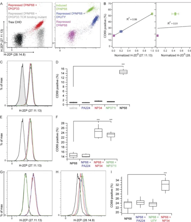
sequence and MHC heavy chain. The sc-DbUTY clones used
in our assays stained strongly with 28.14.8 but relatively weakly with 27.11.13 (Fig. 6 A). Linear regression analysis of CD69hi F5 T cells after activation with repressed sc-DbNP68,
repressed sc-DbNP68 plus sc-DbUTY, or induced sc-DbNP68
revealed a very strong correlation between the percentage of CD69hi T cells and 27.11.13 staining of the
correspond-ing CHO cells (Fig. 6 B). No such correlation existed for these same CD69 data plotted against 28.14.8 staining, sug-gesting that coagonism for the F5 TCR was sensitive to the conformation of the 12 domains of the nonstimulatory pMHC and/or to the peptide presented. This result sug-gested that there are fundamental differences in coagonism/ activation enhancement between the DbNP68-F5 and KbOVA–
OT-I systems.
To probe the question of the specificity of coagonism of
F5 T cells for nonstimulatory H-2Db pMHC, we screened
a small library of 40 H-2Db–binding peptides (Table S1)
for H-2Db binding and the ability to act as coagonists for
F5 T cells when presented by RMA-S, identifying only two variant influenza peptides as coagonists (not depicted). Neither of these peptides (NP34, ASNENMETM; and NP371I, ASNENIDTM) stimulates F5 T cells (Townsend et al., 1986; Price et al., 2000), but NP34 can act as an antagonist (Williams et al., 1998) and possibly so can NP371I (Price et al., 2000). We therefore carefully tested these peptides for direct activation of F5 T cells. RMA-S cells were loaded with 100 µM each of NP34, NP371I, and PA224
(SSLEN-FRAYV, a Db-binding peptide which did not act as
coago-nist in our screen) or with 10 nM NP68 agocoago-nist peptide.
H-2Db was stabilized on RMA-S cells by binding of NP34,
NP371I, and PA224, but the H-2Db loaded with 10 nM
NP68 agonist was undetectable (Fig. 6 C). There was no significant difference in T cell activation between saline-treated RMA-S cells and those presenting high amounts of
PA224, NP34, or NP371I on H-2Db (Fig. 6 D). RMA-S cells
treated with 10 nM NP68 peptide activated 15% of F5 T cells. To test for coagonism by PA224, NP34, and NP371I, we first loaded RMA-S cells with 10 nM NP68 agonist, followed by 100 µM each of PA224, NP34, and NP371I, as previ-ously described (Yachi et al., 2007). After peptide loading,
these RMA-S cells expressed comparable amounts of H-2Db
(Fig. 6 E), and when used to stimulate F5 T cells, both NP34 and NP371I, but not PA224, showed strong coago-nist activity (Fig. 6 F).
Because of the positive coagonist results with sc-DbUTY–
expressing CHO cells, we also tested the ability of the UTY peptide to support activation enhancement when presented on RMA-S cells. UTY peptide loaded at 100 µM did stabi-lize H-2Db on RMA-S cells as measured by anti-Db staining
with antibody clone 28.14.8, but staining with the comple-mentary anti-Db antibody 27.11.13 was negative for UTY
but positive for NP34 and PA224 (Fig. 6, G and H). The fraction of F5 CD8+ T cells that up-regulated CD69
expres-sion showed that the UTY peptide did not support activation enhancement (Fig. 6 I).
expression in the nonstimulatory sc-KbVSV and sc-Kb
VSV-TCRm were comparable (Fig. 5 C). The CD69 expression pro-files for OT-I CD8+ T cells (Fig. 5 D) and quantification of
CD69hi T cells (Fig. 5 E) clearly showed the lack of statistically
significant coagonism for sc-KbOVA-Db3 plus sc-Kb
VSV-TCRm compared with either of the significant responses found for the sc-KbOVA-Db3 plus wild-type sc-KbVSV or
wild-type sc-KbOVA plus sc-KbVSV-TCRm.
Both repressed and induced sc-KbOVA-Db3–expressing
CHO cells elicited systematically higher CD69 expression than the wild-type sc-KbOVA in the responder OT-I T cells.
This result was unexpected, but we examined several different clones of the inducible sc-KbOVA-Db3 construct and each
one behaved in a similar manner (not depicted). This phe-nomenon could be explained by the serial TCR engagement model (Valitutti, 2012), which proposes that a few agonist pMHC complexes can initiate signaling from many TCRs. Optimum induction of T cell activation occurs in response to agonist pMHC complexes that engage TCR with intermediate affinity, with half-lives sufficient to initiate productive TCR ITAM phosphorylation but short enough to allow an indi-vidual pMHC molecule to serially trigger several TCRs. We suggest that the attenuated CD8 affinity reduces the half-life
of OT-I/CD8 engagement by KbOVA-Db3, bringing it
closer to the optimum values that allow more effective serial TCR engagement and T cell activation. Because of this un-expected result, and to explicitly show the altered behavior of sc-KbVSV-TCRm coagonists for the recognition of
sc-KbOVA-Db3 versus recognition of wild-type sc-KbOVA, we
calculated the activation enhancement of OT-I T cells (percent CD69hi in response to repressed agonist with sc-KbVSV-TCRm
minus percent CD69hi in response to repressed agonist alone;
Fig. 5 F). This enabled comparison of coagonism by sc-Kb
VSV-TCRm with these two different agonists, revealing that the nonstimulatory ligand with reduced TCR interaction provided greater coagonism for recognition of sc-KbOVA than for
chi-meric sc-KbOVA-Db3. The results presented in Fig. 5
sug-gest that reduced CD8 affinity for antigenic pMHCI enforces an increased reliance on TCR interaction with nonstimulatory ligand to achieve coagonism.
Coagonist activity by H-2Db for F5 T cells requires
TCR discrimination of nonstimulatory pMHCs
The two antibodies used to stain sc-H-2Db–transfected CHO
cells in this study recognize different H-2Db epitopes. The
clone 27.11.13 recognizes a peptide-dependent conforma-tional epitope, whereas the 28.14.8 clone recognizes the H-2Db
3 domain independent of peptide (Fig. 6 A; Palmowski et al.,
2009). We prepared sc-DbGP33 and sc-DbGP33-TCRm
sc-MHC molecules, expressed them constitutively in CHO cells, and stained with both 27.11.13 and 28.14.8 anti-Db antibodies.
These results show that the 27.11.13 antibody staining was
diminished by the TCR-binding site mutation on H-2Db
(Fig. 6 A). This offers strong evidence that the 27.11.13 anti-body actually reports on the presence of amino acid residues that are important for TCR recognition, both in the peptide
Figure 6. Contribution of F5 TCR recognition of nonstimulatory H-2Db pMHC to coagonism. Inducible sc-DbNP68 CHO cells were super-transfected
with constitutive sc-DbGP33, sc-DbGP33-TCRm, or sc-DbUTY and used either as APCs to assess coagonism of F5 T cells or as probes of anti-Db antibody
speci-ficity. (A) Dual anti-Db antibody staining (clones 28.14.8 and 27.11.13) of CHO cell APCs. (B) Percentage of CD69hi F5 T cells plotted separately versus the
27.11.13 and 28.14.8 anti-Db stains of corresponding CHO cell APCs in A. The black lines are linear fits to the data with the R2 value for the fit reported on the
graph (n = 6 mice). Error bars represent SEM. RMA-S cell APCs were also used to assess coagonism of F5 T cells by H-2Db–binding peptides. (C) Anti-Db
stain-ing of RMA-S APCs loaded with individual peptides. Gray shadstain-ing shows RMA-S alone, and black shows RMA-S after 10 nM NP68 agonist addition. 100-µM additions of peptides were as follows: blue, PA224; red, NP34; and green, NP371I. (D) Percentage of CD8+CD62L+CD44lo F5 cells expressing CD69hi after 3.5-h
incubation with the RMA-S APCs in C. (E) Anti-Db staining of RMA-S cells loaded with combinations of peptides to test coagonist activity, color coded as
indicated in C. (F) Percentage of CD8+CD62L+CD44lo F5 cells expressing CD69hi after 3.5-h incubation with the RMA-S cells in E. (G and H) Anti-Db antibody
staining with clones 27.11.13 (G) and 28.14.8 (H) of RMA-S cell APCs loaded with trace NP68 agonist with or without coagonists. Black indicates NP68 alone. 100 µM additions of peptides were as follows: blue, PA224; red, NP34; and green, UTY. (I) Percentage of CD8+ F5 cells expressing CD69hi after 3.5-h incubation
with the RMA-S cell APCs in G and H. Statistical significance (***, P < 0.001) results are from ANOVA analysis with Dunnett’s post-test, referencing the sample with saline (D) or agonist alone (F and I) as the standard. Data in A are representative of three independent experiments, and data in B are the combined re-sults from three independent experiments and are the same data as presented in Figs. 2 G and 3 D. For D and F, n = 5; for I, n = 15. For RMA-S assays with
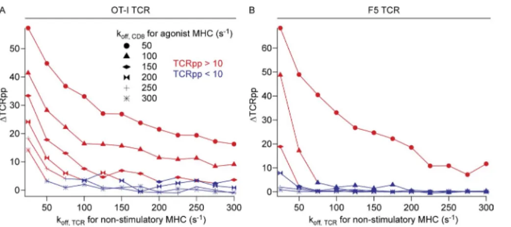
pMHC. At these weak CD8 agonist pMHCI–binding affin-ities, the lowest affinity nonstimulatory peptides provided little signal enhancement, and the absolute TCRpp value for these simulations fell below the activation threshold of TCRpp = 10. For the TCR with F5-like affinity for agonist pMHC (Fig. 7 B), there was a steeper dependence on
non-stimulatory pMHC–TCR off rate at low koff, CD8 (high CD8
affinity) for agonist pMHC. This indicates that the kinetics of the TCR–agonist pMHCI interaction play a role in the re-quirement for specific coagonists. However, at the smallest values of koff, CD8 for agonist pMHC, coagonism of F5 was still
significant and the absolute values of TCRpp for the simula-tions of agonist plus nonstimulatory peptides remained above the threshold of 10. For intermediate koff, CD8 for agonist
pMHC, only the highest affinity nonstimulatory peptides gave any appreciable activation. At the highest values of koff,
CD8 for agonist pMHC, the nonstimulatory pMHC gave no
coagonism for F5. These computational results provide a con-ceptual framework describing our experimental results, in which we found that CD8 affinity for agonist pMHCI is a key factor for the dependence of coagonism on the TCR’s interaction with the nonstimulatory pMHC.
DISCUSSION
Recent work has demonstrated that nonstimulatory peptides bound to MHC can enhance T cell recognition (Krogsgaard et al., 2005; Yachi et al., 2005, 2007; Anikeeva et al., 2006; Davis et al., 2007; Gascoigne, 2008; Gascoigne et al., 2010; Juang et al., 2010). To investigate the parameters of the coago-nist phenomenon for CD8 T cells, we developed a system where very low expression of sc-pMHCI agonists for specific TCRs was coupled with constitutive expression of non-stimulatory sc-pMHCI. Using OT-I and F5 TCRs, we tested combinations of H-2Kb and H-2Db, as well as mutations in
the cognate or nonstimulatory MHC, to identify the inter-actions important in coagonism. In addition, we used computer
Computer simulations reveal the importance of TCR interaction with nonstimulatory pMHC when CD8 binding to agonist pMHC is weak
To provide a mechanistic context to the experimental find-ings described here, that sensitivity of coagonism to the na-ture of the nonstimulatory peptide originates in part from differences in CD8 affinity for agonist pMHCI, we modified the computational model described above to introduce sep-arate koff, CD8 values for agonist and nonstimulatory pMHC
species. This allowed us to systematically vary coreceptor binding affinity to agonist pMHC while keeping coreceptor binding to nonstimulatory pMHC constant. To test the differ-ent requiremdiffer-ents of OT-I and F5 T cells for nonstimulatory pMHC in responses to low amounts of their specific agonist pMHCIs, we performed simulations with agonist peptides only or with agonist plus nonstimulatory peptides. These sim-ulations were performed over a range of high, intermediate, and low CD8 affinities for agonist pMHC (koff, CD8 for agonist
pMHC of 50–300/s; Fig. 7 and not depicted; Cole et al., 2012). Simulations were performed for relevant combinations of parameters while varying the quality of the nonstimulatory pMHC (koff, TCR for nonstimulatory pMHC ranging from 20/s
to 300/s). The results are presented as the increase in TCR/ CD3 phosphorylation TCRpp,
∆TCRpp TCRpp= agonist nonstimulatory, −TCRppagonist only, and color coded according to the TCRpp value of the agonist plus nonstimulatory peptide simulations, where TCRpp val-ues >10 are considered activating (TCRpp denotes the mean number of fully phosphorylated TCR; Artyomov et al., 2010). The results revealed that for TCR with an OT-I–like agonist affinity, coagonism showed little dependence on nonstimu-latory pMHC quality for small values of koff, CD8 for agonist
pMHCI (i.e., high CD8 affinity; Fig. 7 A). With intermediate or weak CD8 binding to agonist pMHCI, however, there was stronger dependence on the quality of the nonstimulatory
Figure 7. Results of computer simula-tions of the kinetic model describing co-agonism as a function of both TCR affinity for nonstimulatory pMHC and CD8 affinity for agonist pMHC. (A) Data
represent simulations for an OT-I–like TCR affinity (modeled as kon, TCR for agonist =
3,720/M/s and koff, TCR for agonist = 0.02/s)
with 10 agonist and 90 nonstimulatory pep-tides. (B) Data represent simulations for an F5-like TCR affinity (twofold lower affinity than OT-I, modeled as kon, TCR for agonist =
72,727/M/s and koff, TCR for agonist = 0.8/s)
with 20 agonist and 80 endogenous pep-tides. TCRpp enhancement values were calculated from two different simulations for each point on each curve. Symbols for different CD8 agonist MHC off rates are indicated, and the curves are color coded according to the TCRpp value of the agonist plus nonstimulatory peptide simulations, where TCRpp values above the activation threshold of 10 (Artyomov et al., 2010) are red and values below that threshold are blue.
and a second mechanism involving the affinity of CD8 for agonist pMHC, consistent with the experimental data de-scribed hereafter and dede-scribed by the kinetic model used in our study. By testing a gradient of CD8 affinities for the agonist sc-KbOVA, using sc-KbOVA-Db3 chimera and CD8m
constructs, we found that the requirement for TCR recogni-tion of the nonstimulatory sc-KbVSV became more stringent
as the ability of CD8 to bind agonist sc-KbOVA was reduced.
sc-KbVSV-TCRm gave slightly reduced coagonism compared
with wild-type sc-KbVSV for agonist sc-KbOVA stimulation,
but sc-KbVSV-TCRm coagonism for sc-KbOVA-Db3
ago-nist was significantly reduced. sc-KbVSV-TCRm was unable
to provide coagonism for sc-KbOVA-CD8m. Thus, loss of
CD8 binding to sc-KbOVA introduced an absolute
require-ment for the OT-I TCR to bind to the nonstimulatory pMHC to drive activation, whereas weak CD8 binding to agonist re-vealed a partial requirement for TCR recognition of the non-stimulatory ligand.
Using the H-2Db–restricted F5 TCR and correlations
with two different anti–H-2Db antibody–mediated measures
of CHO H-2Db expression, the associations that we
uncov-ered suggested that coagonism of F5 cells by sc-DbUTY
non-stimulatory pMHC was dependent on the TCR interaction
with nonstimulatory H-2Db. This requirement for TCR
discrimination of the nonstimulatory pMHC suggests that coagonism for F5 recognition of DbNP68 should be more
peptide specific than OT-I recognition of KbOVA. To further
probe the question of peptide-specific coagonism of F5 T cells
by nonstimulatory peptides bound to H-2Db, we used the
RMA-S cell APC system. From a library of 40 peptides, we found only two H-2Db–binding peptides that acted as
coago-nists for F5 T cells. Both of these peptides were variant influ-enza peptides, each differing in only two amino acid residues from the NP68 agonist. These data show that coagonism for F5 T cells requires TCR binding to nonstimulatory pMHC, where only peptides closely related to the antigen worked as coagonists. In contrast, as described above, when nonstimula-tory peptides are presented to F5 T cells on H-2Kb (along
with agonist NP68 on H-2Db), the peptide specificity of
co-agonism is lost because of the increased CD8 affinity of the
coagonist H-2Kb pMHC (i.e., mechanism 1).
We tested the ability of the UTY peptide bound to RMA-S to act as a coagonist for F5 T cells. As shown in Fig. 6 I, the UTY peptide did not support activation enhancement of F5
T cells when presented on RMA-S cells. However, sc-DbUTY
expressed on CHO cells did give some degree of F5 coago-nism. We can offer two potential explanations for this discrep-ancy. It is possible that coagonism by sc-DbUTY results from
the relatively high H-2Db expression on CHO compared
with RMA-S (Fig. 6, compare A and B with G and H, for example) or is an artifact supplied by the single-chain design, for example by the presence of the stabilizing linker between the peptide and 2m. However, based on the 27.11.13 and 28.14.8 anti-Db antibody correlation analysis of sc-UTY
(Fig. 6, A and B), we concluded that an observable anti-Db
27.11.13 antibody stain correlated with activation enhancement simulations of T cell activation to verify our key conclusions
and develop a conceptual model.
Our results describe two distinct mechanisms for which coreceptor binding to pMHC influences the phenomenon of coagonism. In the first and most obvious mechanism, we find that CD8 affinity for the nonstimulatory pMHC exerts sev-eral effects on coagonism. We found that both H-2Db and
H-2Kb molecules supported coagonism but that activation
enhancement by nonstimulatory Db ligands was less effective
than Kb ligands for both (Kb restricted) OT-I and (Db
re-stricted) F5 T cells. This demonstrated that the phenomenon of coagonism is not restricted to a single MHCI molecule and suggested that coagonism correlated with the CD8 bind-ing affinity for nonstimulatory MHC, as CD8 affinity for H-2Kb is stronger than for H-2Db (Moody et al., 2001b;
Huang et al., 2007; Cole et al., 2012). In addition, when
co-agonist peptides were presented to F5 on H-2Kb with RMA-S
cell APC, we found that the higher CD8 affinity for the non-stimulatory Kb enabled coagonism to occur for all the peptides
tested. To provide a conceptual framework within which to test our experimental results, we modified a kinetic model of T cell activation (Artyomov et al., 2010) to include nonstim-ulatory pMHC. In stochastic computer simulations, we sys-tematically varied the affinity of CD8 for pMHC. The results showed that coagonism depends on the affinity of CD8 for nonstimulatory pMHC, rendering our model consistent with experimental findings showing that higher CD8 affinity for nonstimulatory H-2Kb gives stronger coagonism than does the
lower CD8 affinity for H-2Db.
The direct correlation between CD8 affinity for nonstim-ulatory pMHC and the magnitude of coagonism suggested that CD8 binding to nonstimulatory pMHC is a dominant factor in coagonism. By introducing CD8-binding site muta-tions (CD8m) in the 3 domains of sc-KbVSV and sc-DbUTY,
we found that CD8 binding to nonstimulatory pMHC is an absolute requirement for coagonism. In the F5 system, the sc-DbUTY-CD8m actually manifested a statistically significant
inhibition of activation, which we interpret as competitive inhibition, similar to the finding that coagonist pMHCs be-come antagonists in the absence of CD8 (Stone et al., 2011). The requirements for the interaction of TCR with the nonstimulatory pMHC for coagonism are complex. For OT-I T cells recognizing sc-KbOVA, we detected a very mild
coag-onist defect using sc-KbVSV-TCRm as nonstimulatory pMHC.
This defect was only clear when we accounted quantitatively for T cell activation as a function of total MHC expression on the APCs. The minimal contribution of the affinity of OT-I TCR for nonstimulatory pMHC to coagonism was confirmed by two other findings. First, the TL/Kb chimera that cannot
bind peptide significantly enhanced activation of OT-I T cells and, second, that a peptide designed to have no TCR interaction (poly-serine; Hogquist et al., 1994) was an effective coagonist, similar to other nonstimulatory peptide–Kb complexes.
This complete lack of specific coagonism for OT-I T cells is a convolution of the relatively strong CD8 binding to
of protein mobility and modeled the relative motion of inte-gral membrane proteins. The work presented here not only reveals a consistent set of requirements for CD8 and TCR-binding affinities to the disease-related agonist and the self– nonstimulatory pMHC complexes, but also allows us to formulate predictions about coagonism for systems in which it has not yet been systematically investigated and suggests explanations for some enigmatic reports in the literature. Mouse CD8 affinity for MHC is higher on developing thy-mocytes than on mature CD8+ T cells (Daniels et al., 2001;
Moody et al., 2001a), and we predict that a larger set of pep-tides will be coagonists for thymocytes than for peripheral CD8+ T cells, especially for T cells bearing Db-restricted
TCR. In addition, a study of LCMV CTL responses showed that H-2Db–restricted CTL numbers were decreased in H-2Kb
knockout mice (Kotturi et al., 2008), a result which is likely caused by the action of mechanism 1 as described in this study, on both positive selection and coagonism of H-2Db–restricted
CTL by H-2Kb–bound coagonists. More importantly, human
CD8 affinity for HLA is generally lower than that of mouse CD8 for H-2 MHCI molecules (Cole et al., 2012), so we pre-dict that during human CD8 T cell activation, only a restricted set of nonstimulatory peptides will support activation en-hancement, similar to mouse CD4+ and Db-restricted CD8+
T cells. Moreover, several HLA alleles confer human disease susceptibility or protection, and the work presented here sug-gests that different coagonism potentials of distinct MHCI alleles could contribute to this association.
MATERIALS AND METHODS
Plasmids. Single-chain trimer MHC KbOVA and DbNP68 (Choudhuri
et al., 2005; Palmowski et al., 2009) were cloned into the pcDNA5/TO vec-tor (hygromycin resistance; Invitrogen) for inducible expression of agonist. KbVSV (Yu et al., 2002) and the TL/Kb chimera (Attinger et al., 2005) were
used in the pcDNA3.1 vector. Constitutive expression of DbNP68, DbUTY,
DbUTY-YFP fusion, and DbGP33 was from the pKG4 vector backbone.
D227K-E229K CD8-binding mutations (“CD8m”) and the E166K TCR-binding mutation (“TCRm”) were introduced by site-directed mutagenesis. The 3 domain–swapped construct was made by overlap PCR. All con-structs were prepared by Maxiprep (QIAGEN) before transfection.
Antibodies. CD8 (53-6.7), CD3 (145-2C11), V2 (B20.1), CD44 (IM7),
CD62L (MEL-14), CD69 (H1.2F3), OVA-Kb (25.D1.16), H-2Kb (AF6-88.5),
H-2Db (27.11.13), and H-2Db (28.14.18) were obtained from BD,
eBiosci-ence, BioLegend, or Abcam. Anti–H-2TL (18/20) was a gift of C. Lena and H. Cheroutre (La Jolla Institute for Allergy and Immunology, La Jolla, CA).
Mice. OT-I, OT-I Rag1/, and F5 Rag1/ mice, all on the B6 background
(at least 10 backcross generations), were bred at the Scripps Research Insti-tute (TSRI). F5 mice were provided by K. Walsh and M. Oldstone (TSRI). Protocols were approved by the Institutional Animal Care and Use Commit-tee of TSRI.
Peptides. OVA (SIINFEKL), VSV (RGYVYQGL), UTY (WMHHNMDLI),
and NP371I (ASNENIDTM) were obtained from Peptides International; R4 (SIIRFEKL), poly-serine (SSYSYSSL), and NP68 (ASNENMDAM) were ob-tained from the Scripps Peptide Core Facility; and NP34 (ASNENMETM), PA224 (SSLENFRAYV), and several other H-2Db–binding peptides (Table S1)
were provided by J. Sidney and A. Sette (La Jolla Institute for Allergy and Im-munology) or D. Popkin (TSRI). Slc2a3 (VNTFTVV), Nmt1 (AAYSFYNV), by the UTY peptide (Fig. 6 B). Consistent with this
require-ment, we found that H-2Db bound to UTY peptide on
RMA-S cells did not stain with the 27.11.13 antibody but did stain with the 28.14.8 antibody (Fig. 6, G and H). There-fore, based on the lack of 27.11.13 staining of RMA-S cells loaded with UTY peptide, we would not predict UTY to act as a coagonist.
The picture of mechanism 2 that emerges from the ki-netic model is one in which activation of Lck upon TCR binding to agonist pMHC (Stirnweiss et al., 2013) is followed by this active Lck–CD8 complex finding other TCR–pMHC complexes in the vicinity on the cell membrane. This Lck can phosphorylate even short-lived complexes of TCR with non-stimulatory pMHC that may be nearby. The kinetic model validated in this study suggests that agonists presented by MHCI with high affinity for CD8 promote a higher likeli-hood (or effective concentration) of CD8 complexes with active Lck than do agonists presented by MHC molecules with low CD8 affinity. For coagonism to occur for weakly CD8-binding agonist pMHC, a longer half-life of the ternary TCR–nonstimulatory pMHC–CD8 complex is required because of the lower amount of active Lck. This increase in stability can be achieved with either a higher-affinity TCR– nonstimulatory pMHC interaction or a higher-affinity CD8– nonstimulatory pMHC interaction (i.e., mechanism 1).
Thus, the overarching principle that emerges from our study and from the literature is the relationship between co-receptor affinity for pMHC and the peptide specificity of T cell activation. CD8 affinity for agonist pMHC plays a di-rect role in signaling through the TCR, where increasing the affinity of CD8 increases ligand potency and the number of peptides recognized as agonists, and can even bypass pep-tide specificity requirements altogether (Laugel et al., 2007; Wooldridge et al., 2007, 2010). In addition to greater cross-reactivity, increased CD8 affinity will make a larger propor-tion of nonstimulatory ligands potential coagonists through both mechanisms described here. Collectively, these data therefore suggest that the highest peptide specificity of T cell activation is achieved with MHC molecules with the lowest affinities for coreceptor.
Our results provide a unifying view of coagonism as these conclusions are also applicable to CD4 T cells, in which CD4 affinity for MHCII is lower than CD8 affinities for MHCI
(van der Merwe and Davis, 2003). The lower-affinity H-2Db
MHCI agonists behave more like the previously character-ized CD4 T cell systems, where only a very restricted set of coagonist peptides can support enhancement. Previously pro-posed qualitative and mathematical models of coagonism studied in the context of CD4 T cells are closely related to the model we have studied here (Li et al., 2004; Krogsgaard et al., 2005). These earlier calculations were modeled in the “well-mixed” limit and did not explicitly study the mechanism by which active Lck coreceptor created by agonist pMHC–TCR complexes “found” the vicinal endogenous pMHC–TCR com-plexes and interacted with them via the coreceptor. Here, among other extensions, we have explicitly included considerations
Cytometry Core Facility for support. J.A.H. Hoerter would also like to thank M. Kronenberg for first suggesting the domain-swap strategy.
This work was supported by National Institutes of Health (NIH) grants R01 GM065230 to N.R.J. Gascoigne and AI027568 and AI055849 to J.M. Connolly, the Irving Sigal Postdoctoral Fellowship and NIH grant T32 AI007244 to J.A.H. Hoerter, and a fellowship from the Spanish Ministerio de Ciencia e Innovacion to J. Casas. A.K. Chakraborty, S.M. Abel, and M.N. Artyomov were supported by an NIH Director’s Pioneer award to A.K. Chakraborty. The content is solely the responsibility of the authors and does not necessarily represent the official views of the National Institute of Allergy and Infectious Diseases, the National Institutes of Health, or other funding agencies. This is manuscript 21721 from the Scripps Research Institute.
The authors have no conflicting financial interests.
M.N. Artyomov, S.M. Abel, and A.K. Chakraborty led the computational work described in this paper.
Submitted: 13 November 2012 Accepted: 12 July 2013 REFERENCES
Alam, S.M., P.J. Travers, J.L. Wung, W. Nasholds, S. Redpath, S.C. Jameson, and N.R.J. Gascoigne. 1996. T-cell-receptor affinity and thymocyte positive selection. Nature. 381:616–620. http://dx.doi.org/10.1038/381616a0 Alam, S.M., G.M. Davies, C.M. Lin, T. Zal, W. Nasholds, S.C. Jameson, K.A.
Hogquist, N.R.J. Gascoigne, and P.J. Travers. 1999. Qualitative and quantitative differences in T cell receptor binding of agonist and an-tagonist ligands. Immunity. 10:227–237. http://dx.doi.org/10.1016/ S1074-7613(00)80023-0
Anikeeva, N., T. Lebedeva, A.R. Clapp, E.R. Goldman, M.L. Dustin, H. Mattoussi, and Y. Sykulev. 2006. Quantum dot/peptide-MHC biosen-sors reveal strong CD8-dependent cooperation between self and viral antigens that augment the T cell response. Proc. Natl. Acad. Sci. USA. 103:16846–16851. http://dx.doi.org/10.1073/pnas.0607771103 Artyomov, M.N., M. Lis, S. Devadas, M.M. Davis, and A.K. Chakraborty.
2010. CD4 and CD8 binding to MHC molecules primarily acts to enhance Lck delivery. Proc. Natl. Acad. Sci. USA. 107:16916–16921. http://dx.doi.org/10.1073/pnas.1010568107
Attinger, A., L. Devine, Y. Wang-Zhu, D. Martin, J.H. Wang, E.L. Reinherz, M. Kronenberg, H. Cheroutre, and P. Kavathas. 2005. Molecular basis for the high affinity interaction between the thymic leukemia antigen and the CD8alphaalpha molecule. J. Immunol. 174:3501–3507. Choudhuri, K., D. Wiseman, M.H. Brown, K. Gould, and P.A. van der
Merwe. 2005. T-cell receptor triggering is critically dependent on the dimensions of its peptide-MHC ligand. Nature. 436:578–582. http:// dx.doi.org/10.1038/nature03843
Cole, D.K., B. Laugel, M. Clement, D.A. Price, L. Wooldridge, and A.K. Sewell. 2012. The molecular determinants of CD8 co-receptor func-tion. Immunology. 137:139–148. http://dx.doi.org/10.1111/j.1365-2567 .2012.03625.x
Connolly, J.M., T.A. Potter, E.-M. Wormstall, and T.H. Hansen. 1988. The Lyt-2 molecule recognizes residues in the class I 3 domain in allogeneic cytotoxic T cell responses. J. Exp. Med. 168:325–341. http:// dx.doi.org/10.1084/jem.168.1.325
Connolly, J.M., T.H. Hansen, A.L. Ingold, and T.A. Potter. 1990. Recognition by CD8 on cytotoxic T lymphocytes is ablated by several substitutions in the class I 3 domain: CD8 and the T-cell receptor recognize the same class I molecule. Proc. Natl. Acad. Sci. USA. 87:2137–2141. http://dx.doi .org/10.1073/pnas.87.6.2137
Daniels, M.A., L. Devine, J.D. Miller, J.M. Moser, A.E. Lukacher, J.D. Altman, P. Kavathas, K.A. Hogquist, and S.C. Jameson. 2001. CD8 binding to MHC class I molecules is influenced by T cell maturation and glycosylation. Immunity. 15:1051–1061. http://dx.doi.org/10 .1016/S1074-7613(01)00252-7
Davis, M.M., M. Krogsgaard, M. Huse, J. Huppa, B.F. Lillemeier, and Q.J. Li. 2007. T cells as a self-referential, sensory organ. Annu. Rev. Immunol. 25:681–695. http://dx.doi.org/10.1146/annurev.immunol.24.021605 .090600
and Stat3 (ATLVFHNL) were provided by S. Jameson (University of Minnesota, Minneapolis, MN).
CHO cell culture, transfection, and cloning. CHO cells expressing the
tetracycline repressor (Trex; Invitrogen) under blasticidin selection (10 µg/ml) were grown in Ham’s F12 media with 10% (vol/vol) FCS, 100 U/ml peni-cillin, and 10 mg/ml streptomycin. CHO cells were passaged with trypsin. Transfection of CHO cells was accomplished with FuGENE 6 (Roche) transfection reagent, and the cells were subsequently drug selected (0.3 mg/ml hygromycin and 0.8 mg/ml G418), cloned by limiting dilution, and screened for MHC transgene expression by FACS. To prepare for a T cell activation experiment, CHO cells were trypsinized, counted using a Beckman Coulter Z1 particle counter, and plated overnight at 20,000 cells per well of a 96-well plate for T cell activation or 200,000 cells per well of a 12-well plate for sepa-rate MHC staining. CHO cells were scraped for MHC staining analysis. Doxycycline was added to a final concentration of 50 ng/ml for induction of agonist expression.
RMA-S cell peptide loading. RMA-S cells were maintained in
serum-free Aim V media (Invitrogen). The afternoon before an experiment, RMA-S cells near confluence were washed, diluted to 106 cells/ml in Aim V media,
and plated at 28°C overnight. The next morning, dilute agonist peptide (100 pM OVA or 10 nM for NP68) was added to the cells and incubated for 30 min at 28°C. The cells were washed with Aim V media, counted, diluted to 106
cells/ml in Aim V media, and plated at 100,000 cells per well of replica 96-well plates. RMA-S cells incubated overnight at 28°C but without agonist peptide were plated for controls. Nonstimulatory peptides were added to the RMA-S cells over a final concentration range of 107 to 104 M, mixed well,
and incubated for another 30 min at 28°C. The cells were then washed and resuspended in 100 µl of fresh Aim V media. The cells were then shifted to 37°C, where after 3 h they were ready for the assay. One plate was used for RMA-S MHC staining and one plate used for T cell activation.
T cell activation assays. Lymphocytes were extracted from the TCR
transgenic mice and incubated as single-cell suspensions in cRPMI (RPMI medium supplemented with 10% [vol/vol] FCS, 100 U/ml penicillin, 10 mg/ml streptomycin, 292 mg/ml glutamine, 50 mM 2-mercaptoehthanol, and 25 mM Hepes, pH 7.3) for 1–3 h at 37°C. Lymphocyte suspensions were diluted to 2–2.5 × 10⁶ cells/ml for OT-I cells and 1–1.5 × 10⁶ cells/ml for F5 Rag1/
cells. 100 µl of the lymphocyte suspension was added per well of the 96-well plate for CHO and RMA-S activation assays, mixed, allowed to interact at 37°C for the allotted time (typically 3–4 h), chilled on ice, and stained for FACS analysis of activation. For assays of IL-2 in the supernatant, plates were spun at the designated stopping point, and the supernatant was removed and frozen for later analysis by IL-2 ELISA (EMD Millipore).
Flow cytometry. All flow cytometry was conducted on BD LSR-II
cytom-eters at the Scripps Research Flow Cytometry Core Facility. Flow cytometry data were analyzed in FlowJo (Tree Star) and exported where necessary.
Statistical analysis. Data analysis was performed in Excel (Microsoft) and
Igor Pro (WaveMetrics). All indications of statistical significance stem from p-values derived from ANOVA analysis with Dunnett’s post-test as imple-mented in Igor Pro.
Online supplemental material. Table S1 lists the library of peptides tested
in RMA-S cell assays. The supplemental text includes SSC code, rate param-eters, and a list of chemical species for the computer simulations of the ki-netic model. Online supplemental material is available at http://www.jem .org/cgi/content/full/jem.20122528/DC1.
We thank K. Walsh and M. Oldstone for the F5 mice, J. Sidney and A. Sette for the peptide library, S. Jameson for H-2Kb–binding peptides, C. Lena and H. Cheroutre for
H-2TL–related reagents, D. Brinson for an equipment loan, D. Popkin for reagents, T. Hansen for helpful comments on the manuscript, M. Parker for making the inducible KbOVA cell line, S. Nathenson for H-2Kb mutant cell lines, and TSRI Flow