HAL Id: hal-02677114
https://hal.archives-ouvertes.fr/hal-02677114
Submitted on 3 Jun 2020
HAL is a multi-disciplinary open access
archive for the deposit and dissemination of
sci-entific research documents, whether they are
pub-lished or not. The documents may come from
teaching and research institutions in France or
abroad, or from public or private research centers.
L’archive ouverte pluridisciplinaire HAL, est
destinée au dépôt et à la diffusion de documents
scientifiques de niveau recherche, publiés ou non,
émanant des établissements d’enseignement et de
recherche français ou étrangers, des laboratoires
publics ou privés.
Distributed under a Creative Commons Attribution - NonCommercial - NoDerivatives| 4.0
International License
79-year-old woman with COVID-19 pneumonia
Olivier de Barry, Ahmed Mekki, Caroline Diffre, Martin Seror, Mostafa El
Hajjam, Robert Yves Carlier
To cite this version:
Olivier de Barry, Ahmed Mekki, Caroline Diffre, Martin Seror, Mostafa El Hajjam, et al.. Arterial
and venous abdominal thrombosis in a 79-year-old woman with COVID-19 pneumonia. Radiology
Case Reports, Elsevier, 2020, 15 (7), pp.1054-1057. �10.1016/j.radcr.2020.04.055�. �hal-02677114�
Available
online
at
www.sciencedirect.com
journal
homepage:
www.elsevier.com/locate/radcrCase
Report
Arterial
and
venous
abdominal
thrombosis
in
a
79-year-old
woman
with
COVID-19
pneumonia
✩
,
✩✩
Olivier
de
Barry,
Medical
intern
a ,b ,∗,
Ahmed
Mekki,
MD
a ,b,
Caroline
Diffre,
MD
a ,b,
Martin
Seror,
MD
a,
Mostafa
El
Hajjam,
MD
a,
Robert-Yves
Carlier,
MD
a ,b ,caDMUSmartImaging,MedicalImagingDepartment,AssistancePublique-HôpitauxdeParis,GHUniversité
Paris-Saclay,AmbroiseParé TeachingHospital,9AvenueCharlesdeGaulle,92100Boulogne-Billancourt,France
bDMUSmartImaging,MedicalImagingdepartment,AssistancePublique-HôpitauxdeParis,GHUniversité
Paris-Saclay,RaymondPoincaré TeachingHospital,Garches,France
cUMR1179End-icap,Université VersaillesSaint-Quentin-en-Yvelines/Paris-Saclay,Versailles,France
a
r
t
i
c
l
e
i
n
f
o
Articlehistory:
Received16April2020 Revised21April2020 Accepted21April2020 Availableonline29April2020
Keywords:
COVID-19 coronavirus CTscan
Severeacuterespiratorysyndrome coronavirus2
Thrombosis
a
b
s
t
r
a
c
t
Ascoronaviruspandemiccontinuetospreadovertheworld,wehavetobeawareof poten-tialcomplicationsonhospitalizedpatients.Wereportacaseofa79-year-oldwomanwith COVID-19pneumoniacomplicatedbycombinedarterialandvenousthrombosisofupper mesentericvessels.AsunenhancedchestCTscanplaysakeyroleinmanagingthe COVID-19pandemic,weshouldpayattentiontoindirectsignsofthrombosis.
© 2020TheAuthors.PublishedbyElsevierInc.onbehalfofUniversityofWashington. ThisisanopenaccessarticleundertheCCBY-NC-NDlicense. (http://creativecommons.org/licenses/by-nc-nd/4.0/)
Introduction
Coronavirus disease(COVID-19), ahighlyinfectiousdisease causedbysevereacuterespiratory syndromecoronavirus2 (SARS-CoV-2),wasfirst reportedinWuhan,HubeiProvince, China,andrapidlyspreadtootherdomesticcitiesandmany countriesbeyondChina.SincechestCTscansarecrucialfor
✩ Funding:None.
✩✩DeclartionofCompetingInterest:Authorsdeclarethattheyhavenocompetinginterest. ∗Correspondingauthor.
E-mail addresses: olivier.debarry@aphp.fr (O. de Barry), ahmed.mekki@aphp.fr (A. Mekki), caroline.diffre@aphp.fr (C. Diffre),
martin.seror@aphp.fr(M.Seror),mostafa.elhajjam@aphp.fr(M.El Hajjam),robert.carlier@aphp.fr(R.-Y.Carlier).
thediagnosisandmanagementofCOVID-19patients[1],our activityasradiologistshaschangeddrasticallywiththe num-berofnonenhancedchesttomodensitometryincreasing sig-nificantly[2].Pulmonarymanifestationsarenowwidely de-scribedbutfewrelateaboutabdominalandthromboembolic complicationsapartfrompulmonaryembolism.Wereportthe imagingfeaturesofsimultaneousarterialandvenous
throm-https://doi.org/10.1016/j.radcr.2020.04.055
1930-0433/© 2020The Authors.Publishedby ElsevierInc.on behalfof UniversityofWashington.Thisisanopenaccessarticleunderthe CCBY-NC-NDlicense.(http://creativecommons.org/licenses/by-nc-nd/4.0/)
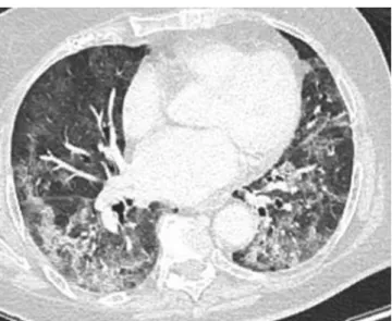
Fig.1– COVID-19pneumonia.Transversesectionof unenhancedchestCTscansshowingtypicallesionsof COVID-19pneumonia:groundglassassociatedwith consolidationandintralobularlines,sometimesarcuate, withperipheralanddecliningpredominance.
bosisina79-year-oldpatientaffectedbyCOVID-19-induced pneumonia.
Clinical
and
radiologic
observation
A79-year-oldwoman, withoutknown medicalhistory,was sufferingfrom fever,deteriorationofhergeneralcondition, andabdominalpainlocatedintheepigastricareaassociated with diarrheaover the previous 8days. As acute dyspnea addedtoitssymptoms,shewenttotheemergencyroom. Ini-tially,thepatient washemodynamicallystable(Blood pres-sure:168/89mmHg),showedsymptomsofacuterespiratory
insufficiencywithpolypnea(22cyclesperminuteswith86%of bloodoxygensaturationinambientair),andtachycardia(100 bpm).Bloodtestsrevealed C-reactiveprotein(125mg/L) in-creaseandahyperleukocytosis(12,600/mm3)with lymphope-nia.Bloodgasanalysisshowedcompensatedlacticacidosis (pH 7.43 withhyperlactatemia: 5.36 mmol/L).Nasopharynx PCRwasnegativeforSARS-CoV-2.
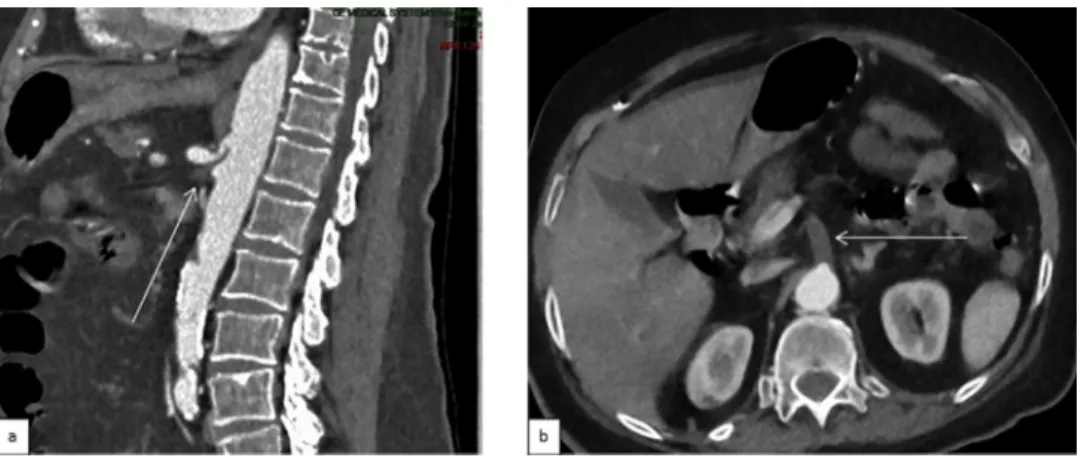
Twohoursafteradmission,anunenhancedchestCTscan displayedtypicalsignsofCOVID-19pneumonia[13](mostly ground-glass opacity without CT-backed evidence of an-other infection) with critical extent (>75% of pulmonary parenchyma;Fig. 1). Theupperabdominal slices showeda spontaneoushyperdensity(71Hounsfiedunits(HU)vs38HU intheportalvein)inthe rightportalvein(Figs.2aand4b). Further examination withenhanced CT scan of the chest, abdomen,and pelvisatthearterialand portalphases, per-formedatthesametime,confirmedaright-portalvein throm-bosis(Fig.2b)originatingfromthrombosisofthedistalpartof theuppermesentericveinextendedtothespleno-mesaraic trunk.Aproximalthrombosisoftheuppermesentericartery (Figs.3aand b)andjejunal artery werealsoobserved with subsequent featuresof bowel ischemiaof thecaecum and smallintestine(Figs.4aandb)withsmallamountofliquidin theperitonealcavity.Nopulmonaryembolismwasrevealed. Laparotomywasundertakenfewhourslaterandconfirmed ischemia,where ameterofnecrotic ileum andright colon were removed.Thrombolysisand thrombectomyofthe up-permesentericarterywerealsoperformedduringthesame procedure. Despitetimely treatment, hismedicalcondition stayedprecariousbecauseofextendedbowelischemiain ad-ditiontoseverelungdamagecausedbyCOVID-19.Palliative careswasimplementedandthepatientpassedaway4days later.
Discussion
COVID19isassociatedwithalargeand misleadingfield of symptoms[3]andcomplicationsincludingcoagulopathy[4]as
Fig.2– Imagingfeaturesofvenousthromboembolicdiseaseina79-year-oldCOVID-19patient.a:Nativetransverseupper abdominalsliceoftheunenhancedchestCTscanshowingaspontaneoushyperdensityintherightportalvein(white arrowhead).b:NativetransverseportalabdominalCTscanimageshowinganintraluminalthrombusoftherightportal vein(whitearrowhead).
Fig.3– Imagingfeaturesofarterialthromboembolicdiseaseina79-year-oldCOVID-19patient.a:Sagittalreconstructionof arterialabdominalCTscanshowingathromboticostialocclusionoftheuppermesentericartery(thinarrow).b:Native transversearterialabdominalCTscanimageshowingathromboticostialocclusionoftheuppermesentericartery(thin arrow).
Fig.4– Imagingfeaturesofbowelischemiaina79-year-oldCOVID-19patient.a:Obliquetransversereconstructionofportal abdominalCTscan.b:ObliquecoronalreconstructionofportalabdominalCTscanimage.Bothimagesshowaperfusion defectoftherightlargeintestine—lowerandmiddlethirdincludingcaecum—(whitearrowheads)relativetoanormalloop ofthesmallintestine(largewhitearrows)andanormalupperoftherightlargeintestine(thinwhitearrow).Right-portal veinthrombosiscanalsobeseen(blackarrowhead).
oftenseeninacuteinfection[5]includingInfluenza[6].This hypercoagulation status,which results indiseases suchas pulmonary embolism [7–9], is leading learned societies to askthemselves the questionofan anticoagulationtherapy atprophylaxis dose oreven higher[10,11]. Gastrointestinal symptomsreported withCOVID-19arenot specificand in-clude nausea, vomiting, diarrhea, and raised liver enzyme
[12].
Inourcase,NasopharynxPCRwasnegativefor SARS-CoV-2althoughCTscandisplayedtypicalsignsofCOVID-19 pneu-monia.Thisscenarioisfrequentlyreported[1]anddonot dis-provethediagnosis.
Asknown,chestCTscanisanessentialexamforthe diag-nosis,evaluationofextensionandcaremanagementmostly innonambulatorypatients.Moreover,itcouldhelpinpatient follow-upwithworseningclinicalconditions.
When CTscancontrolisneeded,itseemsreasonableto performwholebodyenhancedCTscanatarterialandvenous phasesespeciallyincaseofpulmonaryembolismsuspicion orabdominalpain.
Conclusion
Chest CT scan is essential in managing the COVID-19 pandemic and allows to uncover potential complications suchasthromboembolicdiseases.Closeattentionshouldbe payed to indirect signs of thrombosis on unenhanced CT scan.
R E F E R E N C E S
[1]AiTao,YangZhenlu,HouHongyan,ZhanChenao, ChenChong,LvWenzhi,etal.CorrelationofchestCTand RT-PCRtestingincoronavirusdisease2019(COVID-19)in China:areportof1014cases.Radiology2020.
doi:10.1148/radiol.2020200642.
[2]KimHyungjin.Outbreakofnovelcoronavirus(COVID-19): whatistheroleofradiologists?EurRadiol2020.
doi:10.1007/s00330-020-06748-2.
[3]ZhouMin,ZhangXinxin,QuJieming.Coronavirusdisease 2019(COVID-19):aclinicalupdate.FrontMed2020. doi:10.1007/s11684-020-0767-8.
[4]ZhangY,CaoW,XiaoM,LiYJ,YangY,ZhaoJ,etal.Clinical andcoagulationcharacteristicsof7patientswithcritical COVID-2019pneumoniaandacro-ischemia.ZhonghuaXue YeXueZaZhi2020;41(0):E006.
doi:10.3760/cma.j.issn.0253-2727.2020.0006.
[5]SmeethLiam,CookClaire,ThomasSara,JHallAndrew, HubbardRichard,VallancePatrick.Riskofdeepvein thrombosisandpulmonaryembolismafteracuteinfection inacommunitysetting.Lancet2006.
doi:10.1016/S0140-6736(06)68474-2.
[6]YangYan,TangHong.Aberrantcoagulationcausesa hyper-inflammatoryresponseinsevereinfluenza pneumonia.CellMolImmunol2016;13:432–42. doi:10.1038/cmi.2016.1.
[7] DanziGianBattista,Loffi Marco,GaleazziGianluca, GherbesiElisa.AcutepulmonaryembolismandCOVID-19 pneumonia:arandomassociation?EurHeartJ2020. doi:10.1093/eurheartj/ehaa254.
[8] FabreOlivier,RebetOlivier,CarjaliuIonut,RadutoiuMihai, GautierLaurence,HysiIllir.Severeacuteproximal
pulmonaryembolismandCOVID-19:awordofcaution.Ann ThoracSurg2020.doi:10.1016/j.athoracsur.2020.04.005.
[9] CellinaM,OlivaG.Acutepulmonaryembolisminapatient withCOVID-19pneumonia.DiagnIntervImaging2020. doi:10.1016/j.diii.2020.04.001.
[10]TangNing,BaiHuan,ChenXing,GongJiale,LiDengju, SunZiyong.Anticoagulanttreatmentisassociatedwith decreasedmortalityinseverecoronavirusdisease2019 patientswithcoagulopathy.JThrombHaemost2020 AcceptedAuthorManuscript.doi:10.1111/jth.14817.
[11]MariettaMarco,AgenoWalter,ArtoniAndrea, DeCandiaErica,GreselePaolo,MarchettiMarina,etal. COVID-19andhaemostasis:apositionpaperfromItalian SocietyonThrombosisandHaemostasis(SISET).Blood Transfus2020.doi:10.2450/2020.0083-20.
[12]WongSunnyH,LuiRashidNS,SungJosephJY.Covid-19and thedigestivesystem.JGastroenterolHepatol2020.
doi:10.1111/jgh.15047.
[13]YeZheng,ZhangYun,WangYi,ZixiangHuang,BinSong. ChestCTmanifestationsofnewcoronavirusdisease2019 (COVID-19):apictorialreview.EurRad2020.