HAL Id: hal-01081777
https://hal.archives-ouvertes.fr/hal-01081777
Submitted on 11 Nov 2014HAL is a multi-disciplinary open access archive for the deposit and dissemination of sci-entific research documents, whether they are pub-lished or not. The documents may come from teaching and research institutions in France or abroad, or from public or private research centers.
L’archive ouverte pluridisciplinaire HAL, est destinée au dépôt et à la diffusion de documents scientifiques de niveau recherche, publiés ou non, émanant des établissements d’enseignement et de recherche français ou étrangers, des laboratoires publics ou privés.
Fiber Optic Dielectric Nanoparticles Characterization
by Atom Probe Microscopy
H Francois-Saint-Cyr, I Martin, W Blanc, P Lecoustumer, C Hombourger, D
Neuville, D.J. Larson, T.J. Prosa, C Guillermier
To cite this version:
H Francois-Saint-Cyr, I Martin, W Blanc, P Lecoustumer, C Hombourger, et al.. Fiber Optic Dielectric Nanoparticles Characterization by Atom Probe Microscopy. International Microscopy Congress, Sep 2014, Prague, Czech Republic. �10.1039/C3CS60305A�. �hal-01081777�
Fiber Optic Dielectric Nanoparticles Characterization by Atom Probe
Microscopy
H. Francois-Saint-Cyr1, I. Martin1, W. Blanc2, P. LeCoustumer3, C. Hombourger1, D. Neuville4, D. J. Larson1, T. J. Prosa1, C. Guillermier5
1 CAMECA Instruments Inc., 5500 Nobel Drive, Suite 100, Madison, WI, 53711, USA 2 Université Nice Sophia Antipolis, CNRS, LPMC, UMR7336, 06100 Nice, France
3 Université Bordeaux 3, Géo-ressources et Environnement, EA4592, 33607 Pessac, France 4 Institut de Physique du Globe de Paris, 1 rue Jussieu, 75005 Paris, France
5 National Resource for Imaging Mass Spectroscopy, Cambridge, MA 02139, USA
The engineered processing of dielectric nanoparticles (DNPs) in optical fibers via luminescent ion-doping of silica-based glass aims at providing an enhanced spectroscopic behavior compared to pure silica. These DNPs should positively impact applications in high power fiber lasers, light sources with new wavelengths and telecommunications.
The prevalence of large phase immiscibility domains in silicate systems containing divalent metal oxides (Mg for instance) promotes the formation of DNPs through phase separation since heat treatments take place during the MCVD process.
Even after 60 years of glass-ceramics research, lack of experimental data concerning early nucleation stages imposes variations in composition and heat treatments as processing steps [1]. Although classical nucleation theory was the first model proposed to explain those phenomena, growth rate mismatches remain wide. According to this capillary assumption-based model, nuclei and bulk share similar structure-composition relationship. Recent articles disprove assumption of structure, pointing toward DNPs structural changes [2] and transition from amorphous nuclei to crystalline DNPs [3]. Compositional changes for small particle sizes (~1-10 nm) have been measured in alloys with Anomalous Small Angle X-Ray Scattering (ASAXS) [4] and in steels with Atom Probe Tomography (APT) [5]. Recent developments in APT has allowed the extension of such studies to glass-ceramics [6], and in the current work, we report experimental data disproving the second capillary assumption at the early stage of nucleation-growth process.
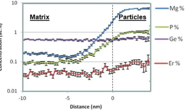
The atomic distribution map of Mg DNPs in silica-based glass doped with Mg, P, Ge and Er is reported in Figure 1 after APT analysis. In addition, quantitative assessment of Mg, P and Er content levels in DNPs smaller than 10nm in diameter (Figure 2) could refine the theories behind nucleation and growth mechanisms.
References
[1] N. Karpukhina et al., Chemical Society Review (2014), DOI 10.1039/C3CS60305A.
[2] S.Y. Chung et al, Nature Physics 5 (2009), p. 68. [3] P. Tan et al, Nature Physics 10 (2014), p. 73
[4] D. Tatchev et al, Journal of Applied Crystallography 38 (2005), p. 787. [5] M.D. Mulholland, D.N. Siedman, Acta Materialia 59 (2011), p. 1881.
Figure 1. Mg-based dielectric nano-particles (pink) surrounded by silica matrix (blue).
Figure 2. Proximity histogram displaying the evolution of Mg, Er, P, and Ge concentrations