HAL Id: hal-00085677
https://hal.archives-ouvertes.fr/hal-00085677
Submitted on 23 Jan 2007
HAL is a multi-disciplinary open access
archive for the deposit and dissemination of sci-entific research documents, whether they are pub-lished or not. The documents may come from teaching and research institutions in France or abroad, or from public or private research centers.
L’archive ouverte pluridisciplinaire HAL, est destinée au dépôt et à la diffusion de documents scientifiques de niveau recherche, publiés ou non, émanant des établissements d’enseignement et de recherche français ou étrangers, des laboratoires publics ou privés.
Near-UV Resonant Two-Photon Ionization Spectroscopy
of Gas Phase Guanine: Evidence for the Observation of
Three Rare Tautomers
M. Mons, F. Piuzzi, I. Dimicoli, L. Gorb, J. Leszczynski
To cite this version:
M. Mons, F. Piuzzi, I. Dimicoli, L. Gorb, J. Leszczynski. Near-UV Resonant Two-Photon Ionization Spectroscopy of Gas Phase Guanine: Evidence for the Observation of Three Rare Tautomers. Journal of Physical Chemistry A, American Chemical Society, 2006, 110, pp.10921-10924. �10.1021/jp063738x�. �hal-00085677�
Near-UV Resonant Two-Photon Ionization Spectroscopy of Gas Phase Guanine:
Evidence for the Observation of Three Rare Tautomers
Michel Monsa*, François Piuzzia, Iliana Dimicoli,a Leonid Gorbb,c,d and Jerzy Leszczynskib
aLaboratoire Francis Perrin (URA CEA CNRS 2453), Service des Photons, Atomes et Molécules, CEA Saclay, Bât. 522, 91191 Gif-sur-Yvette Cedex, France
bComputational Centre for Molecular Structure and Interactions, Dept. of Chemistry,bJackson State University, P.O. Box 17910, 1325 Lynch St., Jackson, MS 39217,USA
cUkrainian-American Laboratory of Computational Chemistry, Kharkiv, Ukraine, and Jackson, Mississippi
dDepartment of Molecular Biophysics, Institute of Molecular Biology and Genetics, National Academy of Science of Ukraine, 150 Zabolotnogo Street, Kyiv 03143, Ukraine
In the light of a recently published study on the IR spectroscopy of guanine in He droplets (Choi and Miller, JACS 2006, 128, 7320), the present letter proposes a new interpretation of the resonant two-photon ionization (R2PI) experiments on gas phase guanine, which is supported by quantum chemistry calculations. Whereas He droplet experiments detect the most stable forms, only one of these forms is observed (very marginally) in the R2PI spectrum, which is actually dominated by three less stable “rare” tautomers, whose stabilities lie in the 3-7 kcal/mol range. The absence of the most stable forms in the R2PI spectrum suggests that a tautomer-dependent ultra-fast relaxation process takes place in the excited state of these stable tautomers. The present reinterpretation modifies qualitatively the picture of the excited state of guanine tautomers and should contribute to understand the deactivation mechanisms taking place in the excited state of DNA bases.
Understanding the near-UV absorption of DNA bases is a key issue for the study of photoinduced damages to DNA material. In this context, the near-UV spectrum of guanine in a supersonic expansion has been widely investigated using nanosecond laser resonant two-photon ionization (R2PI).1-8 Using the IR/UV double resonance technique, Nir et al. found vibrationally resolved contributions of three different tautomers (A, B and C in Fig. 1) and assigned them to 3 forms among the most stable of guanine : one hydroxy-amino (enol) form (either 3 or 4), and two oxo-amino (keto) forms 2 and 1 respectively, by comparison between IR experimental and calculated spectra.3 Using the
same experimental technique, our group confirmed the observation of the three forms A-C and identified the
contribution of a fourth tautomer D.6,8 Based on a
comparison with the UV spectra of 7- and 9-methylated species6 and of hydrates,8 we concluded to different
assignments in terms of 7/9H tautomerism for the A, B and C features: i.e., 5, 1 and 2, respectively. In particular, the red-most intense band system (A), assigned to the N9H enol form (either 3 or 4) by the de Vries’ group was assigned by us to a higher energy form: 7H enol syn 5. Finally, the fourth tautomer D observed was assigned to the 9H enol
form 4.6,8 Paradoxically, the R2PI spectrum of
9-methylguanine (9MG) is a much simpler, narrow-range, band system (Fig. 1) and found to be due to a unique form: the same 9H enol anti 4 tautomer as the D form of
Chart 1. Most stable9-11 tautomers of guaninea
a: 1,2 and 6 are oxo-amino (keto); 3-5: hydroxy-amino (enol) and 7-8: oxo-imino forms. Relative energies 9-11 are in the following energy ranges:
1-4 : 0-1 kcal/mol, 5 ~3 kcal/mol and 6-8 ~ 7 kcal/mol. The next forms are
found to be in the 12 kcal/mol range.9 Although the precise relative energies depend on the level of theory used, these data provide an overview of the conformational landscape.
guanine.6,8 The enol syn counterpart 3 and the
biologically relevant keto form 2, calculated to be of similar stability, are surprisingly missing, which suggested that 9-methylation could have a dramatic effect on the excited
dynamics of these tautomers.8 However, the several
theoretical studies focused on this point failed to account for this so-called 9-methylation effect.12,13 The issue was
left at this point, until a very recent work by Roger Miller’s group14 provided relevant enlightening data, which enables
Figure 1. R2PI spectrum of guanine and 9-methylguanine as obtained with
a ns-UV laser.6,8 The origins of the four tautomers identified are indicated by arrows. Assignments of A and D to forms 5 and 4, respectively, are based upon comparison with 7- and 9-methylated species. B and C features are assigned to oxo-imino conformers, 8 and 7 respectively, by comparison with theoretical calculations, see Fig. 2.
us to propose an elegant way to rationalize the puzzling UV R2PI spectroscopy of guanine.
R. Miller and his coworkers, working with molecules embedded in He droplets, have developed a very powerful method to assign conformational contributions in vibrational spectra, based upon comparison between the vibrational transition moment angles determined from experiment and theory.15 Applied to adenine and cytosine,
these studies led to IR spectra nearly identical to those obtained in the gas phase,16 as expected from a soft matrix
like suprafluid He. For guanine, the same methodology14
provides an unambiguous evidence for population of four tautomers under the He droplet environment. The very
convincing agreement of the complete experimental set of data with the quantum chemistry calculations enables Choi and Miller to assign the IR spectrum (NH stretch region) to a mixture of the four most stable forms (1-4), namely the 7Hketo, 9Hketo, 9Henol syn and 9Henol anti tautomers (Fig. 2).14 The comparison with IR/UV double resonance
spectroscopy,3,4,6,8 however, is quite intriguing. In contrast to what is generally observed for other molecules, the IR He droplet data are not found to match the IR/UV spectra (Fig. 2). Only one form observed in He droplets14 is also found in
the R2PI experiment under isolated conditions:6,8 the 9H
enol anti 4 tautomer observed in He droplets exhibits IR spectral features nearly identical to those of the D form observed in R2PI experiments, with relative frequency differences of the order of 1 cm-1 (Table 1). No such close counterpart can be found among the IR/UV spectra for the three other conformers (1-3) observed in He droplets (Fig. 2). Even the A form, assigned to an oxo-amino (enol) form,3,6 does not match the second oxo-amino rotamer 3 in
He droplets. This demonstrates clearly that the A-C forms
observed in the R2PI experiment are not the same tautomers as those observed in He droplets: in other
words, the two experiments actually do detect different tautomer populations.
Since the R2PI spectra reported are all mass-selected, the influence of impurities or of fragmenting clusters, whose UV signature would be seen on both cluster and guanine ion channels is easily ruled out. Therefore, the immediate consequence is that the forms (A-C) which are responsible for the intense features observed in the R2PI spectrum of guanine should actually be due to high-energy “rare” tautomers, whose weak population cannot be detected under cold He droplet conditions. In the following, we will propose an assignment of A-C in terms of tautomers and
discuss the possible reasons for the absence of the most stable tautomers in the R2PI spectrum.
Assignment of the A-D forms observed in the R2PI spectra. Our assignment of the D species to the 9Henol anti
form 46,8 by comparison with the UV spectrum of 9MG is
in agreement with the independent analysis of Choi and
Miller.14 This form is the only form observed
simultaneously in both experiments. Concerning A, again the comparison with 7MG led us to assign it to the 7H enol syn form 5.6 The low stability argument of this form, which
could be opposed so far, now obviously disappears, since, apart from D, the observed tautomers must be rare forms. With respect to B and C features, the assignment in terms of oxo tautomerism is not questionable because of the absence
of the remarkable features in the 3580 cm-1 region,
characteristic of the hydroxy forms (Fig. 2).3,4,6,8 Again,
comparison with the IR spectra of tautomers 1 and 2, measured by Choi and Miller, is informative, in particular
concerning the symmetric and asymmetric NH2 stretch
bands (Fig. 2). This NH2 doublet, easily detected by Choi
and Miller,14 is not present in the IR/UV spectra of forms
B and C.3,6 This suggests that the rare tautomers
responsible for these features might be oxo-imino species, like 7 or 8, rather than oxo-amino forms, as considered so far.3,4,6,8
In order to confirm this point and to propose a more refined assignment, quantum chemistry calculations of the optimized geometries and IR harmonic frequencies of the
1-8 forms have been carried out at the B3LYP/6-31+G(d)
level of theory (Table S1), which was previously successfully used to account for the enol rotamers (3-5) of guanine derivatives.8 The results, depicted to the relative
weakness of the imino NH bond, was not observed.4,6 The
N3H band, however, gives rise to a characteristic prominent absorption in the 3490 cm-1 range. In spite of a strong
similarity between the two IR spectra, the details of the band patterns3 are well reproduced by theory, suggesting to
assign the B and C band systems of Fig. 1 to the 8 and 7 forms respectively. One will notice that the other rare 7H oxo-amino form 6 does not match any IR spectrum experimentally available and is therefore observed neither in He droplets, nor in R2PI spectra.
The impressive match of the theoretical IR spectra with their experimental counterparts provides a good confidence for the assignment and illustrates the level of prediction which can be expected from quantum chemistry. The poor agreement observed previously, together with spectroscopic inconsistencies discussed in Ref 6, were actually the
indication of a misassignment. These clues were discarded on the legitimate basis that species observed under supersonic expansion conditions should be a priori the most stable species, namely forms 1 and 2.
The present reassignment also solves the puzzling so-called 9-methylation effect8: the most intense band systems
in R2PI spectra do not correspond to the most stable forms but to rare 7H tautomers: the biologically relevant 9H keto
2 and the 9Henol syn 3 forms are therefore missing in both
the R2PI spectrum of 9-methylguanine and that of guanine.
Gas phase R2PI vs. He droplets. There is no obvious
reason to expect that the tautomer populations should be so different in the two experiments and the explanation for the dichotomy observed should be found elsewhere. The detection schemes in both experiments are quite different.
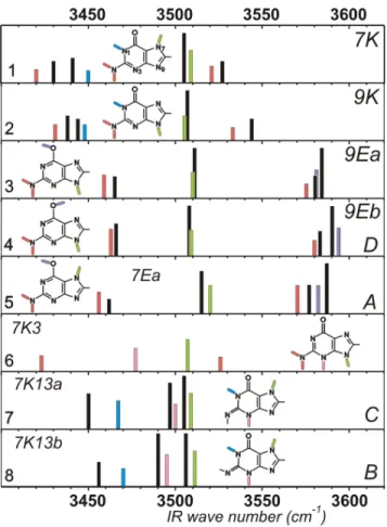
Figure 2. Theoretical IR spectra (B3LYP/6-31+G(d) harmonic frequencies
of the NH/OH stretches) of the 8 most stable forms of Chart 1 (color) compared to the spectra of the seven tautomers of guanine observed experimentally (black). The top four panels correspond to the 4 tautomers observed by Choi and Miller in He droplets (order of decreasing stability).14 Panels 5, 7 and 8 match the experimental IR data of forms A, C and B, respectively, observed in the R2PI spectrum of guanine.3,6,8 The D form observed in R2PI corresponds to the G9Eb form observed by Choi and Miller and exhibits the same band positions at the scale of the figure (the weak band at 3466 cm-1 could not be observed in our modest signal-to-noise ratio IR/UV spectrum6). Colors indicate the nature of the stretching modes: NH2: red; N7/9H: green; N1H: blue, OH: violet, N3H: pink. Labeling of the spectral features on the right are those of Choi and Miller14 (He droplet data) as well as those of Mons et al.6 (R2PI data). Calculated NH and OH stretch frequencies are scaled by adjusted specific factors (0.963 and 0.971 respectively). Calculated spectra are displayed on an arbitrary smaller intensity scale for the sake of clarity.
In He droplet studies, IR absorption spectra are measured as a directly consequence of the energy absorbed. In contrast, gas phase IR/UV double resonance measures population depletions, which are monitored using a selective detection process based on UV absorption, namely by collecting either fluorescence from the excited state or ions produced through a R2PI scheme. This procedure implicitly requires that the lifetime of the excited state is long enough for allowing fluorescence emission or photoionisation during the laser pulse. In the R2PI case, the ionization potential (IP) of the species probed should also not be too high to be reached by two-photon excitation.
The absence of the most stable forms (1-3) in the R2PI experiments can be due a priori to several factors: i) a high IP for these species could forbid the two-photon ionization, ii) unfavorable Franck-Condon overlap might forbid the adiabatic excitation to the ππ* state and therefore lead to a significantly blue-shifted, congested and complicated
spectrum iii) additionally, an intrinsic ultrafast relaxation process17 can quench the excited state population either as
soon as the origin of the ππ* state or after some excess energy and make therefore the absorption spectrum either severely broadened or quite narrow-ranged, and, finally, iiii) the absorption features may not be located in the spectral region probed so far. All these factors might combine together to forbid us the observation of the stable tautomers in the previous experiments, even if, so far, a wide spectral range has been investigated. Using R2PI, the 32150-35700 cm-1 range has been scanned, including a two-color R2PI scheme,1 in order to keep the capability to photoionize high
IP species (second color at 193 nm). The upper part of this range (32700 to 35700 cm-1) region was also probed by fluorescence, which is not impeded by the high IP issue. A possible explanation for the missing features might be that the keto forms 1 and 2 exhibit very red-shifted UV transitions, in a region not yet investigated, even if this does not seem to be a definitive argument for three reasons. First, the ππ* adiabatic transitions of the 1-4 conformers have been predicted to lie in the same energy range.12,18 Second,
calculations also suggest a very large change in equilibrium geometry between the ground and the first ππ* state. 11-13,17-21 Consequently, the Franck-Condon envelop is expected to
be broad, comparable in spread to that observed by R2PI for the A-C forms.1,3-8 Finally, the second rotamer of the 9H
enol-amino 3 form, whose spectrum is expected to lie in the vicinity of its twin form 4, could not be detected, neither in guanine nor in 9MG.
All these facts suggest the occurrence of a fast non-radiative relaxation of the excited state of the 1-3 forms which would prevent their observation in both the R2PI and fluorescence experiment. This is supported by the following points: First, the weird narrow range spectrum of the D form (4), composed of a few fluorescent bands in guanine and 9MG, compared to the rich and long spectra of forms A or C (Fig. 1), suggests a dramatically sudden onset of relaxation occurring a few hundreds of wavenumbers above the ππ* origin transition. Recent calculations indicate that in the biologically relevant 2 form, an internal conversion process, controlled by a conical intersection (CI) with the ground state, is expected to occur close to the origin of the excited state (~1.7 kcal/mol).17 Such a process would
qualitatively account for the sudden quenching of the excited state of D and also possibly for the 3 other forms since one can expect the location of the conical intersection to be tautomer-dependent. A CI located closer to the origin would make the excited state ultrashort-lived, forbidding its observation by R2PI or fluorescence. This is also consistent with the ultrashort lifetime measured for the ππ* excited state of guanine (actually a mixture of its four most stable tautomers) at 267 nm, which is the shortest of all the DNA bases.22
In summary, the present letter rationalizes the gas phase and He droplet measurements of the literature on guanine in terms of observation of seven tautomers. Whereas He droplet experiments detect the most stable (1-4) tautomers, only one of these forms (4) is observed (very marginally) in the R2PI spectrum, which is actually dominated by three less stable “rare” tautomers (5, 7 and 8), whose stabilities lie in a 3-7 kcal/mol range. The present finding radically modifies the qualitative picture of the excited state dynamics of the guanine tautomers suggesting the
Table 1. Experimental IR band frequencies vs. theoretical harmonic NH stretch frequencies (B3LYP/6-31+G(d) level) for the four tautomers observed in the R2PI experiments.3,6 Tautomer / UV band system origin transition6 Experimental Frequency He droplet 14 Experimental Frequency R2PI Scaled Harmonic frequency (cm-1) a Intensity (km/mol) Assignment 4 N9H hydroxy-amino 3590.6 3590 3594 81 OH
(9H enol anti) 3583.2 3583 3580 45 NH2 antisym.
D 3509.6 3508 3509 73 N9H
origin : 34755 cm-1 3466.1 not observed 3463 75 NH2 sym.
5 N7H hydroxy-amino 3587 3582 80 OH
(7H enol syn) 3577 3570 40 NH2 antisym.
A 3516 3 ; 3515 6 3520 83 N7H
origin : 32864 cm-1 3462 3456 58 NH2 sym.
7 N7H oxo-imino 3505 3; 3503 6 3509 104 N7H
C 3497 3; not observed in 6 3501 66 N3H
origin : 33910 cm-1 3450 3; 3449 6 3467 76 N1H
not observed 3375 11 NH imino
8 N7H oxo-imino 3506 3; 3504 6 3511 103 N7H
B 3490 3; not observed in 6 3495 94 N3H
origin : 33269 cm-1 3456 3470 49 N1H
not observed 3386 13 NH imino
a: NH and OH stretches are scaled by specific factors (0.963 and 0.971 respectively).
occurrence of a tautomer-dependent fast relaxation process in the excited state. Additional experiments will be useful in order to support the present interpretation. In particular systematic gas phase scans either using two-color R2PI or fluorescence will be carried out to find spectral features of the “missing” tautomers. In this context, He droplets experiments on guanine and 9MG as well could also be welcome in order to determine the electronic absorption region of the 4 most stable forms.
The assignment of the R2PI spectra in terms of rare species is expected to be of broad significance since it may affect any flexible system whose excited state dynamics is strongly conformation-dependent. This should be expected in particular for complexes of guanine.23-27 As an example, the IR spectra of three conformers of guanine-water detected by R2PI26 exhibit common IR features with the
minor tautomers of the guanine R2PI spectrum, suggesting that one observes hydrates of the rare tautomers. In the same spirit, guanine-containing dimers might be formed between one of the most stable forms and one of those actually detected by R2PI, which suggests enlarging the set of possible structures to account for observations.23,27
Finally, the difficulties encountered in recording the near-UV spectrum of the most stable tautomers of guanine, including the biologically relevant form, emphasize the importance of theoretical studies as a relevant alternative to experiment. Providing accurate electronic transition energies of the seven tautomers observed would greatly help spectroscopists to find the probably weak features of the “missing” tautomers. Alternatively, the presently revised experimental data pertaining to an enlarged set of tautomers should help understanding and refining the mechanism controlling the excited state relaxation dynamics in the DNA bases,2,11-13,17-20,22,28 in particular its dependence with
the tautomeric form.
Acknowledgment. This work is dedicated to the memory
of R.E. Miller, deceased in 2005.
Supporting Information Available: Table S1: Calculated
harmonic NH/OH stretch frequencies of the 8 forms of Chart 1. This material is available free of charge via the Internet at http://pubs.acs.org.
References
(1) Nir, E.; Grace, L.; Brauer, B.; de Vries, M. S. J. Am. Chem. Soc. 1999,
121, 4896.
(2) Nir, E.; Kleinermanns, K.; Grace, L.; de Vries, M. S. J. Phys. Chem. A
2001, 105, 5106.
(3) Nir, E.; Janzen, C.; Imhof, P.; Kleinermanns, K.; de Vries, M. S. J. Chem.
Phys. 2001, 115, 4604.
(4) Nir, E.; Plutzer, C.; Kleinermanns, K.; de Vries, M. Eur. Phys. J. D 2002,
20, 317.
(5) Piuzzi, F.; Mons, M.; Dimicoli, I.; Tardivel, B.; Zhao, Q. Chem. Phys.
2001, 270, 205.
(6) Mons, M.; Dimicoli, I.; Piuzzi, F.; Tardivel, B.; Elhanine, M. J. Phys.
Chem. A 2002, 106, 5088.
(7) Chin, W.; Mons, M.; Dimicoli, I.; Piuzzi, F.; Tardivel, B.; Elhanine, M.
Eur. Phys. J. D 2002, 20, 347.
(8) Chin, W.; Mons, M.; Piuzzi, F.; Tardivel, B.; Dimicoli, I.; Gorb, L.; Leszczynski, J. J. Phys. Chem. A 2004, 108, 8237.
(9) Ha, T. K.; Keller, H.-J.; Gunde, R.; Gunthard, H.-H. J. Phys. Chem. A
1999, 103, 6612.
(10) Hanus, M.; Ryjacek, F.; Kabelac, M.; Kubar, T.; Bogdan, T. V.; Trygubenko, S. A.; Hobza, P. J. Am. Chem. Soc. 2003, 125, 7678.
(11) Gorb, L.; Kaczmarek, A.; Gorb, A.; Sadlej, A. J.; Leszczynski, J. J. Phys.
Chem. B 2005, 109, 13770.
(12) Langer, H.; Doltsinis, N. L. Phys. Chem. Chem. Phys. 2004, 6, 2742. (13) Cerný, J.; Špirko, V.; Mons, M.; Hobza, P.; Nachtigallová, D. Phys.
Chem. Chem. Phys. 2006, accepted; doi: 10.1039/b601629d.
(14) Choi, M. Y.; Miller, R. E. J. Am. Chem. Soc. 2006, 128, 7320 (15) Dong, F.; Miller, R. E. Science 2002, 298, 1227.
(16) Plützer, C.; Nir, E.; de Vries, M. S.; Kleinermanns, K. Phys. Chem.
Chem. Phys. 2001, 3, 5466.
(17) Chen, H.; Li, S. H. J. Chem. Phys. 2006, 124, 154315. (18) Shukla, M. K.; Leszczynski, J. J. Phys. Chem. A 2005, 109, 7775. (19) Langer, H.; Doltsinis, N. L.; Marx, D. ChemPhysChem 2005, 6, 1734. (20) Sobolewski, A. L.; Domcke, W.; Hattig, C. Proc. Natl. Acad. Sci. U. S.
A. 2005, 102, 17903.
(21) Mennucci, B.; Toniolo, A.; Tomasi, J. J. Phys. Chem. A 2001, 105, 7126.
(22) Canuel, C.; Mons, M.; Piuzzi, F.; Tardivel, B.; Dimicoli, I.; Elhanine, M.
J. Chem. Phys. 2005, 122, 074316.
(23) Nir, E.; Janzen, C.; Imhof, P.; Kleinermanns, K.; de Vries, M. S. Phys.
Chem. Chem. Phys. 2002, 4, 732.
(24) Nir, E.; Janzen, C.; Imhof, P.; Kleinermanns, K.; de Vries, M. S. Phys.
Chem. Chem. Phys. 2002, 4, 740.
(25) Abo-Riziq, A.; Grace, L.; Nir, E.; Kabelac, M.; Hobza, P.; de Vries, M. S. Proc. Natl. Acad. Sci. U. S. A. 2005, 102, 20.
(26) Crews, B.; Abo-Riziq, A.; Grace, L.; Callahan, M.; Kabelac, M.; Hobza, P.; de Vries, M. S. Phys. Chem. Chem. Phys. 2005, 7, 3015.
(27) Bakker, J. M.; Compagnon, I.; Meijer, G.; von Helden, G.; Kabelac, M.; Hobza, P.; de Vries, M. S. Phys. Chem. Chem. Phys. 2004, 6, 2810. (28) Fulscher, M. P.; SerranoAndres, L.; Roos, B. O. J. Am. Chem. Soc.