134 Dahah et al.
RESEARCH PAPER OPEN ACCESS
Antimicrobial, antioxidant and hemolytic effects of Pyocyanin
produced by
Pseudomonas aeruginosa
isolated from saline soil
of Mina river, Algeria
Hicham Dahah
*1, Rachid Djibaoui
1, Saïd Nemmiche
21
Laboratory of Microbiology and Vegetal-Biology, Department of Biology, Faculty of SNV,
University of Mostaganem, Mostaganem , Algeria
2
LSTPA Laboratory, Department of Biology, Faculty of SNV, University of Mostaganem,
Mostaganem , Algeria
Key words:Pseudomonas aeruginosa, Pyocyanin, Antimicrobial, Antioxidant effect
http://dx.doi.org/10.12692/ijb/9.5.134-143
Article published on November 30, 2016
Abstract
Pseudomonas aeruginosa is a common inhabitant of soil, fresh-water and marine environments. Pyocyanin is
one of the stronger antimicrobial substance produced by this bacterium inhibiting a wide range of pathogenic microorganisms. In order to exploit the antimicrobial and antioxidant effects of pyocyanin and abrogate some pathogenic microbes responsible for several diseases, three strains (P1, P2 and P3) of Pseudomonas aeruginosa were isolated from saline soil of Mina river region (Relizane, Algeria). The higher producer of pyocyanin (P3) was selected for further studies. Pyocyanin was produced in King A broth medium, extracted with chloroform then purified by silica gel chromatography. The characterization of the metabolite by UV spectrum, TLC, IR and HPLC revealed a similarity with the pyocyanin standard. The effect of NaCl on pyocyanin production was determined by using different concentrations and the maximum amount was detected in the medium containing 20g/l. The antimicrobial effect results showed varying degrees of inhibition zones against the microorganisms tested and the remarkable effect were detected against Candida albicans ATCC 10231 and Bacillus cereus ATCC10876 with zones diameters of 26 mm and 14 mm respectively. Hemolytic activity using human blood was obtained above 5mg/ml and the IC50 value of the antioxidant activity of pyocyanin with DPPH method was evaluated at 3.15µg/ml.
*
Corresponding Author: Hicham Dahah hicham.dahah@univ-mosta.dz.
International Journal of Biosciences | IJB |
ISSN: 2220-6655 (Print) 2222-5234 (Online) http://www.innspub.net Vol. 9, No. 5, p. 134-143, 2016
135 Dahah et al.
Introduction
Pseudomonas aeruginosa is a Gram negative, obligate
aerobe, rod shaped bacterium belonging to the family of Pseudomonadaceae. It is largely widespread in the environment by an ability to develop in soil, plants, fresh and marine water. The metabolism of P. aeruginosa was significantly controlled in diverse ecological niches by the degree of salinity and other environmental factors. It is also known to produce pyocyanin (5-N-methyl-1-hydroxy phenazine (PCN) which is the major phenazine compound in this species (Rangarajan et al., 2003; Prabhakaran et al., 2014).
Interest is now growing in the use of antimicrobial drugs that directly target the expression of virulence factors. PCN has various pharmacological effects on prokaryotic cells; its biologicalactivity is related to similarity in the chemical structure to isoalloxazine, lipoproteins, flavin mononucleotide and flavin adenine dinucleotide compounds (Ohfuji et al., 2004). A variety of potential biotechnological applications of PYO were found, as production of the antitumor (Laursen and Nielsen, 2004; Mavrodi et al., 2006) and the ability to control the pathogenic fungi and bacteria. This secondary metabolite has been studied intensively and has drawn the attention of the researchers for its broad spectrum antibiotic properties against fungi (Candida
albicans, Aspergillus funmigatus) (Costa and Cusmane,
1975; Kerr et al., 1999) and a high antibacterial activity against (Bacillus cereus, Micrococcus luteus, Staphylococcus aureus, Escherichia coli) and many
pathogenic microbes infecting human body.
The broad spectrum of pyocyanin is basically due to its ability to regenerate an exceed of O2- and H2O2 during respiration in cells membranes of other microorganisms inhibiting the energy process and active transport of aqueous solution (Baron et al., 1989; Usher et al., 2002). Pyocyanin increases intracellular oxidant stress and exhibits a redox cycle under aerobic condition. However,
Pseudomonas aeruginosa protect themselves against
pyocyanin production with high level of superoxide dismutase and catalase (Price-Whelan et al, 2007). In additional, the major role of pyocyanin in the producing bacterium is its important persistence in absence of other competitors (Whelan et al., 2006; Price-Whelan et al., 2009).
The study was designed to investigate antimicrobial and antioxidant activities of PCN against a number of pathogenic microorganisms, specifically, Bacillus cereus ATCC 10876, Escherichia coli ATCC 25922, Proteus mirabilis ATCC 35659, Proteus vulgaris ATTC 6380 and Candida albicans ATCC 10231.
Material and methods
Isolation of Pseudomonas aeruginosa
Fluorescents Pseudomonas was isolated from saline soil of Mina river region (Relizane, Algeria). Soil samples were collected from different areas in sterile plastic bags then transported to the laboratory. The bacteria were isolated by suspending 10g of soil in a sterile flask containing 90ml of sterile physiological water. Isolates and standard Pseudomonas aeruginosa strain were grown in King B agar
medium (King et al., 1954). The plates were incubated for 48 hours at 28°C. Only isolates producing fluorescence on King B agar medium were streaked on King A agar medium to select only those producing bleu pigment.
Pseudomonas strains identification
Fourteen isolates were identified by phenotypic characterization to Pseudomonas. Therefore we realized the Gram stain, motility test and oxidase test (growth at 42°C and 4°C). All isolates were identified with API 20 NE identification system (API NE, bio Merieux, France) and kept in freezer at -20°C. The strains identified as
Pseudsomonas aeruginosa by biochemical and
physiological characterizations were approved by the genotypic identification.
The genomic DNA of isolates were extracted, purified and amplified with primers 27F (5’AGAGTTTGATC MTGGCTCAG-3’) and 1492R (5’-TACGGYTACCTTG TTACGACTT-3’) and using thermocycler ABI 9700. The PCR products were sequenced. The sequence determined was compared with the reference species of Pseudomonas contained in genomic database banks, using the ‘‘NCBI Blast’’.
136 Dahah et al.
Extraction of pyocyaninTo extract the crude pyocyanin, we select one isolate belonging to P. aerations and showing high bleu pigmentation on King A agar medium. The selected strain was inoculated in the King A broth medium, incubated for 3 days in rotary shaker (180 rpm at 30°C). The culture was centrifuged and the supernatant was extracted with 1v/1v chloroform. The organic phase was concentrated in a rotary evaporator at 50ºC.
Antimicrobial activity
The Antimicrobial effect of purified pyocyanin was determined by the disc diffusion assay (Barry and Thornsberry, 1985). Bacterial turbidity was adjusted to McFarland standard (0.5 McFarland: 108 UFC/ ml) and suspension was spread on the solid media plates (Mueller–Hinton). The paperdiscs (Whatman paper Grade AA) were impregnated with the solution of extracted pyocyanin, placed on the plates and incubated for 24 h at 37°C. Antibacterial activity was determined by the diameter of inhibition zones (mm) around the wells. All tests were performed in triplicate.
Determination of the Minimal Inhibiting Concentration (MIC)
The minimum inhibitory concentration of pyocyanin was evaluated by broth microdilution. All pathogens microorganisms were suspended in broth cultures for 18 hours and adjusted to obtain a 0.5 McFarland standard turbidity. The purified pyocyanin was added in serial concentrations (8 µg/mlto 128µg/ml) for all inoculums suspensions prepared and incubated at 37°C for 24 hours. The MIC was determined as the lowest concentration of pyocyanin able to inhibit any visible growth of each microorganism in the broth medium.
Effect of salinity on the growth and pyocyanin production
To verify the influence of salinity on the growth and pyocyanin production of the isolate; the bacterium was inoculated in King A broth supplemented with 2.5, 5, 7.5, 10, 12.5, 15, 17.5,20 and 30g/l of NaCl. Cultures were incubated in a shaker incubator (180 rpm at 30°C) for 48h.
The growth was determined visually and the pyocyanin production was evaluated using the method described by Kurachi (1958). All experiments were done in triplicate.
Hemolytic activity of pyocyanin pigment
The human blood was added in tube containing EDTA and centrifuged at 5000rpm for 10min at 4°C. The supernatant was discarded and the packed RBC was washed by normal saline. 1ml of the packed RBC was resuspended in normal saline to obtain 1% RBC suspension. The assay was carried in microtiter plate and the pyocyanin compound was assayed at different concentration. 100µl of normal saline and 100µl of 1% RBC were added to each well, and then different concentrations of pyocyanin were added. The microtiter plate was incubated for 3h at room temperature. The negative reaction was indicated by observing a fin button cell with regular margin and the uniform red colored suspension indicated positive of the lyses RBC (Samanta et al., 2008).
Antioxidant activity of pyocyanin
DPPH radical scavenging activity of purified pyocyanin was measured as described by Liyana and Shahidi (2005). Various concentrations of pyocyanin (2.8, 1.4, 0.7, 0.350 and 0.175µg/ml) were prepared in methanol. 2 mL of 24µg/ml DPPH solution was mixed with 0.5ml of each concentration of pyocyanin and incubated at room temperature for 30min. Ascorbic acid was also used as positive control. The absorbance of all solutions was read at 517nm by using a spectrophotometer.
Analysis of pyocyanin pigment with UV-VIS spectrophotometer
Extraction and purification of Pyocyanin
3 ml of chloro form was added to 5ml culture supernatant. After extraction, the chloroform layer was transferred to a fresh tube and mixed with1ml 0.2 M HCl.
After centrifugation, the red top layer was analyzed in range 200 to 500nm using UV-V is spectrophotometer (JASCO V-530) and the maximum absorbance was detected.
137 Dahah et al.
The crude extract was purified by using silica gel column and the obtained bleu fraction was analyzed by thin layer chromatographic method (TLC) to determine the purity of PCN compound. A standard pyocyanin was spotted on the first position and the purified PCN was spotted on the second position. The TL Cplat was developed in chloroform-methanol (1:1 v/v) and after migration of compounds the Rf values were calculated and compared.
Identification of the pyocyanin compounds by high-performance liquid chromatography (HPLC/DAD)
The purified pyocyanin was performed at 280nm on analytical HPLC/DAD with C18 column (250 x 4.6 mm) and DAD Shimatzu SPD-M20A detector. The analysis was controlled by Lab Solution LC-PDA software. In the method of elution samples we used two solvents A and B. The first was water-trifluroacetic acid (100:0.04, v/v) and the second was acetonitrile-water-trifluroacetic acid (90:10:0.04, v/v/v). The flow rate was 1ml/min, and the injected volume was 1µL. All chromatographic analysis was performed at 30°C (Fernández and Pizarro, 1997).
Identification of pyocyanin by FTIR
The structure of pyocyanin extract was confirmed with Jasco 4200 FT-IR spectrophotometer by analysis of the functional groups of the substance used. The KBr was heated at 110°C to eliminate humidity and results were treated with Jasco Spectra Manager II software.
Results
Pseudomonas identification
The obtained isolates are rode shaped gram-negative, motile and showed positive oxidase reaction. They also grow aerobically and all of them produce yellow green pigment on King B medium. Consequently they are belonged to the group of fluorescent
Pseudomonas and only the isolates Pa1, Pa2 and Pa3,
produced a bleu pigment, showed a positive reaction with gelatinase and grow at 42°C (but not at 4°C). The API 20NE system used confirms by APIW eb software that Pa1, Pa2 and Pa3 have a similarity of 99.9%, 91.9% and 99.9% respectively to Pseudomonas
aeruginosa.
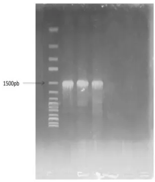
The numerical profiles were 1354475, 1356575 and 1356457 successively. The amplification of 16S rDNA of P1, P2, and P3 using primers revealed a fragment DNA of 1.5kb (Fig. 1) which was identified by sequencing as P. aeruginosa F 9670 with a similarity of 99%. Only the isolate P1 was selected for the further experiments.
Fig. 1. Agarose gel electrophoresis of the polymerase
chain reaction (PCR) amplified 16S-rRNA gene for P1, P2 and P3.
Antimicrobial activity of the pyocyanin
The spectrum of antimicrobial activity againsta Gram-positive bacteria (Bacillus cereus ATCC 10876), Gram negative bacteria (Proteus mirabilis ATCC 35659, Proteus vulgaris ATCC 6380 and Escherichia
coli ATCC 25922) and yeast (Candida albicans ATCC
10231) is presented in Table 1.
The results indicate that pyocyanin antibiotic showed antimicrobial activity against all pathogenic microorganisms tested. Pyocyanin exhibited a highest activity against Candida albicans ATCC 10231 and
Bacillus cereus ATCC 10876 with average inhibition
zones diameters of 26mm and 14mm respectively. We also found that pyocyanin revealed a weak antibacterial activity was against Escherichia coli ATCC 25922 (11mm), Proteus vulgaris ATCC 6380 (10mm) and Proteus mirabilis ATCC 35659 (09mm).
138 Dahah et al.
Table 1. Antimicrobial activity of purified pyocyanin.
Zones inhibition (mm) of the crude pyocyanin produced by P. aeruginosa on tested pathogenic microorganisms.
Candida albicans ATCC10231 26 ± 0.057
Bacillus cereus ATCC 10876 14 ± 0.057
Escherichia coli ATCC25922 11 ± 0.000
Proteus mirabilis ATCC35659 09 ± 0.057
Proteus vulgaris ATCC6380 10 ± 0.057
Minimal Inhibitory Concentration (MIC)
determinations
Minimal inhibition concentration values were given in Table 2. The MIC of pyocyanin against
Candida albicans.
ATCC 10231 and Bacillus cereus ATCC 10876 was
found at 16µg/ml and 32 µg/ml whereas the MIC of
Escherichia coli ATCC 25922, Proteus mirabilis
ATCC 35659 and Proteus vulgaris was observed at 64µg/ml.
Table 2. Minimal inhibitory concentrations (MIC) of purified pyocyanin.
Pathogen microorganisms Concentration of Pyocyanin crude extract (µg/ml)
8 16 32 64 128
Candida albicans ATCC10231 + - - - -
Bacillus cereus ATCC 10876 + + - - -
Escherichia coli ATCC25922 + + + - -
Proteus mirabilis ATCC35659 + + + - -
Proteus vulgaris ATCC6380 + + + - -
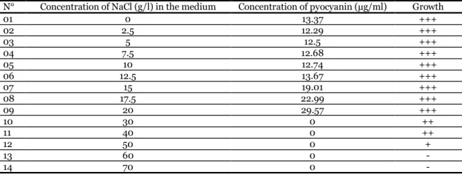
Effect of salinity on the growth and pyocyanin production
The isolate Pa was notably influenced by different concentration of salinity. The production of pyocyanin increased proportionally with salinity concentration.
The high amount of pyocyanin (29.57µg/ml) was obtained with 20g/L of NaCl. The result also indicated that salinity above 20g/L have totally affected pyocyanin production but did not affect the bacterium growth which was completely inhibited at 60 g/L of NaCl (Table 3).
Table 3. Effect of salinity on producing pyocyanin by P. aeruginosa.
N° Concentration of NaCl (g/l) in the medium Concentration of pyocyanin (µg/ml) Growth
01 0 13.37 +++ 02 2.5 12.29 +++ 03 5 12.5 +++ 04 7.5 12.68 +++ 05 10 12.74 +++ 06 12.5 13.67 +++ 07 15 19.01 +++ 08 17.5 22.99 +++ 09 20 29.57 +++ 10 30 0 ++ 11 40 0 ++ 12 50 0 + 13 60 0 - 14 70 0 - Hemolytic assay
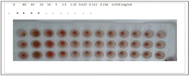
The effect of pyocyanin on the lysis of human red cells was tested to check its toxicity and side effects when used as curing agent at concentration ranging
from 0.156 to 5mg/ml. A negative hemolytic activity was observed at this rang and the effect was detected at all concentration exceeding 5mg/ml of pyocyanin (Fig. 2).
139 Dahah et al.
Fig. 2. Hemolytic activity of pyocyanin (+: hemolysis, - : no hemolysis).
Antioxidant activity of pyocyanin
The DPPH (1,1-diphenyl-2-picrylhydrazyl) radical scavenging activity of pyocyanin is given in Fig. 3. In the DPP Hmethod, the result of antioxidant efficiency is expressed as EC 50 determined as the concentration of substrate that causes 50% loss in absorbance (DPPH activity). This activity was increased by increasing the concentration of sample substrate. The IC50 value of the pyocyanin was 3.15µg/ml, as opposed to that of ascorbic acid (IC507.79µg/ml), which is a well-known antioxidant.
Fig. 3. DPPH radical scavenging activity of the
pyocyanin. Values are the average of triplicate experiments and represented as mean ± standard deviation.
Characterization of pyocyanin by UV spectrum and TLC
The UV spectrum of partially purified compound showed three peaks, two major of theme were found at 204 nm and 277.5 nm. However, the minor was observed at 386.5nm (Fig. 4).
These observations confirm pyocyanin compound characters in the examined solution. The visualization of TLC revealed only one spot after migration and the RF was found to be 0.9 (Fig. 5). The spot showed a similarity between the standard and the used molecule of pyocyanin with the same RF.
Fig. 4. UV absorption spectra of pyocyanin showing
ʎ max 277.5.
Fig. 5. Identification of pyocyanin by TLC (A) sample
140 Dahah et al.
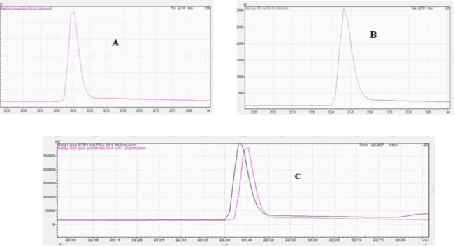
Characterization of pyocyanin by HPLC and FTIR
The analysis of purified extract and the pyocyanin standard were performed in HPLC. The two compounds were identified by comparison of their retention time. The results represented in Fig. 6, show a peak of purified.
extract of analyzed pyocyanin with a retention time of 22.746 min (Fig. 6A) which was highly identical to that of the pyocyanin standard 22.701min (Fig. 6B). Fig. 7 summarizes the characteristics of the studied pyocyanin using the FTIR technique.
Fig. 6. Identification of pyocyanin by HPLC. (A) Sample of extract pyocyanin from Pseudomonas aeruginosa
(B) standard of pyocyanin and (C) extrapolation of two graphs A and B.
Fig. 7. FTIR measurements (A) standard of pyocyanin, (B) sample of pyocyanin from P. aeruginosa and
141 Dahah et al.
Discussions
The present work is focusing a selection of environmental species of Pseudomonas aeruginosa to produce pyocyanin. The antagonistic activity of purified PCN compound was evaluated by measuring the zones of inhibition. It was found active against all tested pathogens microorganisms. The results showed high activity against Candida albicans ATCC 10231,
Bacillus cereus ATCC 10876 and exhibit a moderate
activity against gram-negative bacteria tested. PCN, a blue colored phenazine exotoxin, can easily penetrate biological membranes. Because the difference in lipid content in their cell wall, the Gram-positive are more sensitive topyocyan in antibiotic than Gram-negative bacteria. El-Fouly et al. (2015) found that the MIC of purified pyocyanin (20µg/ml) exhibited by Staphylococcus aureus; whereas the highest MIC (50µg/ml) was recorded by E. coli. The broad-spectrum antibiotic of pyocyaninobtained in the present study is in agreement with several works (Kerr et al., 1999; Preetha et al., 2010; Barakat, 2012; El-Fouly et al., 2015).The antibiotic effect of PCN is also due to its power in generating toxic effect of O 2-and H2O2 during respiration (Hassan and Fridovich, 1980; Mavrodi et al., 2006), and the capacity to arrest the electron transport chain of the fungi to exhibit antifungal activity (Wilson et al., 1987).
In the present work we found that production of pyocyanin was salinity-dependent when
Pseudomonas aeruginosa was grown in King A
medium supplemented with 2.5 to 20g/l of NaCl. The highest amount of PCN was obtained at 20g/l and the growth of P. aeruginosa was completely inhibited at 50 g/l of NaCl. However; a recent study shows that maximum of productivity was found in a medium salinity ranging from 5-10g/l (Prabhakaran et al., 2014).
These findings can be explained by the role of stress salinity in increasing metabolites production and to the capacity of this bacterium to adapt in different environmental conditions (Selezska et al., 2012). In the experience of antioxidant activity of pyocyanin we obtained a very high free radical scavenging at very low concentration as also obtained by Liyana and Shahidi (2005) and Laxmi et al. (2016).
In addition, this substance showed no hemolytic activity against human erythrocytes at less than 5mg/ml. The same results were cited by Park et al. (2004). These findings give us the possibility to check the way of using this compound in in vivo experiments and further more in a therapeutic treatment.
The pyocyan in chloroform extract separated as a blue color compound in organic phase showed a red color after addition of 0.2N HCl indicating its membership to pyocyanin. The UV-spectrophotometric analysis showed that the maximum absorption was found at 277.5nm which confirm one of pyocyanin characteristics as indicated by Kerr et al. (1999) and Sudhakar et al. (2013). The purified extract of pyocyanin showed one spot on TLC plate with RF 0.90 identical to the standard used. Our result was in accordance with Sudhakar et al. (2013). The obtained RF value was probably correlated to the kind of the solvent used.
- The results of HPLC analysis revealed only a major peak with retention time of about 22.746 which was the same as observed with the standard. This finding identifies the extracted pyocyanin and confirms its purity. The obtained FTIR spectrare present vibrations characteristic of the various connections constituting the structure of studied pyocyanin. The majority of the peaks appear in the field 400 with 2000cm-1 and absorption bands correspond to those of pyocyanin An intense band located at 3433 cm-1, corresponding respectively to the asymmetrical elongation of grouping OH of the water molecule, A band located between 2853 and 2922 cm-1, associated the elongation of the CH grouping, An intense band located between 1634 and 1698 cm-1, associated the elongation of grouping C=C and C=N respectively, -
- Bands located à1458 and at 1560 cm-1, associated the vibrations of the Benzene cycle, A band located between 1286 and 1258 cm-1, associated the elongation of the grouping C-n (primary Amine), A band located at 1336 cm-1, associated the elongation of the grouping CO, A band located at 1022 cm-1, associated the elongation of the grouping C-n (Tertiary Amine).
142 Dahah et al.
Conclusion
Despite the striking success of the pharmaceutical industries in creating new antibiotics, finding new broad spectrum antimicrobial agents is still a priority because of resistant bacterial infections. In this study
Pseudomonas aeruginosa isolated from saline soil
was selected to produce a high amount of pyocyanin substance under saline conditions. The evaluation of purified extract using different analytical methods confirmed the similitude of the obtained substance with pyocyanin reference. The obtained pyocyanin was active against human pathogenic microbes and showed an important antioxidant activity. The hemolytic activity was not shown at less than 5mg/ml, opening an issue to use weak concentrations of pyocyanin as therapeutic agent. Finally, we can conclude that the present work needs to be continued with in vivo studies to clarify the clinical and therapeutic effects of pyocyanin.
Conflict of interest
The authors declare that there are no conflicts of interest.
References
Barakat R. 2012. Etude des propriétés biologiques
et antimicrobiennes de la pyocyanine, pigment redoxactif produit par Pseudomonas aeruginosa. Université de La Rochelle 226 p.
Baron SS, Terranova G, Rowe JJ. 1989.
Molecular mechanism of the antimicrobial action of pyocyanin. Current Microbiol 18, 223-230.
Barry AL, Thornsberry C. 1985. Susceptibility
tests: Diffusion tests procedures. In: Lennette E.H., Ballows A.W. Shadomy J.R.H. (Eds). Manual of Clinical Microbiology pp. 1000-1008. American Society for Microbiology, Washington DC USA.
Costa AL, Cusmane V. 1975. Antimycotic activity
of pyocyanin in vitro and in vivo an pathogenic strain of Candida albicans. Gen. Bacteriol. Viro. Immunol
66, 297-308.
El-Fouly MZ, Sharaf AM, Shahin AAM, El-Bial.
2015. Biosynthesis of pyocyanin pigment by
Pseudomonas aeruginosa. J. Rad. Res. Appl. Sci
8, 36-48.
Fernández RO, Pizarro RA, 1997.
High-performance liquid chromatographic analysis of
Pseudomonas aeruginosa Phenazines. J. Chromatogr.
A 771, 99-104.
Hassan HM, Fridovich I, 1980. Mechanism of the
antibiotic action of pyocyanin. J. Bacteriol 141, 156-163.
Hassett DJ, Sutton MD, Schurr MJ, Herr AB,
et al. 2009. Pseudomonas aeruginosa hypoxic or anaerobic biofilm infections within cystic fibrosis airways. Trends Microbiol 17, 130-138.
Kerr JR, Taylor GW, Rutman A, Hoiby. 1999.
Pseudomonas aeruoginosa pyocyanin and
1-hydroxyphenazine inhibit fungal growth. J. Clin. Pathol 52, 385-387.
King ED, Ward MK, Raney DE. 1954. Two simple
media for demonstration of pyocyanin and fluorescein. J. Lab. Clin. Med 44, 301-307.
Kurachi M. 1958. Studies on the biosynthesis of
pyocyanin, isolation and determination of pyocyanine. Bull. Inst. Chem. Res. Kyoto Univ. 36, 163-173.
Laursen JB, Nielsen L. 2004. Phenazine natural of
products: biosynthesis, synthetic analogs, and biological activity. Chem. Rev 104, 1683-1685.
Laxmi M, Sarita and Bhat G. 2016.
Characterization of pyocyanin with radical scavenging and antibiofilm properties isolated from Pseudomonas
aeruginosa strain BTRY 1.3 Biotech 6, 27.
DOI 10.1007/s13205-015-0350-1.
Liyana PCM, Shahidi F. 2005. Antioxidant activity
of commercials oft and hard wheat (Triticuma
estivum L.) as affected by gastric pH conditions. J.
143 Dahah et al.
Mavrodi DV, Blankenfeldt W, Thomashow LS.
2006. Phenazine compounds in fluorescent
Pseuodomonas spp. biosynthesis and regulation.
Annu. Rev. Phytopathol 44, 417-445.
Ohfuji K, Sato N, Hamada-Sato N, Kobayashi
et al. 2004. Construction of a glucose sensor based on a screen-printed electrode and a novel mediator pyocyanin from Pseudomonas aeruginosa. Biosens. Bioelectron 19, 1237-1244.
Park Y, Park SN, Park SC, Park JY. 2004.
Antibiotic activity and synergistic effect of antimicrobial peptide against pathogens from a patient with gallstones. Biochem. Biophys. Res. Commun 321, 631-637.
Prabhakaran P, Puthumana J, Neil SC, Balachandran. 2014. Antagonistic effect of
Pseudomonas aeruginosa isolates from various
ecological niches on Vibrio species pathogenic to crustaceans. J. Coast Life Med 2(1), 76-84.
Preetha R, Jose S, Prathapan S, Vijayan KK.
2010. An inhibitory compound produced by Pseudomonas with effectiveness on Vibrio harveyi. Aquac. Res 41, 1452-1461.
Price-Whelan A, Dietrich LE, Newman DK.
2006. Rethin king ‘secondary’ metabolism: physiological roles for phenazine antibiotics. Nature Chem. Biol 2, 71-78.
Price-Whelan A, Dietrich LE, Newman DK.
2007. Pyocyanin alters redox homeostasis and carbon flux through central metabolic pathways in
Pseudomonas aeruginosa PA14. J. Bacteriol 189,
6372-6381.
Rangarajan S, Saleena LM, Vasudevan P, Nair S. 2003. Biological suppression of rice diseases by
Pseudomonas spp. under saline salt condition. Plant Soil 251,73-82.
Samanta SR, Thavasi Jayalakshmi S. 2008.
Phenazine pigments from Pseudomonas aeruginosa and their application as antibacterial agent and food colorants. Res. J. Microbiol 3(3), 122-128.
Selezska K, Kazmierczak M, Müsken M, Garbe
et al. 2012. Pseudomonas aeruginosa population structure revisited under environmental focus: impact of water quality and phage pressure. Environ. Microbiol 14, 1952-1967.
Sudhakar T, Karpagam S, Shiyama S, 2013.
Analysis of pyocyanin compound and its antagonistic activity against phytopathogens. Int. J. Chem. Tech. Res 5, 1101-1106.
Usher LR, Lawson RA, Geary I., Taylor. 2002.
Induction of neutrophil apoptosis by the
Pseudomonas aeruginosa exotoxin pyocyanin: a
potential mechanism of persistent infection. J. Immun 168, 1861-1868.
Wilson R, Pitt T, Tayler G, Walson D. 1987.
Pyocayanin and hydroxyphenazine produced by
Pseudomonas aeruginosa inhibit the beating of human
respiratory cilia in vitro. J. Investig 79, 221-229.
![[PDF] Utilisation des Algorithmes dans le Calcul Formel | Cours informatique](data:image/gif;base64,R0lGODlhAQABAIAAAP///wAAACH5BAEAAAAALAAAAAABAAEAAAICRAEAOw==)