HAL Id: hal-02548462
https://hal.sorbonne-universite.fr/hal-02548462
Submitted on 20 Apr 2020
HAL is a multi-disciplinary open access
archive for the deposit and dissemination of
sci-entific research documents, whether they are
pub-lished or not. The documents may come from
teaching and research institutions in France or
abroad, or from public or private research centers.
L’archive ouverte pluridisciplinaire HAL, est
destinée au dépôt et à la diffusion de documents
scientifiques de niveau recherche, publiés ou non,
émanant des établissements d’enseignement et de
recherche français ou étrangers, des laboratoires
publics ou privés.
old drugs to fight accelerated ageing
Solenn Guilbert, Déborah Cardoso, Nicolas Lévy, Antoine Muchir, Xavier
Nissan
To cite this version:
Solenn Guilbert, Déborah Cardoso, Nicolas Lévy, Antoine Muchir, Xavier Nissan. Hutchinson-Gilford
progeria syndrome: Rejuvenating old drugs to fight accelerated ageing. Methods, Elsevier, 2020,
�10.1016/j.ymeth.2020.04.005�. �hal-02548462�
Contents lists available atScienceDirect
Methods
journal homepage:www.elsevier.com/locate/ymeth
Hutchinson-Gilford progeria syndrome: Rejuvenating old drugs to
fight
accelerated ageing
Solenn M. Guilbert
a, Déborah Cardoso
b, Nicolas Lévy
c, Antoine Muchir
b, Xavier Nissan
a,⁎aCECS, I-STEM AFM, Institute for Stem Cell Therapy and Exploration of Monogenic Diseases, 28 rue Henri Desbruères, 91100 Corbeil-Essonnes, France bSorbonne Université, UPMC Paris 06, INSERM UMRS974, Center of Research in Myology, Institut de Myologie, F-75013 Paris, France
cAix-Marseille Université, UMRS910: Génétique médicale et Génomique fonctionnelle, Faculté de médecine Timone, Marseille, France
A R T I C L E I N F O
Keywords: Progeria Aging
Pluripotent stem cells Drug repurposing High-throughput screening
A B S T R A C T
What if the next generation of successful treatments was hidden in the current pharmacopoeia? Identifying new indications for existing drugs, also called the drug repurposing or drug rediscovery process, is a highly efficient and low-cost strategy. First reported almost a century ago, drug repurposing has emerged as a valuable ther-apeutic option for diseases that do not have specific treatments and rare diseases, in particular. This review focuses on Hutchinson-Gilford progeria syndrome (HGPS), a rare genetic disorder that induces accelerated and precocious aging, for which drug repurposing has led to the discovery of several potential treatments over the past decade.
1. Hutchinson-Gilford progeria syndrome 1.1. A premature aging disease
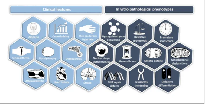
With a prevalence of 1 in 20 million births, Hutchinson-Gilford progeria syndrome (HGPS) is an extremely rare and consistently fatal genetic disorder characterized by accelerated aging. Clinical symptoms usually appear in thefirst 18 months after birth, and include growth retardation, facial dysmorphic changes (long narrow nose, prominent outer ears, wrinkled skin), alopecia, loss of subcutaneous fat, bone and joint abnormalities and cardiovascular pathology. Death occurs at a median age of 14.6 years, mainly due to atherosclerosis, cardiovascular failure and stroke[1,2](Fig. 1).
The genetic origin of HGPS was identified in 2003 by two in-dependent research groups led by Nicolas Lévy and Francis S. Collins, respectively[3,4]. This autosomal dominant disease is caused by a de novo mutation in the LMNA gene which encodes A-type lamins, inner nuclear membrane proteins represented mainly by lamins A and C. A-type lamins play crucial roles in nuclear structure and shape, as well as in chromatin organization, nuclear pore and cytoskeleton organization
[5], and mutations in LMNA were reported to cause various genetic disorders known as laminopathies. They include a wide spectrum of diseases, with or without overlapping symptoms, such as lipodystro-phies, Emery-Dreifuss muscular dystrophy, Charcot-Marie-Tooth dis-ease, dilated cardiomyopathies and progeroid syndromes, including HGPS[6].
Over the past decade, several groups have explored the molecular causes of HGPS, ultimately leading to the identification of the first therapeutic strategies. In physiological conditions, lamin A is produced from its prelamin A precursor, which undergoes complex post-transla-tional modifications (Fig. 2). A cysteine in the C-terminal of prelamin A is first farnesylated, cleaved and finally carboxymethylated by the metalloprotease STE24 (ZMPSTE24) and isoprenylcysteine carboxy-transferase (ICTM). The farnesyl group is then removed through clea-vage of the 15C-terminal amino acids, leading to the production of the mature lamin A. The most common mutation in HGPS (c.1824C > T), although apparently silent (LMNA p.G608G), activates an alternative splice site, leading to the deletion of 150 nucleotides at the end of exon 11, which encode the endoprotease cleavage site. As a result, a pre-lamin A variant lacking 50 aa residues is produced in a permanently
https://doi.org/10.1016/j.ymeth.2020.04.005
Received 15 November 2019; Received in revised form 6 April 2020; Accepted 7 April 2020
Abbreviations: HGPS, Hutchinson-Gilford progeria syndrome; ZMPSTE24, metalloprotease STE24; ICTM, isoprenylcysteine carboxytransferase; hES, human em-bryonic stem cells; IPS, induced-pluripotent stem cells; MSC, mesenchymal stem cell; VSMC, vascular smooth muscle cell; miRNA, micro ribonucleic acid; DNA, Deoxyribonucleic acid; mRNA, messenger ribonucleic acid; FTI, farnesyl-transferase inhibitor; NPY, Neuropeptide Y; HMGCR, 3-hydroxy-3-methylglutaryl-coenzyme A reductase; SRFS1, splicing factor serine/arginine-rich splicing factor 1; ROS, reactive oxygen species; SIRT1, NAD+-dependent sirtuin 1; HSP, heat shock protein;
NAC, N-acetyl cysteine drug; HTS, high throughput screening; AON, antisense oligonucleotide; Mono-AP, mono-aminopyrimidine; FPPS, farnesyl pyrophosphate synthase; FT, farnesyl-transferase
⁎Corresponding author.
E-mail address:xnissan@istem.fr(X. Nissan).
Methods xxx (xxxx) xxx–xxx
1046-2023/ © 2020 The Authors. Published by Elsevier Inc. This is an open access article under the CC BY-NC-ND license (http://creativecommons.org/licenses/BY-NC-ND/4.0/).
farnesylated form, and the resultant protein, called progerin, exerts toxic effects in HGPS cells[7,8].
1.2. Phenotypic characteristics of HGPS
Due to the structural role of lamins in the nucleus, accumulation of progerin is accompanied by dramatic changes in nuclear structure and function (Fig. 1). In contrast to the mature lamin A, progerin remains anchored to the inner nuclear membrane, leading to shape abnormal-ities with the appearance of “blebs”, disorganization of the hetero-chromatin (e.g. tri-methylation on lysine 47 of histone (H3K27))[7–9], as well as abnormal chromosome segregation and telomere degradation [10–12]. Among the cellular phenotypes reported in HGPS, premature senescence as a result of genomic instability[10,13,14]and accumu-lation of DNA double-strand breaks, notably through the decrease in recruitment of major DNA repair factors[7,15–17], have been widely described. Cells expressing progerin also exhibit mitochondrial defects
[18], increased oxidative stress[19–21], decreased stress tolerance[9], stem cell exhaustion[22], alteration of proteolysis [23–25] and in-flammation[26,27]. Since these phenotypes are commonly observed in physiological ageing [28], HGPS is considered a genetically induced model of accelerated ageing. First evidence came from the observation that progerin was expressed at low levels in physiologically aged-cells [29–32], but its possible role in tissue dysfunction during physiological aging has not been demonstrated and its role or its contribution toward tissue dysfunction remains unanswered.
1.3. In vitro models to study HGPS
For almost a decade, the main biological material available for the in vitro study of HGPS werefibroblasts isolated from skin biopsies or generated following progerin overexpression [7,29,33,34]. Even though skinfibroblasts were useful to assess pathological phenotypes, their limited proliferation capacities and lack of clinical relevance have Fig. 1. Hallmarks of HGPS. In light blue, the principal clinical features of HGPS are recapitulated whereas in dark blue, the major in vitro pathological phenotypes are represented.
Fig. 2. Defective processing of prelamin A in HGPS and associated nuclear shape disorganization. Lamin A protein is obtained after several post-translational modifications, including the addition of a farnesyl group at the C-terminal and further cleavage by ZMPSTE24 endonuclease. The mutation on the LMNA gene that causes HGPS is responsible for the activation of an alternative splicing site that results in the deletion of 50 amino acids from a lamin A protein, including the cleavage site for ZMPSTE24. Consequently, the resulting mutant protein, called progerin, remains permanently farnesylated and thus induces nuclear shape ab-normalities and disorganization.
delayed the identification of the tissue-specific mechanism of action and specific treatments. The discovery of human embryonic stem cells (hES) and, more recently, the possibility of reprogramming somatic cells into pluripotent stem cells (iPS)[35], have opened up the possi-bility of studying some of the phenotypes associated with diseases“in a dish”. Pluripotent stem cells have the unique properties of being able to self-renew and to differentiate into any cell type, allowing the pro-duction of an“unlimited” quantity of cells for disease modeling and drug screening[36]. In 2011, the groups led by J.C Belmonte and A. Colman pioneerly reported the derivation of thefirst HGPS iPS cell lines [15,37]. Interestingly, in agreement with previous studies conducted with human embryonic stem cells[38], these two groups reported that neither lamin A nor progerin were expressed in undifferentiated HGPS iPS cells, making it possible to expand and differentiate these cells with no bias relating to the disease. Through a mechanism that remains unknown, these two groups have also reported that lamin A and pro-gerin were re-expressed upon differentiation into different cell types, inducing pathological features such as nuclear abnormalities, reduced telomere length and premature senescence[15,37]. In addition to these findings, Zhang et al. also demonstrated that progerin was mainly ex-pressed in mesenchymal stem cells (MSC) and vascular smooth muscle cells (VSMC), two cell types of particular relevance for the disease, but was absent in neuronal cells[37]. In 2012, our group discovered the origin of this specificity, identifying that miR-9, a miRNA pre-dominantly expressed in neural cells, was capable to target the 3’UTR of progerin and decrease its expression in neurons [39]. Later, several other studies have subsequently elucidated some pathological me-chanisms occurring in progerin-expressing cells in different cell types using iPS cells[15,17,27,37,40,41]. For example, Zhang et al. proposed in 2014 that the loss of proliferation in VSMCs could be attributed to a decrease in PARP-1 expression through an increase in chromosomal aberrations[17]and Xiong et al. demonstrated in 2013 a role of pro-gerin in the deregulation of PPARγ2 and C/EBPα expression, two fac-tors implicated in the differentiation in adipocytes[41].
1.4. In vivo models of HGPS
Several animal models have been developed to elucidate the pa-thological mechanisms of HGPS and to evaluate potential therapeutic strategies. Thefirst living HGPS model was developed in 2002 through the depletion of ZMPSTE24 (FACE-1), which encodes the enzyme re-sponsible for the cleavage of the prelamin A farnesylated residue. Zmpste24-/- mice display several progeroid features, such as growth
retardation, alopecia, cardiomyopathy, lipodystrophy, muscular dys-trophy and premature death[42,43]. This model was used as the gold standard for HGPS and related disorders for almost a decade, demon-strating that the accumulation of the farnesylated protein induces nu-clear abnormalities in vascular and osteogenic tissues, as well as p53 hyperactivation, defective DNA repair, cellular senescence and stem cell dysfunction[14,16,44]. Since this model expresses the full-length version of farnesylated prelamin A, 2nd generation models were based on the knock-in of a mutant allele of LMNA using selective or ubiqui-tous promoters, leading to the specific expression of progerin with or without lamin A and C[45,46]. More recently, Osorio et al. generated a knock-in mouse strain carrying the HGPS mutation in LMNA. This mouse model produces progerin through aberrant splicing of its en-dogenous LMNA mRNA and recapitulates the main features of HGPS disease at both molecular and clinical levels, including reduced life-span, as well as vascular calcification, and cardiovascular and bone defects[47,48]. Even though mouse models are essential and widely used to depict molecular mechanisms of the disease and to test different therapeutic strategies, some key differences remain between these models and humans. To bridge the gap between mice and humans, and thanks to new gene editing methodologies, the group led by Vicente Andrès has recently reported the generation of a minipig model of HGPS carrying, by knock-in, the heterozygous LMNA c1824C > T
mutation. This model has the advantage of having a cardiovascular system with strong similarities to that in humans and is therefore par-ticularly relevant to HGPS[49].
2. Repurposing of old drugs for HGPS
2.1. Why we should consider repositioning drugs for ultra-rare diseases HGPS is one of the rare or orphan diseases, defined as disorders affecting less than 5/100,000 people in Europe or fewer than 200,000 Americans at any point in time (around 650 in 1 million people). Mostly genetic in origin, more than 7,000 disorders were classified as rare, with no available treatment for most of them. In this context, “drug repurposing” represents a valuable strategy for bridging the gap be-tween the need for treatment for patients with HGPS and the limited profits expected by pharmaceutical companies from developing new chemical entities. Drug repurposing– also called repositioning - consists of identifying new indications for existing or abandoned pharmacolo-gical drugs. This strategy takes advantage of previous data collected for a compound during clinical trials, notably on its bioavailability and safety, thus reducing the risks linked to the development of an entirely new product, which consequently accelerates access to the market. While these strategies present clear advantages, some challenges re-main. The principle of repurposing depends not only on knowledge of the nature of the drugs, but also on knowledge on the disease, with the latter condition that is not always fulfilled in rare diseases.
2.2. Different strategies for pharmacological repositioning
The principle of drug repurposing is not novel. First successes were historically due to serendipity, as described with sildenafil, that was initially indicated as an anti-hypertensive drug before its successful use in erectile dysfunction, or with thalidomide, initially developed for insomnia or morning sickness treatment and then repurposed for mul-tiple myeloma, other forms of cancer or leprosy[50]. One of the most striking example of successful repurposing based on drugs’ side effects observations was recently described when french physicians located in Bordeaux observed the unexpected effect of Propanolol on the he-mangioma present in the patient’s face[51]whereas it was initially used to treat his heart condition. Ever since, other approaches have led to the development of more systematic strategies of drug repurposing, which can be classified into two groups: experimental and computa-tional approaches. Experimental approaches mainly comprise two kinds of assays, binding assays to identify target interactions (not described here) or assays to rescue a phenotype.
In computational approaches, knowledge about the drug and dis-eases, and the analysis of data from a variety of origins, form the basis for the discovery of potential new drug-disease associations[52]. First, a“target-centric approach” could be envisaged in repurposing a drug, by focusing on the biological role that a specific component plays in disease. This requires the identification of potential genes implicated in the pathological phenotypes and searching in the pharmacopeia for existing drugs known to target them. Another relevant strategy is “pathway or network mapping”, which consists of targeting a pathway upstream or downstream from the causative gene, but with strong re-levance for the disease. The identification of such pathways could arise from the study of in vitro or in vivo models, with the development of “omics” data and transcriptomic analysis, in particular, being of great interest to the discovery of new deregulated genes. Transcriptomic data generated to identify misregulated pathways in the context of disease or drug treatment might also be useful for another approach called “sig-nature mapping”. Other methods exist and rely mostly on similarities between drugs to identify new possible indications. For example, a comparable chemical structure in different drugs suggests a shared biological activity and therefore the possibility to be repurposed. The search for similarities in side effects has also been reported as a possible
S.M. Guilbert, et al. Methods xxx (xxxx) xxx–xxx
approach to drug repurposing, based on the hypothesis that similar side effects result from shared target or protein pathways and could thus lead to the discovery of new drug indications. These various strategies are not exhaustive, but reflect the major in silico methods that are currently being used in repurposing. Because several of these strategies have been successfully applied to HGPS, we will discuss the main findings of these reports below.
2.3. Repurposing old drugs in HGPS: From thefirst “success” with farnesylation inhibitors to promising compounds targeting progerin
Farnesylation, and more generally prenylation, is a common cellular mechanism that concerns a large number of proteins, including small GTPases, proteins implicated in the regulation of important cellular events like proliferation or cell motility. Targeting of the farnesylation process, which is required for the malignant activities of the RAS on-cogenic family, has led to the development of farnesyl-transferase in-hibitors (FTIs) as anti-cancer drugs[53]. Since 2000, several clinical trials using FTIs (lonafarnib, tipifarnib, BMS-214662 and L-778123) have evaluated their toxicity and efficacy in various cancer indications and revealed acceptable tolerance in humans[54–56]. Based on this knowledge, several FTIs were tested in vitro for HGPS, where an im-provement in the nuclear shape was demonstrated[57–60], and also in vivo, revealing an improvement in the symptoms of the disease, in ad-dition to a decrease in nuclear blebbing,[46,61–63]and an extension to lifespan[62](Figs. 3 and 4).
In 2008, a similar pharmacological approach targeting the entire
prenylation pathway was employed in repurposing for HGPS using the combination of zoledronate, a member of the amino-bisphosphonates class mainly used to treat osteoporosis[64], and pravastatin, that be-longs to a class of inhibitors of 3-hydroxy-3-methylglutaryl-coenzyme A reductase (HMGCR) used to reduce cholesterol levels and prevent car-diovascular disease[65]. Treatment of the Zmpste24-/- mouse model
with this combination led to an improvement in several hallmarks of HGPS, including lifespan[66].
These pioneering preclinical studies have successfully led to the design of several clinical trials, highlighting the efficiency of the re-purposing of these drugs. Thefirst ever clinical trial was launched in 2007 (ClinicalTrials.gov, NCT00425607) using the FTI lonafarnib on a cohort of 25 patients for a minimum period of 2 years, showing en-couraging results with an improvement in weight gain, vascular stiff-ness and bone density[67]. In 2008, following the identification of the zoledronate and pravastatin effect, a second clinical trial was initiated using these two drugs in 12 patients (ClinicalTrials.gov, NCT00731016) followed by a tri-therapy clinical trial combining lonafarnib, zole-dronate and pravastatin in 37 patients (ClinicalTrials.gov, NCT00879034). Results of this last clinical trial was reported describing no additional improvement of the tri-therapy as compared to lonafarnib alone[68]. More recently, in late 2015, another phase I/II clinical trial combining the existing drugs lonafarnib and everolimus (Clinical-Trials.gov, NCT02579044) was started in 60 patients, for which results are expected in October 2020 (https://www.progeriaresearch.org/ clinical-trials/). Everolimus is an analog of the antibiotic macrolide drug rapamycin, an mTOR inhibitor, already used against cancer or for immunosuppression and implicated in the regulation of several cellular functions such as cell proliferation, protein synthesis, transcription, cytoskeleton rearrangement and autophagy[69]. Previous studies had suggested that rapamycin improved lifespan, notably in aged mice, through activation of autophagy, a process that is down-regulated during ageing[70–74]. This led to the hypothesis that autophagy in-duction could decrease the accumulation of the toxic progerin through a complementary mechanism to lonafarnib and improve cell pheno-types in progeria. Indeed, in HGPSfibroblasts treated with rapamycin or with temsirolimus, a decrease was observed in progerin through autophagy activation, accompanied by an improvement in abnormal nuclear shape, a decrease in senescence[75]and a reduction in DNA damage[76]. More recently, Neuropeptide Y (NPY), a neuronal peptide evaluated in Humans to treat feeding difficulties, acute stress disorders or posttraumatic stress disorders, was also shown to decrease progerin expression and alleviate several in vitro hallmarks of HGPS through autophagy induction[77]. In parallel to the evaluation of autophagy activators, several other studies have investigated the possibility to induce progerin clearance by modulating other degradation processes. To date, the most advanced and promising strategy to target this pro-cess is the use of proteasome inhibition. First evidence was described in 2017 by the group led by Nicolas Lévy, showing that MG132 treatment could lead to progerin clearance by an indirect induction of autophagy [78]. Finally, modulating alternative splicing regulation was also de-scribed to be a valuable strategy to target progerin content. Successfully applied to HGPS, the main target of these studies is the splicing factor serine/arginine-rich splicing factor 1 (SRFS1), previously shown to enhance the aberrant splicing of lamin A pre-mRNA involved in the production of progerin[79]. Thefirst results were reported in 2016, when our group demonstrated that treatment with metformin, an an-tidiabetic drug with a good safety profile, could lead to a decrease in SRSF1 and progerin in both iPS cell-derived MSCs and HGPS fibro-blasts. In this study, we also reported that metformin treatment was associated with a reduction in several defects, such as a decrease in nuclear shape abnormalities, in premature osteoblastic differentiation and in DNA damage[80]. Interestingly, MG-132 was also reported to induce the downregulation of SRSF1, suggesting a second additive level of regulation of progerin expression[78].
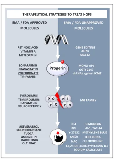
Progerin
RETINOIC ACID VITAMIN A METFORMIN TIPIFARNIB LONAFARNIB PRAVASTATIN ZOLEDRONATE EVEROLIMUS RAPAMYCIN TEMSIROLIMUS NEUROPEPTIDE Y OLTIPRAZP
r
o
g
e
r
i
n
RESVERATROL TUDCA QUERCETIN BARICITINIB GENE EDITING AONs MG132 MONO-APs GGTI-2147 shRNAs against ICMTMG FAMILY
AI-1, TAT-14 Y-27632 METHYLENE BLUE
NAC TERT mRNA 1ɲ,2ϱ-DIHYDROyYVITAMIN D3 CHLOROQUINE PPi REMODELIN JH4 SODIUM SALICYLATE tASOs
THERAPEUTICAL STRATEGIES TO TREAT HGPS EMA / FDA APPROVED
MOLECULES
EMA / FDA UNAPPROVED MOLECULES
Fig. 3. Therapeutic strategies in HGPS. Several therapeutic strategies to treat HGPS has been proposed targeting either the production of the mutant protein progerin, its degradation or its pathological consequences. Here are represented all the therapeutical strategies, FDA or EMA approved or not, that have been investigated for the treatment of HGPS. The underlined compounds have been clinically tested.
2.4. Rescue of progerin-downstream pathological phenotypes
Among repurposing strategies, targeting the phenotypic con-sequences of progerin expression is another possibility that could in-directly reduce the burden caused by the aberrant protein in HGPS cells. The recent development of a mouse model for HGPS and of more relevant cellular models, such as based on iPS-derived cells, has allowed the identification of such pathological phenotypes and pathways and thus led to the identification of other possible drugs for repurposing (Figs. 3, 4).
First evidences came from the identification of the beneficial effect of resveratrol in HGPS, an anti-ageing polyphenol compound reported to have antioxidant properties [81–83]. Resveratrol is an activator of the NAD+-dependent sirtuin 1 (SIRT1), a histone deacetylase protein with implied involvement in several cell processes, such as antioxidant and stress responses, mitochondrial biogenesis and metabolism [84] that was initially tested for the treatment of cancer, diabetes, obesity, neurological or cardiovascular disorders [85]. Using the Zmpste24 -/-mouse model treated with resveratrol, Liu et al demonstrated a decrease in the abnormal lamin A-SIRT1 association observed in HGPS, which was thus accompanied by a restoration of HGPS defects, including a decrease in stem cell loss and an extension to lifespan[86]. Whereas the
clinical use of resveratrol is currently limited because of its poor bioavailability, a micronized form with a good safety profile was de-veloped (SRT501)[87]opening new perspectives for HGPS.
In 2015, another example was reported by Gabriel et al[23]. With the goal of restoring proteolytic machinery activity in HGPS cells [24,25]and consequently inducing progerin degradation, authors has evaluated the effect of sulforaphane, an antioxidant molecule found in cruciferous vegetables clinically used for its capability to improve symptoms of chronic inflammatory diseases and for the treatment of cancer [88]. Sulforaphane’s mechanism of action was reported to be mediated by an improvement in proteostasis, including an increase in heat shock protein (HSP) and components of the proteasome and au-tophagy machineries[89,90], and through the targeting of Keap1/Nrf2 pathways[91]. As described in this study, treatment of HGPS cells with this compound was accompanied by an increase in proteolytic activity, a reduction in progerin, a decrease in DNA damage, and an increase in cell proliferation, suggesting its possible use as a therapy for HGPS [23,92].
Attempts to target the reactive oxygen species (ROS) accumulation were also made with several compounds, such as methylene blue[93], that improve mitochondrial dysfunction, and oltipraz, an antiparasitic drug that targets the antioxidant Nrf2 pathways and was also evaluated Fig. 4. Recapitulation of the therapeutic compounds described in HGPS. This scheme recapitulates the therapeutic approaches for the treatment of HGPS depending on their target. Non-repurposable strategies are represented in white, whereas repurposable drugs are highlighted inbold or underlined for the one used in clinical trials for HGPS. Progerin expression was decreased directly at gene level through gene therapy (CRISPR Cas 9 and AON) or through small molecules like retinoids and metformin. Strategies targeting progerin at a protein level or its prenylation have also been evaluated, among them small molecules that have reached the clinic, such as FTIs like lonafarnib, and rapamycin analogs like everolimus. Finally, targeting the downstream effects of progerin has been described as a valuable strategy for HGPS, with some repurposable drugs that have proven effective in correcting the disease-associated phenotypes in cellular and/or animal models.(See above-mentioned references for further information.)
S.M. Guilbert, et al. Methods xxx (xxxx) xxx–xxx
for its effect against cancer cells[19]. Interestingly, in relation to this second compound, both sulforaphane and resveratrol, mentioned above, also depend at least partially on Nrf2 for their beneficial anti-oxidant effects, suggesting a common mechanism of action.
Senolytics represents a promising anti-ageing therapeutic strategy that targets senescent cells. Recently, a study performed in the context of Werner syndrome, another premature ageing disease, identified the benefits of quercetin, a polyphenol derived from plants and known for its anti-inflammatory and antioxidant properties. Authors of this study reported that quercetin treatment was able to rescue the replicative senescence in Werner syndrome MSCs through the transcriptional regulation of several cellular processes, such as cell cycle and anti-oxidation pathways. Interestingly, a similar effect on senescence were observed in HGPS cells, suggesting a common geroprotective effect of quercetin that could be relevant for premature and physiological aging [94].
In the past decade, several other pharmacological compounds have been reported to decrease progerin-induced pathological phenotypes (see review[95]) by several ways, such as a decrease in ROS production through N-acetyl cysteine drugs[96], decreasing vascular calcification through pyrophosphate treatment[48], targeting telomere dysfunction [97,98]or the restoration of nuclear organization through SUN1-asso-ciated acetyl transferase protein NAT10 inhibition with Remodelin[99] (Figs. 3 and 4). Recently, a promising strategy was reported by Hamczyk et al[21]showing that the treatment with a chemical cha-perone tauroursodeoxycholic acid (TUDCA), successfully used in hu-mans to treat cholestatic liver disease[100–103], was able to alleviate endoplasmic reticulum stress delaying medial VSMC loss and extending lifespan of progeroid mice.
3. Drug repurposing by drug screening
Even if previous research has led to the identification of a large number of potential therapeutic interventions by small molecules to correct HGPS defects, there is still a need for the discovery of new drugs that are suitable for trials in humans. To this end, another approach is to use high throughput screening (HTS) to evaluate the effect of old drugs on new phenotypes. This drug discovery strategy corresponds to the unbiased experimental testing of a compound library in order to find positive “hits” capable of correcting a defective mechanism of ac-tion or phenotype. This strategy will be discussed below in the light of three different studies reported in recent years.
3.1. Challenges in drug screening for HGPS
The first and most obvious challenge in high-throughput drug screening is the need for a robust and relevant assay that can be used to test the compounds. Such strategies could be developed based on sev-eral ways, with either“target-based”, also called “mechanism-based”, or “phenotypic-based” screening. A second challenge that has to be taken into consideration in drug screening is the choice of the cellular model. High-throughput screening requires a large quantity of stable cells that are relevant to the disease in order to work in reproducible conditions. In the case of HGPS, the use of primary cells from patients is challenging, firstly, because their growth, and thus the production of large quantities of cells, is limited by premature senescence and, sec-ondly, because they are also associated with a phenotypic heterogeneity that could interfere with the identification of drugs of interest. Consequently, immortalizedfibroblasts are generally preferred for HTS to bypass the use of primary cells. However, as it has been demon-strated in HGPS, progerin expression is progressively lost following immortalization, thus limiting its use for pharmacological interventions in HGPS[104]. Alternative strategies have been described to overcome this obstacle, involving inducing progerin in non-HGPS cell lines by overexpression [105] or using antisense oligonucleotides (AON) [106,107]. Finally, in this context, iPS cell derivatives have emerged as
a model of choice for drug screening, in addition to their role in disease modeling. Their high capacity for proliferation and the possibility of differentiating iPS cells into different cell types allows the generation of large banks of cells that recapitulate the main phenotypes of HGPS. To date, only three drug screening processes have been performed to identify new drugs using different cell models in the case of HGPS. These are described in more detailed in the following section. 3.2. Use of HTS to repurpose old drugs for HGPS
In 2016, our group reported thefirst high-throughput screening conducted on HGPS cells using MSCs derived from iPS cells. This screening led to the identification of a new class of compounds that decreases the farnesylation of prelamin A[108]. In this study, HGPS iPS-derived MSCs were used to evaluate the effects of 21,608 com-pounds on the subcellular localization of prelamin A by immuno-fluorescence, assuming that this would reflect a decrease in its farne-sylation. From an initial list of 59 hits, 11 were ultimately validated regarding their efficacy in relation to increasing prelamin A staining in the nuclear membrane, their safety and the correction of HGPS phe-notypic defects, as assessed in a secondary assay. Among these, a statin was identified, which highlights the relevance of screening as this class of compounds was previously shown to target progerin prenylation [66]. Most importantly, a new class of compounds with a common mono-aminopyrimidine group (Mono-AP) was found to improve os-teogenic differentiation and nuclear shape organization. Thanks to docking experiments, it was later proposed that Mono-AP decreases farnesylation by simultaneously inhibiting farnesyl pyrophosphate synthase (FPPS) and FTs. Therefore, even if this class of drugs is not directly repurposable, it could represent an advantageous form of treatment for HGPS by targeting farnesylation at different stages.
In 2016, Tom Misteli’s group reported on further high-throughput screening conducted on HGPS, using immortalized skinfibroblasts that overexpressed progerin in an inducible manner[105]. Several pheno-types of HGPS, such as reduced levels of the nuclear protein lamin B1, and LAP2, disorganization of the heterochromatin and an increase in DNA damage, were assessed at different points in time. Detection of lamin B1 and ofγH2AX foci, a DNA damage marker, was selected to develop a multi-parametric assay and screen 2,816 FDA-approved drugs and bioactives. Among these, 27 compounds were identified as poten-tial hits, including two members of the retinoids, previously used for the treatment of acne or psoriasis, which exhibited strong and sig-nificant effects on all parameters that were tested.
Retinoids were also identified independently by our group in a third high-throughput screening performed on HGPS iPS-derived MSCs [109]. In this study, the premature osteogenic differentiation observed in HGPS iPS-derived MSCs[110] was used as a readout to screen a chemical library of 2800 drugs. 10 compounds were identified and, ultimately, the retinoids emerged as potential drug candidates. Results of this screening also confirmed the anti-progeroid effect of this class of drugs that was previously identified by Kubben et al. using another cellular model [105], also demonstrating its efficiency in reducing other pathological defects in HGPS MSCs. Furthermore, this work has added a proof of concept, showing that drug screening using iPSC-de-rived cells could be helpful in the discovery of drugs that target cell-types specific to phenocell-types in HGPS.
4. Conclusions
Since the discovery of the genetic origin of HGPS in 2003, much progress has been made, leading to greater in-depth knowledge on the disease, its mechanisms of action and the cellular consequences that ultimately drive the disease phenotypes. Given this short period of time, it is very surprising to see that several successful clinical translations of drug repurposing have already been reported, as shown for lonafarnib, zoledronate and pravastatin. Drug repurposing allowed researchers to
progress rapidly to phase II clinical trials and to evaluate the efficacy of a limited number of drugs in a time and cost-effective manner. As discussed in this review, identification of these treatments was mainly based on hypothesis-driven approaches and unbiased high-throughput screening. To date, around 35 molecules have been reported to have a positive effect on HGPS by either limiting progerin expression, mod-ulating alternative splicing, decreasing progerin accumulation or its pathological consequences. This situation is almost unique making impossible to evaluate the clinical benefit of all these individual drugs because of a limited number of patients. In the future, the systematic evaluation of the combination of drugs targeting in one hand progerin and in the other hand its downstream consequences appears to be a valuable strategy to identify an efficient treatment for this disease that remains uncured.
Acknowledgements
I-Stem and the Center of Research in Myology are part of the Biotherapies Institute for Rare Diseases (BIRD), supported by the Association Française contre les Myopathies (AFM-Téléthon). This re-search was funded by grants from INSERM, the domaine d’intéret ma-jeur (DIM) Biothérapies, Genopole and the European Commission: the laboratoire d’Excellence Revive (Investissement d’Avenir; ANR-10-LABX-73), NeurATRIS: A Translational Research Infrastructure for Biotherapies in Neurosciences (Investissement d’Avenir - ANR-11-INBS-0011), INGESTEM: the National Infrastructure Engineering for Pluripotent and differentiated Stem cells (Investissement d’Avenir -ANR-11-INBS-0009), the University of Paris-Saclay (BiotherAlliance) and the French National Agency (ANR-18-CE14-0036).
Appendix A. Supplementary data
Supplementary data to this article can be found online athttps:// doi.org/10.1016/j.ymeth.2020.04.005.
References
[1] M. Gerhard-Herman, L.B. Smoot, N. Wake, M.W. Kieran, M.E. Kleinman, D.T. Miller, A. Schwartzman, A. Giobbie-Hurder, D. Neuberg, L.B. Gordon, Mechanisms of premature vascular aging in children with hutchinson-gilford progeria syndrome, Hypertension 59 (2012) 92–97,https://doi.org/10.1161/ HYPERTENSIONAHA.111.180919.
[2] M.A. Merideth, V. Sachdev, C.C. Brewer, B. Solomon, M.L. Turner, J. Graf, J.A. Yanovski, F.S. Collins, W.A. Gahl, Phenotype and course of hutchinson-gilford progeria syndrome, N Engl. J. Med. (2008) 13.
[3] A. De Sandre-Giovannoli, Lamin A truncation in hutchinson-gilford progeria, Science 300 (2003) 2055,https://doi.org/10.1126/science.1084125.
[4] M. Eriksson, W.T. Brown, L.B. Gordon, M.W. Glynn, J. Singer, L. Scott, M.R. Erdos, C.M. Robbins, T.Y. Moses, P. Berglund, A. Dutra, E. Pak, S. Durkin, A.B. Csoka, M. Boehnke, T.W. Glover, F.S. Collins, Recurrent de novo point mutations in lamin A cause Hutchinson-Gilford progeria syndrome, Nature 423 (2003) 293–298,
https://doi.org/10.1038/nature01629.
[5] C.Y. Ho, J. Lammerding, Lamins at a glance, J. Cell Sci. 125 (2012) 2087–2093,
https://doi.org/10.1242/jcs.087288.
[6] S. Kang, M.-H. Yoon, B.-J. Park, Laminopathies; Mutations on single gene and various human genetic diseases, BMB Rep. 51 (2018) 327–337,https://doi.org/ 10.5483/BMBRep.2018.51.7.113.
[7] R.D. Goldman, D.K. Shumaker, M.R. Erdos, M. Eriksson, A.E. Goldman, L.B. Gordon, Y. Gruenbaum, S. Khuon, M. Mendez, R. Varga, F.S. Collins, Accumulation of mutant lamin A causes progressive changes in nuclear archi-tecture in Hutchinson-Gilford progeria syndrome, PNAS 101 (2004) 8963–8968,
https://doi.org/10.1073/pnas.0402943101.
[8] P. Scaffidi, T. Misteli, Reversal of the cellular phenotype in the premature aging disease Hutchinson-Gilford progeria syndrome, Nat Med. 11 (2005) 440–445,
https://doi.org/10.1038/nm1204.
[9] M. Paradisi, D. McClintock, R.L. Boguslavsky, C. Pedicelli, H.J. Worman, K. Djabali, Dermalfibroblasts in Hutchinson-Gilford progeria syndrome with the lamin A G608G mutation have dysmorphic nuclei and are hypersensitive to heat stress, BMC Cell Biol. 6 (2005) 27,https://doi.org/10.1186/1471-2121-6-27. [10] E.K. Benson, S.W. Lee, S.A. Aaronson, Role of progerin-induced telomere
dys-function in HGPS premature cellular senescence, J. Cell Sci. 123 (2010) 2605–2612,https://doi.org/10.1242/jcs.067306.
[11] T. Dechat, T. Shimi, S.A. Adam, A.E. Rusinol, D.A. Andres, H.P. Spielmann, M.S. Sinensky, R.D. Goldman, Alterations in mitosis and cell cycle progression
caused by a mutant lamin A known to accelerate human aging, Proc. Natl. Acad. Sci. U.S.A. 104 (2007) 4955–4960,https://doi.org/10.1073/pnas.0700854104. [12] M.L. Decker, E. Chavez, I. Vulto, P.M. Lansdorp, Telomere length in
Hutchinson-Gilford progeria syndrome, Mech. Ageing Dev. 130 (2009) 377–383,https://doi. org/10.1016/j.mad.2009.03.001.
[13] S. Gonzalo, R. Kreienkamp, DNA repair defects and genome instability in Hutchinson-Gilford progeria syndrome, Curr. Opin. Cell Biol. 34 (2015) 75–83,
https://doi.org/10.1016/j.ceb.2015.05.007.
[14] I. Varela, J. Cadiñanos, A.M. Pendás, A. Gutiérrez-Fernández, A.R. Folgueras, L.M. Sánchez, Z. Zhou, F.J. Rodríguez, C.L. Stewart, J.A. Vega, K. Tryggvason, J.M.P. Freije, C. López-Otín, Accelerated ageing in mice deficient in Zmpste24 protease is linked to p53 signalling activation, Nature 437 (2005) 564–568,
https://doi.org/10.1038/nature04019.
[15] G.-H. Liu, B.Z. Barkho, S. Ruiz, D. Diep, J. Qu, S.-L. Yang, A.D. Panopoulos, K. Suzuki, L. Kurian, C. Walsh, J. Thompson, S. Boue, H.L. Fung, I. Sancho-Martinez, K. Zhang, J.Y. Iii, J.C.I. Belmonte, Recapitulation of premature ageing with iPSCs from Hutchinson-Gilford progeria syndrome, Nature 472 (2011) 221–225,https://doi.org/10.1038/nature09879.
[16] Y. Liu, A. Rusinol, M. Sinensky, Y. Wang, Y. Zou, DNA damage responses in pro-geroid syndromes arise from defective maturation of prelamin A, J. Cell Sci. 119 (2006) 4644–4649,https://doi.org/10.1242/jcs.03263.
[17] H. Zhang, Z.-M. Xiong, K. Cao, Mechanisms controlling the smooth muscle cell death in progeria via down-regulation of poly(ADP-ribose) polymerase 1, Proc. Nat. Acad. Sci. 111 (2014) E2261–E2270,https://doi.org/10.1073/pnas. 1320843111.
[18] J. Rivera-Torres, R. Acín-Perez, P. Cabezas-Sánchez, F.G. Osorio, C. Gonzalez-Gómez, D. Megias, C. Cámara, C. López-Otín, J.A. Enríquez, J.L. Luque-García, V. Andrés, Identification of mitochondrial dysfunction in Hutchinson-Gilford progeria syndrome through use of stable isotope labeling with amino acids in cell culture, J. Proteomics 91 (2013) 466–477,https://doi.org/10.1016/j.jprot.2013. 08.008.
[19] N. Kubben, W. Zhang, L. Wang, T.C. Voss, J. Yang, J. Qu, G.-H. Liu, T. Misteli, Repression of the antioxidant NRF2 pathway in premature aging, Cell 165 (2016) 1361–1374,https://doi.org/10.1016/j.cell.2016.05.017.
[20] Y. Ao, J. Zhang, Z. Liu, M. Qian, Y. Li, Z. Wu, P. Sun, J. Wu, W. Bei, J. Wen, X. Wu, F. Li, Z. Zhou, W.-G. Zhu, B. Liu, Z. Wang, Lamin A buffers CK2 kinase activity to modulate aging in a progeria mouse model, Science Advances. 5 (2019),https:// doi.org/10.1126/sciadv.aav5078eaav5078.
[21] M.R. Hamczyk, R. Villa-Bellosta, V. Quesada, P. Gonzalo, S. Vidak, R.M. Nevado, M.J. Andrés-Manzano, T. Misteli, C. López-Otín, V. Andrés, Progerin accelerates atherosclerosis by inducing endoplasmic reticulum stress in vascular smooth muscle cells, EMBO Mol Med. 11 (2019),https://doi.org/10.15252/emmm. 201809736.
[22] J. Halaschek-Wiener, A. Brooks-Wilson, Progeria of stem cells: stem cell exhaus-tion in Hutchinson-Gilford progeria syndrome, J. Gerontol. Ser. A 62 (2007) 3–8,
https://doi.org/10.1093/gerona/62.1.3.
[23] D. Gabriel, D. Roedl, L.B. Gordon, K. Djabali, Sulforaphane enhances progerin clearance in Hutchinson-Gilford progeriafibroblasts, Aging Cell 14 (2015) 78–91,
https://doi.org/10.1111/acel.12300.
[24] G. Mariño, A.P. Ugalde, N. Salvador-Montoliu, I. Varela, P.M. Quirós, J. Cadiñanos, I. van der Pluijm, J.M.P. Freije, C. López-Otín, Premature aging in mice activates a systemic metabolic response involving autophagy induction, Hum. Mol. Genet. 17 (2008) 2196–2211,https://doi.org/10.1093/hmg/ddn120. [25] G. Viteri, Y.W. Chung, E.R. Stadtman, Effect of progerin on the accumulation of oxidized proteins infibroblasts from Hutchinson Gilford progeria patients, Mech. Ageing Dev. 131 (2010) 2–8,https://doi.org/10.1016/j.mad.2009.11.006. [26] F.G. Osorio, C. Barcena, C. Soria-Valles, A.J. Ramsay, F. de Carlos, J. Cobo,
A. Fueyo, J.M.P. Freije, C. Lopez-Otin, Nuclear lamina defects cause ATM-de-pendent NF-B activation and link accelerated aging to a systemic inflammatory response, Genes Dev. 26 (2012) 2311–2324,https://doi.org/10.1101/gad. 197954.112.
[27] J. Ribas, Y.S. Zhang, P.R. Pitrez, J. Leijten, M. Miscuglio, J. Rouwkema, M.R. Dokmeci, X. Nissan, L. Ferreira, A. Khademhosseini, Biomechanical strain exacerbates inflammation on a progeria-on-a-chip model, Small 13 (2017) 1603737,https://doi.org/10.1002/smll.201603737.
[28] C. López-Otín, M.A. Blasco, L. Partridge, M. Serrano, G. Kroemer, The hallmarks of aging, Cell 153 (2013) 1194–1217,https://doi.org/10.1016/j.cell.2013.05.039. [29] P. Scaffidi, Lamin A-dependent nuclear defects in human aging, Science 312
(2006) 1059–1063,https://doi.org/10.1126/science.1127168.
[30] D. McClintock, D. Ratner, M. Lokuge, D.M. Owens, L.B. Gordon, F.S. Collins, K. Djabali, The Mutant form of lamin A that causes Hutchinson-Gilford progeria is a biomarker of cellular aging in human skin, PLoS ONE 2 (2007) e1269, ,https:// doi.org/10.1371/journal.pone.0001269.
[31] M.I. Miyamoto, K. Djabali, L.B. Gordon, Atherosclerosis in ancient humans, ac-celerated aging syndromes and normal aging, Global Heart 9 (2014) 211–218,
https://doi.org/10.1016/j.gheart.2014.04.001.
[32] S. Rodriguez, F. Coppedè, H. Sagelius, M. Eriksson, Increased expression of the Hutchinson-Gilford progeria syndrome truncated lamin A transcript during cell aging, Eur. J. Hum. Genet. 17 (2009) 928–937,https://doi.org/10.1038/ejhg. 2008.270.
[33] D. Constantinescu, A.B. Csoka, C.S. Navara, G.P. Schatten, Defective DSB repair correlates with abnormal nuclear morphology and is improved with FTI treatment in Hutchinson-Gilford progeria syndromefibroblasts, Exp. Cell Res. 316 (2010) 2747–2759,https://doi.org/10.1016/j.yexcr.2010.05.015.
[34] D.K. Shumaker, T. Dechat, A. Kohlmaier, S.A. Adam, M.R. Bozovsky, M.R. Erdos, M. Eriksson, A.E. Goldman, S. Khuon, F.S. Collins, T. Jenuwein, R.D. Goldman, S.M. Guilbert, et al. Methods xxx (xxxx) xxx–xxx
Mutant nuclear lamin A leads to progressive alterations of epigenetic control in premature aging, Proc. Natl. Acad. Sci. 103 (2006) 8703–8708,https://doi.org/ 10.1073/pnas.0602569103.
[35] K. Takahashi, S. Yamanaka, Induction of pluripotent stem cells from mouse em-bryonic and adultfibroblast cultures by defined factors, Cell 126 (2006) 663–676,
https://doi.org/10.1016/j.cell.2006.07.024.
[36] Y. Shi, H. Inoue, J.C. Wu, S. Yamanaka, Induced pluripotent stem cell technology: a decade of progress, Nat. Rev. Drug Discov. 16 (2017) 115–130,https://doi.org/ 10.1038/nrd.2016.245.
[37] J. Zhang, Q. Lian, G. Zhu, F. Zhou, L. Sui, C. Tan, R.A. Mutalif, R. Navasankari, Y. Zhang, H.-F. Tse, C.L. Stewart, A. Colman, A human iPSC model of Hutchinson Gilford progeria reveals vascular smooth muscle and mesenchymal stem cell de-fects, Cell Stem Cell 8 (2011) 31–45,https://doi.org/10.1016/j.stem.2010.12. 002.
[38] D. Constantinescu, H.L. Gray, P.J. Sammak, G.P. Schatten, A.B. Csoka, Lamin A/C expression is a marker of mouse and human embryonic stem cell differentiation, Stem Cells. 24 (2006) 177–185,https://doi.org/10.1634/stemcells.2004-0159. [39] X. Nissan, S. Blondel, C. Navarro, Y. Maury, C. Denis, M. Girard, C. Martinat, A. De
Sandre-Giovannoli, N. Levy, M. Peschanski, Unique Preservation of neural cells in Hutchinson-Gilford progeria syndrome is due to the expression of the neural-specific miR-9 MicroRNA, Cell Reports. 2 (2012) 1–9,https://doi.org/10.1016/j. celrep.2012.05.015.
[40] L. Atchison, H. Zhang, K. Cao, G.A. Truskey, A tissue engineered blood vessel model of Hutchinson-Gilford Progeria syndrome using human iPSC-derived smooth muscle cells, Sci Rep. 7 (2017) 8168, https://doi.org/10.1038/s41598-017-08632-4.
[41] Z.-M. Xiong, C. LaDana, D. Wu, K. Cao, An inhibitory role of progerin in the gene induction network of adipocyte differentiation from iPS cells, Aging. 5 (2013) 288–303,https://doi.org/10.18632/aging.100550.
[42] M.O. Bergo, B. Gavino, J. Ross, W.K. Schmidt, C. Hong, L.V. Kendall, A. Mohr, M. Meta, H. Genant, Y. Jiang, E.R. Wisner, N. van Bruggen, R.A.D. Carano, S. Michaelis, S.M. Griffey, S.G. Young, Zmpste24 deficiency in mice causes spon-taneous bone fractures, muscle weakness, and a prelamin A processing defect, Proc. Natl. Acad. Sci. 99 (2002) 13049–13054,https://doi.org/10.1073/pnas. 192460799.
[43] A.M. Pendás, Z. Zhou, J. Cadiñanos, J.M.P. Freije, J. Wang, K. Hultenby, A. Astudillo, A. Wernerson, F. Rodríguez, K. Tryggvason, C. López-Otín, Defective prelamin A processing and muscular and adipocyte alterations in Zmpste24 me-talloproteinase–deficient mice, Nat Genet. 31 (2002) 94–99,https://doi.org/10. 1038/ng871.
[44] F.G. Osorio, Á.J. Obaya, C. López-Otín, J.M.P. Freije, Accelerated ageing: from mechanism to therapy through animal models, Transgenic Res. 18 (2009) 7–15,
https://doi.org/10.1007/s11248-008-9226-z.
[45] R. Varga, M. Eriksson, M.R. Erdos, M. Olive, I. Harten, F. Kolodgie, B.C. Capell, J. Cheng, D. Faddah, S. Perkins, H. Avallone, H. San, X. Qu, S. Ganesh, L.B. Gordon, R. Virmani, T.N. Wight, E.G. Nabel, F.S. Collins, Progressive vascular smooth muscle cell defects in a mouse model of Hutchinson-Gilford progeria syndrome, Proc. Natl. Acad. Sci. 103 (2006) 3250–3255,https://doi.org/10.1073/ pnas.0600012103.
[46] S.H. Yang, A farnesyltransferase inhibitor improves disease phenotypes in mice with a Hutchinson-Gilford progeria syndrome mutation, J. Clin. Invest. 116 (2006) 2115–2121,https://doi.org/10.1172/JCI28968.
[47] F.G. Osorio, C.L. Navarro, J. Cadiñanos, I.C. López-Mejía, P.M. Quirós, C. Bartoli, J. Rivera, J. Tazi, G. Guzmán, I. Varela, D. Depetris, F. de Carlos, J. Cobo, V. Andrés, A. De Sandre-Giovannoli, J.M.P. Freije, N. Lévy, C. López-Otín, Splicing-directed therapy in a new mouse model of human accelerated aging, Sci. Transl. Med. 3 (2011),https://doi.org/10.1126/scitranslmed.3002847106ra107. [48] R. Villa-Bellosta, J. Rivera-Torres, F.G. Osorio, R. Acín-Pérez, J.A. Enriquez,
C. López-Otín, V. Andrés, Defective extracellular pyrophosphate metabolism pro-motes vascular calcification in a mouse model of Hutchinson-Gilford progeria syndrome that is ameliorated on pyrophosphate treatment, Circulation 127 (2013) 2442–2451,https://doi.org/10.1161/CIRCULATIONAHA.112.000571. [49] B. Dorado, G.G. Pløen, A. Barettino, A. Macías, P. Gonzalo, M.J. Andrés-Manzano,
C. González-Gómez, C. Galán-Arriola, J.M. Alfonso, M. Lobo, G.J. López-Martín, A. Molina, R. Sánchez-Sánchez, J. Gadea, J. Sánchez-González, Y. Liu, H. Callesen, D. Filgueiras-Rama, B. Ibáñez, C.B. Sørensen, V. Andrés, Generation and char-acterization of a novel knockin minipig model of Hutchinson-Gilford progeria syndrome, Cell Discov. 5 (2019) 16,https://doi.org/10.1038/s41421-019-0084-z. [50] S. Pushpakom, F. Iorio, P.A. Eyers, K.J. Escott, S. Hopper, A. Wells, A. Doig,
T. Guilliams, J. Latimer, C. McNamee, A. Norris, P. Sanseau, D. Cavalla, M. Pirmohamed, Drug repurposing: progress, challenges and recommendations, Nat Rev Drug Discov. 18 (2019) 41–58,https://doi.org/10.1038/nrd.2018.168. [51] L.-L. Christine, Propranolol for severe Hemangiomas of infancy, N. Engl. J. Med.
(2008) 3.
[52] R.A. Hodos, B.A. Kidd, K. Shameer, B.P. Readhead, J.T. Dudley, In silico methods for drug repurposing and pharmacology: Computational approaches to drug re-purposing and pharmacology, WIREs Syst. Biol. Med. 8 (2016) 186–210,https:// doi.org/10.1002/wsbm.1337.
[53] N. Berndt, A.D. Hamilton, S.M. Sebti, Targeting protein prenylation for cancer therapy, Nat. Rev. Cancer 11 (2011) 775–791,https://doi.org/10.1038/nrc3151. [54] M.W. Kieran, R.J. Packer, A. Onar, S.M. Blaney, P. Phillips, I.F. Pollack, J.R. Geyer, S. Gururangan, A. Banerjee, S. Goldman, C.D. Turner, J.B. Belasco, A. Broniscer, Y. Zhu, E. Frank, P. Kirschmeier, P. Statkevich, A. Yver, J.M. Boyett, L.E. Kun, Phase I and pharmacokinetic study of the oral farnesyltransferase inhibitor lona-farnib administered twice daily to pediatric patients with advanced central ner-vous system tumors using a modified continuous reassessment method: a pediatric
brain tumor consortium study, JCO 25 (2007) 3137–3143,https://doi.org/10. 1200/JCO.2006.09.4243.
[55] E. Van Cutsem, H. van de Velde, P. Karasek, H. Oettle, W.L. Vervenne, A. Szawlowski, P. Schoffski, S. Post, C. Verslype, H. Neumann, H. Safran, Y. Humblet, J. Perez Ruixo, Y. Ma, D. Von Hoff, Phase III trial of gemcitabine plus tipifarnib compared with gemcitabine plus placebo in advanced pancreatic cancer, JCO 22 (2004) 1430–1438,https://doi.org/10.1200/JCO.2004.10.112. [56] B.C. Widemann, R.J. Arceci, N. Jayaprakash, E. Fox, P. Zannikos, W. Goodspeed,
A. Goodwin, J.J. Wright, S.M. Blaney, P.C. Adamson, F.M. Balis, Phase 1 trial and pharmacokinetic study of the farnesyl transferase inhibitor tipifarnib in children and adolescents with refractory leukemias: a report from the Children’s Oncology Group, Pediatr. Blood Cancer. 56 (2011) 226–233,https://doi.org/10.1002/pbc. 22775.
[57] B.C. Capell, M.R. Erdos, J.P. Madigan, J.J. Fiordalisi, R. Varga, K.N. Conneely, L.B. Gordon, C.J. Der, A.D. Cox, F.S. Collins, Inhibiting farnesylation of progerin prevents the characteristic nuclear blebbing of Hutchinson-Gilford progeria syn-drome, Proc. Natl. Acad. Sci. 102 (2005) 12879–12884,https://doi.org/10.1073/ pnas.0506001102.
[58] M.W. Glynn, T.W. Glover, Incomplete processing of mutant lamin A in Hutchinson-Gilford progeria leads to nuclear abnormalities, which are reversed by farnesyltransferase inhibition, Hum. Mol. Genet. 14 (2005) 2959–2969,https:// doi.org/10.1093/hmg/ddi326.
[59] M.P. Mallampalli, G. Huyer, P. Bendale, M.H. Gelb, S. Michaelis, Inhibiting far-nesylation reverses the nuclear morphology defect in a HeLa cell model for Hutchinson-Gilford progeria syndrome, Proc. Natl. Acad. Sci. 102 (2005) 14416–14421,https://doi.org/10.1073/pnas.0503712102.
[60] J.I. Toth, S.H. Yang, X. Qiao, A.P. Beigneux, M.H. Gelb, C.L. Moulson, J.H. Miner, S.G. Young, L.G. Fong, Blocking protein farnesyltransferase improves nuclear shape infibroblasts from humans with progeroid syndromes, Proc. Natl. Acad. Sci. 102 (2005) 12873–12878,https://doi.org/10.1073/pnas.0505767102. [61] B.C. Capell, A farnesyltransferase inhibitor prevents both the onset and late
pro-gression of cardiovascular disease in a progeria mouse model, Proc. Natl. Acad. Sci. 106 (2009) 13143,https://doi.org/10.1073/pnas.0907672106.
[62] S. Yang, Treatment with a farnesyltransferase inhibitor improves survival in mice with a Hutchinson-Gilford progeria syndrome mutation, Biochimica et Biophysica Acta (BBA)– Mol. Cell Biol. Lipids 1781 (2008) 36–39,https://doi.org/10.1016/j. bbalip.2007.11.003.
[63] S.H. Yang, M.O. Bergo, J.I. Toth, X. Qiao, Y. Hu, S. Sandoval, M. Meta, P. Bendale, M.H. Gelb, S.G. Young, L.G. Fong, Blocking protein farnesyltransferase improves nuclear blebbing in mousefibroblasts with a targeted Hutchinson-Gilford progeria syndrome mutation, Proc. Nat. Acad. Sci. 102 (2005) 10291–10296,https://doi. org/10.1073/pnas.0504641102.
[64] R.G.G. Russell, Bisphosphonates: mode of action and pharmacology, Pediatrics 119 (2007) S150–S162,https://doi.org/10.1542/peds.2006-2023H. [65] J.K. Liao, Isoprenoids as mediators of the biological effects of statins, J. Clin.
Invest. 110 (2002) 285–288,https://doi.org/10.1172/JCI0216421.
[66] I. Varela, S. Pereira, A.P. Ugalde, C.L. Navarro, M.F. Suárez, P. Cau, J. Cadiñanos, F.G. Osorio, N. Foray, J. Cobo, F. de Carlos, N. Lévy, J.M.P. Freije, C. López-Otín, Combined treatment with statins and aminobisphosphonates extends longevity in a mouse model of human premature aging, Nat. Med. 14 (2008) 767–772,https:// doi.org/10.1038/nm1786.
[67] L.B. Gordon, M.E. Kleinman, D.T. Miller, D.S. Neuberg, A. Giobbie-Hurder, M. Gerhard-Herman, L.B. Smoot, C.M. Gordon, R. Cleveland, B.D. Snyder, B. Fligor, W.R. Bishop, P. Statkevich, A. Regen, A. Sonis, S. Riley, C. Ploski, A. Correia, N. Quinn, N.J. Ullrich, A. Nazarian, M.G. Liang, S.Y. Huh, A. Schwartzman, M.W. Kieran, Clinical trial of a farnesyltransferase inhibitor in children with Hutchinson-Gilford progeria syndrome, Proc. Nat. Acad. Sci. 109 (2012) 16666–16671,https://doi.org/10.1073/pnas.1202529109.
[68] L.B. Gordon, M.E. Kleinman, J. Massaro, R.B. D’Agostino, H. Shappell, M. Gerhard-Herman, L.B. Smoot, C.M. Gordon, R.H. Cleveland, A. Nazarian, B.D. Snyder, N.J. Ullrich, V.M. Silvera, M.G. Liang, N. Quinn, D.T. Miller, S.Y. Huh, A.A. Dowton, K. Littlefield, M.M. Greer, M.W. Kieran, Clinical trial of the protein farnesylation inhibitors lonafarnib, pravastatin, and zoledronic acid in children with Hutchinson-Gilford progeria syndrome, Circulation 134 (2016) 114–125,
https://doi.org/10.1161/CIRCULATIONAHA.116.022188.
[69] F. Boutouja, C.M. Stiehm, H.W. Platta, mTOR: a cellular regulator interface in health and disease, Cells. 8 (2019) 18,https://doi.org/10.3390/cells8010018. [70] J.J. Graziotto, K. Cao, F.S. Collins, D. Krainc, Rapamycin activates autophagy in
Hutchinson-Gilford progeria syndrome: implications for normal aging and age-dependent neurodegenerative disorders, Autophagy. 8 (2012) 147–151,https:// doi.org/10.4161/auto.8.1.18331.
[71] D.E. Harrison, R. Strong, Z.D. Sharp, J.F. Nelson, C.M. Astle, K. Flurkey, N.L. Nadon, J.E. Wilkinson, K. Frenkel, C.S. Carter, M. Pahor, M.A. Javors, E. Fernandez, R.A. Miller, Rapamycin fed late in life extends lifespan in genetically heterogeneous mice, Nature 460 (2009) 392–395,https://doi.org/10.1038/ nature08221.
[72] C.-Y. Liao, S.S. Anderson, N.H. Chicoine, J.R. Mayfield, E.C. Academia, J.A. Wilson, C. Pongkietisak, M.A. Thompson, E.P. Lagmay, D.M. Miller, Y.-M. Hsu, Y.-M.A. McCormick, Y.-M.N. O’Leary, B.K. Kennedy, Rapamycin reverses me-tabolic deficits in Lamin A/C-deficient mice, Cell Rep. 17 (2016) 2542–2552,
https://doi.org/10.1016/j.celrep.2016.10.040.
[73] R.A. Miller, D.E. Harrison, C.M. Astle, J.A. Baur, A.R. Boyd, R. de Cabo, E. Fernandez, K. Flurkey, M.A. Javors, J.F. Nelson, C.J. Orihuela, S. Pletcher, Z.D. Sharp, D. Sinclair, J.W. Starnes, J.E. Wilkinson, N.L. Nadon, R. Strong, Rapamycin, but not resveratrol or simvastatin, extends life span of genetically heterogeneous mice, J. Gerontol: Ser. A 66A (2011) 191–201,https://doi.org/10.
1093/gerona/glq178.
[74] F. Neff, D. Flores-Dominguez, D.P. Ryan, M. Horsch, S. Schröder, T. Adler, L.C. Afonso, J.A. Aguilar-Pimentel, L. Becker, L. Garrett, W. Hans, M.M. Hettich, R. Holtmeier, S.M. Hölter, K. Moreth, C. Prehn, O. Puk, I. Rácz, B. Rathkolb, J. Rozman, B. Naton, R. Ordemann, J. Adamski, J. Beckers, R. Bekeredjian, D.H. Busch, G. Ehninger, J. Graw, H. Höfler, M. Klingenspor, T. Klopstock, M. Ollert, J. Stypmann, E. Wolf, W. Wurst, A. Zimmer, H. Fuchs, V. Gailus-Durner, M. Hrabe de Angelis, D. Ehninger, Rapamycin extends murine lifespan but has limited effects on aging, J. Clin. Invest. 123 (2013) 3272–3291,https://doi.org/ 10.1172/JCI67674.
[75] K. Cao, J.J. Graziotto, C.D. Blair, J.R. Mazzulli, M.R. Erdos, D. Krainc, F.S. Collins, Rapamycin reverses cellular phenotypes and enhances mutant protein clearance in Hutchinson-Gilford progeria syndrome cells, 89ra58-89ra58, Sci. Transl. Med. 3 (2011),https://doi.org/10.1126/scitranslmed.3002346.
[76] D. Gabriel, L.B. Gordon, K. Djabali, Temsirolimus partially rescues the hutchinson-gilford progeria cellular phenotype, e0168988, PLoS ONE 11 (2016),https://doi. org/10.1371/journal.pone.0168988.
[77] C.A. Aveleira, M. Ferreira-Marques, L. Cortes, J. Valero, D. Pereira, L. Pereira de Almeida, C. Cavadas, Neuropeptide Y enhances progerin clearance and amelio-rates the senescent phenotype of human Hutchinson-Gilford progeria syndrome cells, glz280, J. Gerontol: Ser. A (2020),https://doi.org/10.1093/gerona/glz280. [78] K. Harhouri, C. Navarro, D. Depetris, M. Mattei, X. Nissan, P. Cau, A. De Sandre-Giovannoli, N. Lévy, MG132-induced progerin clearance is mediated by autophagy activation and splicing regulation, EMBO Mol. Med. 9 (2017) 1294–1313,https:// doi.org/10.15252/emmm.201607315.
[79] I.C. Lopez-Mejia, V. Vautrot, M. De Toledo, I. Behm-Ansmant, C.F. Bourgeois, C.L. Navarro, F.G. Osorio, J.M.P. Freije, J. Stévenin, A. De Sandre-Giovannoli, C. Lopez-Otin, N. Lévy, C. Branlant, J. Tazi, A conserved splicing mechanism of the LMNA gene controls premature aging, Hum. Mol. Genet. 20 (2011) 4540–4555,
https://doi.org/10.1093/hmg/ddr385.
[80] A.-L. Egesipe, S. Blondel, A.L. Cicero, A.-L. Jaskowiak, C. Navarro, A.D. Sandre-Giovannoli, N. Levy, M. Peschanski, X. Nissan, Metformin decreases progerin ex-pression and alleviates pathological defects of Hutchinson-Gilford progeria syn-drome cells, NPJ Aging Mech. Dis. 2 (2016),https://doi.org/10.1038/npjamd. 2016.26.
[81] J.L. Barger, T. Kayo, J.M. Vann, E.B. Arias, J. Wang, T.A. Hacker, Y. Wang, D. Raederstorff, J.D. Morrow, C. Leeuwenburgh, D.B. Allison, K.W. Saupe, G.D. Cartee, R. Weindruch, T.A. Prolla, A low dose of dietary resveratrol partially mimics caloric restriction and retards aging parameters in mice, PLoS ONE 3 (2008) e2264, ,https://doi.org/10.1371/journal.pone.0002264.
[82] C.-L. Kao, L.-K. Chen, Y.-L. Chang, M.-C. Yung, C.-C. Hsu, Y.-C. Chen, W.-L. Lo, S.-J. Chen, H.-H. Ku, S.-S.-J. Hwang, Resveratrol protects human endothelium from H2O2-induced oxidative stress and senescence via SirT1 activation, JAT. 17 (2010) 970–979,https://doi.org/10.5551/jat.4333.
[83] R.S. Khan, Z. Fonseca-Kelly, C. Callinan, L. Zuo, M.M. Sachdeva, K.S. Shindler, SIRT1 activating compounds reduce oxidative stress and prevent cell death in neuronal cells, Front. Cell. Neurosci. 6 (2012),https://doi.org/10.3389/fncel. 2012.00063.
[84] H.-C. Chang, L. Guarente, SIRT1 and other sirtuins in metabolism, Trends Endocrinol. Metab. 25 (2014) 138–145,https://doi.org/10.1016/j.tem.2013.12. 001.
[85] A.P. Singh, R. Singh, S.S. Verma, V. Rai, C.H. Kaschula, P. Maiti, S.C. Gupta, Health benefits of resveratrol: evidence from clinical studies, Med Res Rev. 39 (2019) 1851–1891,https://doi.org/10.1002/med.21565.
[86] B. Liu, S. Ghosh, X. Yang, H. Zheng, X. Liu, Z. Wang, G. Jin, B. Zheng, B.K. Kennedy, Y. Suh, M. Kaeberlein, K. Tryggvason, Z. Zhou, Resveratrol rescues SIRT1-dependent adult stem cell decline and alleviates progeroid features in la-minopathy-based progeria, Cell Metab. 16 (2012) 738–750,https://doi.org/10. 1016/j.cmet.2012.11.007.
[87] L.M. Howells, D.P. Berry, P.J. Elliott, E.W. Jacobson, E. Hoffmann, B. Hegarty, K. Brown, W.P. Steward, A.J. Gescher, Phase I Randomized, Double-Blind Pilot Study of Micronized Resveratrol (SRT501) in patients with hepatic metastases-safety, pharmacokinetics, and pharmacodynamics, Cancer Prevention Res. 4 (2011) 1419–1425,https://doi.org/10.1158/1940-6207.CAPR-11-0148. [88] N. Mazarakis, K. Snibson, P.V. Licciardi, T.C. Karagiannis, The potential use of
l-sulforaphane for the treatment of chronic inflammatory diseases: a review of the clinical evidence, S0261561419301360, Clin. Nutrition. (2019),https://doi.org/ 10.1016/j.clnu.2019.03.022.
[89] N. Gan, Y.-C. Wu, M. Brunet, C. Garrido, F.-L. Chung, C. Dai, L. Mi, Sulforaphane activates heat shock response and enhances proteasome activity through up-reg-ulation of Hsp27, J. Biol. Chem. 285 (2010) 35528–35536,https://doi.org/10. 1074/jbc.M110.152686.
[90] M.-K. Kwak, J.-M. Cho, B. Huang, S. Shin, T.W. Kensler, Role of increased ex-pression of the proteasome in the protective effects of sulforaphane against hy-drogen peroxide-mediated cytotoxicity in murine neuroblastoma cells, Free Radical. Biol. Med. 43 (2007) 809–817,https://doi.org/10.1016/j.freeradbiomed. 2007.05.029.
[91] E. Kubo, B. Chhunchha, P. Singh, H. Sasaki, D.P. Singh, Sulforaphane reactivates cellular antioxidant defense by inducing Nrf2/ARE/Prdx6 activity during aging and oxidative stress, Sci Rep. 7 (2017) 14130, https://doi.org/10.1038/s41598-017-14520-8.
[92] D. Gabriel, D.D. Shafry, L.B. Gordon, K. Djabali, Intermittent treatment with far-nesyltransferase inhibitor and sulforaphane improves cellular homeostasis in Hutchinson- Gilford progeriafibroblasts, Oncotarget. 8 (2017),https://doi.org/ 10.18632/oncotarget.19363.
[93] Z.-M. Xiong, J.Y. Choi, K. Wang, H. Zhang, Z. Tariq, D. Wu, E. Ko, C. LaDana,
H. Sesaki, K. Cao, Methylene blue alleviates nuclear and mitochondrial abnorm-alities in progeria, Aging Cell 15 (2016) 279–290,https://doi.org/10.1111/acel. 12434.
[94] L. Geng, Z. Liu, W. Zhang, W. Li, Z. Wu, W. Wang, R. Ren, Y. Su, P. Wang, L. Sun, Z. Ju, P. Chan, M. Song, J. Qu, G.-H. Liu, Chemical screen identifies a ger-oprotective role of quercetin in premature aging, Protein Cell. 10 (2019) 417–435,
https://doi.org/10.1007/s13238-018-0567-y.
[95] K. Harhouri, D. Frankel, C. Bartoli, P. Roll, A. De Sandre-Giovannoli, N. Lévy, An overview of treatment strategies for Hutchinson-Gilford Progeria syndrome, Nucleus. 9 (2018) 265–276,https://doi.org/10.1080/19491034.2018.1460045. [96] S.A. Richards, J. Muter, P. Ritchie, G. Lattanzi, C.J. Hutchison, The accumulation
of un-repairable DNA damage in laminopathy progeriafibroblasts is caused by ROS generation and is prevented by treatment with N-acetyl cysteine, Hum. Mol. Genet. 20 (2011) 3997–4004,https://doi.org/10.1093/hmg/ddr327.
[97] J. Aguado, A. Sola-Carvajal, V. Cancila, G. Revêchon, P.F. Ong, C.W. Jones-Weinert, E. Wallén Arzt, G. Lattanzi, O. Dreesen, C. Tripodo, F. Rossiello, M. Eriksson, F. d’Adda di Fagagna, Inhibition of DNA damage response at telo-meres improves the detrimental phenotypes of Hutchinson–Gilford progeria syn-drome, Nat Commun. 10 (2019) 4990, https://doi.org/10.1038/s41467-019-13018-3.
[98] Y. Li, G. Zhou, I.G. Bruno, N. Zhang, S. Sho, E. Tedone, T. Lai, J.P. Cooke, J.W. Shay, Transient introduction of human telomerase mRNA improves hallmarks of progeria cells, Aging Cell 18 (2019),https://doi.org/10.1111/acel.12979. [99] D. Larrieu, S. Britton, M. Demir, R. Rodriguez, S.P. Jackson, Chemical inhibition of
NAT10 corrects defects of laminopathic cells, Science 344 (2014) 527–532,
https://doi.org/10.1126/science.1252651.
[100] A. Crosignani, P.M. Battezzati, K.D. Setchell, P. Invernizzi, G. Covini, M. Zuin, M. Podda, Tauroursodeoxycholic acid for treatment of primary biliary cirrhosis. A dose-response study, Dig. Dis. Sci. 41 (1996) 809–815,https://doi.org/10.1007/ bf02213140.
[101] A. Larghi, A. Crosignani, P.M. Battezzati, G. De Valle, M. Allocca, P. Invernizzi, M. Zuin, M. Podda, Ursodeoxycholic and tauro-ursodeoxycholic acids for the treatment of primary biliary cirrhosis: a pilot crossover study, Aliment. Pharmacol. Ther. 11 (1997) 409–414,https://doi.org/10.1046/j.1365-2036.1997. 124295000.x.
[102] oneyear pilot study for TUDCA-Angelico M.ris, (n.d.).
[103] P. Invernizzi, K.D. Setchell, A. Crosignani, P.M. Battezzati, A. Larghi, N.C. O’Connell, M. Podda, Differences in the metabolism and disposition of ur-sodeoxycholic acid and of its taurine-conjugated species in patients with primary biliary cirrhosis, Hepatology 29 (1999) 320–327,https://doi.org/10.1002/hep. 510290220.
[104] K. Cao, C.D. Blair, D.A. Faddah, J.E. Kieckhaefer, M. Olive, M.R. Erdos, E.G. Nabel, F.S. Collins, Progerin and telomere dysfunction collaborate to trigger cellular se-nescence in normal humanfibroblasts, J. Clin. Invest. 121 (2011) 2833–2844,
https://doi.org/10.1172/JCI43578.
[105] N. Kubben, K.R. Brimacombe, M. Donegan, Z. Li, T. Misteli, A high-content ima-ging-based screening pipeline for the systematic identification of anti-progeroid compounds, Methods 96 (2016) 46–58,https://doi.org/10.1016/j.ymeth.2015. 08.024.
[106] L.G. Fong, T.A. Vickers, E.A. Farber, C. Choi, U.J. Yun, Y. Hu, S.H. Yang, C. Coffinier, R. Lee, L. Yin, B.S.J. Davies, D.A. Andres, H.P. Spielmann, C.F. Bennett, S.G. Young, Activating the synthesis of progerin, the mutant pre-lamin A in Hutchinson-Gilford progeria syndrome, with antisense oligonucleo-tides, Hum. Mol. Genet. 18 (2009) 2462–2471,https://doi.org/10.1093/hmg/ ddp184.
[107] K. Harhouri, C. Navarro, C. Baquerre, N. Da Silva, C. Bartoli, F. Casey, G. Mawuse, Y. Doubaj, N. Lévy, A. De Sandre-Giovannoli, Antisense-based progerin down-regulation in HGPS-like patients’ cells, Cells. 5 (2016) 31,https://doi.org/10. 3390/cells5030031.
[108] S. Blondel, A.-L. Egesipe, P. Picardi, A.-L. Jaskowiak, M. Notarnicola, J. Ragot, J. Tournois, A. Le Corf, B. Brinon, P. Poydenot, P. Georges, C. Navarro, P.R. Pitrez, L. Ferreira, G. Bollot, C. Bauvais, D. Laustriat, A. Mejat, A. De Sandre-Giovannoli, N. Levy, M. Bifulco, M. Peschanski, X. Nissan, Drug screening on Hutchinson Gilford progeria pluripotent stem cells reveals aminopyrimidines as new mod-ulators of farnesylation, Cell Death Dis. 7 (2016),https://doi.org/10.1038/cddis. 2015.374e2105-e2105.
[109] A. Lo Cicero, A.-L. Jaskowiak, A.-L. Egesipe, J. Tournois, B. Brinon, P.R. Pitrez, L. Ferreira, A. de Sandre-Giovannoli, N. Levy, X. Nissan, A High Throughput phenotypic screening reveals compounds that counteract premature osteogenic differentiation of HGPS iPS-derived mesenchymal stem cells, Sci Rep. 6 (2016) 34798,https://doi.org/10.1038/srep34798.
[110] S. Blondel, A.-L. Jaskowiak, A.-L. Egesipe, A. Le Corf, C. Navarro, V. Cordette, C. Martinat, Y. Laabi, K. Djabali, A. de Sandre-Giovannoli, N. Levy, M. Peschanski, X. Nissan, Induced pluripotent stem cells reveal functional differences between drugs currently investigated in patients with Hutchinson-Gilford progeria syn-drome, STEM CELLS Transl. Med. 3 (2014) 510–519,https://doi.org/10.5966/ sctm.2013-0168.
[111] O. Santiago-Fernández, F.G. Osorio, V. Quesada, F. Rodríguez, S. Basso, D. Maeso, L. Rolas, A. Barkaway, S. Nourshargh, A.R. Folgueras, J.M.P. Freije, C. López-Otín, Development of a CRISPR/Cas9-based therapy for Hutchinson-Gilford progeria syndrome, Nat. Med. 25 (2019) 423–426, https://doi.org/10.1038/s41591-018-0338-6.
[112] E. Beyret, H.-K. Liao, M. Yamamoto, R. Hernandez-Benitez, Y. Fu, G. Erikson, P. Reddy, J.C. Izpisua Belmonte, Single-dose CRISPR–Cas9 therapy extends life-span of mice with Hutchinson-Gilford progeria syndrome, Nat. Med. 25 (2019) 419–422,https://doi.org/10.1038/s41591-019-0343-4.
S.M. Guilbert, et al. Methods xxx (xxxx) xxx–xxx
[113] M.X. Ibrahim, V.I. Sayin, M.K. Akula, M. Liu, L.G. Fong, S.G. Young, M.O. Bergo, Targeting isoprenylcysteine methylation ameliorates disease in a mouse model of progeria, Science 340 (2013) 1330–1333,https://doi.org/10.1126/science. 1238880.
[114] H.T. Kang, J.T. Park, K. Choi, H.J.C. Choi, C.W. Jung, G.R. Kim, Y.-S. Lee, S.C. Park, Chemical screening identifies ROCK as a target for recovering mi-tochondrial function in Hutchinson-Gilford progeria syndrome, Aging Cell 16 (2017) 541–550,https://doi.org/10.1111/acel.12584.
[115] R. Kreienkamp, M. Croke, M.A. Neumann, G. Bedia-Diaz, S. Graziano, A. Dusso, D. Dorsett, C. Carlberg, S. Gonzalo, Vitamin D receptor signaling improves Hutchinson-Gilford progeria syndrome cellular phenotypes, Oncotarget. 7 (2016),
https://doi.org/10.18632/oncotarget.9065.
[116] M. Qian, Z. Liu, L. Peng, X. Tang, F. Meng, Y. Ao, M. Zhou, M. Wang, X. Cao, B. Qin, Z. Wang, Z. Zhou, G. Wang, Z. Gao, J. Xu, B. Liu, Boosting ATM activity alleviates aging and extends lifespan in a mouse model of progeria, ELife. 7 (2018)
e34836, ,https://doi.org/10.7554/eLife.34836.
[117] B.A. Kudlow, M.N. Stanfel, C.R. Burtner, E.D. Johnston, B.K. Kennedy, Suppression of proliferative defects associated with processing-defective lamin A mutants by hTERT or Inactivation of p53, MBoC 19 (2008) 5238–5248,https://doi.org/10. 1091/mbc.e08-05-0492.
[118] S.-J. Lee, Y.-S. Jung, M.-H. Yoon, S. Kang, A.-Y. Oh, J.-H. Lee, S.-Y. Jun, T.-G. Woo, H.-Y. Chun, S.K. Kim, K.J. Chung, H.-Y. Lee, K. Lee, G. Jin, M.-K. Na, N.C. Ha, C. Bárcena, J.M.P. Freije, C. López-Otín, G.Y. Song, B.-J. Park, Interruption of progerin–lamin A/C binding ameliorates Hutchinson-Gilford progeria syndrome phenotype, J. Clin. Invest. 126 (2016) 3879–3893,https://doi.org/10.1172/ JCI84164.
[119] Arnold Liu, Henriques Djabali, Inhibition of JAK-STAT signaling with baricitinib reduces inflammation and improves cellular homeostasis in progeria cells, Cells 8 (2019) 1276,https://doi.org/10.3390/cells8101276.