HAL Id: hal-02387179
https://hal.archives-ouvertes.fr/hal-02387179
Submitted on 29 Dec 2020
HAL is a multi-disciplinary open access
archive for the deposit and dissemination of
sci-entific research documents, whether they are
pub-lished or not. The documents may come from
teaching and research institutions in France or
abroad, or from public or private research centers.
L’archive ouverte pluridisciplinaire HAL, est
destinée au dépôt et à la diffusion de documents
scientifiques de niveau recherche, publiés ou non,
émanant des établissements d’enseignement et de
recherche français ou étrangers, des laboratoires
publics ou privés.
filopodia adhesion
N. Alieva, A. Efremov, S. Hu, D. Oh, Z. Chen, M. Natarajan, H. Ong,
Antoine Jegou, Guillaume Romet-Lemonne, J. Groves, et al.
To cite this version:
N. Alieva, A. Efremov, S. Hu, D. Oh, Z. Chen, et al.. Myosin IIA and formin dependent
mechanosen-sitivity of filopodia adhesion. Nature Communications, Nature Publishing Group, 2019, 10 (1),
�10.1038/s41467-019-10964-w�. �hal-02387179�
Myosin IIA and formin dependent
mechanosensitivity of
filopodia adhesion
N.O. Alieva
1,8
, A.K. Efremov
1,2,8
, S. Hu
1
, D. Oh
1
, Z. Chen
1,3
, M. Natarajan
1
, H.T. Ong
1
, A. Jégou
4
,
G. Romet-Lemonne
4
, J.T. Groves
1,3
, M.P. Sheetz
1,5
, J. Yan
1,2,6
& A.D. Bershadsky
1,7
Filopodia, dynamic membrane protrusions driven by polymerization of an actin
filament core,
can adhere to the extracellular matrix and experience both external and cell-generated pulling
forces. The role of such forces in
filopodia adhesion is however insufficiently understood.
Here, we study
filopodia induced by overexpression of myosin X, typical for cancer cells. The
lifetime of such
filopodia positively correlates with the presence of myosin IIA filaments at the
filopodia bases. Application of pulling forces to the filopodia tips through attached
fibronectin-coated laser-trapped beads results in sustained growth of the filopodia.
Phar-macological inhibition or knockdown of myosin IIA abolishes the
filopodia adhesion to the
beads. Formin inhibitor SMIFH2, which causes detachment of actin
filaments from formin
molecules, produces similar effect. Thus, centripetal force generated by myosin IIA
filaments
at the base of
filopodium and transmitted to the tip through actin core in a formin-dependent
fashion is required for
filopodia adhesion.
https://doi.org/10.1038/s41467-019-10964-w
OPEN
1Mechanobiology Institute, National University of Singapore, T-lab, 5A Engineering Drive 1, Singapore 117411, Singapore.2Center for BioImaging Sciences, National University of Singapore, 14 Science Drive 4, Singapore 117557, Singapore.3Department of Chemistry, University of California, Berkeley, CA 94720, USA.4Institut Jacques Monod, CNRS, Université de Paris, 15 rue Helene Brion, F-75013 Paris, France.5Department of Biological Sciences, Columbia University, New York, NY 10027, USA.6Department of Physics, National University of Singapore, Singapore 117542, Singapore.7Weizmann Institute of Science, Herzl St 234, Rehovot 7610001, Israel.8These authors contributed equally: N.O. Alieva, A.K. Efremov. Correspondence and requests for materials
should be addressed to A.D.B. (email:alexander.bershadsky@weizmann.ac.il)
123456789
F
ilopodia are ubiquitous cell extensions involved in cell
motility,
exploration
of
the
microenvironment,
and
adhesion
1,2. These
finger-like membrane protrusions help
cells to determine the direction of movement
3, establish contacts
with other cells
4, and capture inert particles or living objects
(bacteria), which cells subsequently engulf
5–7. Filopodia are
involved in numerous processes of embryonic development, as
well as in cell migration in adult organisms. Moreover,
aug-mented
filopodia activity is a hallmark of tumor cells, which use
them in the processes of invasion and metastasis
1.
The main element of
filopodia is the actin core, which consists
of parallel actin
filaments with barbed ends oriented towards the
filopodium tip, and pointed ends toward the cell body
1,2. Actin
filaments are connected to each other by several types of
cross-linking proteins
8,9. The
filopodia grow via actin polymerization at
the tip, in a process driven by formin family proteins such as
mDia2
10,11, FMNL2 and 3
12,13, as well as by actin elongation
protein Ena/VASP
14,15. In addition to proteins that cross-link
and polymerize actin,
filopodia also contain actin-based
mole-cular motors. In partimole-cular, myosin X, localized to the tips of the
filopodia is known to be required for filopodia growth, and its
overexpression promotes
filopodia formation
16–18.
Adhesion of the
filopodia to the extracellular matrix (ECM) is
mediated by the integrin family of receptors (e.g.,
αvβ3)
19, which
are localized to the tip area. In addition to integrins,
filopodia tips
have been shown to contain other proteins involved in
integrin-mediated adhesion, such as talin and RIAM
19. The hallmark of
integrin-mediated adhesions of focal adhesion type is their
mechanosensitivity
20,21. They grow in response to pulling forces
applied to them, either by the actomyosin cytoskeleton, or
exo-genously by micromanipulations, and may play a role in matrix
rigidity sensing
22. Filopodia also may participate in matrix
rigidity sensing. For example, it was demonstrated that cell
dur-otaxis, a preferential cell movement along a gradient of substrate
rigidity, is mediated by
filopodia
23. However, force dependence of
filopodia adhesion has not yet been explored.
In the present study, we
find that lifetime of filopodia induced by
myosin X depends on forces generated by myosin IIA localized to
filopodia bases. To further investigate the force dependence of
filopodia growth and adhesion, we monitored filopodia attached to
beads coated with matrix protein
fibronectin under conditions of
pulling with a constant rate. We demonstrate that adhesion of
filopodia to fibronectin strongly depends on myosin IIA activity.
Moreover, formin family protein function at the
filopodia tips is
also requires for
filopodia adhesions, most probably through a role
in the transmission of force through the actin core, from the
filo-podium base to the
filopodium tip. Thus, filopodia are elementary
units demonstrating adhesion-dependent mechanosensitivity.
Results
Myosin X-induced
filopodia are associated with myosin IIA.
Transfection of HeLa-JW cells with either GFP-myosin X or
mApple-myosin X promoted
filopodia formation in agreement
with previous studies
16. Myosin X was concentrated at the
filo-podia tips, forming characteristic patches also called puncta or
comet tails (Fig.
1
). Here, we focused on
filopodia originating
from cell edges and extending along the
fibronectin-coated
sub-strate. These
filopodia demonstrated periods of persistent growth,
with an average velocity of 67 ± 6 nm/s (mean ± s.e.m., n
= 89)
interrupted by pauses and periods of shrinking with an average
velocity of 28 ± 3 nm/s (mean ± s.e.m., n
= 100), consistently with
previously published results
24. In addition to myosin X, the
expression of
fluorescent fusion protein constructs of other
characteristic proteins such as mDia2, VASP, and talin revealed
the preferential localization of these proteins to the
filopodia tips
(Fig.
1
a, b) in agreement with localization of endogenous
pro-teins
25–27.
Expression of GFP-labeled myosin light chain in HeLa-JW cells
showed that myosin II does not localize to the
filopodia tips or
shafts, but is often seen at the proximal ends of the
filopodia
(Fig.
1
c, d, Supplementary Movie 1). Structured illumination
microscopy (SIM) visualizes individual bipolar myosin II
filaments as pairs of fluorescent dots (corresponding to myosin
II head regions of the
filaments); the filament length was
estimated to be 300 nm
28. One or few myosin II
filaments were
usually located at the
filopodium base (Fig.
1
d).
Cos-7 cells provide a convenient model for the studies of
myosin IIA localization and function, since these cells express
myosin IIB and IIC, but not myosin IIA
29,30. Upon transfection
with myosin X, the wild-type Cos-7 cells form
filopodia with a
short life-time that apparently do not adhere to the
fibronectin-coated substrate (Fig.
1
f, Supplementary Movie 2).
Co-transfection of these cells with myosin IIA-GFP heavy chain
(GFP was localized at the N-terminus adjacent to myosin IIA
head) resulted in formation of numerous bipolar myosin IIA
filaments visualized as doublets of fluorescent dots. These
doublets were often localized to the bases of
filopodia (Fig.
1
e,
g and Fig.
2
a, b, Supplementary Movie 3). The life imaging of 102
filopodia from 3 cells (including the cell shown in Supplementary
Movie 6) revealed (Fig.
2
d) that 54 of them had one or more
myosin IIA doublets at their bases during the whole duration of
the movie (240 s) as in Fig.
2
b. Fourty-five filopodia had no
myosin IIA as in Fig.
2
c, and in the remaining three
filopodia the
myosin IIA
filaments appeared during part of the observation
period.
Myosin IIA mutant N93K, which has reduced myosin ATPase
and motor activity, but preserves the ability to bind to actin
filaments
31,32, was still localized to the bases of myosin X-induced
filopodia, similar to wild-type myosin IIA (Fig.
1
h,
Supplemen-tary Movie 4). Out of 56, 52
filopodia contained N93K-positive
filaments at their bases during observation period (240 s) as
shown in Supplementary Movie 4. Unlike myosin IIA,
Emerald-myosin IIB was weakly localized to the
filopodia bases in these
cells (Fig.
1
i, Supplementary Movie 5). Only 19 of 90 assessed
filopodia contained myosin IIB at their bases during period of
observation (240 s).
Myosin IIA augments
filopodia lifetime in Cos-7 cells.
Com-parison between dynamics of
filopodia associated and
non-associated with myosin IIA
filaments showed that the presence of
myosin IIA at the
filopodia bases positively correlates with the
duration of lifetime of
filopodia (Fig.
2
d). Among
filopodia having
myosin IIA at their bases, the majority (45 of 54) existed for more
than 240 s, while among
filopodia nonassociated with myosin IIA
only 4 survived for this period. Lifetime of other
filopodia
non-associated with myosin IIA was only 102 ± 2 s (mean ± s.e.m.,
n
= 45). In the Fig.
2
d, the bars representing the lifetimes of
filopodia associated with myosin IIA filaments during entire
period of observations, part of this period, and nonassociated,
were marked by magenta, yellow, and cyan colors, respectively.
The difference between lifetimes of myosin IIA-associated and
nonassociated
filopodia for three assessed cells (pooled data,
Fig.
2
d) were significant according to nonparametric Wilcoxon
rank-sum test (p < 0.001). Thus, these data show that the presence
of one or few myosin
filaments at the filopodium base locally
augments the lifetime of this
filopodium.
We then compared the lifetimes of
filopodia in Cos-7 cells
transfected with different constructs of myosin X and myosin II
(Fig.
2
e, f). In these experiments we did not distinguish between
filopodia associated and nonassociated with myosin II, but
compared the survival curves in the cohorts of
filopodia from
cells treated as indicated (Fig.
2
e) and calculated the
correspond-ing lifetimes (Fig.
2
f). This analysis revealed that average lifetime
of
filopodia in cells expressing myosin IIA and myosin X was
significantly higher than in cells expressing myosin X only
(Fig.
2
f). Unlike the wild type, mutant myosin IIA N93K only
slightly enhanced the lifetime of
filopodia (Fig.
2
e, f). Thus, motor
activity of myosin IIA is critically important for its effect on
filopodia lifetime.
In agreement with previous studies
33, formation of
filopodia in
Cos-7 cells can be also induced by expression of truncated myosin
X lacking FERM domain (Supplementary Movie 7).
Co-expression of myosin IIA significantly enhanced the lifetime of
the
filopodia induced by this truncated myosin X (Supplementary
HeLa-JW
Cos-7
g
Myosin X Myosin XMyosin IIA MyosinXMyosin IIB
f
HeLa-JW
d
Myosin XMyosin IIAN93K
a
Distance, μm 0 0 0 2 2 2 4 4 4 6 6 6 8 8 8 0.0 0.0 0.0 0.2 0.2 0.2 0.4 0.4 0.4 0.6 0.6 0.6 0.8 0.8 0.8 1.0 1.0 1.0 GFP-myosin X Cherry-VASP GFP-myosin X Cherry-talin Nor maliz ed intensity Nor maliz ed intensity Nor maliz ed intensity HeLa-JW Cos-7b
e
Myosin X Myosin II RLC lifeactmyo X m y o IIA m y o X m y o II RLC GFP-myosin X Cherry-mDia2 Cherry-VASP Cherry-talin Cherry-mDia2 GFP-myosin X
c
f
g
h
i
Movie 8) showing that binding of myosin X to integrin via FERM
domain
18is not required for myosin IIA-driven enhancement of
filopodia lifetime.
Finally, expression of Emerald-myosin IIB heavy chain in
Cos-7 cells did not affect the
filopodia lifetime (Fig.
2
e, f) consistent
with the lack of localization of the myosin IIB to the
filopodia
bases in these cells (Fig.
1
i, Supplementary Movie 5).
Pulling force induces growth of matrix-attached
filopodia. The
lifetime of
filopodia attached to the ECM is a complex parameter,
which depends on
filopodia adhesion to the matrix and protrusion
activity of adherent
filopodia. To observe the dynamics of
filo-podia adhesion and protrusion under controlled experimental
conditions, we monitored the growth of
filopodia that were
adhered to
fibronectin-coated beads trapped by optical tweezers
(Fig.
3
a, see Methods for more details). These experiments were
performed on HeLa-JW cells, which generated distinct
filopodia
extending from stable cell edges, more suitable for optical tweezers
manipulation than
filopodia of Cos-7 cells. Two micrometre
dia-meter
fibronectin-coated polystyrene beads were placed onto
filopodia tips by the optical tweezers. About 20–30 s was sufficient
for the initial attachment of the bead to the
filopodium, since after
that time the bead remained attached to the
filopodium tip even
after the optical trap was switched off. After this attachment test,
the movement of microscope piezo stage in the direction from the
tip to the base of
filopodium was initiated (Fig.
3
b, Supplementary
Movies 9–11). Immediately after attachment of the bead to the
filopodium tip, a portion of myosin X demonstrated transient
retrograde movement with an approximate velocity of 31 ± 5 nm/s
(mean ± s.e.m., n
= 42, 6 cells) (Fig.
3
c, Fig.
6
b). The original
amount of myosin X at the tip was fully restored after several
minutes (Fig.
3
c, top panel, Supplementary Movie 10).
The pulling force exerted by
filopodium on the bead was
monitored by measuring the bead displacement from the center
of the trap (ΔX). In order to preserve the structural integrity of
the
filopodia, the velocity of the stage movement was set to
approximately 10–20 nm/s, which is slower than the average
velocity of spontaneous
filopodia growth in these cells. With this
setup we observed sustained
filopodia growth for more than
10 min, during which time the F-Tractin-tdTomato labeled actin
core remained intact (Figs.
3
b,
4
a, Supplementary Movie 10).
During the
first 3 min after stage movement commenced, the
exerted force approached 3–5 pN, then dropped to the 1.5–2 pN
for a further 1–3 min, after which it rapidly increased again
(Fig.
3
c, middle and bottom panels). In a typical experiment, we
detected 2–4 such peaks with a mean peak force value of 3 pN
(Fig.
3
c inset) alternating with the 1–3 min periods of lower force
(1.5–2 pN).
Filopodia growth in these experiments continued until one of
three type of events occurred: (i) withdrawal of the bead from the
trap by
filopodium, (ii) detachment of filopodium from the bead
detected by returning of the bead to the center of the trap, and
(iii) formation of a membrane tether lacking F-actin between the
bead and
filopodium tip (Fig.
4
a–d and Supplementary
Movies 10–16). The fractions of these outcomes for each type
of treatment are represented on pie charts (Fig.
4
e) by red, blue,
and white, respectively.
Pulling-induced
filopodia growth was depended on
integrin-mediated adhesion of
filopodia tips to fibronectin-coated beads.
When the beads were coated with concanavalin A instead of
fibronectin, application of force never induced the growth of
filopodia actin cores. Instead, an immediate detachment of
filopodia tips from the beads, withdrawal of the beads from the
trap, or, in majority of cases, formation of membrane tethers
(Fig.
4
e, Supplementary Movie 12) occurred.
Role of myosin IIA in
filopodia adhesion. We further studied
how the presence and activity of myosin IIA affects force-induced
filopodia growth and adhesion. The function of myosin II was
suppressed in HeLa-JW cells by three different methods: through
the inhibition of ROCK by Y-27632, by siRNA-mediated
knockdown of myosin IIA heavy chain (siMIIA HC), and
through the inhibition of myosin II ATPase activity by
light-insensitive S-nitro-blebbistatin.
Inhibition of ROCK blocks myosin II regulatory light chain
(RLC) phosphorylation, which interferes with myosin II
filament
assembly
28,34–36. HeLa-JW cells treated with 30
μM of ROCK
inhibitor Y-27632 lost their myosin II
filaments in less than half
an hour. siMIIA HC knockdown also resulted in a loss of most of
the myosin II
filaments (Supplementary Fig. 1). Inhibition of
myosin II ATPase activity by S-nitro-blebbistatin did not disrupt
myosin II
filaments
28, although this treatment did result in
profound changes to the organization of the actomyosin
cytoskeleton, including a loss of stress
fibers. Myosin X-positive
comet tails persisted at the tips of
filopodia in HeLa-JW cells
irrespective of inhibition or lack of myosin IIA.
While the morphological integrity of
filopodia was preserved in
myosin II inhibited or depleted cells, adhesion of
filopodia to the
ECM was significantly impaired. First, in all cells with suppressed
myosin II function, the prevalent outcome of pulling experiments
(more than 70%) was detachment of the tip of
filopodium from the
bead (Fig.
4
b–e). The time intervals between the start of pulling and
bead detachment were, on average, shorter in myosin II impaired
cells than in control ones (Fig.
4
b–e). Furthermore, while
fibronectin-coated bead pulling experiments revealed that the
maximal forces exerted on the bead approached ~5 pN in
filopodia
from control cells (Figs.
3
c,
4
a, f), in cells with impaired myosin II
activity these forces were in a range of 2 pN (Fig.
4
b–f,
Supplementary Movies 13 and 14). We also examined the
immediate effect of Y-27632 during the pulling-induced sustained
growth of
filopodia. Shortly after the drug was added to the
experimental chamber,
filopodia detached from the bead after a
transient increase of the force (Supplementary Fig. 2a, b,
Supplementary Movie 15). Altogether, these results suggest that
Fig. 1 Molecular composition offilopodia induced by myosin X expression. a Images of filopodia in HeLa-JW cells co-expressing GFP-myosin X with mCherry fusion constructs of mDia2, VASP, and talin, respectively.b Line scans of thefluorescence intensities through the filopodia indicated by dashed boxes in images in (a). Intensities of the myosin X, mDia2, VASP, and formin were normalized to their maximal values at thefilopodia tips. c Visualization of mApple-myosin X (red), myosin II RLC-GFP (green), and actin (mTagBFP-Lifeact, blue) in HeLa-JW cell.d–e Zoomed images of bipolar myosin IIA filaments at the bases of filopodia. d Myosin IIA and myosin X labeled as indicated in (c). e Cos-7 cell expressing mApple-myosin X (red) and myosin IIA-GFP heavy chain (green). Arrows indicate the doublets offluorescent spots corresponding to heads of myosin II mini-filaments. f–i Images of Cos-7 cells expressing various constructs of myosin X (red) and myosin II (green):f GFP-myosin X, g myosin X and myosin IIA-GFP heavy chain, h mApple-myosin X and GFP-mApple-myosin IIA N93K,i mApple-myosin X and Emerald-myosin IIB heavy chain. Note, that both myosin IIA wild-type- and N93K-containing filaments, but not myosin IIB, can be seen at the bases of filopodia. See also Supplementary Movies 1–5, which correspond to images (c, f–i), respectively. Images were collected using spinning disk confocal microscopy (SDCM) (a, f) and structural illumination microscopy (SIM) (c–e and g–i). Scale bars, 5 μm (a), 2μm (c–i)filopodia are unable to establish a proper adhesion contact with the
fibronectin matrix in the presence of inhibitors of myosin II
mechanochemical activity or in the absence of myosin IIA
filaments.
In addition to the studies of
filopodia growth in response to
pulling forces, we examined the effects of myosin II inhibition on
the dynamics of free, unconstrained
filopodia (Fig.
5
). We found
that knockdown of myosin IIA or cell treatment with Y-27632 or
S-nitro-blebbistatin significantly reduced elongation and
retrac-tion rates of
filopodia, growing along the fibronectin-coated
substrate. Of note,
filopodia that originated from the upper cell
surface and did not attach to the substrate were apparently
unaffected by treatments impairing myosin II activity.
34 s 122 s
a
0 s 99 s 0 s 88 s 2 1b
c
Filopodia # 0 10 20 30 40 50 60 70 80 90 100 Lif e time , s 0 40 80 120 200 ≥ 240d
Filopodia lif etime , s Suviv a l fr action 0.0 0.2 0.4 0.6 0.8 1.0 myoX myoX, myoIIA myoX, myoIIA N93K 0 50 100 150 200 250 Time, s myoX, myoIIB myoX ΔFERM myoX ΔFERM, myoIIAe
f
myoX
myoX, myoIIAmyoX
ΔFERM
myoX
ΔFERM, myoIIAmyoX, myoIIB
myoX, myoIIA N93K 0 100 200 300 400 500 600 160
Myosin X Myosin IIA lifeact
Cos-7
Filopodium 1
Filopodium 2
Fig. 2 Effects of myosin IIA on lifetime of myosin X-inducedfilopodia. a A region of Cos-7 cell with myosin X positive filopodia either associated or non-associated with myosin IIAfilaments. The cell expressed mApple-myosin X (red), myosin IIA-GFP heavy chain (green), and mTagBFP-Lifeact (blue). Dashed boxes outlinefilopodia associated (1) and nonassociated (2) with myosin IIA filaments. See also Supplementary Movie 6. b, c High magnification of frames showingfilopodium 1 (b) and 2 (c) boxed in (a) at different time points. The purple 1.5 μm diameter circles at the left-most frames illustrate how the presence of myosin IIAfilaments at the filopodia bases was assessed (see Methods). Images were collected using SIM. Scale bars 2 μm. d Lifetime values offilopodia associated and nonassociated with myosin IIA filaments. Pooled data showing 102 filopodia from 3 cells (including the cell shown in (a)). Each bar corresponded to individualfilopodium; the bars labeled by one, two, or three black dots represent filopodia taken from first (shown in (a) and (b)), second and third cell, respectively. The bars are arranged in ascending order according tofilopodia lifetime. The filopodia, which survived longer than the observation period of 240 s, are shown as bars with the uniform maximal height (240 s). Color code:filopodia nonassociated with myosin IIA filaments— cyan,filopodia associated with myosin IIA filaments part time—yellow, filopodia associated with myosin IIA filaments during entire period of observations —magenta. e Dynamics of survival fractions of the filopodia cohorts in Cos-7 cells transfected with the indicated constructs. Number of assessed filopodia (n) and cells for each type of transfection were: myo X (n = 72, 3 cells), myoX, myoIIA (n = 86, 3 cells), myoX ΔFERM (n = 104, 3 cells), myoX ΔFERM, myoIIA (n = 100, 3 cells, myoX, myoIIB (n = 93, 3 cells) and myoX, myoIIA N93K (n = 104, 4 cells). f Average lifetimes of filopodia (mean ± s.d.) calculated byfitting graphs shown in (e) to exponential decay function
Actin-formin interaction is required for
filopodia adhesion. As
mentioned before, in myosin X-induced
filopodia of HeLa-JW
cells, the formin mDia2 is localized to the
filopodia tips, and
overlaps with myosin X patches (Fig.
1
a, b, top panels). Small
molecular inhibitor of formin homology domain 2 (SMIFH2)
37was used to investigate the role of formins in attachment of
filopodia to fibronectin-coated beads. We found that in
SMIFH2-treated cells (40
μM, 1 h), filopodia behavior mimics that
observed in myosin II inhibited/depleted cells. Majority of
filo-podia from SMIFH2 treated cells also detached from the beads
rather than withdrew them from the trap (Fig.
4
d, e,
Supple-mentary Movie 16). The duration of contacts between
filopodia
and beads was significantly shorter (Fig.
4
d, e), and the maximal
force exerted by
filopodia to the bead was significantly weaker
than in control cells (Fig.
4
f). Similarly, the growth of
uncon-strained
filopodia was inhibited in cells treated with SMIFH2 as
compared to control cells (Fig.
5
).
While the number of
filopodia visualized by actin labeling in
myosin X expressing cells treated with SMIFH2 remained the
same as in control cells (0.36 ± 0.01 (mean ± s.e.m., n
= 34 cells)
and 0.39 ± 0.02 (mean ± s.e.m., n
= 31 cells) per micrometre of
cell boundary, respectively, the difference was insignificant
according to unpaired two-tailed t test with Welch’s correction,
p
= 0.17), only about 25% of these filopodia preserved myosin X
comet tails at their tips 1–2 h following SMIFH2 addition
(Supplementary Fig. 3). We found that SMIFH2 induced
disintegration of the comet tails into myosin X patches, which
rapidly moved centripetally toward the cell body (Fig.
6
a top and
middle panel, 6b, Supplementary Movies 17 and 18). Such
movement was sometimes observed in
filopodia of control cells
(Fig.
3
c), where it proceeds with the same velocity as retrograde
movement of actin visualized by photoactivated PAGFP-β-actin
construct (Supplementary Fig. 4, Supplementary Movie 19). In
filopodia of cells treated with SMIFH2 the retrograde movement
of myosin X patches was much more prominent, and led to the
gradual disappearance of myosin X from the
filopodia tips
(Fig.
6
a middle). Patches of VASP co-localized with the patches of
myosin X and moved centripetally together with them in
SMIFH2-treated cells (Supplementary Fig. 5, Supplementary
Movie 20).
The velocity of retrograde movement of myosin X patches in
filopodia of cells treated with SMIFH2 was 84 ± 22 nm/s (mean ±
s.e.m., n
= 45) (Fig.
6
b) versus 31 ± 5 nm/s (mean ± s.e.m., n
=
42) in control
filopodia pulled by the beads (Figs.
3
c,
6
b). This
rapid movement of myosin X patches in
filopodia of
SMIFH2-treated cells might be a result of the detachment of myosin
X-bearing actin
filaments from the filopodia tips. Once free, their
subsequent retrograde movement is driven by myosin II located
at the bases of the
filopodia. Indeed, incubation of
SMIFH2-treated cells with Y-27632 efficiently stopped the retrograde
movement of the myosin X-positive patches, reducing their
average velocity to 0.66 ± 0.14 nm/s (mean ± s.e.m., n
= 17), see
Figs.
6
a (bottom panel),
6
b and Supplementary Movies 17, 18,
and 21.
We also performed an experiment with addition of SMIFH2 to
the
filopodium attached to the bead, during the force-induced
a
b
58 s 1461 sc
Count Peak force, pN Mean ± s.e. = 3 ± 0.05 pN Time, s 0 1 2 3 4 5 6 7 0 2 4 6 8 10 0 200 400 600 800 1000 1200 1400 1600 2 1 0 3 4 2 0 4 6 8 10 12 Net filopodium length change ( Δ L) GFP-myosin X F-tractin-tdTomato μm Force, pN Δx Stage displacement, S Fibronectin Optically trapped bead 16 μ mFig. 3 Dynamics of pulling-inducedfilopodia growth. a Experimental setup used to observe force-induced filopodia growth. Optical tweezers were used to trapfibronectin-coated microbeads attached to filopodia tips of HeLa-JW cells. b Confocal images of a typical cell expressing GFP-myosin X and F-Tractin-tdTomato with an attached bead, taken immediately after starting of stage movement (top) and in the course of sustained growth (bottom). Note that both myosin X and actin remain at thefilopodium tip during growth. See also Supplementary Movies 9 and 10. Scale bar, 5 μm. c Top panel: A kymograph showing the dynamics of myosin X and actin in thefilopodium shown in (b). This kymograph is composed of two parts following the change of the filopodium direction at 256 s from the beginning of observation. Middle panel: The length of new pulling-induced segment of filopodia is plotted versus time. Bead position in the coordinate system associated with moving microscope stage is indicated. The origin of the coordinate system corresponds to the bead position in the center of the laser trap at the initial time point. The netfilopodium length change (ΔL) was calculated as ΔL = S − ΔX, where S is the microscope piezo stage displacement from its initial position, whileΔX is the deviation of the bead from the center of the optical trap. Bottom panel: Forces experienced by the bead. Note the discrete peak force values corresponding to the moments offilopodia growth cessation (seen in the middle panel) as marked with dotted lines. Inset: The distribution of peak force values, based on the pooled measurements of 21 peaks from 6 beads. Graphs were obtained by Origin software package
sustained growth of
filopodia. Within about 10 s after addition of
the drug, myosin X patch disintegration, disappearance of actin
from the tip and appearance of membrane tether instead were
observed. The force generated by a
filopodium dropped to a value
of zero in ~3 min (Supplementary Fig. 2c, d, Supplementary
Movie 22). The
fibronectin-coated bead, however, remained
associated with the
filopodium tip via the membrane tether
(Supplementary Movie 22), and did not detach even upon switching
the trap off. This suggests that SMIFH2 treatment disrupted the link
between actin core and the integrin adhesion receptors, which still
remain associated with the membrane tether at the
filopodium tip.
To prove that SMIFH2 treatment can indeed detach actin
filaments from formin located at the filopodia tips, we performed
experiments where the actin
filaments were growing from
immobilized formin mDia1 construct containing FH1, FH2,
and DAD domains of formin under
fluid shear conditions
38in
the absence or presence of SMIFH2. Treatment with 100
μM
SMIFH2 did not significantly affect the rate of actin filament
elongation (35 ± 6 vs. 32 ± 8 subunits/s, mean ± s.d., n
= 40
filaments, p = 0.01 according to unpaired two-tailed t test with
Welch’s correction, without and with SMIFH2, respectively) in
agreement with the original study (see Fig.
3
in Rizvi et al.
37, in
which it was shown that 100 µM SMIFH2 reduced the fraction of
formin-associated
filaments but not their elongation rate). At the
same time, addition of 100
μM SMIFH2 resulted in a rapid
decrease in the fraction of
filaments remaining associated with
a
b
0 s Control 98 s 242 s 480 s 598 s 638 s 776 s 428 s 944 s 1010 s S-nitro-Blebbistatin 0 s 130 s 338 s 598 s 758 s 784 s siMIIA HC 0 s 58 s 124 s 208 s 260 sd
SMIFH2 0 s 68 s 132 s 178 s 246 s 292 se
Control ConA siMIIA HC
S-nitro-blebbistatin Y-27632 SMIFH2 3 2 6 1 10 2 2 10 4 15 9 2 1 2 3 3 8
f
(n = 13) (n = 16) (n = 15) (n = 22) (n = 14) 0 1 2 3 4 5 6 7 Max force, pN p < 0.0001 Time, s 10,000 1000 100 10Control siMIIA HC S-nitro-blebbistatin Y-27632 SMIFH2
c
immobilized formins under conditions of mild shear
flow (Fig.
7
a,
Supplementary Movies 23 and 24). Fitting the curve of the
fraction of the actin
filaments that remain associated with
immobilized formin (survival fraction) during a period of 400 s
(Fig.
7
b) with single exponential decay fraction revealed that
SMIFH2 treatment increased the koff
from (9.3 ± 1.5) × 10
−05s
−1(mean ± s.d., n
= 40) for control to (1.0 ± 0.16) × 10
–03s
−1(mean
± s.d., n
= 40) for SMIFH2-treated filaments. In addition, to
quantitatively estimate the difference between survival curves for
control conditions and SMIFH2 treatment, the nonparametric
logrank test
39was used, which revealed the highly significant
difference (p
= 0.0009). Thus, SMIFH2 treatment disrupted
physical contacts between formin molecules and the actin
filaments.
Altogether, our in vitro and in vivo data suggest that
SMIFH2-induced rapid centripetal movement of myosin X occurs due to
detachment of actin
filaments with associated myosin X
molecules from the
filopodia tips, and is driven by myosin
II-mediated pulling of these
filaments towards the base of
filopodium. This, in turn, suggests that in nontreated cells formin
participates in transduction of the force generated by myosin II at
the
filopodium base to the filopodium tip.
Regulation of
filopodia adhesion by the substrate rigidity. To
check whether
filopodia adhesion mechanosensitivity can be used
by cell for its orientation during migration, we compared the
behavior of unconstrained
filopodia on a rigid substrate with that
on
fluid supported lipid bilayer (SLB) where the traction forces
cannot develop
40. To this end, we created a composite substrate,
on which a rigid surface was covered by orderly patterned small
islands (D
= 3 μm) of SLB. Both rigid and fluid areas were coated
with the integrin ligand, RGD peptide, at the same density
(Supplementary Fig. 6). We found that
filopodia encountered
with the SLB islands could not attach properly and as a result
spent a significantly shorter time over the SLB substrate than over
a rigid area of the same geometry (Fig.
8
a, b, Supplementary
Movie 25). Thus, we demonstrate that proper adhesion of
filo-podia can be prevented not only by inhibition of myosin II or
formin, but also by micro-environmental conditions under which
filopodia tips do not develop traction force.
Other types of
filopodia. All experiments described in Results
were performed on
filopodia induced by overexpressing myosin X
in HeLa-JW or Cos-7 cells. The results related to
filopodia of
other types (induced by constitutively active Cdc42 or mDia2
formin) are presented in Supplementary Information.
Discussion
In the present study, we have demonstrated that adhesion of
myosin X-induced
filopodia to the extracellular matrix depends
on the forces generated by associated myosin IIA
filaments. Our
interest in
filopodia induced by myosin X is in part justified by
the fact that this protein is overexpressed in many types of cancer
cells and adhesion of myosin X containing
filopodia to the matrix
may play an important role in the cancer cells invasion and
metastasis
41–43.
SIM revealed the presence of individual myosin IIA
filaments at
the bases of myosin X-induced
filopodia in HeLa-JW and Cos-7 cells.
In particular, experiments with Cos-7 cells, which do not contain
endogenous myosin IIA, demonstrated that
filaments containing
exogenous myosin IIA, but not IIB, are associated with
filopodia.
The dependence of
filopodia adhesion and growth on myosin
IIA-generated forces was revealed in two types of experimental
systems. First,
filopodia of Cos-7 cells with exogenous myosin IIA
filaments at their bases have a longer lifetime than filopodia
lacking myosin IIA
filaments. We compared lifetime durations of
myosin IIA associated and nonassociated
filopodia in the same
cell and found that the effect of myosin IIA
filaments on filopodia
is local and not related to overall activation of cell contractility
upon myosin IIA overexpression. Myosin IIB overexpression does
not increase the
filopodia lifetime and this myosin does not
localize to
filopodia bases. At the same time, myosin IIA mutant
with compromised motor activity (myosin IIA N93K)
31localizes
to
filopodia bases, but only slightly enhances the filopodia
life-time. Thus, the local motor function of myosin IIA is required for
augmentation of
filopodia lifetime.
This increase of the
filopodia lifetime can be explained by
stabilization of adhesion of the
filopodia to the substrate and
promotion of growth of adherent
filopodia. Therefore, our second
experimental system was designed to follow the pulling
force-induced growth of
filopodia that were adhered to
fibronectin-coated beads trapped by optical tweezers. This system permitted
us to investigate key molecular players in force dependent
filo-podia adhesion and growth. In control HeLa-JW cells,
filopodia
attached to optically trapped
fibronectin-coated beads underwent
growth upon slow movement of the microscope stage and
developed periodic 3–5 pN forces. Such forces can be in principle
developed by individual myosin IIA
filaments (Supplementary
Note 2). The average duration of growth was about 13 min, after
which majority of
filopodia withdrew the bead from the optical
trap, thereby preserving the adhesion integrity. Myosin
IIA-depletion or cell treatment with inhibitors of myosin II assembly
(Y-27632) or motor activity (blebbistatin) led to significant
decrease of the forces exerted by
filopodia and shortening of the
duration of
filopodia growth upon pulling.
Fig. 4 Inhibition of myosin II or formin reducesfilopodia adhesion. a–d Growth of filopodia attached to laser-trapped beads upon stage movement in HeLa-JW cells treated as indicated (see Supplementary Movies 11, 13, 14, and 16). Cells were transfected with GFP-myosin X (green) and F-Tractin-tdTomato (red). Position of the center of laser trap is indicated by blue dashed line.a At 944 sfilopodia started to retract and at 960 s (dark red arrow) pulled the bead out of the trap.b–f Filopodia in cells treated with: 20 μM of S-nitro-blebbistatin (b); myosin IIA siRNA (c); 40 μM of SMIFH2 (d). The detachment of thefilopodium from the bead, detected by returning of the bead to the center of the trap, occurred at 404 s (b), 230 s (c) after starting the stage movement (blue arrows). Upon switching off the trap (at 760, 256 s, respectively) the detached beads disappeared from thefield of view (b, c). In (d) the return of bead to the center of trap at 200 s (white arrow) was accompanied by formation of membrane tether and the bead remained within thefield of view after switching off the trap at 284 s (Supplementary Movie 16).e Statistics of the outcomes of the pulling experiments. Color pie charts represent the fractions of outcomes for each treatment. Red—bead withdrawal from the trap, blue—filopodia detachment from the bead, white—formation of membrane tether. The numbers of assessedfilopodia indicated in each slice. Each dot on the graphs underneath the pie charts represents time interval between start of pulling and corresponding outcome for individualfilopodium in cell treated as indicated; color-coded bars represent the mean values. The filopodia were attached tofibronectin-coated or ConA-coated beads as indicated. Concentration of Y-27632 was 30 μM. f Peak values of the forces exerted by filopodia on thefibronectin-coated beads during the stage movement. Mean values (horizontal lines) and s.e.m. (error bars) are shown. Numbers of measured filopodia (n) are indicated. p Values calculated according to unpaired two-tailed t test with Welch’s correction were all less than 0.0001
Since myosin II is located at the bases of
filopodia, a question
requiring further clarification is how the pulling force is
trans-mitted to the
filopodia tips. Our data suggest that this force is
transmitted by the
filaments of the actin core attached to formin
molecules at the
filopodia tips. We have shown that formin
inhibition by SMIFH2 suppresses
filopodia adhesion to the beads
in the same manner as inhibition of myosin II. SMIFH2
treat-ment efficiently detaches actin filatreat-ments from formin molecules
in vitro and most probably detaches actin core
filaments from the
tips of
filopodia, which triggers rapid retrograde myosin II
dependent movement of myosin X and VASP patches.
Alto-gether, our experiments suggest that myosin II inhibition, or
inhibition of the formin-mediated association between actin
filaments and the filopodia tips, result in filopodia that are unable
to form stable adhesions with
fibronectin-coated beads.
Formins could function in force/myosin IIA dependent
filo-podia adhesion and growth not only as linkers between actin
filaments and filopodia tips. Recent studies demonstrate that
formin-driven actin polymerization can be enhanced by pulling
forces
38,44–46. Thus, myosin II-generated force transmitted via
actin core to formins at the
filopodium tip can in principle
sti-mulate actin polymerization, promoting
filopodia growth and
reinforcing adhesion. Such a mechanism is not addressed in the
present study and deserves further investigation.
Force-driven growth of
filopodia attached to the bead is an
integrin-dependent process and was not observed in experiments
with integrin-independent adhesion of
filopodia to beads coated
by concanavalin A. A major linker between integrin and actin
filaments, talin, has been detected at the filopodia tips in this and
other publications
19,27. Previously, it was established that
force-driven unfolding of talin facilitates interaction of talin with
another adhesion complex component, vinculin, resulting in
reinforcement of the association between talin and actin
fila-ments
47–50. Since vinculin enrichment was detected in shafts but
not tips of
filopodia
51, the question whether this mechanism is
applicable to
filopodia adhesion reinforcement requires additional
studies. A protein, RIAM, that competes with vinculin for
talin-binding and localizes to
filopodia tips
19is mainly involved in
activation of
β2 integrin
52. Another mechanosensory protein
involved in integrin adhesion, p130cas
53, could also play a role in
integrin signaling in
filopodia
54. In some types of
filopodia the
attachment of
filopodia tips to the substrate was shown to
gen-erate a signal (possibly force-dependent), which, instead of
fur-ther elongation of
filopodia, triggers the formation of associated
lamellipodia
55,56.
Force dependence of
filopodia adhesion and dynamics was
documented here mainly for
filopodia induced by overexpression
of myosin X. Our pilot experiments revealed that
filopodia
induced by constitutively active mDia2 formin or Cdc42
demonstrate different morphology and dynamics (see
Supple-mentary Note 1, SuppleSupple-mentary Fig. 7, SuppleSupple-mentary Movies 26
and 27). It was suggested that myosin X is required for the
delivery of integrins to the
filopodia tips
18. However, association
of integrin with myosin X FERM domain was dispensable for the
myosin IIA-driven augmentation of the
filopodia lifetime in
Cos-7 cells. This suggests that the link between integrin and myosin X
is not critically important for force-dependent
filopodia adhesion
stabilization. An interesting possibility might be a potential
GFP-myosin X F-Tractin-tdTomato –200 –100 0 Probability Velocity, nm/s Control siMIIA HC S-nitro-blebbistatin Y-27632 SMIFH2 0.3 0.2 0.1 0.0 1.0 0.8 0.6 0.4 0.2 0.0 1.0 0.8 0.6 0.4 0.2 0.0 1.0 0.8 0.6 0.4 0.2 0.0 1.0 0.8 0.6 0.4 0.2 0.0 200 100a
b
Fig. 5 Dynamics of unconstrainedfilopodia Inhibition of myosin II or formins interfered with growth of unconstrainedfilopodia. a A cell expressing GFP-myosin X and F-Tractin-tdTomato representative of those used in experiments assessingfilopodia growth. The filopodium attached to the laser trappedfibronectin-coated bead is indicated by the red arrowhead. Suchfilopodia were excluded from the score. Scale bar 2 μm. b Histograms showing the probability distribution of growth/retraction velocities (nm/s) of unconstrainedfilopodia in HeLa-JW cells, measured in the same experiments as those assessing growth offilopodia attached to thefibronectin-coated beads. Positive values of the velocities correspond to growth, and negative—to retraction of filopodia. Probabilities represent the mean fractions of time, during which the velocity value belonged to a given interval with a width 15 nm/s. The numbers of assessedfilopodia (with number of cells in parenthesis) were 24 (5), 24 (4), 43 (6), 24 (5), and 42 (8) for (from top to bottom) control (gray), myosin IIA HC siRNA knockdown (blue), S-nitro-blebbistatin (orange), Y-27632 (green), and SMIFH2 (purple) treatments, respectively
a
No treatment 401s 14 μ m SMIFH2 SMIFH2 + Y-27632 GFP-myosin XControl SMIFH2 SMIFH2 + Y27632 0 100 200 300 400 500 600 700
Myosin X patches velocities, nm
/s
b
p = 0.0227 p = 0.0004 (n = 45, 10 cells) (n = 42, 6 cells) (n = 17, 1 cell)Fig. 6 Formin inhibitor SMIFH2 promotes intrafilopodial centripetal movement. a Filopodium of HeLa-JW cell is shown before SMIFH2 treatment (top panel), 15 min following the addition of 20μM SMIFH2 (middle panel) and 15 min after subsequent addition of 30 μM Y-27632 (bottom panel). Myosin X patches are shown in the left images (see also Supplementary Movies 17, 18, and 21), and kymographs representing the movement of the patches along the boxedfilopodium—in the images on the right. Note that treatment with SMIFH2 resulted in formation of numerous myosin X patches moving from the tip to the base offilopodium. Addition of ROCK inhibitor Y-27632 stopped this movement. Images for analysis were obtained with SDCM. Scale bars, 5 μm. b Graph displaying the myosin X patches velocities infilopodia of control cells (left, gray dots), of cells treated with SMIFH2 for 15 min (middle, purple dots), and in SMIFH2-treated cells 15 min following the addition of Y-27632 (right, green dots). Each dot corresponds to individual myosin X patch; numbers of analyzed patches (n) and cells, as well as significance of the differences (p values) are indicated. The p values were calculated using unpaired two-tailedt test with Welch’s correction
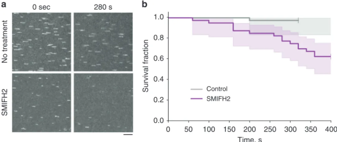
No treatment SMIFH2 0 sec 280 s 100 150 200 250 0.0 0 50 300 350 400 0.2 0.4 0.6 0.8 1.0 Survival fraction Time, s Control SMIFH2
a
b
Fig. 7 SMIFH2 enhances the detachment of actinfilaments from mDia1 formin in vitro. a The constitutively active mDia1 formin construct (FH1FH2DAD) was anchored to the glass surface of a microfluidic chamber by one of their FH2 domains using an anti-His antibody (as in ref.38, see Methods). Actin filaments were grown first in the presence of Alexa488-labeled actin to form fluorescent segments at their tips. Once the formin had nucleated a filament, it was exposed, from time zero onward, to 1µM unlabeled actin and 4 µM profilin, in the absence or presence of 100 µM SMIFH2. Images show frames corresponding to the beginning (time zero, left column) and 280 s (right column) following addition of unlabeled actin into theflow chamber in the absence (top row) and presence (bottom row) of SMIFH2 inhibitor in solution. The representative fragments of epifluorescent image field of view of the labeled segments of actinfilaments are seen. The number of filaments decreased with time due to their detachment from immobilized formin (see Supplementary Movies 23 and 24). Scale bar, 10μm. b The time at which each filament detached was recorded and the survival fraction of the filaments at each time point was calculated. See Methods for more details. Graphs of the survival fraction curves with 95% confidence interval shaded area around the actual data are presented. To estimate the significance of difference between the two survival fraction curves, the logrank test39was used. Calculated p value (Python software) was equal to 0.0009 (***)
collaboration between myosin X and actin polymerization
machinery including VASP
26and, perhaps, formins, but
corre-sponding mechanisms remain to be elucidated.
Our
finding that filopodia adhesion is force-dependent explains
how
filopodia could respond differently to substrates of varying
stiffness. On a stiff substrate, the force generated by myosin II and
applied to the adhesion complex will develop faster than on a
compliant substrate
57. Accordingly,
filopodia adhesion should be
more efficient on stiff substrates than on compliant substrates.
Indeed, we showed that the contacts of
filopodia with RGD
ligands associated with
fluid membrane bilayer were less stable
than with the areas of rigid substrate covered with RGD of the
same density. These considerations can explain involvement of
filopodia in the phenomenon of durotaxis
23, a preferential cell
movement towards stiffer substrates
58. This may provide a
mechanism to rectify directional cell migration.
Orientation based on
filopodia adhesion is characteristic for
several cell types, in particular for nerve cells. Application of
external force can regulate the direction of growth cone
advancement
59. The growth cones of most neurites produce
numerous
filopodia, and the adhesion of these filopodia can
determine the direction of neurite growth
60,61. The results from
these experiments can now be explained by preferential adhesion
of
filopodia, which experience larger force. Interestingly, the
filopodia-mediated traction force in growth cones is myosin
IIB-dependent
62, suggesting that preferential use of one or another
myosin II isoforms for
filopodia mediated mechanosensing could
be cell type specific.
The mechanosensitivity of
filopodia adhesion provides a
mechanism of cell orientation that complements that mediated by
focal adhesions. Focal adhesions are formed by cells attached to
rigid two-dimensional substrates, whereas
filopodia adhesion can
be formed by cells embedded in three-dimensional
fibrillar ECM
network. Further investigation of
filopodia mechanosensitivity
could shed a new light on a variety of processes related to tissue
morphogenesis.
Methods
Cell culture, transfection, and constructs. Hela-JW, a subline of a HeLa cervical carcinoma cell line derived in the laboratory of J. Willams (Carnegie-Mellon University, USA) on the basis of better attachment to plastic dishes63, was obtained from the laboratory of B. Geiger64. Cos-7 (African green monkeyfibroblast-like cell line) was obtained from ATCC (ATCC®CRL-1651™). Both cell lines were grown in Dulbecco’s Modified Eagle Medium (DMEM) supplemented with 10% fetal bovine serum (FBS), 100 U ml−1penicillin/streptomycin, 2 mM glutamine, and 1 mM sodium pyruvate at 5% CO2at 37 °C. HeLa-JW cells were transfected with DNA
plasmids by electroporation (two pulses of 1005 V for 35 ms) using the Neon transfection system (Thermofisher), while Cos-7 cells were transfected by jet-PRIME transfection reagent (Polyplus) according to the manufacturer’s protocol. Both cell lines were tested for mycoplasma contamination. No cell lines used in this study were found in the database of commonly misidentified cell lines that is maintained by ICLAC and NCBI Biosample. We did not attempt to authenticate them. Expression vectors encoding thefluorescent fusion proteins used in this study are described in the Supplementary Note 3.
Live cell imaging and confocal microscopy. Following electroporation or chemical-based transfection, cells were seeded at a density of 2 × 10⁴cells ml−1in 2 ml onto 35 mm glass based dishes with a bottom base cover glass #1 either 12 or 27 mm in diameter (Iwaki, Japan) coated with 10 µg ml−1fibronectin
a
b
D w ell time , s SLB-RGD (fluid) n = 1036 5 cells PLL-PEG-RGD (rigid) n = 1477 5 cells 1 10 100 1000 p < 0.0001Fig. 8 Attachment offilopodia to RGD-coated rigid and fluid substrate. a HeLa-JW cells expressing GFP-myosin X were plated on micropatterned coverslips covered with circular islands (D = 3 μm) of SLB conjugated to RGD (orange circles), organized into a square lattice. The glass between the islands was covered with poly-L-lysine-PEG (PLL-PEG) conjugated to RGD at the same density (Supplementary Fig. 6). Trajectories of GFP-positive filopodia tips acquired during a 14–36 min time interval are shown. The cell border is shown by a yellow dashed line. For comparison of the trajectories on rigid andfluid substrates, the circles of similar diameter were drawn by computer in the centers of the square lattice formed by SLB islands (outlined by gray contours). The segments of the trajectories located inside either the SLB islands or the drawn circles on the rigid substrate are shown in red, and the remaining parts of the trajectories are shown in white. See also Supplementary Movie 25. Scale bar, 5μm. b Dwelling time that myosin X-positive filopodia tips spent insidefluid (orange) or rigid (green) circles defined above. Pooled data for 5 cells; n—number of filopodia tip trajectories analyzed. Aligned dot plots and mean ± s.e.m. values are shown. Thep value was calculated using unpaired two-tailed t test with Welch’s correction
(Calbiochem) for 20 min. Cells were imaged in Leibovitz’s L-15 medium without Phenol Red containing 10% FBS. Samples were mounted in a humidified cell culture chambers and maintained at 37 °C with 5% CO2.
Snapshots or time-lapse images were acquired with a spinning-disc confocal microscopy system (PerkinElmer Ultraview VoX) based on an Olympus IX81 inverted microscope, equipped with a 100× oil immersion objective (1.40 NA, UPlanSApo), an EMCCD camera (C9100-13, Hamamatsu Photonics), and Volocity control software (PerkinElmer).
Photoactivation with instant imaging was performed by activation of a defined region insidefilopodia of live cells using a blue diode laser (405 nm, 100 mW) on a CSU-W1 spinning disk confocal system attached to a Nikon Eclipse Ti-E inverted microscope with Perfect Focus System, controlled by MetaMorph software (Molecular device), supplemented with a 100× oil 1.45 NA CFI Plan Apo Lambda oil immersion objective and sCMOS camera (Prime 95B, Photometrics).
For SIM, two types of equipment were used: (1) Live SR (Roper Scientific) module on a Nikon Eclipse Ti-E inverted microscope (specifications of setup described above), (2) Nikon N-SIM microscope, based on a Nikon Ti-E inverted microscope with Perfect Focus System controlled by Nikon NIS-Elements AR software supplemented with a 100× oil immersion objective (1.40 NA, CFI Plan-ApochromatVC) and EMCCD camera (Andor Ixon DU-897).
All SIM images with obtained with system (1) except for images on Fig.1c, d, which were obtained with set up (2).
Local presence of myosin IIAfilaments at filopodia bases. The presence of myosin IIAfilaments at filopodia bases was determined manually by examining of allfilopodia in time-lapse movies of Cos-7 cells transfected with mApple-myosin X and Myosin IIA-GFP constructs. The base offilopodium were defined as a space occupied by the circle with the diameter of 1.5μm, which is internally tangent to the plasma membrane at the area of negative curvature manifesting thefilopodia orifice. The myosin IIA filaments located inside this circle and above it (inside the filopodia) were scored as filopodia-associated (see Fig.2b, c, left panels). Lifetime analysis. The average lifetime offilopodia, λ, was calculated by fitting the time-dependentfilopodia survival fraction to the exponential decay function, e−t/λ, where t is the time. Thefitting was done by using the Levenberg–Marquardt algorithm built in Origin software package. Standard deviation (s.d.) of the average filopodia lifetime was estimated based on the standard error computed by the fitting algorithm.
Transfection of siRNA and immunoblotting. Cells were seeded into a 35 mm dish on day 0 and transfected with 50 nM of myosin IIA HC MYH9 ON-TARGET plus SMART pool siRNA (L-007668-00-0005, Dharmacon) using ScreenfectTMA (WAKO, Japan) on day 1. Control cells were transfected with scrambled control ON-TARGET plus Non-targeting pool siRNA (D-001810-10, Dharmacon). Transfection of plasmid GFP-myosin X and F-Tractin-tdTomato was performed in the evening of day 1 using Jet Prime transfection reagent (Polyplus) and cells were imaged on day 2. For assessment of myosin IIA heavy chain expression, transfected cells were lysed in RIPA buffer on day 2 (exactly 24 h following siRNA transfec-tion) and analyzed by Western blotting with primary rabbit antibodies to the myosin IIA tail domain (M8064, Sigma-Aldrich, dilution 1:1000); staining of α-tubulin with mouse monoclonal DM1A antibody (T6199, Sigma-Aldrich, dilution 1:5000) was used as a loading control. HRP-conjugated anti-rabbit IgG (Bio-Rad, 1706515, dilution 5000) and anti-mouse IgG (A4416, Sigma-Aldrich, dilution 1:10000) were used as secondary antibodies, respectively.
Immunofluorescence antibody staining. Cells were pre-fixed by addition of warm 20% PFA (Tousimis) into medium (to make afinal 2% solution) and subsequent incubation for 15 min at room temperature. This was followed byfixation and permeabilization by 3.7% PFA, 0.2% glutaraldehyde, and 0.25% Triton X-100 in PBS for 15 min. Thefixed cells were then washed two times with PBS and blocked with 5% bovine serum albumin (BSA) for 30 min. To assess endogenous level of myosin IIA cells were incubated with anti-myosin IIA tail domain (M8064, Sigma-Aldrich, dilution 1:800) 1 h at 4 °C, and 405 Alexa-Fluor-conjugated secondary antibodies (A31556, Molecular Probes, dilution 1:200) for 1 h at room temperature. Drug treatment. For formin drug inhibition studies, cells were incubated with 20 or 40μM SMIFH2 (4401, TOCRIS, UK) in serum containing DMEM for 1–2 h at 5% CO2, 37 °C. In in vitro experiments, SMIFH2 (340316-62-3, Sigma-Aldrich)
was used at a concentration of 100μM (see below). For myosin II inhibition studies, 30μM or 50 μM Rho-kinase (ROCK) inhibitor Y-27632 (Y0503, Sigma-Aldrich), or 20μM S-nitro-blebbistatin (13013-10, Cayman Chemicals) was added for 10–20 min before the experiments or directly during observation. All inhibitors remained in the medium during the entire observation period.
Optical tweezers and data acquisition. All experiments involvingfilopodia pulling were carried out on a Nikon A1R confocal microscope adapted for the use of laser tweezers. Totally, 2.19μm diameter polystyrene beads (PC05N, Bangs Laboratories) were coated withfibronectin (341635, Calbiochem) according to a
previous protocol65. For bead trapping we used an infrared laser (λ = 1064 nm, power 0.5–1 W, YLM-5-LP-SC Ytterbium Fiber Laser, IPG photonics).
To determine the forces, F, applied to the bead by the optical trap, we measured the displacement of the bead from the trap center,ΔX, and with knowledge of the stiffness of the trap, K, the force was calculated as F= KΔX. The trap stiffness was calibrated using the equipartition method66by tracking thefluctuations of a bead trapped by optical tweezers, using an Andor Neo sCMOS camera, at 100 fps. The displacement of the beads from the center of the optical trap in confocal microscopy observations was monitored using a piA640–210gm camera (Basler) at 0.5–1 fps and Metamorph software for tracking. The smallest detectable bead displacement was ~5 nm, corresponding to the smallest force measured of ~0.04 pN. The moment of detachment offilopodia from the bead was detected by the bead returning to the center of the optical trap. To confirm the detachment, we then switched the trap off, which resulted in release of the bead and its disappearance from thefield of view unless it remained connected to filopodia via the membrane tethers.
For laser trap experiments, HeLa-JW cells transfected by electroporation with GFP-myosin X and F-Tractin-tdTomato, were seeded at a density of 2 × 10⁴cells ml−1in 750μl onto chambered #1 borosilicate cover glasses (155383, Lab-Tek) coated withfibronectin (341635, Calbiochem) by incubation in 1 μg ml−1solution in PBS for 20 min at 37.0 °C. A reduced concentration offibronectin (compared to that used for regular cell observations) was used to prevent the beads sticking to the cover slip. Chambers with cells were mounted on a P-545.3R7 stage equipped with E-545 Plnano piezo controller (Physik Instrumente), which was moved in order to generate the pulling force betweenfilopodia and trapped beads. The velocity of the stage movement was maintained in a range of 10–20 nm/s by PIMikroMove 2.15.0.0 software. The specimen were incubated in a custom-built microscope hood at 37.0 °C, 5% CO2humidified environment. Simultaneously with pulling, the cells
were imaged using lasersλ = 488 and 561 nm for excitation of GFP and tdTomato, respectively. Collected experimental data were processed by particle tracking algorithms of the Metamorph software.
In vitro assay for formin-dependent actin polymerization. We used a micro-fluidics based assay to assess formin processivity in the course of formin-driven actin polymerization38. Formin construct Snap-mDia1(FH1FH2DAD)-6×HisTag was specifically anchored to the bottom surface of a microchamber, using a bio-tinylated pentaHis antibody (Qiagen) and streptavidin to bind to a biotin-BSA-functionalized glass surface which was further passivated with BSA. The micro-fluidics PDMS chamber height was 60 µm. Immobilized formin constructs were allowed to nucleate actinfilaments by exposing them for 30 s to 2 µM 20% Alexa488-labeled Lys328actin67and 0.4 µM profilin in a buffer containing 10 mM Tris-HCl (Euromedex) pH 7.8, 1 mM MgCl2 (Merck), 200 µM ATP (Roche), 50 mM KCl (VWR Chemicals) and supplemented with 5 mM DTT (Euromedex), 1 mM DABCO (1,4-diazabicyclo[2.2.2]octane, Sigma-Aldrich) to reduce photo-bleaching. Recording of actinfilament elongation was started after the buffer was changed to contain 1 µM unlabeled actin and 4 µM profilin, with or without 100 µM SMIFH2 (Sigma-Aldrich). The microfluidics flow was kept low enough to ensure that the viscous drag had no impact onfilament detachment from formin. Images were acquired every 10 s on a Nikon Ti-E microscope using a 60× objective and a Hamamatsu Orca Flash 4.0 V2+ sCMOS camera. Actin was purified from rabbit muscle68. Recombinant Profilin I and Snap-mDia1(FH1FH2DAD)-6×His-Tag were expressed in E. coli and purified69. Formin elongation rates were obtained by measuring the slope of kymograph from individualfilament traces38. Survival fraction curves representing the fractions of actinfilaments remaining associated with immobilized formins were calculated for cohorts of 40filaments for control and SMIFH2 treatment. The koffrates obtained by exponential decayfitting, and
the significance of the difference between survival fraction curves by logrank test were calculated using the Python software.
Unconstrainedfilopodia growth study. The same image sequences, which were used in experiments with optical trap, were then analyzed to track the tips of filopodia, which were neighboring to filopodia analyzed in pulling experiments and hence not attached to the beads. To trackfilopodia tips, the Imaris 8.3.1 spots tracking procedure was used. The trajectories of unconstrainedfilopodia tips were manually segmented into periods of linear growth, shrinking, and pausing. Elon-gation and shrinking rates were obtained using the linear regressionfitting function of Origin software package.
Analysis offilopodia adhesion to rigid and fluid substrates. The cleaned glass coverslips were coated with PLL-g-PEG-biotin (Susos) for 2 h followed by UV etching using 3 µm circular shape arrays of chromium (Cr) photomask70, which resulted in removal of PLL-g-PEG-biotin polymers on the etched circular regions. After multiple rinses with UHQ water, phospholipid vesicles were introduced to the surface for 5 min, allowing the self-assembly of lipid bilayer on the etched glass surfaces. Phospholipid vesicles were synthesized using the existing protocol70. Specifically, 98% of DOPC (1,2-dioleoyl-sn-glycero-3-phosphocholine) was mixed with 2% of biotin-DOGC (1,2-dioleoylsn-glycero-3-[(N-(5-amino-1-carbox-ypentyl)iminodiacetic acid)-succinyl]) biotin lipids. The lipid solution was further diluted with tris buffered saline (Sigma-Aldrich) at a ratio of 1:1 before