ORIGINAL ARTICLE
Treatment of residual pockets with photodynamic therapy,
diode laser, or deep scaling. A randomized, split-mouth
controlled clinical trial
Isabelle Cappuyns&Norbert Cionca&Philipp Wick&
Catherine Giannopoulou&Andrea Mombelli
Received: 13 July 2011 / Accepted: 28 October 2011 / Published online: 22 November 2011 # Springer-Verlag London Ltd 2011
Abstract The objective of this work was to compare the effects of antimicrobial photodynamic therapy (PDT), diode soft laser therapy (DSL), and thorough deep scaling and root planing (SRP) for treatment of residual pockets. Thirty-two subjects with a history of non-surgical treatment for chronic periodontitis were included. Residual pockets >4 mm and bleeding upon probing were debrided with an ultrasonic device and then subjected to either PDT, DSL, or SRP. Pocket probing depth (PPD), bleeding on probing (BOP), and gingival recession were monitored over 6 months. Counts of four microorganisms were determined by direct hybridization with RNA probes. PPD decreased from 5.6±1.0 to 3.8±1.1 in 6 months (p<0.001), and BOP decreased from 100% to 52% (p<0.01). The risk for a site to remain >4 mm with BOP depended on initial PPD (p=0.036) and was higher if treated with DSL (p=0.034). Frequencies of three microorganisms were significantly lower in PDT- and SRP-treated than in DSL-treated quadrants (p=0.02) after 14 days, but not at months 2 and 6. All three treatments resulted in a significant clinical improvement. PDT and SRP suppressed Porphyr-omonas gingivalis, Tannerella forsythia, and Treponema denticola stronger, and resulted in fewer persisting pockets after 6 months, than DSL application.
Keywords Photodynamic . Laser . Scaling/root planing . Non-surgical/mechanical . Therapy/treatment . Periodontitis
Introduction
Periodontal diseases can be treated by thorough, profes-sional, mechanical cleaning of microbially contaminated tooth surfaces [1]. The situation may remain stable over prolonged periods as long as appropriate continuous maintenance care is provided [2]. Complete removal of bacterial deposits can be challenging, especially if the disease has led to the formation of deep periodontal pockets. As an example, in patients with chronic perio-dontitis, full-mouth scaling and root planing within 48 h reduced the average number of pockets greater than 4 mm and bleeding on probing from 30 to three per subject [3]. In pockets initially deeper than 6 mm, the mean pocket probing depth was reduced by 2.3 mm to reach 4.9± 1.4 mm. In advanced cases, clinicians may therefore choose to repeat the treatment in the context of a surgical intervention to lift back the soft tissues for better access. If therapy is supplemented with systemic antibiotics, the results are generally better [4]. However, a limited number of residual pockets may still persist.
Residual pockets carry the risk of continuous pres-ence of periodontal pathogens [5] and may be repopu-lated with a microbiota incompatible with periodontal health [6–8]. Such sites therefore require specific profes-sional attention, and, over the years, may be re-instrumented repeatedly. Hence, maintenance protocols for residual pockets not only need to be efficient but also harmless after repeated application. Conventional scaling and root planing with sharp metal instruments is clearly not the optimal method. This procedure removes tooth sub-stance each time it is applied and will cause significant hard tissue damage cumulatively [9–12]. It should be reserved for initial cause-related therapy aiming at the
I. Cappuyns
:
N. Cionca:
P. Wick:
C. Giannopoulou:
A. Mombelli (*)School of Dental Medicine Division of Periodontology and Oral Pathophysiology, University of Geneva, Rue Barthélemy-Menn 19,
CH-1205 Geneva, Switzerland e-mail: andrea.mombelli@unige.ch
elimination of a combination of mineralized and non-mineralized bacterial deposits. Antibiotics are no alter-native, as their repeated administration would contribute extensively to the development of antimicrobial resis-tance. There is thus a need to carefully evaluate new protocols that are safe and effective for maintenance, especially for the removal of soft bacterial deposits (biofilm) without any adverse effects on host tissues. In a recent study we reported safety, patient acceptance, and short-term microbiological effects of a new air-polishing device in subjects in maintenance care with residual pockets >4 mm [13]. Long-term results will be needed to establish the value of this procedure as an alternative to conventional debridement. In the present paper, we report the effects of two other approaches: Treatment with diode laser and photodynamic therapy. Both methods may have a bactericidal and detoxifying effect and may therefore be useful as an adjunct or alternative to conventional, mechanical instrumentation of residual pockets.
The diode laser, with a wavelength between 655 and 980, does not interact with dental hard tissues. It is used for cutting and coagulating soft tissue, and has been proposed for sulcular debridement and curettage. Moritz et al. [14,
15] reported that pocket irradiation with a diode laser (805 nm) following scaling reduced bacterial counts from periodontal pockets better than the scaling alone. Bleeding on probing scores and pocket depths were better in the laser group as well. Two systematic reviews evaluated 10 years later the accumulated evidence regarding the potential benefit of laser application compared to mechanical debridement in non-surgical periodontal therapy [16, 17]. Due to the heterogeneity of the studies, a meta-analysis could not be performed, and the evidence to support the clinical application of different diode laser wavelengths was inconclusive.
Photodynamic therapy (PDT) is based on the princi-ple that light of a suitable wavelength can activate certain substances, called “photosensitizers”, to produce free oxygen radicals able to destroy bacteria and their products. The effectiveness of PDT therapy for perio-dontitis in adults, as a primary mode of treatment or as an adjunct to non-surgical treatment, was recently evaluated in a systematic review [18]. Five studies could be evaluated. PDT, as an independent treatment or as an adjunct to SRP versus a control group of SRP, did not demonstrate a clear advantage. The authors concluded that more research is necessary for proper evaluation of this therapy.
The purpose of this study was to evaluate in subjects in maintenance care, i.e., in patients having already received periodontal therapy earlier, with residual pockets >4 mm, the clinical and microbiologic effects of diode soft laser and
PDT as compared to conventional mechanical debridement (deep scaling and root planing).
Materials and methods
This was a single-center, examiner-masked, randomized, split-mouth three-arm parallel-design clinical trial of 6-month duration. The Ethical Committee of the University Hospitals of Geneva approved the protocol. Research was conducted according to the principles outlined in the Declaration of Helsinki on experimentation involving human subjects.
Subjects
Between September 2007 and June 2009 we recruited 32 systemically healthy subjects with residual pockets, previously treated for periodontal disease at the School of Dental Medicine of the University of Geneva. The eligibility criteria were: an age of 25–75 years; in maintenance after completion of comprehensive peri-odontal therapy since 3 to 24 months; presence, in the region from the incisors to the mesial aspect of the first molars, of at least one site in each of three quadrants with a probing pocket depth (PPD) >4 mm, bleeding upon probing (BOP positive), with clinical attachment loss (CAL) >1 mm, and without any macroscopic plaque retentive element, such as an insufficient resto-ration margin. The sample size was chosen taking into consideration reported mean differences in PPD in the order of 0.5 to 1 mm between non-surgical periodontal therapies with or without adjunctive antibiotics [19,20]. Assuming that the common standard deviation is 1 mm, a sample of 30 per group would provide 80% power to detect a true difference of 0.75 mm between groups.
Exclusion criteria included major systemic illnesses (i.e., diabetes mellitus, cancer, HIV, bone metabolic diseases or disorders that compromise wound healing, radiation, or immunosuppressive therapy), antibiotics, anti-inflammatory drugs, or other medication taken within the previous 2 months that may affect the outcome of the study, confirmed or suspected intolerance to the test products (i.e., thiazine compounds), and any physical limitations or restric-tions that might preclude normal oral hygiene procedures. The smoking history was recorded, but smoking was not an exclusion criterion. Written informed consent was obtained from all included subjects. A patient number was attributed in ascending order.
Three quadrants were selected as study quadrants. The deepest pocket in the area between the first incisor and the mesial aspect of the first molar with PPD >4 mm, CAL >1 mm and BOP was designated as the study site. Study
sites in contralateral quadrants had to be separated by at least one tooth.
Test products and randomization
Subjects were randomly assigned by a computer-generated table to receive one of the following three treatments in each of the study quadrants.
Treatment 1 (SRP): Root scaling and planning with Gracey curettes until the operator feels that all tooth surfaces are clean, hard, and smooth.
Treatment 2 (DSL): Subgingival irradiation with a diode soft laser (Elexxion CLAROS supplied by the Elexxion Dental Academy) for 60 s. The diode soft laser had a wavelength of 810 nm and a power output of 1 W.
Treatment 3 (PDT):“Photodynamic therapy” carried out according to the instructions of the supplier (Helbo Photodynamic Systems, Walldorf, Germany) as follows: 100 μg/ml phenothiazine chloride (Helbo Blue) sterile solution was instilled into the pockets with a blunt cannula. After 1 min, the pockets were rinsed with water and subsequently irradiated with a diode laser for 1 min. Laser light with a wavelength of 660 nm was applied subgingi-vally by corresponding sterile fiber optics. The effective power output was 40 mW.
Treatments were performed under anesthesia at the discretion of the patient.
Clinical protocol
Two independent clinicians (IC, NC) performed all proce-dures involving a contact with the subjects. The examiner (IC) enrolled the patients and recorded all parameters. The operator (NC) removed any supra-gingival deposits and performed the subgingival treatments. The operator was not involved in any evaluations before or after his intervention. With the exception of the periodontal pocket chart, necessary to deliver the treatment, he was unaware of previously recorded data.
The chronological sequence of the trial was as follows: In a first visit, the examiner recorded the medical history, obtained informed consent, removed supra-gingival depos-its, and gave oral hygiene instructions, if necessary. Within a maximum of 10 days, the patients were scheduled for the subgingival treatment. Immediately before treatment, the examiner recorded the Plaque Index (PlI) [21] and the Gingival Index (GI) [22]. She then took a subgingival plaque sample from each designated study site by inserting one paper point (Dentsply-Maillefer ISO 035) into each site. Afterwards, she recorded the PPD, BOP, and Reces-sion (REC; positive if gingival margin located apical, negative if located coronal to the cemento-enamel junction). Once completed, the operator took over. He opened an envelope with the subject’s number to reveal the treatment
assignment for the three study quadrants and the sequence of treatment. Quadrant by quadrant, he debrided all residual pockets mechanically with an ultrasonic device (EMS, Nyon, Switzerland) and applied the experimental treatment. After each quadrant, the operator noted the time he had used and asked the patient to rate pain/discomfort on a visual analog scale (VAS) by placing a mark on a horizontal line, 100 mm long, labeled with“no pain” at one end and with “worst pain” at the other.
The subjects were recalled 2 weeks, 2 months, and 6 months after treatment by the examiner who was blinded with respect to the allocation of treatments. Medical history, any concomitant medication, and all adverse events were recorded. The oral hygiene was checked and reinforced, if necessary. No attempt was made to re-instrument residual periodontal pockets. Clinical parameters were recorded at 2 and 6 months. Microbial samples were obtained after 2 weeks, 2 months, and 6 months.
Microbiological procedures
The samples were stored in 4 M guanidinium thiocyanate 2-mercaptoethanol at−20°C until processing. The samples were analyzed according to standard procedures [23] with an oligonucleotide probe-based method developed and validated by the laboratory processing the samples (Institut für angewandte Immunologie, Zuchwil, Switzerland). They were hybridized to a specific probe for the ssrRNA of Aggregatibacter actinomycetemcomitans (AA), Porphyro-monas gingivalis (PG), Tannerella forsythia (TF), Trepone-ma denticola (TD), and to a universal bacterial probe (TBL, total bacterial load) (IAI and MicroProbe Corporation, Bothell, WA, USA). Bacterial counts were calculated by comparison with homologous reference standards and expressed as count ×106.
Statistical analysis
The presence or absence of PPD >4 mm and bleeding upon probing was the primary outcome measure. Secondary outcomes included differences between groups for changes in PPD, REC, BOP, detection or no detection of target microorganisms.
Data were entered into a database and were checked for entry errors. The Kruskal–Wallis one-way analysis of variance, the Mann–Whitney U test or Fisher’s exact test were used to determine differences between sites treated with different procedures. The longitudinal changes were analyzed using the Wilcoxon matched-pairs signed-ranks test. Stepwise multiple linear and logistic regression analysis was used to study the relationship between VAS scores and treatment conditions, and between clinical outcomes at months 6 and the other parameters assessed.
One statistical program package (PASW Statistics 18 for Mac OS X, IBM Corporation, Somers, NY, USA) was used for all statistical analyses. p values <0.05 were accepted for statistical significance.
Results
All 32 enrolled patients could be followed up to month 2. Three subjects were unavailable for the 6-month evaluation. The reason was moving away from the Geneva region. Hence, 32 subjects were included in the analysis up to month 2, and 29 up to month 6. The mean age of the participants was 52 (range 36 to 74), females accounted for 28% of the participants, and 41% were smokers. Table 1
displays the baseline characteristics of the 32 participants. Only three out of all 96 study sites showed a PlI of 2, and none a value of 3 at baseline. Eleven sites scored GI=2 and none higher. No significant differences in clinical status were found between the three groups.
Table2shows the mean treatment time per quadrant and per tooth, and the perception of pain/discomfort rated on a VAS from 1 to 100 mm. Treatment times were about 6 min per tooth, and about 9 min per quadrant overall, with no significant differences between treatment methods. The patients’ perception of discomfort or pain was also similar. Mean VAS scores, as well as the number of subjects giving a score >40 mm, were not significantly different. Five subjects scored only one treatment above 40 (2× SRP, 3× DSL), four subjects scored to two therapies above this threshold
(2× DSL + PDT, 2× SRP + PDT). Using a threshold of 30 mm, twice as many subjects complained about SRP in comparison to PDT. Six of the ten subjects scoring SRP >30 mm complained about SRP only. Four of the seven subjects scoring DSL >30 mm complained about DSL only. Just one of the five subjects scoring PDT >30 mm complained about PDT only. In a model that also included total treatment time per quadrant and use of local anesthetics, logistic regression analysis indicated no significant influence of the treatment method on VAS scores >30 mm. An additional analysis was carried out to test if there was a sequence effect from the treatments. The impact of sequence was not significant.
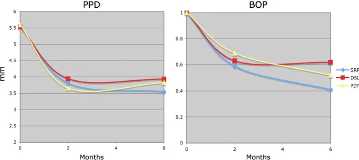
Figure 1 shows the longitudinal development of mean PPD and BOP over time. All three treatments resulted in a sustained, significant, and clinically relevant improvement of PPD (p<0.001) and BOP (p<0.005). Table3shows the clinical status in the study sites after 2 months. Only two sites in two different subjects showed a PlI of 2. All three sites in one subject, plus two isolated sites in two other subjects scored GI=2. The mean PPD amounted to 3.8 mm, 3.9 mm, and 3.6 mm after SRP, DSL, and PDT, respectively. A total of 16% of the sites treated with SRP, 16% of those treated with DSL, and 19% of those treated with PDT were still deeper than 4 mm, and bled upon probing. Eight subjects continued to have one study site in this category. In four subjects, two sites, treated with a different treatment modality, persisted. Differences between groups were not significant.
Table4 shows the clinical status in the study sites after 6 months. Only one site in two subjects scored PlI=2. Score 3 was never recorded. Only one site in two different subjects scored GI=2, and none were above. The pocket depth reduction obtained after 3 months was maintained, and differences between groups were not significant. Irrespective of treatment modality, roughly half of the study sites were BOP-positive. A total of 9% of the sites treated with SRP, 25% of those treated with DSL, and 9% of those treated with PDT were deeper than 4 mm and bled
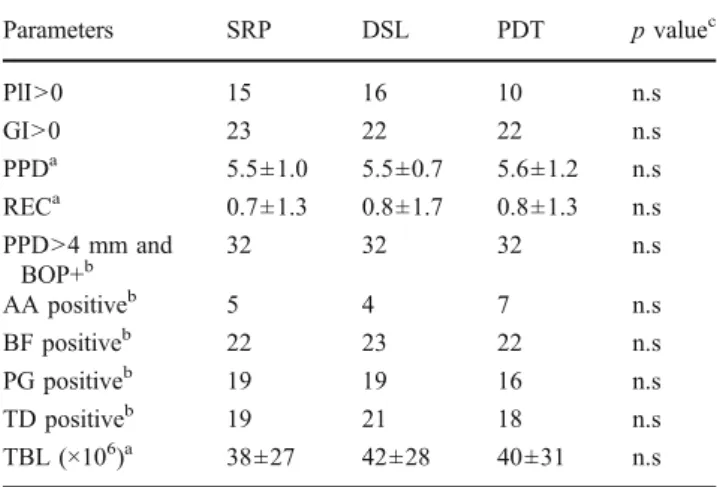
Table 1 Baseline characteristics of the study sites per treatment group (SRP scaling and root planing, DSL diode soft laser, PDT photody-namic therapy), n=32 Parameters SRP DSL PDT p valuec PlI>0 15 16 10 n.s GI>0 23 22 22 n.s PPDa 5.5±1.0 5.5±0.7 5.6±1.2 n.s RECa 0.7±1.3 0.8±1.7 0.8±1.3 n.s PPD>4 mm and BOP+b 32 32 32 n.s AA positiveb 5 4 7 n.s BF positiveb 22 23 22 n.s PG positiveb 19 19 16 n.s TD positiveb 19 21 18 n.s TBL (×106)a 38±27 42±28 40±31 n.s
PlI Plaque Index; GI Gingival Index; PPD probing pocket depth; REC recession; BOP bleeding upon probing. AA A. actinomycetemcomi-tans; TF T. forsythia; PG P. gingivalis; TD T. denticola
a
Mean ± standard deviation, bNumber of subjects, cDifference between groups
Table 2 Treatment time (TT) per quadrant and per tooth (seconds) and pain/discomfort (VAS 0–100 mm), for scaling and root planing (SRP), diode soft laser (DSL) or photodynamic therapy (PDT). n=32
Parameters SRP DSL PDT p valuec TT/quadranta 590±331 490±206 575±214 n.s TT/tootha 329±105 332±115 378±116 n.s VAS (mm)a 24±18 22±20 17±16 n.s VAS >40 mmb 5 5 4 n.s VAS >30 mmb 10 7 5 n.s a
Mean ± standard deviation, bNumber of subjects, cDifference between groups
upon probing. Twelve subjects continued to have one, and one subject two study sites of this category. Using backward stepwise logistic regression, the impact of the following variables on the persistence of PPD >4 mm and BOP positive was evaluated: initial PPD, treatment modal-ity, smoking, and gender. Two variables were retained in the final equation: The risk for being deeper than 4 mm and BOP positive rose with increasing initial PPD (p=0.036) and was higher if treated with DSL instead of SRP or PDT (p=0.034).
The total bacterial loads (TBL ×106) of microbial samples taken in study sites before treatment and 2 weeks, 2 months, and 6 months thereafter are given in Table5.
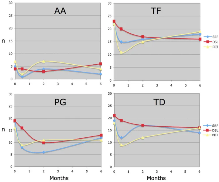
The numbers of subjects testing positive for AA, BF, PG, and TD are shown in Fig.2. Detection frequencies of
PG, TF, and TD were significantly lower 2 weeks after treatment by PDT and SRP than by DSL (p=0.02). After 2 and 6 months, these differences were no longer significant.
Discussion
The aim of the present study was to evaluate the clinical and microbiologic effects of diode soft laser and PDT as compared to conventional mechanical debridement specif-ically in subjects in maintenance care, i.e., in patients having already received periodontal therapy earlier. It should be noted that this represents a clinical condition more difficult to improve than untreated periodontal disease, the situation most often addressed in clinical trials.
Fig. 1 Longitudinal development of mean PPD (mm probing pocket depth) and BOP (1 bleeding, 0 no bleeding upon probing). n =29. PDT photodynamic therapy, DSL diode soft laser, SRP scaling and root planing. For statistical significance, see Tables3and4
Table 3 Clinical findings 2 months after scaling and root planing (SRP), diode soft laser (DSL), or photodynamic therapy (PDT), n=32
Parameters SRP DSL PDT p value‡ PlI>0 12 9 10 n.s GI>0 21 14c 22 n.s PPD* 3.8±1.0a 3.9±1.3a 3.6±1.2a n.s REC* 1.3±1.5b 1.2±1.6 1.2±1.1c n.s PPD>4 mm and BOP+† 5a 5a 6a n.s BOP positive† 19a 20b 22b n.s
PlI Plaque Index; GI Gingival Index; PPD probing pocket depth; REC recession; BOP bleeding upon probing. *Mean ± standard deviation, †Number of subjects, ‡Difference between groups, a
Different from baseline, p<0.001,bDifferent from baseline, p<0.01,cDifferent from baseline, p<0.05
Table 4 Clinical findings 6 months after scaling and root planing (SRP), diode soft laser (DSL), or photodynamic therapy (PDT), n=29
Parameters SRP DSL PDT p value‡ PlI>0 12 12 14 n.s GI>0 20 14C 19 n.s PPD* 3.6±1.1a 3.9±1.0a 3.8±1.2a n.s REC* 0.7±1.3 1.3±1.8b 1.0±1.3 n.s PPD>4 mm and BOP+† 3a 8a 3a p=0.034 BOP positive† 12a 18b 15a n.s
PlI Plaque Index; GI Gingival Index; PPD probing pocket depth; REC recession; BOP bleeding upon probing. *Mean±standard deviation, †Number of subjects, ‡Difference between groups,a
Different from baseline, p<0.001,bDifferent from baseline, p<0.01,cDifferent from baseline, p<0.05
The present study showed significant and clinically relevant improvements of mean PPD and BOP achieved with all
three treatment modalities, without a significant difference between groups. Of the test sites, 100% were bleeding upon probing at baseline. At month 6, roughly half of these sites were still bleeding. BOP may persist because of tooth position or other local difficulties limiting the access for therapy and optimal plaque control. The risk of a site to remain BOP-positive and deeper than 4 mm was higher after treatment with DSL than with SRP or PDT. Persisting pockets >4 mm bleeding upon probing are commonly perceived as needing further treatment in clinical practice. Table 4 indicates that more than twice as many sites remained in this category after DSL. Therefore, with regards to further treatment needs, we conclude that SRP and PDT were better than DSL.
It is clear that intra-individual comparisons of local periodontal therapies must be interpreted with caution. The
Table 5 Mean total bacterial loads (TBL ×106) of microbial samples taken in study sites at baseline, 2 weeks, 2 months, and 6 months after scaling and root planing (SRP), diode soft laser (DSL) or photody-namic therapy (PDT), n=29
Time point SRP DSL PDT p value‡
Baseline* 38±27 42±28 40±31 n.s
Week 2* 24±20c 32±23 27±21b n.s
Month 2* 28±19 28±25c 26±21b n.s
Month 6* 28±20 37±29 33±21 n.s
*Mean±standard deviation,†Number of subjects, ‡Difference between groups,bDifferent from baseline, p<0.01,cDifferent from baseline, p < 0.05
Fig. 2 Number of subjects testing positive at baseline, after 2 weeks, 2 months, and 6 months for A. actinomycetemcomitans (AA), T. forsythia (TF), P. gingivalis (PG), and T. denticola (TD). n=29. PDT
photodynamic therapy, DSL diode soft laser, SRP scaling and root planing. Detection frequencies of PG, TF, and TD significantly lower 2 weeks after treatment by PDT and SRP than by DSL (p=0.02)
photosensitizing agent was instilled into the pockets taking great care to avoid spillover to other dentition areas. If minimal amounts of the colorant should nevertheless have reached other parts of the dentition, it is very unlikely that it penetrated into other residual pockets. Furthermore, it has been shown that the photosensitizer has no effect if it is not activated by light of the corresponding wavelength [24]. Following the recommendations for photodynamic therapy issued by the manufacturer, all sites received preliminary debridement with an ultrasonic device before the experimen-tal treatment. It can therefore not be excluded that a part of the observed effects in all three groups may be attributed to this preliminary cleaning. On the other hand, it should not be forgotten that these treatments were given to sites that in the past had already received mechanical treatments with ultrasonic and other instruments multiple times.
On the basis of five papers, a systematic review [18] was unable to demonstrate a clear advantage of PDT, either as an independent treatment or as an adjunct to SRP, versus a control group of SRP. These studies concerned cases with early to mild [25], moderate to advanced [26], chronic [27,
28], or aggressive periodontitis [29]. As different photody-namic devices and procedures were used in these trials and clinical diagnoses were variable, a clear over-all conclusion cannot be drawn. The patients with chronic periodontitis treated by Braun et al. [28] did show a significantly better improvement of CAL and BOP if treated with PDT plus SRP instead of SRP alone. A study not identified by the above-mentioned review concerned 24 patients receiving supportive periodontal therapy [30]. After 3 and 6 months, there were no statistically significant differences of mean PPD and CAL between patients treated with SRP with or without adjunctive PDT, but BOP was found to improve better in the test group. A further, recent study [31] evaluated the adjunctive effects of PDT to SRP in the treatment of chronic periodontitis in 56 patients 6 and 12 weeks after treatment. There were again no differences between groups for PPD and CAL, however, in sites with PPD >4 mm, the BOP was lower if treated by PDT.
In the present study, detection frequencies of PG, TF, and TD were significantly lower 2 weeks after treatment by PDT and SRP than by DSL (p=0.02). After 2 and 6 months, these differences were no longer significant. Yilmaz et al. [25] reported short-term microbiological and clinical results of treatment with soft laser in conjunction with methylene blue and/or SRP in ten patients. Within the limits of this study, PDT provided no additional microbiological and clinical benefits over conventional mechanical debridement. Chondros et al. [30] reported a statistically significant reduction of Fusobacterium nucleatum and Eubacterium nodatum in the test group at month 3. The levels of the microorganisms investigated also in our study (AA, PG, TF, TD) were not significantly different. Theodoro et al. [32]
evaluated the long-term clinical and microbiological effects of PDT associated with nonsurgical periodontal treatment in 33 patients. Although no statistically significant benefit in terms of clinical outcome could be demonstrated, PDT treatment led to a significant reduction in the percentage of sites positive for all bacteria compared to SRP alone.
The next point to discuss is patient acceptance. Consen-sus is lacking about the statistical analysis of VAS data and their interpretation [33]. Comparison of parametric and nonparametric analysis of VAS pain scores suggested that parametric tests were accurate and had the greatest power to detect a difference [34]. In the present study, mean VAS scores and the number of scores >40 mm were similar in all three groups. A tendency for more frequent VAS scores >30 mm after SRP did not reach the level of statistical significance. The total treatment time per quadrant, the use of local anesthetics, and the sequence of the treatments had no significant impact. For acute pain, the minimal clinically significant VAS change is 16 mm [35]. The results of the study are compatible with the notion that pain associated with dental care is a subjective reaction strongly influenced by factors such as gender, personality, and especially previous general and dental experience [36].
Within the limits of our study, we conclude that all three treatments resulted in a significant clinical improvement. Although significant differences with regards to the suppres-sion of PG, TF, and TD were noted 14 days after treatment, a clear superiority of one procedure could not be demonstrated. PDT seemed to suppress TF and TD better initially, but levels increased again thereafter. As this study was not designed to prove equivalence, clear-cut recommendations for clinical practice cannot be made. One should, however, consider that in the context of maintenance care, a procedure that is well tolerated and has minimal side-effects even when repeated multiple times, has a potential. Clinical performance in comparison to standard maintenance protocols remains to be demonstrated in long-term studies.
Acknowledgements The authors gratefully acknowledge the support of the following companies: Helbo Photodynamic Systems GmbH & Co KG, Walldorf, Germany, who provided the material for the photodynamic therapy and the 660-nm diode laser free of charge, Elexxion AG, Radolfzell, Germany who provided the 810-nm diode laser free of charge, and the Institut für angewandte Immunologie, Zuchwil, Switzerland, who provided the microbiological analyses free of charge.
Conflict of interest The authors declare that there are no conflicts of interest in this study.
References
1. van der Weijden GA, Timmerman FA (2002) A systematic review on the clinical efficacy of subgingival debridement in the
treatment of chronic periodontitis. J Clin Periodontol 29(Suppl 3):55–71
2. Axelsson P, Nystrom B, Lindhe J (2004) The long-term effect of a plaque control program on tooth mortality, caries and periodontal disease in adults. Results after 30 years of maintenance. J Clin Periodontol 31:749–757
3. Cionca N, Giannopoulou C, Ugolotti G, Mombelli A (2009) Amoxicillin and metronidazole as an adjunct to full-mouth scaling and root planing of chronic periodontitis. J Periodontol 80:364–371 4. Mombelli A, Cionca N, Almaghlouth A (2011) Does adjunctive antimicrobial therapy reduce the perceived need for periodontal surgery? Periodontol 2000 55:205–216
5. Mombelli A, Schmid B, Rutar A, Lang NP (2000) Persistence patterns of Porphyromonas gingivalis, Prevotella intermedia/ nigrescens, and Actinobacillus actinomycetemcomitans after me-chanical therapy of periodontal disease. J Periodontol 71:14–21 6. Magnusson I, Lindhe J, Yoneyama T, Liljenberg B (1984)
Recolonization of a subgingival microbiota following scaling in deep pockets. J Clin Periodontol 11:193–207
7. Quirynen M, Bollen CML, Vandekerckhove BNA, Dekeyser C, Papaioannou W, Eyssen H (1995) Full- vs. partial-mouth disinfection in the treatment of periodontal infections: short-term clinical and microbiological observations. J Dent Res 74:1459–1467
8. Sbordone L, Ramaglia L, Gulletta E, Iacono V (1990) Recoloni-zation of the subgingival microflora after scaling and root planing in human periodontitis. J Periodontol 61:579–584
9. Kocher T, Fanghanel J, Sawaf H, Litz R (2001) Substance loss caused by scaling with different sonic scaler inserts—an in vitro study. J Clin Periodontol 28:9–15
10. Flemmig TF, Petersilka GJ, Mehl A, Hickel R, Klaiber B (1998) The effect of working parameters on root substance removal using a piezoelectric ultrasonic scaler in vitro. J Clin Periodontol 25:158–163 11. Ritz L, Hefti AF, Rateitschak KH (1991) An in vitro investigation on the loss of root substance in scaling with various instruments. J Clin Periodontol 18:643–647
12. Zappa U, Smith B, Simona C, Graf H, Case D, Kim W (1991) Root substance removal by scaling and root planing. J Periodontol 62:750–754
13. Moëne R, Décaillet F, Andersen E, Mombelli A (2010) Subgin-gival plaque removal using a new air-polishing device. J Periodontol 81:79–88
14. Moritz A, Schoop U, Goharkhay K, Schauer P, Doertbudak O, Wernisch J, Sperr W (1998) Treatment of periodontal pockets with a diode laser. Lasers Surg Med 22:302–311
15. Moritz A, Gutknecht N, Doertbudak O, Goharkhay K, Schoop U, Schauer P, Sperr W (1997) Bacterial reduction in periodontal pockets through irradiation with a diode laser: a pilot study. J Clin Laser Med Surg 15:33–37
16. Schwarz F, Aoki A, Becker J, Sculean A (2008) Laser application in non-surgical periodontal therapy: a systematic review. J Clin Periodontol 35:29–44
17. Karlsson MR, Diogo Lofgren CI, Jansson HM (2008) The effect of laser therapy as an adjunct to non-surgical periodontal treatment in subjects with chronic periodontitis: a systematic review. J Periodontol 79:2021–2028
18. Azarpazhooh A, Shah PS, Tenenbaum HC, Goldberg MB (2010) The effect of photodynamic therapy for periodontitis: a systematic review and meta-analysis. J Periodontol 81:4–14
19. Haffajee AD, Socransky SS, Gunsolley JC (2003) Systemic anti-infective periodontal therapy. A systematic review. Ann Perio-dontol 8:115–181
20. Herrera D, Sanz M, Jepsen S, Needleman I, Roldán S (2002) A systematic review on the effect of systemic antimicrobials as an adjunct to scaling and root planing in periodontitis patients. J Clin Periodontol 29:136–159
21. Silness J, Löe H (1964) Periodontal disease in pregnancy II. Correlation between oral hygiene and periodontal condition. Acta Odontol Scand 22:121–135
22. Löe H, Silness J (1963) Periodontal disease in pregnancy I. Prevalence and severity. Acta Odontol Scand 21:533–551 23. Dix K, Watanabe SM, McArdle S, Lee DI, Randolph C, Moncla
B, Schwartz DE (1990) Species-specific oligodeoxynucleotide probes for the identification of periodontal bacteria. J Clin Microbiol 28:319–323
24. Sigusch BW, Engelbrecht M, Volpel A, Holletschke A, Pfister W, Schutze J (2010) Full-mouth antimicrobial photodynamic therapy in Fusobacterium nucleatum-infected periodontitis patients. J Periodontol 81:975–981
25. Yilmaz S, Kuru B, Kuru L, Noyan U, Argun D, Kadir T (2002) Effect of gallium arsenide diode laser on human periodontal disease: a microbiological and clinical study. Lasers Surg Med 30:60–66
26. Andersen R, Loebel N, Hammond D, Wilson M (2007) Treatment of periodontal disease by photodisinfection compared to scaling and root planing. J Clin Dent 18:34–38
27. Christodoulides N, Nikolidakis D, Chondros P, Becker J, Schwarz F, Rossler R, Sculean A (2008) Photodynamic therapy as an adjunct to non-surgical periodontal treatment: a randomized, controlled clinical trial. J Periodontol 79:1638–1644
28. Braun A, Dehn C, Krause F, Jepsen S (2008) Short-term clinical effects of adjunctive antimicrobial photodynamic therapy in periodontal treatment: a randomized clinical trial. J Clin Perio-dontol 35:877–884
29. de Oliveira RR, Schwartz-Filho HO, Novaes AB Jr, Taba M Jr (2007) Antimicrobial photodynamic therapy in the non-surgical treatment of aggressive periodontitis: a preliminary randomized controlled clinical study. J Periodontol 78:965–973
30. Chondros P, Nikolidakis D, Christodoulides N, Rossler R, Gutknecht N, Sculean A (2009) Photodynamic therapy as adjunct to non-surgical periodontal treatment in patients on periodontal maintenance: a randomized controlled clinical trial. Lasers Med Sci 24:681–688
31. Ge L, Shu R, Li Y, Li C, Luo L, Song Z, Xie Y, Liu D (2011) Adjunctive effect of photodynamic therapy to scaling and root planing in the treatment of chronic periodontitis. Photomed Laser Surg 29:33–37
32. Theodoro LH, Silva SP, Pires JR, Soares GH, Pontes AE, Zuza EP, Spolidorio DM, de Toledo BE, Garcia VG (2011) Clinical and microbiological effects of photodynamic therapy associated with nonsurgical periodontal treatment. A 6-month follow-up. Lasers Med Sci
33. Hauser K, Walsh D (2008) Visual analogue scales and assessment of quality of life in cancer. J Support Oncol 6:277–282
34. Dexter F, Chestnut DH (1995) Analysis of statistical tests to compare visual analog scale measurements among groups. Anesthesiology 82:896–902
35. Gallagher EJ, Bijur PE, Latimer C, Silver W (2002) Reliability and validity of a visual analog scale for acute abdominal pain in the ED. Am J Emerg Med 20:287–290
36. Williams JM, Murray JJ, Lund CA, Harkiss B, de Franco A (1985) Anxiety in the child dental clinic. J Child Psychol Psychiatry 26:305– 310