Imaging
Imaging the right heart: the use of integrated
multimodality imaging
Emanuela R. Valsangiacomo Buechel
1
*
and Luc L. Mertens
2
1
Division of Paediatric Cardiology and Children’s Research Centre, University Children’s Hospital Zurich, Steinwiesstrasse 75, 8032 Zurich, Switzerland; and2
The Labatt Family Heart Center, The Hospital for Sick Children, University of Toronto, Toronto, ON, Canada
Received 16 August 2011; revised 11 October 2011; accepted 12 December 2011; online publish-ahead-of-print 8 March 2012
During recent years, right ventricular (RV) structure and function have been found to be an important determinant of outcome in different
cardiovascular and also pulmonary diseases. Currently, echocardiography and cardiac magnetic resonance (CMR) imaging are the two
imaging modalities most commonly used to visualize the RV. Most structural abnormalities of the RV can be reliably described by
echocardi-ography but due its complex geometrical shape, echocardiographic assessment of RV function is more challenging. Newer promising
echocar-diographic techniques are emerging but lack of validation and limited normal reference data influence their routine clinical application. Cardiac
magnetic resonance is generally considered the clinical reference technique due to its unlimited imaging planes, superior image resolution, and
three-dimensional volumetric rendering. The accuracy and reliability of CMR measurements make it the ideal tool for serial examinations of RV
function. Multidetector computed tomography (MDCT) plays an important role in the diagnosis of pulmonary emboli but can also be used for
assessing RV ischaemic disease or as an alternative for CMR if contra-indicated. Radionuclide techniques have become more obsolete in the
current era. The different imaging modalities should be considered complimentary and each plays a role for different indications.
-Keywords
Right ventricle † Multimodality imaging † Echocardiography † Cardiac magnetic resonance imaging
Introduction
The right ventricle (RV) has for a long time been the neglected side
of the heart, but its role in different cardiovascular diseases has
been increasingly recognized. This is obvious for structural
con-genital heart defects (CHD) involving the RV such as pulmonary
valve stenosis, tetralogy of Fallot (TOF), and Ebstein
malforma-tion.
1Beyond this, RV function has been shown to be one of the
most important outcome determinants in patients with pulmonary
arterial hypertension (PAH).
2,3Also in patients with
cardiomyop-athy and ischaemic heart disease, RV dysfunction has been
shown to be a strong predictor of adverse events, independent
from left ventricular (LV) function and the presence of
ischae-mia.
4–6Different imaging modalities can be used for imaging the
RV. Due to technological advances, the role and clinical use of
these techniques is evolving. In different conditions, each technique
provides complementary information and this influences its use in
clinical practice. Currently, echocardiography and cardiac magnetic
resonance (CMR) imaging are the two most commonly used
imaging techniques for structural and functional evaluation of the
RV.
7Other imaging modalities such as multidetector-computed
tomography (MDCT) and radionuclide techniques are valuable
alternatives in selected patients.
Right ventricular structure and
function
Correct identification of the morphologic RV is the first important
step for RV assessment, independently from the imaging modality
used. The segmental approach to cardiac anatomy helps define
the cardiac structures and segments based on constant anatomical
features.
8–11The gross anatomy of the RV differs from the LV as it
has a more complex geometrical shape being ‘wrapped around’ the
LV. This complex geometry precludes imaging the inflow and
outflow tract in a single two-dimensional plane. Compared with
the LV, the RV myocardium is significantly more trabeculated,
and the RV wall much thinner with a normal compacted wall
thick-ness of 3 – 5 mm in the adult population.
The structural organization of the myocardial cells has a
charac-teristic complex three-dimensional myofiber arrangement.
12,13The
LV wall has a three-layered structure with the epicardial cells
*Corresponding author. Tel:+41 1 266 7339, Fax: +41 1 266 7981, Email:[email protected]
oriented obliquely, the mid-myocardial cells more
circumferential-ly, and the endocardial cells again obliquely. The well-developed
midwall circumferential layer is responsible for the predominance
of circumferential shortening and radial thickening in the LV. The
RV epicardial fibres are oriented obliquely and contiguous with
epicardial LV fibres, the midwall circumferential layer is poorly
developed and the endocardial fibres are oriented longitudinally.
This fibre structure explains why RV ejection is determined by
lon-gitudinal shortening rather than by circumferential deformation.
The normal RV contraction results in a peristaltic contraction
going from the inflow to the outflow part of the RV.
14In case of
RV hypertrophy, the hypertrophied fibres seem to be oriented
more circumferentially and circumferential and radial shortening
contribute more to RV ejection.
15,16The RV is also functionally different from the LV.
17–19Right
ven-tricular pressure – volume loops are more triangular compared
with the rectangular LV loops and have very short or absent
isovo-lumetric contraction and relaxation periods.
18The RV responds
differently to acute and chronic stressors as well as to
pharmaco-logical agents. The RV is more sensitive to both acute and chronic
pressure loading and is at risk for acute and chronic RV failure.
19Recent data have suggested that in both ventricles, different
mo-lecular pathways are involved in the adaptive myocardial response
to changes in loading conditions.
20–22This could be related to the
different embryologic origin of RV and LV myocardial cells
23,24and
has potential implications for targeted pharmacological treatment
of RV failure.
Interventricular interaction is another important aspect of RV
disease: RV hypertrophy and/or dilatation affect LV function.
25–28The increased transseptal gradient associated with RV
hyperten-sion causes bowing of the interventricular septum towards the
LV. In RV dilatation, this occurs during diastole influencing LV
filling, and in the case of elevated RV systolic pressure, this can
affect LV systolic function and mechanics. Thus, RV abnormalities
may indirectly affect LV output and overall cardiac performance.
Imaging modalities
Echocardiography
Right ventricular morphology can generally be adequately
described by transthoracic echocardiography in most patients.
Only when transthoracic imaging windows are poor and RV
disease is suspected, additional imaging may be required.
Depend-ing on patient age and clinical problem, transoesophageal
echocar-diography, CMR, or MDCT can be used.
Assessment of RV size and function should be part of every
echocardiographic examination at the time of first diagnosis and
during serial follow up, particularly in patients with chronic
condi-tions, such as PAH and cardiomyopathy. Guidelines for the
echo-cardiographic evaluation of the RV have been published for the
adult
29and paediatric population.
30Both guidelines stress the
im-portance of combining different 2D echocardiographic views for
obtaining full coverage of the different RV segments. Different
apical views as well as subcostal and parasternal long-axis and
short-axis views should be acquired (Figure
1
). Echocardiographic
evaluation should also include 2D measurements of right atrial
dimensions and RV wall thickness. The most commonly used
normal values for the adult population reported in the
echocardio-graphic guidelines as a summary of several studies are shown in
Table
1
. These proposed normal data have some limitations: the
re-producibility of different measurements needs to be tested and the
normal data will have to be stratified for age ranges, body size, and
gender. Assessment of RV volumes using 2D echocardiography is
more challenging. Two-dimensional RV measurements as well as
different geometrical formula proposed for volume calculation
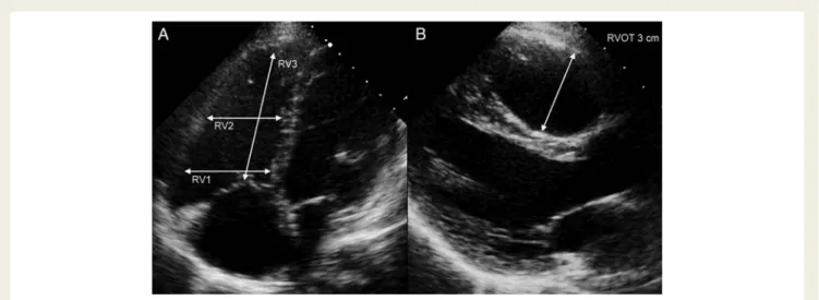
Figure 1
Measurement of right ventricular (RV) dimensions using echocardiography. Two-dimensional measurements should be made in
dif-ferent parts of the RV. (A) From the apical four-chamber view, the inflow part of the RV is measured as the maximal short-axis dimension in the
basal one-third of the RV (RV1). The midcavity dimension is measured in the middle third of the RV at the level of the papillary muscles (RV2).
This represents the trabecular part of the RV. The longitudinal dimension is measured from the middle of the tricuspid valve to the RV apex
(RV3). (B) From the parasternal long-axis view, the proximal part of the right ventricular outflow tract is measured.
show poor agreement with three-dimensional (3D) volumes
calcu-lated by CMR.
31,32Recently, 3D echocardiography has emerged as
a promising technique for the assessment of RV volumes.
33Differ-ent methods have been proposed for the analysis of 3D volumetric
data sets; the most commonly used being the Beutel technique.
34–38This method has been proven to be reliable and accurate in different
conditions, including CHD and PAH. The challenge of acquiring a
good quality full volumetric 3D data set, including the RV anterior
wall and the RV apical lateral segments, in patients with poor
imaging windows and/or dilated RV is the main limitation of the
method. Moreover, accuracy tends to decrease with increasing RV
dilatation, limiting its application in the more dilated ventricles.
34,39Three-dimensional echocardiography can also be useful for
imaging the tricuspid valve, as all three leaflets and commissures
can be visualized in a single 3D image. The mechanisms of tricuspid
regurgitation can be better understood, which facilitate guiding
sur-gical repair.
40–42Due to the limitations discussed above, echocardiographic
as-sessment of RV function remains challenging in clinical practice
and often is limited to subjective qualitative assessment. Recent
guidelines recommend performing quantitative measurements of
RV function
29,30by using at least one of the following
echocardio-graphic parameters as surrogate of volumetric assessment of RV
function: percent fractional area change (FAC), tricuspid annular
plane systolic excursion (TAPSE), or RV index of myocardial
per-formance (RIMP) (Table
2
). The observation that the combined
use of three different parameters is being proposed probably
indi-cates that no single measurement has been validated for clinical use
. . . .
. . . .
Table 1
Two-dimensional measurements of the right ventricular cavity
Normal value Limitation 2D RV measurements
RV 1 (RV basal diameter) ,4.2 cm Influenced by probe rotation Requires RV-focused view
RV 2 (midcavity diameter) ,3.5 cm Level of measurement not clearly defined (level LV papillary muscles)
Normal value?
RV 3 (base-apex RV length) ,8.6 cm Influenced by probe rotation Definition of RV apex?
RV end-diastolic area ,28 cm2
Influenced by probe rotation
Definition of trabeculations can be difficult RVOT parasternal short axis just proximal to pulmonary valve 2.7 cm Often difficult endocardial definition
Limited normative data
RVOT parasternal long axis 3.3 cm Anterior wall definition can be difficult Limited normative data
2D right atrial dimensions
RA major dimension (apical 4C) ,5.3 cm Influenced by imaging plane Relatively few normative data RA minor dimension ,4.4 cm Imaging plane will affect measurement
RA end-systolic area ,20 cm2
Few normative data
. . . .
Table 2
Right ventricular functional measurements
Measurement Normal value Limitation
%FAC .35% Endocardial border detection can be difficult especially in systole Load-dependent
No imaging of the outflow tract
TAPSE .16 mm Influenced by direction of motion (alignment) Influenced by loading and tricuspid regurgitation No imaging of the outflow tract
Peak systolic tissue Doppler velocity tricuspid annulus
.10 cm/s Influenced by alignment with the motion Influenced by loading conditions
Only one single segment used for global function Peak systolic longitudinal strain of the
RV lateral wall
No good reference value Needs further validation Normative data are needed
in different conditions and that their impact on management and
outcome remains uncertain.
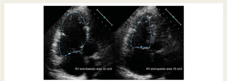
Fractional area change (Figure
2
) has been shown to correlate
with RV EF calculated by CMR and to be an independent predictor
for outcome after myocardial infarction.
43However, image quality
and visualization of the endocardial borders are often limited
espe-cially in the RV lateral wall and RV apex. Tricuspid annular plane
systolic excursion (Figure
3
) is easy to measure and represents
lon-gitudinal shortening of the RV lateral wall, but normal values for
different age groups and its validation are limited.
29In addition,
the presence of tricuspid regurgitation may influence the values
obtained. Right ventricular index of myocardial performance is a
Doppler method combining measurement of isovolumic
contrac-tion and relaxacontrac-tion times.
44Right ventricular index of myocardial
performance is load dependent and due to the short RV
isovolu-mic time intervals, its use remains controversial.
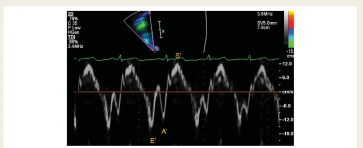
45Emerging echocardiographic techniques like tissue Doppler
(TDI) and strain imaging have been applied to the assessment of
RV function. Peak tissue Doppler systolic velocity in the tricuspid
annulus is a measurement of RV longitudinal function (Figure
4
).
Figure 2
Measurement of fractional area change (FAC). Right ventricular end-diastolic and end-systolic area is measured from an apical
four-chamber view. FAC is calculated as (end-diastolic area-end-systolic area)/end-diastolic area. In the example shown FAC is 50% (normal value
.35%).
Figure 3
Tricuspid annular planar systolic excursion (TAPSE). From the apical four-chamber view, an M-mode is put through the tricuspid
annulus with as good alignment as possible with longitudinal motion of the annulus. The systolic displacement of the tricuspid annulus is
mea-sured. To optimize timing, we use tissue colour M-mode which allows to well define end-diastole and end-systole on the tracings. TAPSE should
be .1.6 cm.
This technique is easy and reproducible but has the limitations of
being angle-dependent, load-dependent, and influenced by the
global cardiac translation and tricuspid regurgitation. In the young
adult, a normal cut-off value of
≥10 cm/s has been proposed;
however, normal data for all different age ranges and gender are
lacking.
29The introduction of speckle-tracking echocardiography made
the measurement of strain and strain rate easier. Although
devel-oped for the LV, speckle tracking has been applied to the RV.
7,46Peak longitudinal strain and strain rate measurement are
independ-ent from global cardiac motion and allow quantifying regional
myo-cardial deformation in the different RV segments. Right ventricular
strain has been shown to be reduced in patients with PAH,
25,47,48a
systemic RV,
49,50and after TOF repair
51,52(Figure
5
). Although very
promising, myocardial deformation imaging has significant
limita-tions. Strain values are influenced by loading conditions, as it has
been demonstrated in patients with PAH, in whom RV longitudinal
strain was related to pulmonary arterial systolic pressures.
25Add-itionally, strain values are influenced by RV size and stroke volume.
Feasibility is not always given in the thin RV wall; normal values for
different age ranges, body size, and gender still have not been
established yet; standardization among different software solutions
is still being investigated. Therefore, TDI and speckle tracking are
not ready yet for routine clinical use.
53Figure 4
Pulsed tissue Doppler of the tricuspid annulus. This a pulsed Doppler tracing obtained in the tricuspid annulus from the apical
four-chamber view. A systolic peak velocity (S
′), early diastolic velocity (E
′), and atrial contraction velocity (A
′) can be measured. Notice on this trace
the near absence of isovolumetric periods in this normal right ventricle. After systole, the early diastolic wave starts, after diastolic, during the
isovolumetric period there is a short isovolumetric peak corresponding to changes in the myocardium during the isovolumetric period.
Figure 5
Longitudinal strain measurement in the right ventricular (RV) free wall. From the apical four-chamber view, strain curves are
obtained using speckle tracking echocardiography. On the left panel, strain in the RV free wall was measured in a postoperative tetralogy of
Fallot patient. This patient underwent pulmonary valve replacement and the right-hand panel reflects strain measurements 6 months after
the surgery. A reduction in strain may result from changes in RV stroke volume, RV preload, and RV geometry.
Cardiovascular magnetic resonance
Cardiovascular magnetic resonance is the second-line modality
after echocardiography for comprehensive RV evaluation.
Cardio-vascular magnetic resonance is currently considered the reference
standard for functional RV studies as it allows visualizing anatomy,
quantifying function, and calculating flows.
Anatomical assessment is usually performed with T1-weighted
black-blood turbo spin-echo sequence or with the steady-state
free precession (SSFP) sequence. Standard axial images allow
seg-mental analysis of cardiac anatomy and visualization of the
pulmon-ary arteries, pulmonpulmon-ary veins, aorta, and systemic veins. Detailed
description of the intra- and extracardiac anatomy can be achieved
by 3D rendering techniques, including contrast-enhanced MR
angi-ography and 3D SSFP. This is important for detailed description of
complex cardiac anatomy and for preoperative planning.
54,55Cardiovascular magnetic resonance also provides advanced
imaging of the RV myocardium inclusive tissue characterization.
Different T1- and T2-weighted sequences combined with
late-enhancement imaging after gadolinium administration can be
used for tissue characterization. Tissue characterization is used
for assessment and differentiation of different cardiomyopathies
affecting the RV, including arrhythmogenic RV
cardiomyopathyopa-thy (ARVC), metabolic storage diseases, and cardiac tumours.
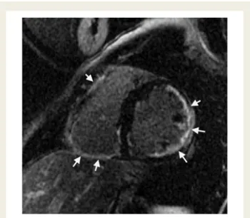
56–59Late enhancement imaging shows intramyocardial fibrosis,
inflam-mation, scars, and fat accumulation (Figure
6
). In CHD, the
pres-ence of myocardial scars in the RV is supposed to be a risk
factor for adverse events during follow-up.
60,61Interpretation of
the images can be challenging due to the thin RV wall and the
sur-rounding epicardial fat and pericardium. The prognostic
signifi-cance of myocardial late-enhancement in various diseases needs
further investigation.
Cardiovascular magnetic resonance is considered the clinical
ref-erence technique for accurate assessment of global RV function.
Short-axis or axial SSFP images and the summation disc method
are used for calculation of RV volumes and ejection fraction (EF)
without any geometrical assumption (Figure
7
). Appropriate
spatial and temporal resolution of the images is important for
ac-curacy of the results and can be achieved by adjusting acquisition
parameters to the patient’s size and heart rate.
62Normal
age-and gender-specific values for RV volumes age-and function have
been published for the adult and the paediatric population.
63,64Provided adequate standardization, CMR RV measurements
show high reproducibility with an interobserver variability ,7%
for the end-diastolic volume, ,14% for the end-systolic volume,
,7% for EF, and ,20% for RV mass.
63–67Right ventricular
seg-mentation is more challenging than LV segseg-mentation and variability
of the data can be influenced by sternal wires obscuring the RV
an-terior wall, correct identification of the level of tricuspid and
pul-monary valve, the thin RV wall, the complex trabeculated RV
cavity, and the partial volume effect in the RV apex.
64Axial
acqui-sition of the images has been reported to improve accuracy of the
measurements,
68but
normal
data
are
only
available
for
children.
65,68Regional RV function can be evaluated qualitatively at rest and
during pharmacological stress on SSFP short-axis cine loops.
69Re-gional dysfunction can be assessed quantitatively by using
myocar-dial tagging or strain encoding CMR; both techniques have been
shown to be feasible in the RV correlate well with
echocardio-graphic evaluation.
70,71However, their application in the RV is
technically demanding, due to the thin wall, and extensive post
processing, limiting their clinical application.
72Velocity-encoded phase contrast imaging is another important
CMR tool for RV evaluation. Phase contrast imaging enable
quan-tification of RV stroke volume, pulmonary and/or tricuspid valve
regurgitation, intracardiac shunts as well as of differential lung
per-fusion.
73–75Multidetector computed tomography
Multidetector computed tomography is not a routinely used
tech-nique for RV assessment, due to the significant radiation exposure
and the use of iodinated contrast medium.
76Multidetector
com-puted tomography is usually performed when concomitant
thor-acic or pulmonary disorders, such as pulmonary embolism (PE),
are suspected. Multidetector computed tomography is a valuable
alternative to CMR in patients with pacemaker, CMR incompatible
prosthetic material and claustrophobia. Recent improvements in
temporal and spatial resolution affected cardiac visualization.
76The use of MDCT for the RV has mainly been validated for the
de-tection of PE and for work up of pulmonary hypertension.
77Multi-detector computed tomography is increasingly used for detecting
coronary artery disease as its accuracy has been demonstrated
for non-invasive visualization of both coronary arteries.
78,79Re-cently, radiation dose could be importantly reduced also in
com-parison to diagnostic coronary angiography.
80Structural evaluation of the RV by MDCT includes measurement
of RV size and volumes, as well as RV free myocardial wall
thick-ness (RV hypertrophy). Septal bowing into the LV indicates RV
volume (diastolic bowing) or pressure overload (systolic bowing).
Figure 6
Late gadolinium enhancement in the right ventricular
free wall. Late enhancement indicating fibrosis of the right
ven-tricular and left venven-tricular myocardium (arrows) in a patient
with Naxos disease, a disease associated with right ventricular
arrhythmogenic ventricular dysplasia.
The diameters of the systemic veins and pulmonary arteries are
in-direct measures of elevated preload and afterload, respectively.
81Normal values for RV structures measured by MDCT have been
recently published.
82ECG-gating during image acquisition is
needed for functional assessment and for CT angiography of the
coronary arteries. Therefore, beta-blockers are usually
adminis-tered in patients with heart rate .75 per min for optimizing
image acquisition.
76Compared with CMR, MDCT has lower
tem-poral resolution and tends to overestimate systolic and
end-diastolic volumes.
83Radionuclide techniques
Radionuclide techniques have historically been the first modalities
used for assessing RV function.
84They have largely been replaced
by CMR and echocardiography. Nonetheless, radionuclide
modal-ities still play a role in assessment of RV myocardial ischaemia and
in patients in whom CMR is contraindicated.
85Among different
techniques tested, gated blood-pool single photon emission
com-puted tomography (SPECT) is the currently recommended
nuclear modality for quantifying RV function, as its 3D nature
over-comes the common limitations of other nuclear techniques.
84,86Gated SPECT is able to provide RV volumetric and functional
data,
87but further studies for validation of automatic measurement
algorithms are still pending.
Radionuclide techniques are of additional particular interest for
assessing myocardial metabolism and perfusion. The use of
posi-tron emission tomography (PET) or SPECT for RV evaluation is
in general limited by the lower overall counts attributable to the
RV compared with the LV causing inconsistent RV visualization.
84In the pathologic RV, hypertrophy leads to an increased RV
tracer uptake and results in improved RV visualization. In PAH,
changes in RV myocardial metabolism and perfusion are thought
to be a precursor of deterioration of systolic function, RV failure,
and/or clinical symptoms, and may be used for guiding therapeutic
decision making.
85Experimental studies using SPECT have shown
that acute or chronic RV pressure overload leads to a myocardial
metabolic shift from fatty acid to glucose.
88Positron emission
tom-ography may be superior to SPECT for visualization of the RV, due
to its superior spatial resolution and attenuation correction. More
recently, 18F-fluorodeoxyglucose PET has been utilized to assess
response to epoprostenol therapy in PAH patients.
89Finally, new hybrid SPECT/CT and PET/CT systems are being
used in the LV for assessing myocardial perfusion, metabolism,
function, and anatomy (coronary arteries) in one single
examin-ation. Their feasibility in the RV still needs to be demonstrated.
90Clinical application of non-invasive
imaging in conditions affecting the
right ventricle
Pulmonary arterial hypertension
Echocardiography plays an important role in the clinical detection
of PAH and in the diagnostic work-up of some of its causes like
Figure 7
Measurement of right ventricular (RV) volume and function by SSFP. (A) A stack of 10 – 12 adjacent slices are acquired on a vertical
long-axis view showing both atria and both ventricles. (B) Endocardial contours (yellow line for RV, red line for LV, green line for LV epicardium)
of each ventricle are traced in the end-systolic and end-diastolic phase during postprocessing. (C ) Data analysis is performed with the
summa-tion disc method, which provides the real shape of the right ventricle (yellow).
left-sided heart disease or CHD. In patients with PAH at the time
of first diagnostic work up, MDCT is an established modality for
exclusion of pulmonary tissue disease, vascular disease, and
PE.
77,91Cardiovascular magnetic resonance adds information
about flow velocities and profiles in the pulmonary arteries and
veins.
Right ventricular function has been shown to be an important
predictor of survival in patients with PAH.
92,93The increased
after-load caused by the increased pulmonary vascular resistance causes
RV hypertrophy and remodelling. With the progression of the
disease, the hypertrophic response becomes inadequate and can
be associated with pathological changes, such as progressive RV
myocardial fibrosis and dysfunction. Different echocardiographic
measurements have been shown to have prognostic value in
patients with PAH and are summarized in Table
3
. Right atrial
and RV size, %FAC, and TAPSE
26,94are good parameters for
mon-itoring the therapeutic effect of pulmonary vasodilator treatment.
Three-dimensional echocardiography has been proposed for
mon-itoring progressive RV dilatation as a marker for disease
progres-sion.
95The effects of vasodilator therapy could be monitored by
CMR, as RV volume, function, and mass can be measured more
re-liably and RV output can be calculated with two different
techni-ques (volumetry and blood flow). Cardiac magnetic resonance
has a higher sensitivity for detecting serial changes.
96The limited
accessibility and cost are probably the main reason why it is not
routinely used for follow-up in most centres. Recent CMR
studies demonstrated a high incidence of fibrotic areas in the RV
myocardium in PAH patients. This finding suggests that a
patho-logical fibrotic response may contribute to progressive RV failure
in these patients.
97Recent echocardiographic studies suggested
that RV apex function may be affected more significantly than
other RV segments.
98All these data show how multimodality
imaging of the RV plays an important role in the diagnosis and
man-agement of patients with PAH.
Multidetector computed tomography has become the most
im-portant imaging test in the diagnosis of acute PE.
91Due to its very
high negative predictive value (around 95%), MDCT can be used
practically as a stand-alone test for the exclusion of acute PE.
99Whereas scintigraphy is still used as the main screening tool for
evaluation of chronic thromboembolic pulmonary hypertension,
in an acute setting and in most centres MDCT has replaced
venti-lation – perfusion scanning because of the high number of
inconclu-sive results of the latest.
100Echocardiography has a low sensitivity
in diagnosing PE (60 – 70%) and is mainly used for risk
stratifica-tion.
101The echocardiographic criteria suspicious for PE include
RV dilatation, hypokinesis, and signs of pulmonary hypertension
such as increased tricuspid regurgitation velocity.
91Patients with
RV dysfunction related to PE have been shown to be at higher
risk for early mortality.
102Ischaemic right ventricular disease and
right ventricular failure
Although isolated RV myocardial infarction is extremely rare, RV
ischaemia complicates up to 50% of inferior myocardial
infarc-tions.
103In acute RV ischaemia, RV free wall hypokinesia or
akin-esia detected by echocardiography is a qualitative and sensitive
parameter for RV dysfunction,
104and in combination with RV
. . . .
Table 3
Prognostic value of echocardiographic measurements in pulmonary hypertension
Imaging parameter Predictive value Limitation
Right atrial size indexed for height136 Increase in 5 cm2/m increases the hazard for death by 1.54 (95% confidence interval: 1.13 – 2.10)
Variability in imaging of the RA RV diameter137 36.5 mm - death rate increase from 6.6/100 person
years (diameter ,36.5 mm) to 15.9/100 person years (diameter .36.5 mm)
Needs further validation
Myocardial performance index138 Normal 0.28 + 0.04 No cut-off value Predictor of adverse outcome (increase by 0.1 unit
increases the hazard ratio 1.3; 95% confidence interval :1.09 – 1.56)
Influenced by loading conditions
TAPSE3 Cut-off value 1.8 cm Angle-dependent
For every 1 mm decrease in TAPSE, the unadjusted risk of death increased by 17% (hazard ratio, 1.17; 95% confidence interval 1.05 – 1.30)
Influenced by overall cardiac motion
Pulmonary vascular capacitance (stroke volume/pulse pressure)139,140
Systolic PA pressure from TR-jet; diastolic pressure from PR-jet and stroke volume from LVOT measurement
Difficult to measure requires: TR-jet; PR-jet; good LVOT alignment Strong independent predictor of mortality
Risk ratio 3.0/mL/mm Hg decrease in PVCAP (95% confidence interval 1.2 – 8.0)
Average free RV wall systolic longitudinal strain141
Cut-off: . 212.5% Further standardization required A 2.9-fold higher rate of death per 5% absolute
decline in RV free wall strain at 1 year
LVOT, left ventricular outflow tract; PR, pulmonary regurgitation; PVCAP, pulmonary vascular capacitance; RA, right atrium; TAPSE, tricuspid annular plane excursion; TR, tricuspid regurgitation.
dilatation defines RV myocardial infarction (RVMI).
Additional
features of RV involvement include paradoxical septal motion
due to increased RV end-diastolic pressure, tricuspid regurgitation,
and severe RA enlargement with possible leftward deviation of the
interatrial septum. Tricuspid annular plane systolic excursion has
been shown to have prognostic value in patients with congestive
heart failure, but its significance in acute RVMI is unclear. Tissue
Doppler studies have demonstrated reduced systolic lateral
tricus-pid velocities in patients with concomitant RVMI and with
ischae-mic RV diastolic dysfunction.
106Recently, the combined used of
lateral tricuspid annulus velocities and RVMPI has been suggested
for detecting RV dysfunction in RVMI in the acute and late
phase.
107Cardiac magnetic resonance is being increasingly used for
diag-nosis and assessment of RV ischaemia. T2-weighted sequences
can depict myocardial oedema and late gadolinium enhancement
suggests fibrosis after RVMI.
108–110A multicentre prospective
study demonstrated that early postinfarction RV ischaemic injury
is common and is characterized by the presence of myocardial
oedema, late gadolinium enhancement, and functional
abnormal-ities. Right ventricular injury is not limited to inferior infarcts but
also occurs in anterior infarcts. During follow-up, RV dysfunction
may be reversible and permanent myocardial damage limited.
59Late after myocardial infarction, CMR evaluation of RV function
can be used for risk-stratification and refined management of
these patients, as RVEF is an important predictor of prognosis.
111In suspected CAD, MDCT enables accurate visualization of the
coronary arteries, not only of the left but also of the right coronary
artery.
79Compared with invasive coronary angiography, new
gen-eration MDCT offer equal high accuracy but delivers a significantly
lower radiation dose to the patient.
80In patients with congestive heart failure, decreased RV function
has been found to be a critical prognostic factor, in addition to
clin-ical parameters, such as NHYA class and exercise
perform-ance.
6,112Identification of the cause for dilated cardiomyopathy
is crucial for clinical management and guiding treatment. Ischaemic
cardiomyopathy can be accurately ruled out by MDCT, nuclear
techniques, and/or by CMR.
79Different late enhancement
pattern at CMR is indicative for different non-ischaemic causes
of cardiomyopathy, including myocarditis, cardiac amyloidosis,
sar-coidosis,
Anderson – Fabry
disease,
and
other
storage
diseases.
58,113,114In summary in ischaemic RV disease and RV failure, the different
imaging modalities provide complementary information.
Echocardi-ography is used for basic evaluation and routine follow-up of RV
function, CMR helps distinguish between non-ischaemic and
is-chaemic RV failure and show myocardial oedema and scars after
myocardial infarct, MDCT provides accurate non-invasive imaging
of the coronary arteries.
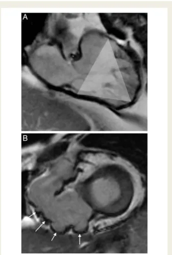
Arrhythmogenic right ventricular
dysplasia
Arrhythmogenic RV cardiomyopathyopathy is a typical myocardial
disorder affecting primarily the RV. In the early stages of the
disease, structural changes may be subtle or absent and confined
to a localized region of the RV, typically within the so-called
triangle of dysplasia (Figure
8
). As ventricular arrhythmia can
occur anytime, affected patients are at risk for sudden death and
timely diagnosis can help preventing arrhythmias and sudden
death.
115The most recent Task Force Criteria (TFC) for diagnosis of
ARVC include global or regional structural and functional
altera-tions, histopathologic tissue characterization, ECG abnormalities,
arrhythmias, and family history.
57Echocardiography and CMR are
the proposed imaging modalities for assessing structural and
func-tional criteria as shown in Table
4
. Echocardiographic abnormalities
of the RV can be found in up to 62% of subjects.
116Dilatation of
the RV outflow tract (RVOT) occurs in all positive subjects and
global RV dysfunction is observed in more than two-thirds.
Meas-urement of RVOT dimension in the parasternal long-axis or
short-axis view should be included in each echocardiographic screening
examination, as regional RV enlargement can be missed in the
apical four-chamber view. Additional abnormalities consist of RV
regional wall motion abnormalities, abnormal trabeculations,
hyperreflective moderator band, and sacculations of the RV free
Figure 8
The triangle of dysplasia in arrhythmogenic right
ven-tricular (RV) cardiomyopathyopathy (ARVC). (A) Structural
anomalies can be observed in a region including the subtricuspidal
RV wall, the RV apex, and the RV outflow tract. (B) Steady-state
free precession images showing severe aneurysmatic
abnormal-ities of the RV free wall subtricuspidal (arrows) in a patient
with ARVC.
wall.
116Tissue Doppler evaluation of myocardial velocities and 3D
echocardiography may be helpful in the early non-invasive
diagno-sis of ARVC.
117,118Cardiac magnetic resonance adds information
to echocardiography, as it enables visualization of subtle changes
and of remote RV segments, such as the RV infero-posterior
wall; RV volumes and function can be quantified (Table
5
).
Qualita-tive criteria such as segmental RV dilatation, presence of RV
micro-aneurysms, or fatty infiltration have been removed from the
revised TFC, as they have been shown to have low sensitivity
and specificity.
57,119,120Similar qualitative findings were observed
in patients with benign RVOT arrhythmias in the absence of
ARVC. Fatty infiltration can be found in normal heart as well.
115In summary, each imaging modality should not be used in
isola-tion as an independent marker. The combined use of
echocardiog-raphy and CMR adds a powerful and accurate piece of information
for the diagnosis of ARVC.
Congenital heart disease
In CHD, the RV is frequently exposed to a chronic volume or
pres-sure overload. This is the case in intracardiac shunts (atrial septal
defect), anomalies of the pulmonary valve, and arteries (pulmonary
atresia) and when the RV is pumping in the systemic circulation
(transposition of the great arteries, single ventricle). Eventually,
RV function is the main determinant of prognosis in these patients.
Tetralogy of Fallot is the most common cyanotic CHD and a good
example on how multimodality imaging of the RV can be used in
CHD. Surgical repair of TOF provides excellent survival, but
re-sidual lesions are frequent and determine long-term morbidity
and mortality.
121Echocardiography correctly identifies the presence
of a residual ventricular septal defect, RVOT obstruction, and
pul-monary regurgitation. In TOF patients, 2D echocardiographic
mea-surements of the RV correlate only moderately with RV
end-diastolic volume (RVEDV) as measured by CMR.
31,122The
strongest relationship is found between RV end-diastolic area
from the apical four-chamber view and RVEDV.
122Right ventricular
systolic function can be estimated using % FAC, TAPSE, and tricuspid
annular tissue Doppler velocities. Three-dimensional
echocardiog-raphy is emerging and may be able to replace CMR-based
volumet-ric measurements at least in patients with a good imaging window.
34Diastolic dysfunction in restrictive RV physiology is demonstrated, if
a late diastolic (during atrial contraction) antegrade flow in the
pul-monary artery is shown at Doppler echocardiography.
123Restrictive
RV physiology has been found to have a protective role against
pro-gressive RV dilatation and to better predict exercise performance
late after TOF repair, although this could only be observed in an
adult cohort and is more controversial at younger age.
123Chronic pulmonary valve regurgitation after TOF repair leads to
progressive RV dilatation and dysfunction. Right ventricular volume
measured by CMR has become an important parameter for trying
to determine the ideal timing for pulmonary valve
replace-ment.
66,124,125Oosterhof
et
al.
126found
that
a
RVEDV
,160 mL/m
2and a RV end-systolic volume ,82 mL/m
2are
pre-dictive for normalization of RV dimensions after pulmonary valve
replacement. In TOF patients, CMR is considered the ideal tool
for serial assessment of RV volumes before and after pulmonary
valve replacement.
66. . . .
. . . .
Table 4
Imaging task force criteria for diagnosing arrhythmogenic right ventricular cardiomyopathyopathy
Structural and functional criteria for ARVC Major criteria
2D echo Regional RV akinesia, dyskinesia, or aneurysm And 1 of the following (end-diastole):
RVOT≥32 mm (19 mm/m2)/parasternal long-axis view RVOT≥36 mm (21 mm/m2)/parasternal short-axis view or RV fractional area change≤33%
CMR Regional RV akinesia or dyskinesia, or dyssynchronous RV contraction And 1 of the following:
RV end-diastolic volume≥110 mL/m2(male) or≥100 mL/m2(female) or RV ejection fraction≤40%
Minor criteria
2D echo Regional RV akinesia or dyskinesia And 1 of the following (end-diastole):
RVOT≥29 mm ,32 mm (≥16 ,19 mm/m2)
RVOT≥32 ,36 mm (≥18 ,21 mm/m2)
or fractional area change .33 to≤40%
CMR Regional RV akinesia or dyskinesia, or dyssynchronous RV contraction And 1 of the following:
RV end-diastolic volume≥100 ,100 mL/m2(male) or≥90 ,100 mL/m2(female) or RV ejection fraction .40≤45%
Adapted from: Marcus et al.57
Proposed modification of the Task Force Criteria. RVOT, RV outflow tract.
More recently, regional myocardial RV function has been used
for advanced functional assessment after TOF repair. Impaired
lon-gitudinal RV deformation indices have been described in the
pres-ence of pulmonary regurgitation by TDI and speckle tracking.
51,127Interestingly, progressive deterioration of RV longitudinal strain has
been found in patients with stable EF, suggesting that myocardial
strain could be a more sensitive parameter for detecting early
ven-tricular dysfunction.
51,128Acute improvement of longitudinal RV
and septal function could be demonstrated by speckle tracking
after
percutaneous
pulmonary
valve
replacement.
129Velocity-encoded CMR imaging of RV myocardium provides
similar myocardial velocities and timing of velocities as TDI. Peak
systolic velocities in the RV free wall and in the RVOT are
reduced in TOF patients compared with normals.
130The importance of the infundibulum has been investigated by
looking at the segmental function in different RV regions,
distin-guishing between the RV inlet, the trabeculated apical part, and
the RV outlet. Not surprisingly, EF is predominantly reduced in
the outlet part where the surgical patch has been inserted.
131This reduced myocardial deformation in the infundibular region
correlates well with areas of late enhancement as well as with
global EF.
132Cardiac magnetic resonance provides important additional
infor-mation, including calculation of pulmonary regurgitant volume and
fraction,
73measurement of differential lung perfusion,
133and
late-enhancement imaging.
61The presence of RV myocardial
scars has been reported to have prognostic relevance in TOF
patients, as it correlates with RV size, function, length of QRS
complex at ECG, and may predict ventricular arrhythmias.
134Contrast-enhanced MR angiography or 3D SFFP provides exact
anatomical evaluation of the RVOT and the pulmonary arteries
and is helpful for planning reinterventions
55particularly for
select-ing patients for percutaneous pulmonary valve replacement.
135Conclusions
The significance of RV function is being increasingly recognized in
the acute phase and during follow-up as prognostic factor in
. . . .
Table 5
Summary of the right ventricular parameters that can be assessed by specific and/or combined imaging
modalities in various diseases
Measurements Imaging modality Disease
RV anatomy Echocardiography All
CMR (SSFP) CHD
MDCT
RV dimensions 2D echocardiography All
CMR (SSFP, volumetry) CHD
MDCT ARVC (RVOT)
Pulmonary valve Echocardiography CHD
CMR (phase contrast imaging for regurgitation quantification)
Pulmonary arteries CMR (angiography/3D SSFP) CHD
MDCT PAH, pulmonary embolism
Echocardiography (proximal segments)
Tricuspid valve Echocardiography All
3D echocardiography
RV volumes CMR (SSFP) All
RV ejection fraction Echocardiography (2D/3D) Gated blood-pool SPECT MDCT
Regional RV function Tissue Doppler imaging All
Myocardial velocities Speckle tracking
Strain/strain rate (investigational) CMR (tagging, velocity encoded sequences)
RV ischaemia CMR (late enhancement, oedema) RV ischaemic disease Echocardiography (wall motion) RV failure
SPECT (perfusion)
RV scars/fibrosis CMR (late enhancement) RV failure
SPECT RV ischaemic disease
Myocarditis CHD
Coronary arteries MDCT RV ischaemic disease
PET/CT
The imaging modalities are represented in the order they should be performed for the specific wanted measurement.
ARVC, arrhythmogenic right ventricular cardiomyopathy; CHD, congenital heart disease; CMR, cardiovascular magnetic resonance; MDCT, multidetector computed tomography; RVOT, right ventricular outflow tract; PAH, pulmonary arterial hypertension; PET, positron emission tomography; SPECT, single-photon electron-computed tomography; SSFP, steady-state free precession sequence.
several cardiac diseases. Echocardiography and CMR are the
imaging modalities of choice for imaging the right heart. In most
patients, both techniques provide complementary information
and can be used in combination for almost complete evaluation
of the RV. Multidetector computed tomography and nuclear
imaging technique are valuable alternative modalities and add
im-portant additional information in selected cases, particularly in
RV ischaemic disease. Emerging new technologies such as 3D
echocardiography, TDI, speckle tracking as well as new CMR
se-quence are enlarging the spectrum of the pathophysiologic
infor-mation obtained, but are still confined to investigational use and
need further clinical validation. Table
5
summarizes the use of all
these imaging modalities for assessing the different RV parameters
in various diseases, and may serve as guide for multimodality
imaging.
Acknowledgements
We thank Prof. C. Attenhofer Jost for his critical appraisal of the
manuscript.
Conflict of interest: none declared.
References
1. Warnes CA. Adult congenital heart disease: importance of the right ventricle. J Am Coll Cardiol 2009;54:1903 – 1910.
2. D’Alonzo GE, Barst RJ, Ayres SM, Bergofsky EH, Brundage BH, Detre KM, Fishman AP, Goldring RM, Groves BM, Kernis JT et al. Survival in patients with primary pulmonary hypertension. Results from a national prospective regis-try. Ann Intern Med 1991;115:343 – 349.
3. Forfia PR, Fisher MR, Mathai SC, Housten-Harris T, Hemnes AR, Borlaug BA,
Chamera E, Corretti MC, Champion HC, Abraham TP, Girgis RE,
Hassoun PM. Tricuspid annular displacement predicts survival in pulmonary hypertension. Am J Respir Crit Care Med 2006;174:1034 – 1041.
4. Haddad F, Doyle R, Murphy DJ, Hunt SA. Right ventricular function in cardiovas-cular disease, part II: pathophysiology, clinical importance, and management of right ventricular failure. Circulation 2008;117:1717 – 1731.
5. Meyer P, Filippatos GS, Ahmed MI, Iskandrian AE, Bittner V, Perry GJ, White M, Aban IB, Mujib M, Dell’Italia LJ, Ahmed A. Effects of right ventricular ejection fraction on outcomes in chronic systolic heart failure. Circulation 2010;121: 252 – 258.
6. de Groote P, Millaire A, Foucher-Hossein C, Nugue O, Marchandise X, Ducloux G, Lablanche J-M. Right ventricular ejection fraction is an independent predictor of survival in patients with moderate heart failure. J Am Coll Cardiol 1998;32:948 – 954.
7. Mertens LL, Friedberg MK. Imaging the right ventricle—current state of the art. Nat Rev Cardiol 2010;7:551 – 563.
8. Haddad F, Hunt SA, Rosenthal DN, Murphy DJ. Right ventricular function in car-diovascular disease, part I: anatomy, physiology, aging, and functional assessment of the right ventricle. Circulation 2008;117:1436 – 1448.
9. Foale R, Nihoyannopoulos P, McKenna W, Kleinebenne A, Nadazdin A, Rowland E, Smith G. Echocardiographic measurement of the normal adult right ventricle. Br Heart J 1986;56:33 – 44.
10. Sheehan FH, Ge S, Vick GW 3rd, Urnes K, Kerwin WS, Bolson EL, Chung T, Kovalchin JP, Sahn DJ, Jerosch-Herold M, Stolpen AH. Three-dimensional shape analysis of right ventricular remodeling in repaired tetralogy of Fallot. Am J Cardiol 2008;101:107 – 113.
11. Anderson RH, Ho SY. Sequential segmental analysis: description and character-ization for the millenium. Cardiol Young 1997;7:98 – 116.
12. Sengupta PP, Korinek J, Belohlavek M, Narula J, Vannan MA, Jahangir A, Khandheria BK. Left ventricular structure and function: basic science for cardiac imaging. J Am Coll Cardiol 2006;48:1988 – 2001.
13. Sengupta PP, Krishnamoorthy VK, Korinek J, Narula J, Vannan MA, Lester SJ, Tajik JA, Seward JB, Khandheria BK, Belohlavek M. Left ventricular form and function revisited: applied translational science to cardiovascular ultrasound imaging. J Am Soc Echocardiogr 2007;20:539 – 551.
14. Haber I, Metaxas DN, Geva T, Axel L. Three-dimensional systolic kinematics of the right ventricle. Am J Physiol Heart Circ Physiol 2005;289:H1826 – H1833.
15. Sanchez-Quintana D, Anderson RH, Ho SY. Ventricular myoarchitecture in tet-ralogy of Fallot. Heart 1996;76:280 – 286.
16. Pettersen E, Helle-Valle T, Edvardsen T, Lindberg H, Smith HJ, Smevik B, Smiseth OA, Andersen K. Contraction pattern of the systemic right ventricle shift from longitudinal to circumferential shortening and absent global ventricu-lar torsion. J Am Coll Cardiol 2007;49:2450 – 2456.
17. Bishop A, White P, Oldershaw P, Chaturvedi R, Brookes C, Redington A. Clinical application of the conductance catheter technique in the adult human right ven-tricle. Int J Cardiol 1997;58:211 – 221.
18. Redington AN, Rigby ML, Shinebourne EA, Oldershaw PJ. Changes in the pressure-volume relation of the right ventricle when its loading conditions are modified. Br Heart J 1990;63:45 – 49.
19. Sheehan F, Redington A. The right ventricle: anatomy, physiology and clinical imaging. Heart 2008;94:1510 – 1515.
20. Bogaard HJ, Abe K, Vonk Noordegraaf A, Voelkel NF. The right ventricle under pressure: cellular and molecular mechanisms of right-heart failure in pulmonary hypertension. Chest 2009;135:794 – 804.
21. Kaufman BD, Desai M, Reddy S, Osorio JC, Chen JM, Mosca RS, Ferrante AW, Mital S. Genomic profiling of left and right ventricular hypertrophy in congenital heart disease. J Card Fail 2008;14:760 – 767.
22. Mital S. Right ventricle in congenital heart disease: is it just a ‘weaker’ left ven-tricle? Arch Mal Coeur Vaiss 2006;99:1244 – 1251.
23. Rochais F, Mesbah K, Kelly RG. Signaling pathways controlling second heart field development. Circ Res 2009;104:933 – 942.
24. Zaffran S, Kelly RG, Meilhac SM, Buckingham ME, Brown NA. Right ventricular myocardium derives from the anterior heart field. Circ Res 2004;95:261 – 268. 25. Puwanant S, Park M, Popovic ZB, Tang WH, Farha S, George D, Sharp J,
Puntawangkoon J, Loyd JE, Erzurum SC, Thomas JD. Ventricular geometry, strain, and rotational mechanics in pulmonary hypertension. Circulation 2010; 121:259 – 266.
26. Raymond RJ, Hinderliter AL, Willis PW, Ralph D, Caldwell EJ, Williams W, Ettinger NA, Hill NS, Summer WR, de Boisblanc B, Schwartz T, Koch G, Clayton LM, Jobsis MM, Crow JW, Long W. Echocardiographic predictors of adverse outcomes in primary pulmonary hypertension. J Am Coll Cardiol 2002; 39:1214 – 1219.
27. Stojnic BB, Brecker SJ, Xiao HB, Helmy SM, Mbaissouroum M, Gibson DG. Left ventricular filling characteristics in pulmonary hypertension: a new mode of ven-tricular interaction. Br Heart J 1992;68:16 – 20.
28. Walker RE, Moran AM, Gauvreau K, Colan SD. Evidence of adverse ventricular interdependence in patients with atrial septal defects. Am J Cardiol 2004;93: 1374 – 1377, A6.
29. Rudski LG, Lai WW, Afilalo J, Hua L, Handschumacher MD, Chandrasekaran K, Solomon SD, Louie EK, Schiller NB. Guidelines for the echocardiographic as-sessment of the right heart in adults: a report from the American Society of Echocardiography endorsed by the European Association of Echocardiography, a registered branch of the European Society of Cardiology, and the Canadian Society of Echocardiography. J Am Soc Echocardiogr 2010;23:685 – 713; quiz 786 – 788.
30. Lopez L, Colan SD, Frommelt PC, Ensing GJ, Kendall K, Younoszai AK, Lai WW, Geva T. Recommendations for quantification methods during the performance of a pediatric echocardiogram: a report from the Pediatric Measurements Writing Group of the American Society of Echocardiography Pediatric and Con-genital Heart Disease Council. J Am Soc Echocardiogr 2010;23:465 – 495; quiz 576 – 577.
31. Lai WW, Gauvreau K, Rivera ES, Saleeb S, Powell AJ, Geva T. Accuracy of guide-line recommendations for two-dimensional quantification of the right ventricle by echocardiography. Int J Cardiovasc Imaging 2008;24:691 – 698.
32. Helbing WA, Bosch HG, Maliepaard C, Rebergen SA, van der Geest RJ, Hansen B, Ottenkamp J, Reiber JH, de Roos A. Comparison of echocardiograph-ic methods with magnetechocardiograph-ic resonance imaging for assessment of right ventrechocardiograph-icular function in children. Am J Cardiol 1995;76:589 – 594.
33. Shimada YJ, Shiota M, Siegel RJ, Shiota T. Accuracy of right ventricular volumes and function determined by three-dimensional echocardiography in comparison with magnetic resonance imaging: a meta-analysis study. J Am Soc Echocardiogr 2010;23:943 – 953.
34. Grewal J, Majdalany D, Syed I, Pellikka P, Warnes CA. Three-dimensional echo-cardiographic assessment of right ventricular volume and function in adult patients with congenital heart disease: comparison with magnetic resonance imaging. J Am Soc Echocardiogr 2010;23:127 – 133.
35. Niemann PS, Pinho L, Balbach T, Galuschky C, Blankenhagen M, Silberbach M, Broberg C, Jerosch-Herold M, Sahn DJ. Anatomically oriented right ventricular volume measurements with dynamic three-dimensional echocardiography vali-dated by 3-Tesla magnetic resonance imaging. J Am Coll Cardiol 2007;50: 1668 – 1676.
36. van der Zwaan HB, Geleijnse ML, Soliman OI, McGhie JS, Wiegers-Groeneweg EJ, Helbing WA, Roos-Hesselink JW, Meijboom FJ. Test-retest vari-ability of volumetric right ventricular measurements using real-time three-dimensional echocardiography. J Am Soc Echocardiogr 2011;24:671 – 679. 37. van der Zwaan HB, Helbing WA, Boersma E, Geleijnse ML, McGhie JS,
Soliman OI, Roos-Hesselink JW, Meijboom FJ. Usefulness of real-time three-dimensional echocardiography to identify right ventricular dysfunction in patients with congenital heart disease. Am J Cardiol 2010;106:843 – 850. 38. van der Zwaan HB, Helbing WA, McGhie JS, Geleijnse ML, Luijnenburg SE,
Roos-Hesselink JW, Meijboom FJ. Clinical value of real-time three-dimensional echocardiography for right ventricular quantification in congenital heart disease: validation with cardiac magnetic resonance imaging. J Am Soc Echocar-diogr 2010;23:134 – 140.
39. Khoo NS, Young A, Occleshaw C, Cowan B, Zeng IS, Gentles TL. Assessments of right ventricular volume and function using three-dimensional echocardiog-raphy in older children and adults with congenital heart disease: comparison with cardiac magnetic resonance imaging. J Am Soc Chocardiogr 2009;22: 1279 – 1288.
40. Takahashi K, Inage A, Rebeyka IM, Ross DB, Thompson RB, Mackie AS, Smallhorn JF. Real-time 3-dimensional echocardiography provides new insight into mechanisms of tricuspid valve regurgitation in patients with hypoplastic left heart syndrome. Circulation 2009;120:1091 – 1098.
41. Nii M, Guerra V, Roman KS, Macgowan CK, Smallhorn JF. Three-dimensional tri-cuspid annular function provides insight into the mechanisms of tritri-cuspid valve regurgitation in classic hypoplastic left heart syndrome. J Am Soc Echocardiogr 2006;19:391 – 402.
42. Nii M, Roman KS, Macgowan CK, Smallhorn JF. Insight into normal mitral and tricuspid annular dynamics in pediatrics: a real-time three-dimensional echocar-diographic study. J Am Soc Echocardiogr 2005;18:805 – 814.
43. Anavekar NS, Gerson D, Skali H, Kwong RY, Yucel EK, Solomon SD. Two-dimensional assessment of right ventricular function: an echocardiographic-MRI correlative study. Echocardiography 2007;24:452 – 456.
44. Tei C, Nishimura RA, Seward JB, Tajik AJ. Noninvasive Doppler-derived myocar-dial performance index: correlation with simultaneous measurements of cardiac catheterization measurements. J Am Soc Echocardiogr 1997;10:169 – 178. 45. Cheung MM, Smallhorn JF, Redington AN, Vogel M. The effects of changes in
loading conditions and modulation of inotropic state on the myocardial per-formance index: comparison with conductance catheter measurements. Eur Heart J 2004;25:2238 – 2242.
46. Dragulescu A, Mertens LL. Developments in echocardiographic techniques for the evaluation of ventricular function in children. Arch Cardiovasc Dis 2010;103: 603 – 614.
47. Dambrauskaite V, Delcroix M, Claus P, Herbots L, D’Hooge J, Bijnens B, Rademakers F, Sutherland GR. Regional right ventricular dysfunction in chronic pulmonary hypertension. J Am Soc Echocardiogr 2007;20:1172 – 1180. 48. Giusca S, Dambrauskaite V, Scheurwegs C, D’Hooge J, Claus P, Herbots L,
Magro M, Rademakers F, Meyns B, Delcroix M, Voigt JU. Deformation imaging describes right ventricular function better than longitudinal displacement of the tricuspid ring. Heart 2010;96:281 – 288.
49. Bos JM, Hagler DJ, Silvilairat S, Cabalka A, O’Leary P, Daniels O, Miller FA, Abraham TP. Right ventricular function in asymptomatic individuals with a sys-temic right ventricle. J Am Soc Echocardiogr 2006;19:1033 – 1037.
50. Eyskens B, Weidemann F, Kowalski M, Bogaert J, Dymarkowski S, Bijnens B, Gewillig M, Sutherland G, Mertens L. Regional right and left ventricular function after the Senning operation: an ultrasonic study of strain rate and strain. Cardiol Young 2004;14:255 – 264.
51. Eyskens B, Brown SC, Claus P, Dymarkowski S, Gewillig M, Bogaert J, Mertens L. The influence of pulmonary regurgitation on regional right ventricular function in children after surgical repair of tetralogy of Fallot. Eur J Echocardiogr 2010;11: 341 – 345.
52. Weidemann F, Eyskens B, Mertens L, Dommke C, Kowalski M, Simmons L, Claus P, Bijnens B, Gewillig M, Hatle L, Sutherland GR. Quantification of regional right and left ventricular function by ultrasonic strain rate and strain indexes after surgical repair of tetralogy of Fallot. Am J Cardiol 2002;90:133 – 138. 53. Koopman LP, Slorach C, Hui W, Manlhiot C, McCrindle BW, Friedberg MK,
Jaeggi ET, Mertens L. Comparison between different speckle tracking and color tissue Doppler techniques to measure global and regional myocardial de-formation in children. J Am Soc Echocardiogr 2010;23:919 – 928.
54. Greil GF, Boettger T, Germann S, Klumpp B, Baltes C, Kozerke S, Bialkowski A, Urschitz MS, Miller S, Wolf I, Meinzer H-P, Sieverding L. Quantitative assessment of ventricular function using three-dimensional SSFP magnetic resonance angiog-raphy. J Magn Reson Imaging 2007;26:288 – 295.
55. Valsangiacomo Bu¨chel ER, DiBernardo S, Bauersfeld U, Berger F.
Contrast-enhanced magnetic resonance angiography of the great arteries in
patients with congenital heart disease: an accurate tool for planning catheter-guided interventions. Int J Cardiovasc Imaging 2005;21:313 – 322.
56. Beroukhim RS, Prakash A, Valsangiacomo Buechel ER, Cava JR, Dorfman AL, Festa P, Hlavacek AM, Johnson TR, Keller MS, Krishnamurthy R, Misra N, Moniotte S, Parks WJ, Powell AJ, Soriano BD, Srichai MB, Yoo S-J, Zhou J, Geva T. Characterization of cardiac tumors in children by cardiovascular mag-netic resonance imaging: a multicenter experience. J Am Coll Cardiol 2011;58: 1044 – 1054.
57. Marcus FI, McKenna WJ, Sherrill D, Basso C, Bauce B, Bluemke DA, Calkins H, Corrado D, Cox MGPJ, Daubert JP, Fontaine G, Gear K, Hauer R, Nava A, Picard MH, Protonotarios N, Saffitz JE, Sanborn DMY, Steinberg JS, Tandri H, Thiene G, Towbin JA, Tsatsopoulou A, Wichter T, Zareba W. Diagnosis of arrhythmogenic right ventricular cardiomyopathy/dysplasia. Eur Heart J 2010; 31:806 – 814.
58. Mahrholdt H, Wagner A, Judd RM, Sechtem U, Kim RJ. Delayed enhancement cardiovascular magnetic resonance assessment of non-ischaemic cardiomyop-athies. Eur Heart J 2005;26:1461 – 1474.
59. Masci PG, Francone M, Desmet W, Ganame J, Todiere G, Donato R, Siciliano V, Carbone I, Mangia M, Strata E, Catalano C, Lombardi M, Agati L, Janssens S, Bogaert J. Right ventricular ischemic injury in patients with acute ST-segment
elevation myocardial infarction/clinical perspective. Circulation 2010;122:
1405 – 1412.
60. Babu-Narayan SV, Goktekin O, Moon JC, Broberg CS, Pantely GA, Pennell DJ, Gatzoulis MA, Kilner PJ. Late gadolinium enhancement cardiovascular magnetic resonance of the systemic right ventricle in adults with previous atrial redirec-tion surgery for transposiredirec-tion of the great arteries. Circularedirec-tion 2005;111: 2091 – 2098.
61. Babu-Narayan SV, Kilner PJ, Li W, Moon JC, Goktekin O, Davlouros PA, Khan M, Ho SY, Pennell DJ, Gatzoulis MA. Ventricular fibrosis suggested by cardiovascu-lar magnetic resonance in adults with repaired tetralogy of Fallot and its relation-ship to adverse markers of clinical outcome. Circulation 2006;113:405 – 413. 62. Miller S, Simonetti O, Carr J, Kramer U, Finn J. MR Imaging of the heart with cine
true fast imaging with steady-state precession: influence of spatial and temporal resolutions on left ventricular functional parameters. Radiology 2002;223: 263 – 269.
63. Maceira A, Prasad S, Khan M, Pennell D. Reference right ventricular systolic and diastolic function normalized to age, gender and body surface area from steady-state free precession cardiovascular magnetic resonance. Eur Heart J 2006;27:2879 – 2888.
64. Buechel E, Kaiser T, Jackson C, Schmitz A, Kellenberger C. Normal right- and left ventricular volumes and myocardial mass in children measured by steady state free precession cardiovascular magnetic resonance. J Cardiovasc Magn Reson 2009;11:19.
65. Sarikouch S, Peters B, Gutberlet M, Leismann B, Kelter-Kloepping A, Koerperich H, Kuehne T, Beerbaum P. Sex-specific pediatric percentiles for ven-tricular size and mass as reference values for cardiac MRI/clinical perspective. Circulation 2010;3:65 – 76.
66. Buechel ERV, Dave HH, Kellenberger CJ, Dodge-Khatami A, Pretre R, Berger F, Bauersfeld U. Remodelling of the right ventricle after early pulmonary valve re-placement in children with repaired tetralogy of Fallot: assessment by cardiovas-cular magnetic resonance. Eur Heart J 2005;26:2721 – 2727.
67. Grothues F, Moon J, Bellenger N, Smith G, Klein H, Pennell D. Interstudy repro-ducibility of right ventricular volumes, function, and mass with cardiovascular magnetic resonance. Am Heart J 2004;147:218 – 223.
68. Fratz S, Schuhbaeck A, Buchner C, Busch R, Meierhofer C, Martinoff S, Hess J, Stern H. Comparison of accuracy of axial slices versus short-axis slices for meas-uring ventricular volumes by cardiac magnetic resonance in patients with cor-rected tetralogy of Fallot. Am J Cardiol 2009;103:1764 – 1769.
69. Robbers-Visser DL, Luijnenburg SE, van den Berg J, Moelker A, Helbing WA. Stress imaging in congenital cardiac disease. Cardiol Young 2009;19:552 – 562. 70. Greil GF, Beerbaum P, Razavi R, Miller O. Imaging the right ventricle. Heart 2008;
94:803 – 808.
71. Youssef A, Ibrahim E-S, Korosoglou G, Abraham MR, Weiss R, Osman N. Strain-encoding cardiovascular magnetic resonance for assessment of right-ventricular regional function. J Cardiovasc Magn Reson 2008;10:33.
72. Menteer JWP, Fogel MA. Quantifying regional right ventricular function in tetral-ogy of Fallot. J Cardiovasc Magn Reson 2005;7:753 – 761.
73. Wald RM, Redington AN, Pereira A, Provost YL, Paul NS, Oechslin EN, Silversides CK. Refining the assessment of pulmonary regurgitation in adults after tetralogy of Fallot repair: should we be measuring regurgitant fraction or regurgitant volume? Eur Heart J 2009;30:356 – 361.
74. Roman KS, Kellenberger CJ, Farooq S, MacGowan CK, Gilday DL, Yoo S-J. Com-parative imaging of differential pulmonary blood flow in patients with congenital heart disease: magnetic resonance imaging versus lung perfusion scintigraphy. Pediatr Radiol 2005;35:295 – 301.