SHORT COMMUNICATION
First description of natural Echinococcus multilocularis
infections in chinchilla (Chinchilla laniger) and Prevost
’s
squirrel (Callosciurus prevostii borneoensis)
Sandra Staebler&Hanspeter Steinmetz&Stefan Keller&Peter Deplazes
Received: 25 June 2007 / Accepted: 7 August 2007 / Published online: 1 September 2007
# Springer-Verlag 2007
Abstract This report describes for the first time the occurrence of alveolar echinococcosis in two exotic rodent species in Europe. A pet chinchilla (Chinchilla laniger) was euthanized due to a painful enlargement of the abdominal cavity, and a Prevost’s squirrel (Callosciurus prevostii borneoensis) was found dead in the enclosure of a zoo. At necropsy, extended liver lesions consisting of small vesicles and cysts were observed in the livers of both animals. Histological examination revealed that these cysts were composed of an outer, homogenous, eosinophilic layer and an inner, cellular germinal layer. The cysts from both animals contained numerous protoscolices. The morpholo-gical diagnosis of Echinococcus multilocularis metacestode infections was confirmed by molecular means.
Introduction
Alveolar echinococcosis (AE) caused by the larval stage of the fox tapeworm Echinococcus multilocularis is a serious
zoonotic disease. The parasite’s distribution spans the northern hemisphere, the endemic area stretching from North America through central and Eastern Europe to central and East Asia including northern parts of Japan. E. multilocularis is maintained in a wild-animal cycle with several fox species, coyotes, raccoon dogs, and in certain endemic areas, domestic dogs as the main definitive hosts. World-wide, many rodent species are known to be suitable intermediate hosts of E. multilocularis with varying signif-icance in the maintenance of the cycle in different regions (Eckert et al. 2001; Rausch 1967; Vuitton et al. 2003). In central Europe, the most common natural intermediate hosts of E. multilocularis are the common vole (Microtus arvalis), the water vole (Arvicola terrestris), and the bank vole (Clethrionomys glareolus). In some regions, muskrats (Ondatra zibethicus) can also be of epidemiological significance (Romig et al. 2006). Most of the described rodent intermediate hosts belong to the family Muridae in the suborder Myomorpha within the order Rodentia (Inte-grated Taxonomic Information System on-line database,
http://www.itis.gov), including the species commonly used and highly susceptible for experimental infections, namely, the cotton rat (Sigmodon hispidus), gerbil (Meriones unguiculatus) and different strains of the house mouse (Mus musculus; Romig and Bilger1999). The two new host species presented in this paper with AE belong to the suborders Hystricognatha and Sciuromorpha, respectively, among which only a few cases of E. multilocularis infections in a few species have been reported. Only the nutria (Myocastor coypus) from the suborder Hystricognatha (Worbes et al. 1989) and different ground, rock, and tree squirrels, as well as the European beaver (Castor fiber) and marmots from the Sciuromorpha suborder have been described (Janovsky et al. 2002; Rausch 1967; Vuitton et al. 2003). Furthermore, many nonrodent mammalian Parasitol Res (2007) 101:1725–1727
DOI 10.1007/s00436-007-0717-2
S. Staebler
:
P. Deplazes (*)Institute of Parasitology, Vetsuisse Faculty, University of Zurich, Winterthurerstr, 266a,
CH-8057 Zurich, Switzerland e-mail: deplazesp@access.uzh.ch H. Steinmetz
Division of Zoo Animals, Exotic Pets and Wildlife, Vetsuisse Faculty, University of Zurich,
Zurich, Switzerland S. Keller
Institute of Veterinary Pathology, Vetsuisse Faculty, University of Zurich,
species and humans have been described as aberrant hosts of E. multilocularis including dogs and monkeys, in which protoscolices fully develop in metacestode tissue mainly located in the liver (Deplazes and Eckert 2001; Rehmann et al.2003; Staebler et al.2006), as well as pigs and horses, in which the parasite develops partially without protoscolex formation (Deplazes et al.2005; Ohbayashi1996).
Results and discussion
A 6-year old chinchilla (Chinchilla lanigera; Hystricognatha) was presented to the veterinarian showing inappetence, tachypnoea, and an enlarged abdomen. The general exam-ination revealed a hard and painful abdomen on the right side caudal to the last ribs. Due to its poor condition, the animal was euthanized, and a necropsy was performed. Pathologic alterations consisted of a liver infiltrated by many small vesicles measuring 1–5 mm in diameter. Microscop-ically, these liver infiltrations were composed of collapsed vesicles containing calcareous corpuscles and living protosco-lices. The laminated layer surrounding the vesicles contained Em2 antigen as visualized with an E. multilocularis-specific monoclonal antibody (Em2G11) by indirect fluorescent antibody test (Deplazes and Gottstein 1991). Histological sections showed an acellular laminated layer with a thin inner germinal layer and protoscolices.
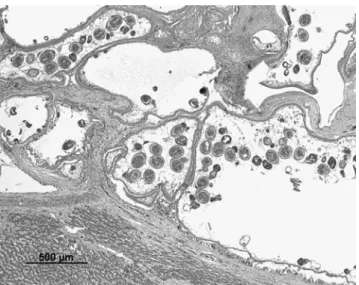
The second case, a 4.5-year-old female Prevost’s squirrel, died unexpectedly. The animal was housed in a 50-m2 enclosure with an outdoor area in the Zürich Zoo. The animal’s diet consisted of fruits, nuts, seeds, flowers, insects, and bird eggs. At necropsy, the liver contained a well-demarcated, white-to-grey mass of 7 cm in diameter which was composed of numerous, small cysts. The mass replaced most of the hepatic tissue and occupied a large portion of the abdominal cavity. In the thoracic cavity, 10 ml of a clear, yellow fluid was found. In hematoxylin– eosin (HE) stained histological sections, a capsule of dense fibrous tissue separated the cystic mass from the liver parenchyma. Cysts were up to 5 mm in diameter and consisted of an outer, homogenous, eosinophilic layer and an inner, cellular, germinal layer. The inner cavity contained protoscolices occasionally arranged into brood capsules as well as numerous calcareous corpuscles (Fig. 1). The connective tissue between the cysts was infiltrated with many lymphocytes, some neutrophils, and occasional multinucleated giant cells. The adjacent liver parenchyma showed moderate hyperplasia of bile ducts, slight cholestasis, and mild periportal infiltration with lymphocytes and plasma cells.
Amplification of specific E. multilocularis DNA by polymerase chain reaction (PCR; Stieger et al. 2002) of fresh (chinchilla) and formalin-fixed, paraffin-embedded
metacestode material (Prevost’s squirrel) resulted positive in both cases and confirmed the morphological and immunological diagnosis of an E. multilocularis metaces-tode infection.
Infections of intermediate and aberrant hosts occur by the ingestion of eggs. In the case of the Prevost’s squirrel, it is suspected that the animal obtained the infection by ingestion of contaminated food, as the squirrel was born and raised in the zoo. Its cage was surrounded by a fox-tight perimeter fence, and hence direct contact with foxes of the highly endemic area of E. multilocularis around Zürich can be excluded. Similar routes of infection have been assumed for zoo monkeys (Deplazes and Eckert2001). The chinchilla was fed with commercially available hay and chinchilla food but got, from time to time, some branches from wild trees or shrubs.
From the epidemiological point of view, the two cases presented can be considered infections without epidemio-logical significance. However, the strong metacestode proliferation in both animal species with the abundant production of protoscolices indicates that these species could be highly susceptible to this parasite. This might be of relevance especially in the case of the chinchilla, which represents one of the largest (up to 1 kg body weight) rodent species described thus far which is susceptible to E. multilocularis and is kept as a pet but also for fur production or as laboratory animal.
Acknowledgments We thank Felizian Kuster for making the putative diagnosis and providing basic data on the diseased chinchilla. Fig. 1 Histological section of the liver of the Prevost’s squirrel (HE stained). Section through a cyst containing protoscolices bordered by the homogenous laminated layer and the cellular inner germinal layer. The adjacent liver parenchyma shows infiltration with lymphocytes and plasma cells
References
Deplazes P, Eckert J (2001) Veterinary aspects of alveolar echinococcosis— a zoonosis of public health significance. Vet Parasitol 98:65–87 Deplazes P, Gottstein B (1991) A monoclonal antibody against
Echinococcus multilocularis Em2-antigen. Parasitology 103:41–49 Deplazes P, Grimm F, Sydler T, Tanner I, Kapel CM (2005) Experimental alveolar echinococcosis in pigs, lesion develop-ment and serological follow up. Vet Parasitol 130:213–222 Eckert J, Gemmel MA, Meslin F-X, Pawlowski ZS (2001) WHO/OIE
manual on echinococcosis in humans and animals: a public health problem of global concern. World Organisation for Animal Health and World Health Organization, Paris
Janovsky M, Bacciarini L, Sager H, Grone A, Gottstein B (2002) Echinococcus multilocularis in a European beaver from Switzerland. J Wildl Dis 38:618–620
Ohbayashi M (1996) Host animals of Echinococcus multilocularis in Hokkaido. In: Uchino J, Sato N (eds) Alveolar echinococcosis: Strategy for eradication of alveolar echinococcosis of the liver. Fuji Shoin, Sapporo, pp 59–64
Rausch RL (1967) On the ecology and distribution of Echinococcus spp. (Cestoda: Taeniidae), and characteristics of their
develop-ment in the intermediate host. Ann Parasitol Hum Comp 42: 19–63
Rehmann P, Grone A, Lawrenz A, Pagan O, Gottstein B, Bacciarini LN (2003) Echinococcus multilocularis in two lowland gorillas (Gorilla g. gorilla). J Comp Pathol 129:85–88
Romig T, Bilger B (1999) Animal models for Echinococcosis. In: Zak O, Sande MA (eds) Handbook of animal models of infection. Academic, San Diego, pp 877–884
Romig T, Dinkel A, Mackenstedt U (2006) The present situation of Echinococcosis in Europe. Parasitol Int 55 Suppl:187–191 Staebler S, Grimm F, Glaus T, Kapel C, Haller M, Hasler A, Hanosset R,
Deplazes P (2006) Serological diagnosis of canine alveolar echinococcosis. Vet Parasitol 141:243–250
Stieger C, Hegglin D, Schwarzenbach G, Mathis A, Deplazes P (2002) Spatial and temporal aspects of urban transmission of Echinococcus multilocularis. Parasitology 124:631–640
Vuitton DA, Zhou H, Bresson-Hadni S, Wang Q, Piarroux M, Raoul F, Giraudoux P (2003) Epidemiology of alveolar echinococcosis with particular reference to China and Europe. Parasitology 127: S87–S107
Worbes H, Schacht KH, Eckert J (1989) Echinococcus multilocularis bei einem Sumpfbiber (Myocastor coypus). Angew Parasitol 30:161–165