Molecular characterization of the fi chain of
the murine interleukin 5 receptor
Seiji Mita, Satoshi Takaki, Yasumichi Hitoshi, Antonius G. Rolink1,
Akira Tominaga, Naoto Yamaguchi, and Kiyoshi Takatsu
Department of Biology, Institute for Medical Immunology, Kumomoto University Medical School, 2-2-1 Honjo, Kumamoto 860, Japan
'Basel Institute for Immunology, Grenzacherstrasse, Basel, Switzerland
Key words: myeloid cell line, membrane receptors, IL-5, high-affinity binding sites, monoclonal antibody
Abstract
Interleukin 5 (IL-5) Is a multifunctional cytoklne that regulates the proliferation and differentiation of hematopoletlc cells including B cells and eosinophlls. The murine IL-5 acts on target cells via an IL-5 specific high-affinity receptor (Kti = 1 5 0 pM) that has been proposed to be composed of
at least two membrane polypeptide chains. The p60 component recognized by anti-murine IL-5 receptor mAbs H7 and T21 binds IL-5 with low affinity (Kd = 10 nM). The other component is
p130, detectable by following cross-linking experiments with IL-5. Using H7, T21, and R52.120 mAbs specific to murine IL-5 receptor, we characterized the molecular nature of the p130 of the high affinity receptor for murine IL-5. R52.120 mAb did not recognize the IL-5 binding
recomblnant p60 expressed on COS7 cells, but reacted with p130/140 on IL-5-dependent cell lines. R52.120 mAb showed partial inhibition of the IL-5-induced proliferation of the
IL-5-dependent early B cell line Y16 at high IL-5 concentrations. Addition of R52.120 mAb together with H7 or T21 mAb caused more striking inhibition of the IL-5-dependent proliferation than that caused by either of them alone. R52.120 mAb down-regulated the number and
dissociation constant of IL-5 binding sites with high affinity without affecting the levels of these with low-affinity. It also preferentially inhibited the formation of the cross-linked complex of p130 with radlolabeled IL-5. These results Indicate that p130/p140, recognized by R52.120 mAb, Is indispensable, together with p60, for the formation of high affinity IL-5 receptor. We propose to designate p60 and p130/p140 as the a and /3 chain of IL-5 receptor, respectively.
Introduction
Interleukin 5 (IL-5) is an inducible glycoprotein cytokine generated mainly by T cells following antigen or mitogen induction (1 - 3 ) . The complementary DNAs encoding both mouse and human IL-5 have already been characterized (4-6). The growth and differentiation of B cells and other hematopoietic cells including eosinophils has been found to be regulated by IL-5 (7-12).
To elucidate how IL-5 mediates multiple functions in various target cells, we attempted to explore the molecular and biochemical characteristics of the receptor. On murine hemato-poietic cells, both high and low affinity membrane IL-5 receptors (IL-5R) have been described with equilibrium dissociation constants (KJ of - 150 pM and - 3 0 nM, respectively (13-16). Because most of the biological effects of IL-5 are observed at picomolar concentrations (13,14), it is clear that high-affinity IL-5R are biologically functional. Chemical cross-linking studies using PSJmethionine labeled IL-5 (FSJIL-S) have suggested that high-affinity IL-5R is composed of at least two polypeptide chains
(14): one is the 60 kd ligand binding peptide (p60) recognized by H7 or T21 mAb (17,18) and the other is a putative membrane peptide of - 1 3 0 kd (p130). Both H7 and T21 mAbs completely inhibited the IL-5 binding to IL-5R under high affinity conditions. Rolink et al. (19) reported another series of mAbs (R52.120 and R52.625) that also recognize mouse IL-5R. It is not well characterized whether or not R52.120 mAb recognizes the p60 of IL-5 binding peptide.
We recently cloned the cDNA encoding p60 of murine IL-5R utilizing a high-efficiency COS7 cell expression cloning system using H7 and T21 mAbs-mediaied cell panning procedures (20). The cDNA transfection experiments in COS7 cells have shown that p60 can be individually expressed and is capable of binding IL-5 with low affinity (Kd = 10 nM) by itself. When FDC-P1 cells
(IL-3-dependent myeloid cell line) (20) were transfected with the IL-5R cDNA, they expressed IL-5R with both high (Kd = 30 pM)
and low affinity (Kd = 6 nM) and were responsive to IL-5,
Correspondence to: K. Takatsu
though parental FDC-P1 cells could neither bind IL-5 nor respond to IL-5. So, we considered that an additional molecule(s) is present which may not bind IL-5 by itself and that may be required to constitute high affinity IL-5R and play an important role in the IL-5 signal transduction.
We describe herein the characterization of the molecular components of murine high-affinity IL-5R, namely p130, utilizing H7, T21, and R52.120 (R52) mAbs. It will be shown that R52 mAb immunoprecipitates a p130/p140 membrane peptide from the IL-5 dependent early B cell line, Y16, but does not recognize p60 on COS7 cells transfected with the murine IL-5R cDNA. R52 mAb down-regulates the number and affinity of high-affinity IL-5R. The results will support the notion that p130/p140 recognized by R52 mAb is an indispensable component of the high affinity IL-5R together with the p60 component, although it does not bind IL-5 by itself. We propose to designate p60 and p130/p140 as the a and /3 chain of IL-5R, respectively.
Methods
Cytokines
Recombinant mouse IL-5 (rlL-5, 2.2 x 10¹⁰ units/g protein) and
PSJIL-S were prepared according to procedures previously described (2,13). The specific activities of FSJIL-S were 4.0-6.2 x 10'5 c.p.m./mmol. Mouse rlL-3 was kindly provided
by Dr T. Sudo (Biomedical Research Center, Kamakura, Japan).
Cell lines
In vitro cell lines were maintained in RPMI 1640 medium
supplemented with heat-inactivated 5% FCS (Cell Culture Technologies, Inc., Toronto, Canada), 50/*M 2-ME, 100^g/ml streptomycin and 100 U/ml penicillin. IL-5-dependent early B cell lines, Y16 and T88-M, were established by long-term bone marrow culture and maintained in vitro in the presence of IL-5 as described (12,20). Both IL-3-dependent IC2 (21) (kindly provided by Dr shin Yonehara, Tokyo Metropolitan Institue of Medical Science) and FDC-P1 cells (22) (kindly provided by Dr Kiyoshi Sakamoto, the Second Department of Surgery, Kumamoto University Hospital, Kumamoto, Japan) were maintained in the presence of 10 units/ml IL-3.
Expression of the IL-5 receptor cDNA
The cDNA encoding the murine IL-5 receptor was transfected into COS7 cells as described (20). In brief, the cDNA of plL-5R.8 was inserted into pCAGGS, a derivative of pAGS-3 (23) (kindly provided by Dr Jun-lchi Miyazaki, Kumamoto University Medical School) using an Xho\ linker, resulting in pCAGGS-5R. COS7 cells were transfected with pCAGGS-5R by the DEAE-dextran method, trypsinized at 8 h post-transfection and re-seeded in Petri dishes. After 2 or 3 days of incubation, cells were harvested after a brief treatment with 0.5 mM EDTA and subjected to further analysis.
Monoclonal antibodies
H7 (IgG^), T21 (lgG1) and R52.120 (R52) (lgG1) anti-murine IL-5R mAbs were prepared and purified from the ascites of H7-and T21-bearing BALB/c-nu/nu mice H7-and from cultured super-natants of R52.120 cells by using Protein G Sepharose (Pharmacia, Uppsala, Sweden) (17,19).
Flow cytometry
Cells (1 x 10⁶) were stained with R52 or H7 mAb and
FITC-coupled F(ab')2 fragments of goat anti-rat IgG (Cappel
Laboratories, Malvern, PA). Negative controls were cells incubated with FITC-coupled F(ab')2 fragments of goat anti-rat
IgG alone. These populations were then analyzed on FACScan (Becton-Dickinson, Sunnyvaie.CA).
Binding assay
Binding assay was performed according to the protocol described previously using biosynthetically labeled PSJIL^ (13). To assess equilibrium radiolabeled IL-5 binding, 1 - 1 0 x 106 Y16 cells were incubated at 37°C for 10 min with various
concentrations of radiolabeled IL-5. At the end of incubation, the cell-associated radioactivity was counted. The specific binding was defined as the difference between total binding and nonspecific binding in the presence of 100-fold molar excess of unlabeled IL-5. The dissociation constant (KJ and an average number of binding sites per cell were calculated by Scatchard plot analysis of the saturation binding data (24).
Chemical cross-linking of IL-5 binding proteins
Disuccmimidyl tartarate (DST) was purchased from Pierce Chemical Co. (Rockford, IL) pS]IL-5 was cross-linked to IL-5 binding proteins as previously described (14). In brief, Y16 cells (107 cells) in 200 jtl of binding medium in the presence or
absence of 100-fold excess unlabeled IL-5 were incubated with PSJIL-S (100 pM) for 10 min at 37°C. After extensive washing, DST (50 mM) in DMSO was added to give a final concentration of 1 mM, and the mixture was incubated for 30 min on ice. The cells were then washed twice with HBSS and resuspended in 300/d of 'lysis buffer' (pH 7.2) (PBS, 1 % Triton X-100, 2 mM EGTA, 2 mM EDTA, 2 mM PMSF, 10 /*M pepstatin, 10 ^M leupeptin, 2 mM O-phenanthroline, 200 KlU/ml aprotinin). The cell lysates were subjected to SDS-PAGE using the stacking gel procedure of Laemmli (25) and analyzed by fluorography (14). Molecular standards (Bio-Rad Laboratories) were myosin (200 kd), (3-galactosidase (116 kd), phosphorylase b (97.4 kd), BSA (66.2 kd), ovalbumm (42.7 kd), and carbonic anhydrase (31 kd).
Immunoprecipitation of membrane protein by mAbs
Cells ( 2 - 6 x 10⁸) were radioiodinated using lodo-Beads
(Pierce) (20). Radiolabeled cells were lysed with 'lysis buffer' (pH 7.2). Detergent extracts were preadsorbed with 50 >d of Protein G-coupled Sepharose (Pharmacia) for 1 h at room temperature. Then resulting samples were incubated with H7 or R52 mAbs followed by the incubation with Protein G-coupled Sepharose beads at 4°C for 12 h. The immunoprecipitates on the beads were washed three times at 4°C with washing buffer containing 1 % Triton X-100,0.1 % SDS, 0.15 M NaCI, and 50 mM HEPES (pH 7.2). The beads were boiled in 30 /il of SDS sample buffer for 3 min. Samples thus obtained were subjected to SDS - PAGE according to the method described by Laemmli (25) using 7.5% polyacrylamide gels with slight modifications and analyzed by autoradiography.
Proliferation assay
IL-5-induced proliferation of Y16 cells was assessed by incorporation of [methyl-3H]thymidine (sp. act., 2 Ci/mmol; New
10
Fluorescence Intensity
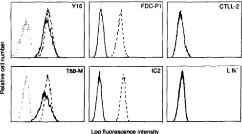
Fig. 1. Flow cytometry analysis of COS7 cells transfected with munne
IL-5R cDNA. COS7 cells transfected with the IL-5R cDNA (pCAGGS-5R) (—) or transfected with control vector (pCAGGS) (—) were stained with either H7 (a) or R52 mAb (b) in combination with 50 iA FITC-coupled F(ab")2 fragments of goat anti-rat IgG (2.5 ^g/ml) The stained cells were
analyzed with a FACScan flow cytometer. Relative fluorescence intensity was expressed by logarithmic amplification.
England Nuclear). In brief, Y16 cells (5 x 1OV0.2 ml/well) were cultured in a microplate in the presence of various concentra-tions of IL-5 and pulsed with 0.2 /iCi//d of [3H]thymidine during
the last 12 h of a 36 h culture. DNA synthesis was determined by means of [3H]thymidine incorporation.
Results
R52 mAb recognizes a different molecule (p130/p140) from p60 of the murine IL-5R
As we reported (17,18), H7andT21 mAbs recognize p60 of IL-5 binding molecule of IL-5R complex. Rolink et al. (19) reported that R52 mAb recognizes p46, p130, and p140. To evaluate whether R52 mAb can recognize molecule(s) identical to that recognized by H7 mAb, we transfected COS7 cells with murine IL-5R cDNA (pCAGGS-5R) and stained transfectants with R52 or H7 mAb in combination with FITC-coupled F(abO2 fragments
of goat anti-rat IgG and analyzed by flow cytometry The COS7 transfectants expressed ~ 8 x 10s IL-5 binding sites per cell
with an apparent Kd of 9.6 nM. H7 mAb immunoprecipitated
p60 from lysates of 125l-labeled COS7 cells as described (20).
As shown in Fig. 1, COS7 transfectants displayed positive staining with H7 (a), but not with R52 mAb (b), indicating that R52 mAb does not recognize the IL-5 binding recombmant p60 protein. We then stained 12 IL-5-dependent cell lines with R52 or H7 mAb. We also examined the reactivity of the mAbs with IL-3-dependent cell lines, IL-2-dependent CTLL2, and L cells. As shown in Fig. 2, all 12 of the IL-5-dependent cell lines represented by Y16 and T88-M reacted with both R52 and H7 mAbs. Neither IL-2-dependent CTLL2 nor L cells showed positive staining to both mAbs. It is of particular interest that IL-3-dependent cell lines represented by FDC-P1 and IC2 were stained with R52 mAb, but not H7 mAb.
To confirm that R52 mAb recognizes molecule(s) different from p60, immunoprecipitation experiments were carried out When Y16 cells were radioiodinated and cell extracts were
immune-I
c . FDC-P1A
CTLL-2A
T88-M IC2 "Log fluorescence intensity
Fig. 2. Row cytometry analysis of cloned cell lines. Various cloned cell lines were stained with 0.5 IIQI50 jd of either H7 (—) or R52 mAb (•—) in
combination with FITC-coupled F(abO2 fragments of goat anti-rat IgG as descnbed in Fig 1. As negative controls ( ), cells were stained with
FITC-coupled F(abO2 fragments of goat anti-rat IgG. The stained cells were analyzed with a FACScan flow cytometer. Relative fluorescence intensity
kDa
130
60
ft
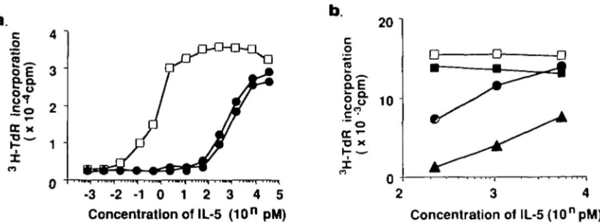
c o 0.1 1 Concentration of IL-5 (pM) 10Fig. 4. Inhibitory effects of mAbs on IL-5-induced DNA synthesis of IL-5-dependent Y16 cells. Y16 cells (5 x 1OV0 2 ml) were distributed into a rmcroplate (0.2 ml/well) together with various concentrations of IL-5 The mAbs to be tested were simultaneously added at a concentration of 10 /jg/ml in triplicate cultures and cells were cultured for 36 h at 37°C. Data were obtained with concentrations of purified antibodies, H7 (O), R52 ( • ) , H7 plus R52 (A), and normal rat IgG ( • ) . Cells were pulsed with 0.2 /iCi [3H]thymidine for the last 12 h of culture. Each point
expresses mean c p m. of triplicate cultures. One of the representative results of three different series of experiments was shown. Y16 cells incorporated 428 c.p.m. in the absence of IL-5
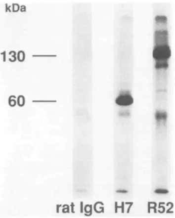
rat IgG H7 R52
Fig. 3. SDS - PAGE and autoradiography of immunoprecipitation using H7 or R52 mAbs. Y16 cells (2 x 10⁷) were radioiodinated andmembrane-fractions were prepared. Membrane-fractions were immuno-precipitated using H7 or R52 mAb. The immunopredpitated samples were analyzed using SDS-PAGE under reducing conditions
precipitated by R52 mAb, two bands of p130 and p140 were observed on SDS - PAGE (Fig. 3). Although the results were in agreement with those reported by Rolink et al. (19), these were in sharp contrast to the detection of a single major band of p60 when lysates were immunoprecipitated with H7 mAb. These results clearly indicate that R52 mAb recognizes membrane proteins p130 and p140 (p130/p140) that are not recognized by H7 or T21 mAbs. As a minor band, a p120 band was also detected, although it is not clear whether it is a degradation product of major immunoprecipitates. It is also unclear whether or not p130 is identical to p140.
Synergistic inhibitory effect of R52 and H7 mAb on IL-5-induced proliferation of Y16 cells
Having obtained the data described above, we anticipated that R52 mAb would inhibit the IL-5-dependent growth of Y16 cells in a manner different from H7 or T21 mAbs. Y16 cells were cultured with a wide range of IL-5 concentrations (0.01 - 1 0 pM) in the presence of R52 mAb (10 /tg/ml; a sufficient concentra-tion to abrogate more than 20% of psSJIL-S binding at 100 pM) or in the presence of H7 mAb (10 /ig/ml; a sufficient concentra-tion to compete with 1 nM psSjIL-S for binding to the p60 alone), or in combination with R52 and H7 mAbs. As shown in Fig. 4, Y16 cells incorporated [3H]thymidine in response to IL-5 in a
dose-dependent manner and its incorporation reached a plateau
beyond 5 pM IL-5. Addition of R52 mAb partially inhibited DNA synthesis of Y16 cells at IL-5 concentrations higher than 0.3 pM, although it completely inhibited the DNA synthesis at lower IL-5 concentrations. In contrast, H7 mAb almost completely inhibited the IL-5-induced proliferation even at 3 pM IL-5 and inhibited by more than 60% at 9 pM IL-5. Similar results to the above were obtained even when 10-fold higher concentrations of each of mAb (100 /tg/ml) were used (data not shown). Intrigumgly, simultaneous addition of both R52 and H7 mAbs caused the cooperative effects resulting in the striking suppression of proliferative response of Y16 cells induced by IL-5 even at 10 pM. The addition of T21 mAb (10 /tg/ml) caused almost complete inhibition of the proliferative response of Y16 cells induced by 10 pM IL-5 (data not shown) that may be due to a higher affinity of T21 mAb than that of H7 mAb to its receptor (18). The inhibitory effect of T21 mAb decreased, however, along with the addition of increased concentrations of IL-5 into the culture of Y16 cells (Fig. 5a) irrespective of the amounts of mAb added. It is of particular interest that simultaneous addition of both T21 and R52 mAbs again caused enhanced inhibition on the proliferation induced by 1 - 5 nM IL-5 (Fig. 5b). We also obtained essentially identical results using T88-M cells in place of Y16 cells. These results strongly suggest that R52 mAb may show a different mode of inibition of IL-5-induced proliferation from that of H7 mAb, besides a much more weak inhibitory activity than that of H7 mAb.
R52 mAb preferentially down-regulates the number of high-affinity IL-5 binding sites
To further evaluate the role of p130/p140 in the high-affinity IL-5R complex, an IL-5 binding study was carried out in the presence or absence of R52 mAb using Y16 cells As shown in Table 1, R52 mAb (10 /ig/ml) caused only partial inhibition (up to 20%) of the binding of [^SlILS (100 pM) to Y16 cells, and the addition of excess amounts of R52 mAb (30 /ig/ml) did not change the extent of the inhibition (data not shown). Both H7 mAb and excess amounts of unlabeled IL-5 also completely
20 o S o 10 T3 - 3 - 2 - 1 0 1 2 3 4 5 Concentration of IL-5 (10n pM) 2 3 4 Concentration of IL-5 (10n pM)
Fig. 5. Inhibitory effects of mAbs on IL-5-induced DNA synthesis of IL-5-dependent Y16 cells. Y16 cells were cultured with various concentrations
of IL-5 as described in Fig. 4. (a) Either 10 ( • ) or 100 ^g/ml ( • ) of T21 mAb was simultaneously added on day 0, or (b) R52 mAb (10 jtg/ml) ( • ) , T21 mAb (10 /ig/ml) (»), both R52 and T21 mAbs (A), or control IgG ( • ) was added at the onset of the culture. Cells were cultured for 36 h at 37°C Cells were pulsed with 0 2 /iCi [3H]thymidme for the last 12 h of culture Each point expresses mean c.p.m. of triplicate cultures. One of
the representative results of three different senes of experiments was shown. Y16 cells incorporated 468 c p m. in the absence of IL-5.
inhibited the IL-5 binding to Y16 cells. Scatchard plot analysis of the saturated binding data showed that Y16 cells consistently showed two IL-5 binding sites (high affinity, Kd 60 pM, - 1 2 0 0
sites per cell; low affinity, Kd 21 nM, -15,000 sites per cell)
(Fig. 6). When we carried out the binding assay in the presence of mAb, the number of high-affinity binding sites were significantly reduced by R52 mAb ( - 600 binding sites per cell) and the affinity decreased to a K6 of - 2 0 0 pM. In contrast, the number and
Kd of low-affinity IL-5 binding sites was not significantly affected
by R52 mAb. Both high and low affinity IL-5 binding sites were hardly detected in the presence of either H7 or T21 mAb under the same conditions employed (data not shown).
We have reported in our previous cross-linking studies that the p130 protein is involved in the formation of a 160 kd cross-linked complex with IL-5, while the p60 protein is involved in the formation of both 100 kd and 160 kd cross-linked complexes (14,17). The 160 kd cross-linked complex is detectable on IL-5-responsive cells under high affinity conditions (14). To examine the effect of R52 mAb on chemical cross-linking, the IL-5 binding protein on the cell surface of Y16 cells was cross-linked to PSJIL-S under high affinity conditions (100 pM PSJIL-S) in the presence or absence of mAbs. As can be seen in Fig. 7, two major cross-linked bands of - 1 0 0 and 160 kd were identified in the absence of mAb. Neither band could be detected if unlabeled and ^S-labeled IL-5 were added together (data not shown). Only the 100 kd cross-linked band was detected when R52 mAb was added before cross-linking, while neither one of the cross-linked complexes was detected when H7 or T21 mAbs were added before cross-linking.
Discussion
The existence of both low-affinity (Kd = 3 0 nM) and high-affinity
(Kd =150 pM) forms of murine IL-5R on early B cells and
eosinophils has raised the question of the structural relationship of one to the other. We have proposed that the murine high-affinity IL-5R is a membrane complex composed of at least two poly-peptide chains: p60 and p130. We also reported, using H7 and T21 mAbs, that the p60 component appears to be the IL-5
Table 1 . Inhibitory effects of mAbs on the binding of radiolabeled
IL-5 to Y16 cells
Inhibitors Specific binding (c.p m.) None IL-5 (1 nM) H7 (10 Mg/ml) R52 (10 ^g/ml) 1396 128 159 1068
Y16 cells (5 x lOS/ml) were cultured for 2 h at 37°C and then pretreated wrth IL-5 or mAb inhibitor at 4°C for another 2 h before the binding assay using 100 pM
2 3 4 5 6 -3 Bound IL-5 molecules (x10 /cell)
Fig. 6. Scatchard plot of equilibrium binding analysis of [35S]IL-5 to
Y16 cells. Cells (1 x 10⁶) were incubated at 37°C with various
concen-trations of [35S]IL-5 for 10 mm. Specific equilibrium binding was
deter-mined after subtraction of nonspecific binding obtained in the presence of a 100-fold molar excess of unlabelled IL-5. Saturation binding data were re-expressed as a Scatchard plot. The binding assay was carried out in the absence (D) or in the presence ( • ) or R52 mAb (10 /ig/ml).
binding molecule (17,18). This was finally demonstrated by isola-tion of the cDNA encoding p60 that can bind IL-5 with low-affinity (Kd =10 nM) (20). Although the identification of p60 has been
kDa
160
- **
100
-mAbs None R52 H7 T21
Fig. 7. Fluorograph of chemical cross-linking of [35S]IL-5 bindingprotein. Y16 cells (2 x 10⁷) were incubated with [35SJIL-5 (0 3 nM, 2
x 10s c.p m ) for 10 min at 37°C in the presence or in the absence of
mAbs (10 fig/ml). The cells were then washed, and DST (50 mM) was added and kept at 4°C for another 30 mm After several washings, cells were lysed with the buffer as described in Methods and the detergent lysates were analyzed on SDS- PAGE with 7.5% acrylamide gel under nonreducing conditions Fluorography was performed after the electrophoresis. The uncross-linked IL-5 ran at the middle of the 7.5% gels, which was - 5 0 kd.
definitively made, the existence of p130 has only been suggested by cross-linking studies using radiolabeled IL-5 (14). The major findings observed in this study are the following, (i) R52 mAb against murine IL-5R does not react with the murine IL-5 binding recombinant p60 protein (Fig. 1), but reacts with all IL-5-responsive cell lines so far tested (Fig. 2). (ii) R52 mAb immunoprecipitates p130/p140 doublets from cell lysates of IL-5-dependent Y16 cells (Fig.3). It is not clear at this moment, however, whether or not p130 is an identical protein to p140 with a slight difference in carbohydrate content, (iii) R52 and H7 mAbs show a little and partial inhibition, respectively, of IL-5-induced proliferation of Y16 cells at high IL-5 concentrations (Fig. 4). However, simultaneous addition of R52 mAb together with H7 mAbs causes higher synergistic inhibition of IL-5-induced proliferation than that caused by the addition of each one of them separately, (iv) R52 mAb partially inhibits IL-5 binding to target cells under high affinity conditions (100 pM PSJIL-5) while H7 or T21 mAbs profoundly inhibit the binding (Table 1). Further-more, R52 mAb preferentially down-regulates the number and Kd of high affinity IL-5-binding sites (Fig. 6). (v) R52 mAb inhibits
the formation of a cross-linked complex between p130/p140 and IL-5 (Fig. 7). Taking all of the results together, p130/p140 recognized by R52 mAb appears to be the most likely candidate for the second chain or 'associated' subunit to form high affinity IL-5R together with the IL-5 binding p60 protein.
There are at least three possibilities to account for the effects of R52 mAb on the IL-5-induced proliferation and down-regulation of the number and Kd of high-affinity IL-5R. If we assume that
IL-5 first binds to p60 and that a complex of p60 and IL-5 then associates with p130/p140 resulting in the formation of high affinity receptor complex, similar to a converted model of the IL-2R system (26), the first possibility is that p130/p140 may bind IL-5 in conjunction with p60, and R52 mAb may inhibit the association of p130/p140 with IL-5. Another possibility is that p130/p140 may associate with epitopes on p60 other than the IL-5-binding epitope. In that case, R52 mAb may recognize epitope(s) with identical or close proximity to the association sites of p130/p140with p60 and inhibit this kind of association by steric hindrance. The third possibility is that R52 mAb recognizes an epitope which is unrelated to the association sites with IL-5 or p60 and that the binding of R52 mAb to p130/p140 may cause conformational changes of the p130/p140 molecule leading to unsuccessful interaction of p130/p140 with a complex of IL-5 and/or p60. We are in favor of the second possibility on the basis of the following findings. R52 mAb could inhibit the formation of the 160 kd cross-linked complex (Fig 7) Furthermore, immunoprecipitation experiments of chemically cross-linked complex revealed that R52 mAb could immunoprecipitate the 160 kd cross-linked complex (data not shown).
There are several reports to support the notion that functional receptors for cytokines consist of two different pdypeptide chains, namely a and /3 subunits. In the case of IL-2R system, the a chain (p55) binds IL-2 with low-affinity (27) and the /3 chain (p75) by itself also binds IL-2 with intermediate affinity (28). Co-expression of both a and /3 chains leads to the formation of a high-affinity receptor, and both chains can be cross-linked with IL-2 (28). In the case of the IL-6R system, transfection of the cDNA encoding the IL-6 binding molecule in COS cells can induce the expression of mainly low-affinity binding sites (29) IL-6 triggers the association of IL-6R and non-ligand binding membrane glyco-protein, gp130, and generates IL-6 signal (30). Soluble IL-6R and IL-6 can also interact with cell surface gp130. Hibi et al. (31) recently cloned the gene encoding gp130 that does not show a binding property to IL-6 by itself. A cloned gp130 can associate with a complex of IL-6 and IL-6R. Hayashida et al (32) recently reported that the low-affinity human GM-CSFR, together with the KH97 protein encoded by a human cDNA homologous to murine IL-3R cDNA, forms a high-affinity receptor for human GM-CSFR, although KH97 protein is able to bind neither human GM-CSF nor IL-3. Because of similarities among the IL-2, GM-CSF, and IL-5 high-affinity receptors, we propose to designate the low-affinity mouse IL-5R as the a chain and the R52 protein (p130) as the /3 chain of mouse IL-5R.
After transfection of COS7 cells with a cDNA encoding p60, cell surface murine IL-5R were expressed at a density of lOS-IO6 per cell as a single binding class of low-affinity (Kd
6 - 9 nM). Despite careful analysis, no high-affinity IL-5-binding was detected (20). It is of particular interest that FDC-5R cells, established from IL-3-dependent FDC-P1 cells by transfection with munne IL-5R cDNA, express - 5 0 0 binding sites per cell for IL-5 with an apparent Kd of 30 pM (high-affinity) and 8000
binding sites per cell with Kd of 6 nM (low affinity), and become
IL-5 responsive for DNA synthesis (20). We have therefore postulated that the IL-5 binding p60 binds to IL-5 with low affinity and can be converted to a high-affinity IL-5R by interaction with
the second chain or 'associated' subunit (p130) that is expressed in FDC-P1 cells. As shown in Fig. 2, FDC-P1 and IC2 cells react with R52 but not with H7 mAb. These IL-3-dependent cell lines neither bound IL-5 determined by the IL-5-binding assay nor responded to IL-5 (data not shown). This may support the notion that the p130/p140 protein is the /3 chain of IL-5R.
Rolink ef a/. (19) reported that R52 mAb inhibits the IL-5-driven proliferation of B,3 cells and the binding of IL-5 to the cells. They
also reported that high concentrations of R52 mAb inhibits the IL-3-driven proliferative response of B,3 cells by 50% but does
not affect the other IL-3-sensitive cell lines and speculated the connection of the IL-3 and IL-5 responsiveness and of the corresponding receptors on B13 (19). We also observed the
partial inhibition by R52 mAb on IL-3-induced proliferation of Y16 and T88-M cells (data not shown). Furthermore, we recently detected similar patterns of protein phosphorylations at serine and tyrosine residues of cellular proteins when T88-M cells were stimulated either with IL-5 or IL-3 (33). Thus, we speculate the presence of interaction between the IL-5R and IL-3R systems via p130/p140.
In summary, we demonstrate that murine high affinity IL-5R is a complex of two polypeptides composed of the IL-5 binding p60 protein and the p130/p140 that has no IL-5 binding ability by itself In any case, identification of p130/p140 of R52 protein is urgently required to clarify the molecular nature of the second chain of high-affinity IL-5R that may be involved in the IL-5 signal transduction. The experimental system described in this study will give us new insights for elucidating IL-5 and the IL-5R system.
Acknowledgements
The authors are grateful to Drs T. Sudo, K. Sakamoto, and S. Yonehara for providing murine rlL-3, FDC-P1, and IC2 cells, respectively. We are also indebted to Dr E Barsoumian for critical reading of this manuscript This work supported in part by a Grant-m-Aid for Scientific Research and for Special Project Research, Cancer Bioscience, from the Ministry of Education, Science, and Culture, and by Special Coordination Funds for Promoting Science and Technology of the Science and Technology Agency, Japan. Abbreviations DST IL IL-5R H7 R52 disuccinimidyl tartarate interleukin IL-5 receptor
rat mAb against murine IL-5 receptor rat mAb against murine IL-5 receptor
References
1 Takatsu, K., Tominaga, A., Harada, N., Mita, S., Matsumoto, M , Takahashi, T., Kikuchi, Y , and Yamaguchi, N. 1988. T cell-replacing factor (TRFyinterleukin 5 (IL-5): molecular and functional properties. Immunol. Rev. 102:107.
2 Tominaga, A., Matsumoto, M , Harada, N., Takahashi, T., Kikuchi, Y , and Takalsu, K. 1988. Molecular properties and regulation of mRNA expression for murine T cell-replacing factor/IL-5. J. Immunol. 140:1175.
3 Honjo, T. and Takatsu, K. 1990. Interleukin 5. In: Sporn, M. B., ed., Peptide Growth Factors and their Receptors (Handbook of Experi-mental Pharmacology), p. 609. Springer-Verlag, Berlin.
4 Kinashi, T., Harada, N., Severinson, E , Tanabe, T., Sideras, P., Konishi, M., Azuma, C , Tominaga, A., Bergstedt-Undqvist, S., Takahashi, M., Matsuda, F., Yaoita, Y., Takatsu, K., and Honjo, T.
1986. Cloning of complementary DNA encoding T-cell replacing factor and identity with B-cell growth factor II. Nature 324:70
5 Azuma, C, Tanabe, T, Konishi, M., Kinashi, T., Noma, T., Matsuda, F., Yaoita, Y., Takatsu, K., Hammerstrom, L., Smith, C. I. E., Sevennson, E , and Honp, T 1986. Cloning of cONA for human T-ceO replacing factor (interleukin-5) and comparison with the munne homologue. Nucleic Adds Res. 14:9149.
6 Yokota, T., Coffman, R. L., Hagiwara, H., Rennick, D. M., Takebe, Y., Yokota, K., Gemmefl, L, Shrader, B., Yang, G., Meyerson, P., Luh, J., Hoy, P., Pene, J., Briere, F., Banchereau, J , Vnes, J. D., Lee, F D., Arai, N., and Arai, K. 1987. Isolation and characterization of lymphokirte cDNA clones encoding mouse and human IgA-enhancing factor and eosinophil colony-stimulating factor activities: Relationship to interleukin 5. Proc. Natl Acad. Sci USA 84:7388.
7 Swain, S. L., Howard, M., Kappler, J., Marrack, P., Watson, J , Booth, R., Wetzel, G. D , and Dutton, R. W. 1983. Evidence for two distinct classes of B cell growth factors with activities in different func-tional assays. J Exp Med. 158:822.
8 Sanderson, C. J., Campbell, H D , and Young, I. G. 1988. Molecular and cellular biology of eosinophils differentiation factor (IL-5) and its effect on human and mouse B cells. Immunol. Rev 102:29. 9 Coffman, R , Seymour, B., Hiraki, D , Christiansen, J., Shrader, B ,
Cherwinsky, H , Savelkoul, H., Fmkelman, J , Bond, M., and Mosmann, T 1988. The role of helper T cell products in mouse B cell differentiation and isotype regulation. Immunol Rev 102:5. 10 Tominaga, A., Takaki, S , Koyama, N , Katoh, S., Matsumoto, R ,
Migita, M., Hitoshi, Y., Hosoya, Y., Yamauchi, S., Kanai, Y., Miyazaki, J.-l., Usuku, G., Yamamura, K -I., and Takatsu, K. 1991 Transgenic mice expressing a B cell growth and differentiation factor (interleukin 5) gene develop eosinophiha and autoantibody produc-tion. J. Exp Med. 173429
11 Yamaguchi, Y., Suda, T., Suda, J , Eguchi, M , Miura, Y., Harada, N., Tominaga, A , and Takatsu, K. 1988. Purified mterleukin-5 (IL-5) supports the terminal differentiation and proliferation of murine eosinophilic precursors J Exp. Med. 167:43.
12 Tominaga, A , Nishikawa, S - I , Mita, S., Ogawa, M., Kikuchi, Y., Hitoshi, Y , and Takatsu, K. 1989 Establishment of IL-5 dependent early B cell lines by long-term bone marrow cultures. Growth Factors 1-135
13 Mita, S., Harada, N., Naomi, S., Hitoshi, Y., Sakamoto, K., Akagi, M., Tominaga, A., and Takatsu, K. 1988. Receptors for T cell-replacing factor/interleukin 5. specificity, quantitation and its implication J. Exp Med. 168863.
14 Mita, S., Tominaga, A , Hitoshi, Y , Honjo, T., Sakamoto, K., Akagi, M., Kikuchi, Y , Yamaguchi, N., and Takatsu, K. 1989 Characterization of high-affinity receptors for interleukin 5 on interleukin 5-dependent cell lines. Proc. Natl Acad. Sci USA 86:2311.
15 Rolink, A. G , Thalmann, P., Kikuchi, Y., and Erdei, A. 1990. Characterization of the interleukin 5-reactive splenic B cell population. Eur. J Immunol 20:1949.
16 Hitoshi, Y., Yamaguchi, N., Korenaga, M., Tominaga, A., and Takatsu, K. 1991 In vivo administration of antibody to murine IL-5 receptor inhibits eosinophiha of IL-5 transgenic mice. Int. Immunol. 3:135.
17 Yamaguchi, N., Hitoshi, Y , Mita, S., Hosoya, Y., MurataY , Kikuchi, Y , Tominaga, A., and Takatsu, K 1990. Characterization of the murine interleukin 5 receptor by using a monoclonal antibody. Int. Immunol. 2-181.
18 Hitoshi, Y., Yamaguchi, N., Mita, S., Takaki, S., Tominaga, A., and Takatsu, K. 1990. Distribution of IL-5 receptor-positive B cells- expres-sion of IL-5-receptor on Ly-1(CD5)+ B cells J. Immunol. 144:4218.
19 Rolink, A. G., Melchers, F., and Palacios, R. 1989. Monoclonal antibodies reactive with the mouse interieukin 5 receptor. J. Exp. Med 169:1693.
20 Takaki, S., Tominaga, A., Hitoshi, Y., Mita, S., Sonoda, E., Yamaguchi, N., and Takatsu, K. 1990. Molecular cloning and expression of the murine interleukin-5 receptor. EMBO J. 9:4367. 21 Koyasu, S., Yodoi, J., Nikaido, T., Tagaya, Y., Tamguchi, Y., Honjo, T.,
and Yahara, I. 1986. Expression of interfeukin 2 receptors on interleukin 3-dependent cell lines. J. Immunol. 136:984.
22 Dexter, T. M., Garland, J., Scott, G. D., Scolnick, E., and Metcalf, D. 1980. Growth of factor-dependent hematopoietJc precursor cell lines. J. Exp. Med. 152:1036.
23 Miyazaki, J.-l., TakakJ, S., Araki, K., Tashiro, F., Tominaga, A., Takatsu, K , and Yamamura, K.-l. 1989. Expression vector system based on the chicken 0-actin promoter directs efficient production of interieukin 5. Gene 79.269.
24 Scatchard, G 1949. The attractions of proteins for small molecules and ions. Ann. NY Acad Sci. 51:660.
25 Laemmli, U. K. 1970. Cleavage of structural proteins during the assembly of the head of bacteriophage T4. Nature 227680 26 Saito, Y., Sabe, H., Suzuki, N., Kondo, S , Ogura, T., Shimizu, A., and
Honjo, T 1988. A larger number of L chains (Tac) enhance the association rate of interieukin 2 to the high affinity site of the interieukin 2 receptor J. Exp. Med. 168.1563.
27 Nikaido, T., Shimizu, A., Ishida, N., Sabe, H., Teshigawara, K., Maeda, M., Uchiyama, T., Yodoi, J., and Honjo, T. 1984. Molecular cloning of cDNA encoding human interleukin-2 receptor. Nature 311-631.
28 Hatakeyama, M., Tsudo, M., Minamoto, S., Kono, T , Doi.T., Miyata, T., Miyasaka, M., and Taniguchi, T. 1989. Interleukin-2 receptor fi chain gene, generation of three receptor forms cloned by human a and |3 chain cDNA's. Science 244551
29 Yamasaki, K., Taga, T , Hirata, Y., Yawata, Y , Kawamshi, Y., Seed, B , Taniguchi, T., Hirano, T., and Kishimoto, T. 1988. Cloning and expression of the human interteukin-6 (BSF-2/IFN/S2) receptor Science 241825.
30 Taga, T, Hibi, M., Hirata, Y., Yamasaki, K., Yasukawa, K , Matsuda, T., Hirano, T, and Kishimoto, T. 1989 lnterleukin-6 triggers the association of its receptor with a possible signal transducer, gp130. Cell 58.573.
31 Hibi, M., Murakami, M., Saito.M., Hirano, T., Taga, T., and Kishimoto, T. 1990. Molecular ctomng and expression of an IL-6 signal transducer, gp130 Cell 63:1149
32 Hayashida, K , Kitamura, T., Gorman, D. M., Arai, K.-l., Yokota, T., and Miyajima, A 1990 Molecular cloning of a second subunit of the receptor for human granulocyte-macrophage colony-stimulating factor (GM-CSF). Reconstitution of a high-affinity GM-CSF receptor. Proc. Natl Acad. Sci. USA 87:9655.
33 Murata, Y., Yamaguchi, N., Hitoshi, Y., Tominaga, A., and Takatsu, K. 1990. Interieukin 5 and interieukin 3 induce senne and tyrosine phosphorylations of several cellular proteins in an interieukin 5-dependent cell line. Biochem. Biophys Res. Commun. 173.1102.
![Fig. 7. Fluorograph of chemical cross-linking of [ 35 S]IL-5 binding protein. Y16 cells (2 x 10 7 ) were incubated with [35SJIL-5 (0 3 nM, 2 x 10 s c.p m ) for 10 min at 37°C in the presence or in the absence of mAbs (10 fig/ml)](https://thumb-eu.123doks.com/thumbv2/123doknet/14885078.646354/6.922.96.443.102.481/fluorograph-chemical-linking-binding-protein-incubated-presence-absence.webp)
