Monitoring mis-acylated tRNA suppression ef®ciency
in mammalian cells via EGFP ¯uorescence recovery
Erwin Ilegems, Horst M. Pick and Horst Vogel*
Institute of Biomolecular Sciences, Swiss Federal Institute of Technology, Lausanne CH-1015, Switzerland
Received August 19, 2002; Revised and Accepted September 27, 2002
ABSTRACT
A reporter assay was developed to detect and quan-tify nonsense codon suppression by chemically aminoacylated tRNAs in mammalian cells. It is based on the cellular expression of the enhanced green ¯uorescent protein (EGFP) as a reporter for the site-speci®c amino acid incorporation in its sequence using an orthogonal suppressor tRNA derived from Escherichia coli. Suppression of an engineered amber codon at position 64 in the EGFP run-off transcript could be achieved by the incorpor-ation of a leucine via an in vitro aminoacylated sup-pressor tRNA. Microinjection of de®ned amounts of mutagenized EGFP mRNA and suppressor tRNA into individual cells allowed us to accurately deter-mine suppression ef®ciencies by measuring the EGFP ¯uorescence intensity in individual cells using laser-scanning confocal microscopy. Control experiments showed the absence of natural sup-pression or aminoacylation of the synthetic tRNA by endogenous aminoacyl-tRNA synthetases. This reporter assay opens the way for the optimization of essential experimental parameters for expanding the scope of the suppressor tRNA technology to different cell types.
INTRODUCTION
The site-speci®c incorporation of unnatural amino acids into proteins in living cells is of importance to analyze in vivo protein structure and function as well as cellular processes using amino acid analogs comprising probes which are photo-activatable, ¯uorescent or chemically reactive (1±6). This emerging technology relies on the suppression of nonsense codon mutations by chemically acylated tRNAs and has been originally developed as an in vitro method (7±12). Meanwhile, several reports for its in vivo application in Xenopus oocytes (4±6,13,14), Escherichia coli (15±18) and COS1 cells (19) appeared in the literature. An expansion of this technology to other cell lines would demand a reporter system permitting the de®nition of optimal parameters for the site-speci®c incorporation of amino acid analogs into proteins.
Here, suppressor tRNA technology was applied to Chinese hamster ovary (CHO) cells which, like other mammalian cell
types, are generally more suitable for structural and functional studies of human-derived proteins if speci®c post-translational modi®cations are important. In addition, certain proteins such as neuro-receptors are optimally expressed only in particular cell lines. We focused on the enhanced green ¯uorescent protein (EGFP) as a reporter to assess the ef®ciency of nonsense codon suppression directly in living cells. An amber stop codon mutation was site-speci®cally introduced in the core position of the EGFP, removing an amino acid essential for the formation of the ¯uorophore. The transfer of that mutagenized in vitro transcript into CHO cells was followed by the expression of an incomplete, non-¯uorescent protein. After co-transfer with a cognate synthetic suppressor tRNA, we could monitor the successful re-incorporation of the missing amino acid by recovery of the EGFP ¯uorescence signal, which could be quanti®ed by using laser-scanning confocal microscopy on living cells.
Unlike other ¯uorescent reporters such as luciferase or b-galactosidase, EGFP does not require the addition of substrate or cofactors nor cell lysis or ®xation. Furthermore, it is stable over a period of several days and, due to its strong ¯uorescence, allows an accurate and sensitive determination of suppression ef®ciencies in individual cells. This strategy could be used to ®nd proper conditions for an ef®cient suppression in a number of different mammalian cell lines.
MATERIALS AND METHODS Materials
Synthetic oligonucleotides were purchased at MWG-Biotech AG (Ebersberg, Germany). Kits for plasmid and DNA-fragment puri®cation were obtained from QIAGEN GmbH (Hilden, Germany). Restriction endonucleases (BsaI, EcoRI and NotI) were provided by New England Biolabs (MA, USA). The MEGAscript kit for in vitro transcription and the cap analog m7G(5¢)ppp(5¢)G were from Ambion (TX, USA).
Puri®ed rEGFP was purchased at Clontech (CA, USA). Octadecyl rhodamine B (R18) and Alexa Fluor 546 C5
maleimide were obtained from Molecular Probes (OR, USA). Other chemicals were purchased at Sigma-Aldrich (MO, USA).
Transcription of reporter gene
The coding sequence of the EGFP (pEGFP-N1, Clontech) was modi®ed by the addition of a T7 promoter site and a poly(A) tail using PCR ampli®cation with synthetic oligonucleotides
(Fig. 1). The resulting 814 bp fragment was ligated into the pCR2.1 vector using the TA cloning kit (Invitrogen, CA, USA) to obtain the plasmid pT7PEGFP.
The pT7PEGFPam64L is a mutated version of the pT7PEGFP plasmid. The CTG codon at position 64 of the EGFP coding sequence was mutated to a nonsense amber (TAG) codon by site-directed mutagenesis using the Quickchange kit (Stratagene, CA, USA). All plasmid con-structs were controlled by restriction mapping and DNA sequencing.
After linearization of the wild-type and mutated plasmids by NotI, in vitro transcription was performed with T7 RNA polymerase using the MEGAscript kit (Ambion). Capping of mRNA was achieved during transcription by replacing 80% of the GTP level with the cap analog m7G(5¢)ppp(5¢)G (Ambion).
After removing the DNA template by DNase I treatment, the resulting mRNAs were puri®ed by successive phenol± chloroform±isoamyl alcohol (25:24:1) and chloroform± isoamyl alcohol (24:1) extractions, precipitation with an equal volume of isopropanol for 1 h at ±20°C, followed by centrifugation at 0°C/20 800 g for 15 min. The mRNA pellet was dried and redissolved in sterile DEPC-treated H2O. The
integrity and size of the mRNAs were assayed by agarose gel electrophoresis under denaturing conditions, and the concen-tration was determined by measuring the optical density at 260 nm.
Transcription of suppressor tRNA gene
A 105 bp synthetic template fragment encoding the E.coli suppressor tRNAAlaCUA was prepared by reannealing and
ligating two synthetic oligonucletides containing the tRNA gene ¯anked by a T7 promoter (Fig. 2). This blunt-end DNA fragment was cloned into pCR4 vector using the TOPO cloning kit (Invitrogen), following the instructions of the manufacturer. The resulting plasmid pEcoliAlaCUA was checked by restriction mapping and sequencing.
Plasmid pEcoliAlaCUA was linearized by successive restriction endonuclease digests with EcoRI and BsaI. After agarose gel puri®cation this DNA fragment was used for the
Figure 1. Scheme of the cloning steps for the wild-type (pT7PEGFP) and mutant EGFP (pT7PEGFPam64L) encoding plasmids used for the in vitro transcrip-tion. PCR ampli®cation with primers 1 and 2 was performed to add a T7 promoter to the 5¢ end and a poly(A) tail to the 3¢ end of the EGFP coding sequence. Primers 3 and 4 were used for replacing the leucine 64 codon by TAG, and providing silent mutations for clonal selection. All nucleotide sequence modi®cations are shown in bold letters.
Figure 2. Scheme for the in vitro synthesis of the suppressor tRNA from a synthetic DNA template by T7 run-off transcription. Two synthetic comple-mentary oligonucleotides encoding the E.coli-derived suppressor tRNAAlaCUA were annealed and cloned into the pCR4 vector, leading to
pEcoliAlaCUA. T7 RNA polymerase run-off transcription was performed on the puri®ed EcoRI and BsaI fragment giving rise to a 76mer suppressor tRNA.
run-off transcription with T7 RNA polymerase following the protocol of the MegaScript kit (Ambion). After removing the template DNA by DNase I treatment the tRNA was puri®ed by following the protocol described above for mRNA. The integrity and purity of the tRNA was assayed by polyacryl-amide gel electrophoresis, and the concentration was determined by measuring the optical density at 260 nm. Deprotection of N-(4-pentenoyl)-S-leucyl-tRNA
Pentenoyl-protected aminoacylated tRNAAlaCUAwas
synthe-sized at Cruachem Ltd (Glasgow, Scotland, UK). Ten micrograms of lyophilized protected tRNA were resuspended in 10 ml of H2O. Deprotection was accomplished by adding
2.5 ml of 25 mM I2 in 1:1 THF±H2O and incubating the
mixture for 10 min at 25°C. The leucyl-tRNA was precipitated by successive additions of 1.25 ml of 3 M NaOAc pH 5.3 and 31.25 ml of cold ethanol. After centrifugation at 0°C/20 800 g for 15 min the pellet was washed once with 70% cold ethanol, dried and dissolved in H2O to a ®nal concentration of
4 mg/ml.
Cell lines and cell culture
Adherent mammalian cells (CHO) were grown in Dulbecco's modi®ed Eagle's medium (DMEM/F12; GIBCO BRL, Rockville, USA). The medium was supplemented with 2.2% fetal calf serum (GIBCO BRL). The cultures were split, distributed in 35 mm Nunc dishes at a density of 50 000 cells/ml 1 day prior to injection, and kept at 37°C in a humidi®ed atmosphere with 5% CO2.
Injections
Microinjections of CHO cells with mRNA and tRNA mixtures diluted in sterile DEPC-treated H2O were performed using an
InjectMan controller and a Transjector 5246 system (both from Eppendorf, Hamburg, Germany) mounted on an Axiovert S100TV inverted microscope (Carl Zeiss AG, Oberkochen, Germany). FemtotipsII capillaries (Eppendorf) were used for all injections.
Laser-scanning confocal microscopy
Laser-scanning confocal microscopy was performed using a Zeiss LSM510 (Carl Zeiss AG). Detection and distinction of the ¯uorescence signals of EGFP (excitation 488 nm/emission 505±530 nm), Alexa546 and R18 (excitation 543 nm/emission >560 nm) was achieved by appropriate ®lter sets using a multitracking mode. Scanning speed and laser intensity were adjusted to avoid photobleaching of the ¯uorophores.
RESULTS
The general scheme used for in vivo visualization of mis-acylated tRNA suppression is presented in Figure 3A. The suppression ef®ciency was determined by measuring the appearance of EGFP ¯uorescence in living cells. The EGFP coding sequence was mutated at position 64 by replacing the CTG (leucine) codon with an amber stop codon in the core of the reporter gene, giving rise to the EGFPam64L in vitro transcript. A successful suppression by co-injection of this mutated mRNA with a suppressor tRNA carrying the `removed' amino acid would lead to the completion of the EGFP translation. Twenty hours after co-injection of suppres-sor leucyl-tRNA with EGFPam64L mRNA, we observed the recovery of the EGFP ¯uorescence in CHO cells by laser-scanning confocal microscopy (see Fig. 3B), whereas the co-injection of the mutated mRNA together with the non-aminoacylated tRNA did not lead to any detectable ¯uorescence within the cells (data not shown).
Because the injected volume may slightly vary between cells, mostly depending on their intrinsic viscosity, it was necessary to quantify this parameter in order to validate further comparisons between different experiments. There-fore, the injections of mRNA and tRNA were performed together with a known concentration of Alexa546. This dye is non-toxic for the cell and its excitation and emission wavelengths are well distinguishable from those of EGFP.
Correlation between the ¯uorescence of the co-loaded dye and the injected volume was obtained by measuring its dilution in the cell and by calculating individual cell volumes.
Figure 3. In vivo suppression visualized by EGFP ¯uorescence recovery. (A) Schematic view of the assay for the amber suppressor tRNA function: co-injec-tion of a mutated EGFP mRNA containing an amber codon at posico-injec-tion 64 with an amber suppressor leucyl-tRNA. A successful suppression leads to complete translation of the mRNA transcript, thus to the appearance of the EGFP molecule. (B) Laser-scanning confocal micrograph of CHO cells visualizing EGFP ¯uorescence 20 h after co-injection of the mutated EGFP-mRNA with the amber suppressor tRNA. Size bar is 10 mm.
We ®rst elaborated a ¯uorescence intensity calibration curve by measurements on droplets containing known concentra-tions of the Alexa546 dye using laser-scanning confocal microscopy. A linear relation between ¯uorescence intensity and dye concentration was found. Fluorescence intensity measurements of the co-injected Alexa546 dye on living cells permitted the determination of the dye concentration within the cells on the basis of this calibration curve.
The injection volume of the Alexa dye together with mRNA and tRNA mixtures can then be determined by calculating the cell volume. Therefore, we detached the adherent cells by addition of 1 mM EDTA and incubation at 37°C for 10 min. Under these conditions the cells became spherical. They were then membrane-stained with R18 (5 mM/10 min incubation at 37°C) to measure their diameter by laser-scanning confocal microscopy, leading to a volume of 1.2 6 0.1 pl (data not shown). The number of EGFP molecules which were expressed during in vivo translation in individual cells can be estimated by comparison of cell-derived EGFP ¯uores-cence intensities with EGFP ¯uores¯uores-cence intensities measured on droplets of known concentrations of puri®ed rEGFP.
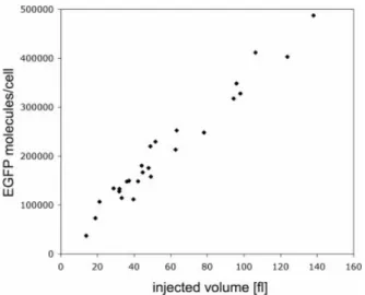
To validate the precision of our injections and concentration calibrations, we also co-injected variable volumes of a solution containing 500 ng/ml rEGFP and 250 ng/ml Alexa546 in CHO cells. As seen in Figure 4, there is a linear
correlation between injected volume, given by the Alexa546 ¯uorescence signal, and the number of EGFP molecules per cell.
On the basis of the presented control experiments we performed all following measurements using concentrations of 250 ng/ml Alexa546, 2 mg/ml mRNA and 2 mg/ml leucyl-tRNAAlaCUA. These concentrations gave the best suppression
ef®ciencies: (i) permitting the highest number of RNA molecules delivery without capillary blocking due to the resulting high solution viscosity, and (ii) favoring the suppression against competing endogenous termination factors at an excess of 10:1 of tRNA:mRNA molecules.
The EGFP ¯uorescence resulting from in vivo suppression was stable between 12 and 24 h post-injection. The injected cells were easily identi®ed under the microscope via ¯uores-cence of the co-injected dye. Thereby we could con®rm that every injected cell turned green and could be re-identi®ed even after 24 h incubation at 37°C. Using the EGFP and Alexa546 ¯uorescence intensity calibration curves, we calcu-lated the number of transcalcu-lated EGFP molecules by confocal microscopy 20 h after microinjection. Because the variation between injection volumes was 0.05 6 0.01 pl, we corrected the number of translated EGFP molecules by normalization to an average injection volume of 0.05 pl (Table 1). We obtained suppression ef®ciencies of 15 6 3% by comparing EGFP ¯uorescence signals resulting from in vivo suppression with ¯uorescence signals derived from the injection of the identical amounts of non-mutated EGFP mRNA.
The microinjection of a non-aminoacylated tRNA together with the mutagenized reporter transcript did not produce any detectable ¯uorescence at the EGFP emission wavelength of 510 nm in CHO cells when we used the identical parameters of confocal microscopy (laser intensity, sensitivity of detection, pinhole diameter, ®lter sets, etc.) as for measuring the EGFP ¯uorescence recovery upon nonsense codon suppression. If we substantially increased the laser power for the excitation at 488 nm and the sensitivity of ¯uorescence signal detection at 510 nm we could hardly detect unspeci®c cellular auto¯uorescence, identical to that of non-injected control cells.
DISCUSSION
A novel assay has been developed using EGFP as a reporter for direct quanti®cation of termination codon suppression ef®ciencies in living mammalian cells. We have shown by ¯uorescence recovery that an aminoacylated tRNA derived from E.coli can suppress an amber mutation at codon 64 of the EGFP mRNA by in vivo translation in CHO cells, whereas the
Figure 4. Correlation between injection volume and number of EGFP molecules per cell by microinjection of variable volumes of a solution con-taining 500 ng/ml rEGFP and 250 ng/ml Alexa546 in CHO cells. The injected volume is calculated on the basis of the known cell volume (see text for detailed description) and the ®nal concentration of Alexa546 dye measured directly in the cell (based on a calibration curve established before).
Table 1. Number of mRNA and tRNA molecules per cell directly calculated after microinjection by comparison of the co-injected Alexa dye with the confocal calibration curve on droplets
0.05 pl injection mRNA
(3103molecules/cell) tRNA(3103molecules/cell) EGFP(3103molecules/cell)
EGFPam64L mRNA + tRNACUA 250 6 20 2500 6 200 ND
EGFPam64L mRNA + leucyl-tRNACUA 250 6 20 2500 6 200 270 6 30
EGFP mRNA 250 6 20 ± 1800 6 200
Expression of EGFP molecules per cell determined on 50 individual cells, 20 h after injection of RNA. The values are corrected for a standard injection volume of 0.05 pl. Co-injections were all processed using concentrations of 2 mg/ml mRNA, 2 mg/ml tRNA and 250 ng/ml Alexa546. ND, not detectable.
non-aminoacylated form of the same tRNA cannot. EGFP ¯uorescence intensities measured in individual cells by laser-scanning confocal microscopy allowed us to quantify sup-pression ef®ciencies. Kohrer et al. (19) have used a different approach to insert amino acid analogs into the chlor-amphenicol acetyltransferase (CAT) by importing a suppres-sor Tyr-tRNA in COS1 cells using lipofection. These authors measured CAT activities in cell extracts to determine average levels of termination codon suppression ef®ciencies in cell samples but not on individual cells.
The use of EGFP as a suppression reporter offers advan-tages compared to other detection techniques such as those based on luciferase, b-galactosidase or CAT. In contrast to these destructive methods, EGFP suppression can be followed in vivo in a single cell without further chemical or enzymatic treatment, due to the suf®ciently high quantum yield of this protein.
To improve the suppression-speci®c ¯uorescent signal, we increased the signal-to-noise ratio by lowering the background signal resulting from possible natural suppressor tRNAs. This was ful®lled by choosing an amino acid in the protein sequence that plays an important role for the ¯uorescence properties of the EGFP. In consequence, a cell line which comprises a natural suppressor tRNA aminoacylated with a different amino acid would provide a background signal reduced to an undetectable value. We selected the leucine at position 64 for its importance in the ¯uorescence properties of the EGFP, in particular because it greatly improves correct protein folding (20,21). Furthermore, due to the fact that it is neither a charged nor a polar amino acid, it is more quickly accepted and processed by the ribosome, thus better compet-ing with the undesirable effect of termination factors which could prevent the completion of the full-length protein (22±26).
To determine the contribution of the unnatural suppression under investigation, the suppressor tRNA should also not be recognized by any endogeneous aminoacyl-tRNA synthetases. This natural aminoacylation of the suppressor tRNA can be minimized by choosing a tRNA from another organism. In our present study, the tRNA sequence was based on that of E.coli tRNAAlaGGC(27) mutated to CUA at the anticodon site and
modi®ed by two other mutations: A38U improves amber suppression ef®ciency (28±30) and C70U renders the non-aminoacylated tRNA a poor substrate for E.coli alanyl-tRNA synthetase (31). This tRNA sequence has been shown by Karginov et al. (9) to have good suppression properties in in vitro translation systems.
Our experiments to determine suppression ef®ciencies by measuring EGFP ¯uorescence intensities in cells and to estimate amounts of injected mRNA and tRNA molecules were based on laser-scanning confocal microscopy measure-ments. Microinjection of de®ned amounts of ¯uorescent dyes in CHO cells demonstrated the suitability of this transfection technique to incorporate, in a controlled way, quanti®able molecule amounts, in contrast to saponin permeabilization (32), electroporation (33) or lipofection (19) of mammalian cells. Furthermore, unlike these other techniques, control of the transfected amount does not depend on cell type or density, allowing a more accurate comparison between different cell types. Finally, microinjection requires only low amounts of
material, and prevents the RNA degradation that can take place during delivery in other transfection techniques.
We were able to observe amber suppression of an amber mutated EGFP mRNA using mis-acylated tRNA in CHO cells, obtaining 15 6 3% of the ¯uorescence signal obtained by injection of the same amount of non-mutated EGFP mRNA. After estimation of the RNA molecules and EGFP protein numbers, we obtained approximately one suppressed transla-tion by mRNA (see Table 1). Referring to a typical injectransla-tion volume in CHO cells of 0.05 pl, the number of proteins resulting from unnatural suppression was in the range of 270 000 6 30 000.
The general applicability of the presented EGFP-based reporter assay will allow us to extend the suppressor tRNA technique for many other cell types, and hence permit the selection of suitable suppressor tRNAs for the site-directed modi®cation of proteins. Of particular interest for applying suppressor tRNA technology are investigations of selective molecular interactions in live biological cells by ¯uorescence techniques with high time, spatial and single molecule resolution, to elucidate the complex cellular biochemical networks (34±36).
ACKNOWLEDGEMENTS
This work was ®nancially supported by grants from the EPFL and the Swiss National Science Foundation.
REFERENCES
1. Gallivan,J.P., Lester,H.A. and Dougherty,D.A. (1997) Site-speci®c incorporation of biotinylated amino acids to identify surface-exposed residues in integral membrane proteins. Chem. Biol., 4, 739±749. 2. Mamaev,S.V., Laikhter,A.L., Arslan,T. and Hecht,S.M. (1996) Fire¯y
luciferase: alteration of the color of emitted light resulting from substitutions at position 286. J. Am. Chem. Soc., 118, 7243±7244. 3. Mendel,D., Cornish,V.W. and Schultz,P.G. (1995) Site-directed
mutagenesis with an expanded genetic code. Annu. Rev. Biophys. Biomol. Struct., 24, 435±462.
4. Turcatti,G., Nemeth,K., Edgerton,M.D., Meseth,U., Talabot,F., Peitsch,M., Knowles,J., Vogel,H. and Chollet,A. (1996) Probing the structure and function of the tachykinin neurokinin-2 receptor through biosynthetic incorporation of ¯uorescent amino acids at speci®c sites. J. Biol. Chem., 271, 19991±19998.
5. Cohen,B.E., McAnaney,T.B., Park,E.S., Jan,Y.N., Boxer,S.G. and Jan,L.Y. (2002) Probing protein electrostatics with a synthetic ¯uorescent amino acid. Science, 296, 1700±1703.
6. Beene,D.L., Brandt,G.S., Zhong,W., Zacharias,N.M., Lester,H.A. and Dougherty,D.A. (2002) Cation-pi interactions in ligand recognition by serotonergic (5-HT(3A)) and nicotinic acetylcholine receptors: the anomalous binding properties of nicotine. Biochemistry, 41, 10262±10269.
7. Cload,S.T., Liu,D.R., Froland,W.A. and Schultz,P.G. (1996) Development of improved tRNAs for in vitro biosynthesis of proteins containing unnatural amino acids. Chem. Biol., 3, 1033±1038. 8. Noren,C.J., Anthony-Cahill,S.J., Suich,D.J., Noren,K.A., Grif®th,M.C.
and Schultz,P.G. (1990) In vitro suppression of an amber mutation by a chemically aminoacylated transfer RNA prepared by runoff transcription. Nucleic Acids Res., 18, 83±88.
9. Karginov,V.A., Mamaev,S.V. and Hecht,S.M. (1997) In vitro suppression as a tool for the investigation of translation initiation. Nucleic Acids Res., 25, 3912±3916.
10. Kurzchalia,T.V., Wiedmann,M., Breter,H., Zimmermann,W., Bauschke,E. and Rapoport,T.A. (1988) tRNA-mediated labelling of proteins with biotin. A nonradioactive method for the detection of cell-free translation products. Eur. J. Biochem., 172, 663±668.
11. Hohsaka,T., Kajihara,D., Ashizuka,Y., Murakami,H. and Sisido,M. (1998) Ef®cient incorporation of nonnatural amino acids with large
aromatic groups into streptavidin in in vitro protein synthesizing systems. J. Am. Chem. Soc., 121, 34±40.
12. Short,G.F.,3rd, Golovine,S.Y. and Hecht,S.M. (1999) Effects of release factor 1 on in vitro protein translation and the elaboration of proteins containing unnatural amino acids. Biochemistry, 38, 8808±8819. 13. Turcatti,G., Nemeth,K., Edgerton,M.D., Knowles,J., Vogel,H. and
Chollet,A. (1997) Fluorescent labeling of NK2 receptor at speci®c sites in vivo and ¯uorescence energy transfer analysis of NK2 ligand-receptor complexes. Receptors Channels, 5, 201±207.
14. Chollet,A., Turcatti,G., Nemeth,K. and Vogel,H. (1998) Probing ligand-receptor recognition in G protein-coupled ligand-receptors through biosynthetic incorporation of ¯uorescent amino acids at speci®c sites and
measurement of distances by ¯uorescence energy transfer. In Slavik,J. (ed.), Fluorescence Microscopy and Fluorescent Probes. Plenum Press, New York, NY, Vol. 2, pp. 87±92.
15. Liu,D.R. and Schultz,P.G. (1999) Progress toward the evolution of an organism with an expanded genetic code. Proc. Natl Acad. Sci. USA, 96, 4780±4785.
16. Wang,L., Brock,A., Herberich,B. and Schultz,P.G. (2001) Expanding the genetic code of Escherichia coli. Science, 292, 498±500.
17. Wang,L., Brock,A. and Schultz,P.G. (2002) Adding
L-3-(2-naphthyl)alanine to the genetic code of E. coli. J. Am. Chem. Soc., 124, 1836±1837.
18. Liu,D.R., Magliery,T.J., Pastrnak,M. and Schultz,P.G. (1997)
Engineering a tRNA and aminoacyl-tRNA synthetase for the site-speci®c incorporation of unnatural amino acids into proteins in vivo. Proc. Natl Acad. Sci. USA, 94, 10092±10097.
19. Kohrer,C., Xie,L., Kellerer,S., Varshney,U. and RajBhandary,U.L. (2001) Import of amber and ochre suppressor tRNAs into mammalian cells: a general approach to site-speci®c insertion of amino acid analogues into proteins. Proc. Natl Acad. Sci. USA, 98, 14310±14315. 20. Cormack,B.P., Valdivia,R.H. and Falkow,S. (1996) FACS-optimized
mutants of the green ¯uorescent protein (GFP). Gene, 173, 33±38. 21. Palm,G.J., Zdanov,A., Gaitanaris,G.A., Stauber,R., Pavlakis,G.N. and
Wlodawer,A. (1997) The structural basis for spectral variations in green ¯uorescent protein. Nature Struct. Biol., 4, 361±365.
22. Drugeon,G., Jean-Jean,O., Frolova,L., Le Goff,X., Philippe,M., Kisselev,L. and Haenni,A.L. (1997) Eukaryotic release factor 1 (eRF1) abolishes readthrough and competes with suppressor tRNAs at all three termination codons in messenger RNA. Nucleic Acids Res., 25, 2254±2258.
23. Le Goff,X., Philippe,M. and Jean-Jean,O. (1997) Overexpression of human release factor 1 alone has an antisuppressor effect in human cells. Mol. Cell. Biol., 17, 3164±3172.
24. Cornish,V.W., Mendel,D. and Schultz,P.G. (1995) Probing protein structure and function with an expanded genetic code. Angew. Chem. Int. Ed. Engl., 34, 621±633.
25. Karginov,V.A., Mamaev,S.V., An,H., Van Cleve,M., Hecht,S.M., Komatsoulis,G. and Abelson,J. (1997) Probing the role of an active site aspartic acid in dihydrofolate reductase. J. Am. Chem. Soc., 119, 8166±8176.
26. Cornish,V.W., Benson,D.R., Altenbach,C.A., Hideg,K., Hubbell,W.L. and Schultz,P.G. (1994) Site-speci®c incorporation of biophysical probes into proteins. Proc. Natl Acad. Sci. USA, 91, 2910.
27. Mims,B.H., Prather,N.E. and Murgola,E.J. (1985) Isolation and nucleotide sequence analysis of tRNAAlaGGC from Escherichia coli K-12. J. Bacteriol., 162, 837±839.
28. Raftery,L.A. and Yarus,M. (1987) Systematic alterations in the anticodon arm make tRNA(Glu)-Suoc a more ef®cient suppressor. EMBO J., 6, 1499±1506.
29. Bruce,A.G., Atkins,J.F., Wills,N., Uhlenbeck,O. and Gesteland,R.F. (1982) Replacement of anticodon loop nucleotides to produce functional tRNAs: amber suppressors derived from yeast tRNAPhe. Proc. Natl Acad. Sci. USA, 79, 7127±7131.
30. Kleina,L.G., Masson,J.M., Normanly,J., Abelson,J. and Miller,J.H. (1990) Construction of Escherichia coli amber suppressor tRNA genes. II. Synthesis of additional tRNA genes and improvement of suppressor ef®ciency. J. Mol. Biol., 213, 705±717.
31. Hou,Y.M. and Schimmel,P. (1988) A simple structural feature is a major determinant of the identity of a transfer RNA. Nature, 333, 140±145.
32. Negrutskii,B.S., Stapulionis,R. and Deutscher,M.P. (1994) Supramolecular organization of the mammalian translation system. Proc. Natl Acad. Sci. USA, 91, 964±968.
33. Negrutskii,B.S. and Deutscher,M.P. (1991) Channeling of aminoacyl-tRNA for protein synthesis in vivo. Proc. Natl Acad. Sci. USA, 88, 4991±4995.
34. Hovius,R., Vallotton,P., Wohland,T. and Vogel,H. (2000) Fluorescence techniques: shedding light on ligand-receptor interactions. Trends Pharmacol. Sci., 21, 266±273.
35. Remy,I. and Michnick,S.W. (2001) Visualization of biochemical networks in living cells. Proc. Natl Acad. Sci. USA, 98, 7678±7683. 36. Luker,G.D., Sharma,V., Pica,C.M., Dahlheimer,J.L., Li,W., Ochesky,J.,
Ryan,C.E., Piwnica-Worms,H. and Piwnica-Worms,D. (2002) Noninvasive imaging of protein±protein interactions in living animals. Proc. Natl Acad. Sci. USA, 99, 6961±6966.